Embed Size (px)
Citation preview

Open Journal of Clinical Diagnostics, 2012, 2, 45-47 OJCD http://dx.doi.org/10.4236/ojcd.2012.23009 Published Online September 2012 (http://www.SciRP.org/journal/ojcd/)
Sensitivity assay of polymerase chain reaction for detection of Canine Parvo Virus infection in dogs
Prashant Sharma*, Amit Rastogi, Kartikaye Kukreti, Partap Singh Narwal
Central Military Veterinary Laboratory, Meerut, India Email: *[email protected] Received 10 June 2012; revised 13 July 2012; accepted 12 August 2012
ABSTRACT
A polymerase chain reaction was performed using reported primers for detection of Canine Parvo Virus (CPV) in the stool sample obtained from repository. The PCR primers were specific to VP1/VP2 gene of CPV. Sensitivity assay of PCR detection was per-formed by making dilutions of CPV positive DNA extracted from fecal sample, carrying out PCR for each dilution and visualizing amplicons in ethidium bromide stained agarose gel under UV radiation. Study was valuable in determining the efficiency of PCR. The sensitivity of PCR in present study was determined to be equivalent to detection of 0.002 pg/µL of CPV DNA. The study was conducted to analyze the variation, sensitivity and repeatability. Keywords: CPV; Canine Parvo Virus; PCR; Polymerase Chain Reaction; Sensitivity
1. INTRODUCTION
CPV is a single stranded DNA virus and is a major pathogen of dogs. The virus is known to cause myocarditis in young puppies and hemorrhagic gastroenteritis in older animals. Over a period of time enteric form of dis-ease has predominated and it persists as a major problem in breeding kennels, or where vaccination is widely prac-ticed [1]. The presence of CPV in India has been con-firmed by Ramadass and Khader [2]. Strain of CPV pre-sent in India has been documented to be CPV 2a [3]. Although the disease is vaccinated against; there is still a chance of vaccine failure amongst pups. If neglected there is always probability of full blown disease. In or-ganized kennel, an outbreak may result in huge morbid-ity and mortality; hence it is essential that presence of infectious agent is detected in shortest possible time, before organism can cause disease. DNA based detection using PCR is one of the most precise and rapid method
for detection of CPV. Present study was undertaken to estimate the minimum detection limit of PCR established in laboratory using reported primers for CPV.
2. MATERIALS AND METHODS
2.1. Sample
Portion of CPV positive feces from Pup number 18 was taken from repository and utilized for the study. DNA was extracted and PCR was carried out to check Pup number 18 positive DNA for CPV. The concentration of extracted DNA was estimated to be 8.3 ng/µL by spectrophotomet-ric analysis carried out at Department of Biotechnology, All India Institute of Medical Sciences, (AIIMS), New Delhi, India.
2.2. Primers for PCR
Already reported primers C4A (5’-CAAATAGAGCAT- TGGGCTTACC-3’) and C4B (5’-CAATCTCCTTCT- GGATATCTTC-3’) [4] amplifying VP1/VP2 gene of CPV were used.
2.3. Amplification
DNA was extracted using Qiagen DNA Extraction kit according to the protocol supplied with it. The PCR as-say mixture (25 µL) contained primers (2 µM/L each), 1X-PCR buffer [750 mM/L Tris HCL (pH 8.8 at 25˚C), 200 mM (NH4)2SO4, 0.1% Tween-20], 1.5 mM/L MgCl2, 160 µM/L dNTPs and 1.5 units Taq DNA Polymerase and 5 µL of extracted DNA as template. Thermal cycling parameters were 94˚C for 5 minutes and 35 cycles of each 1 minute at 94˚C, 1 minute at 55˚C and 1 minute at 72˚C followed by final extension at 72˚C for 10 minutes. Amplified product obtained in PCR assay was of 400 bp.
2.4. Detection of PCR Amplified Products
For gel based PCR detection amplicons were detected by standard ethidium bromide staining and UV illumination of 2% agarose gels. *Corresponding author.
OPEN ACCESS
P. Sharma et al. / Open Journal of Clinical Diagnostics 2 (2012) 45-47 46
2.5. Sensitivity of Assay
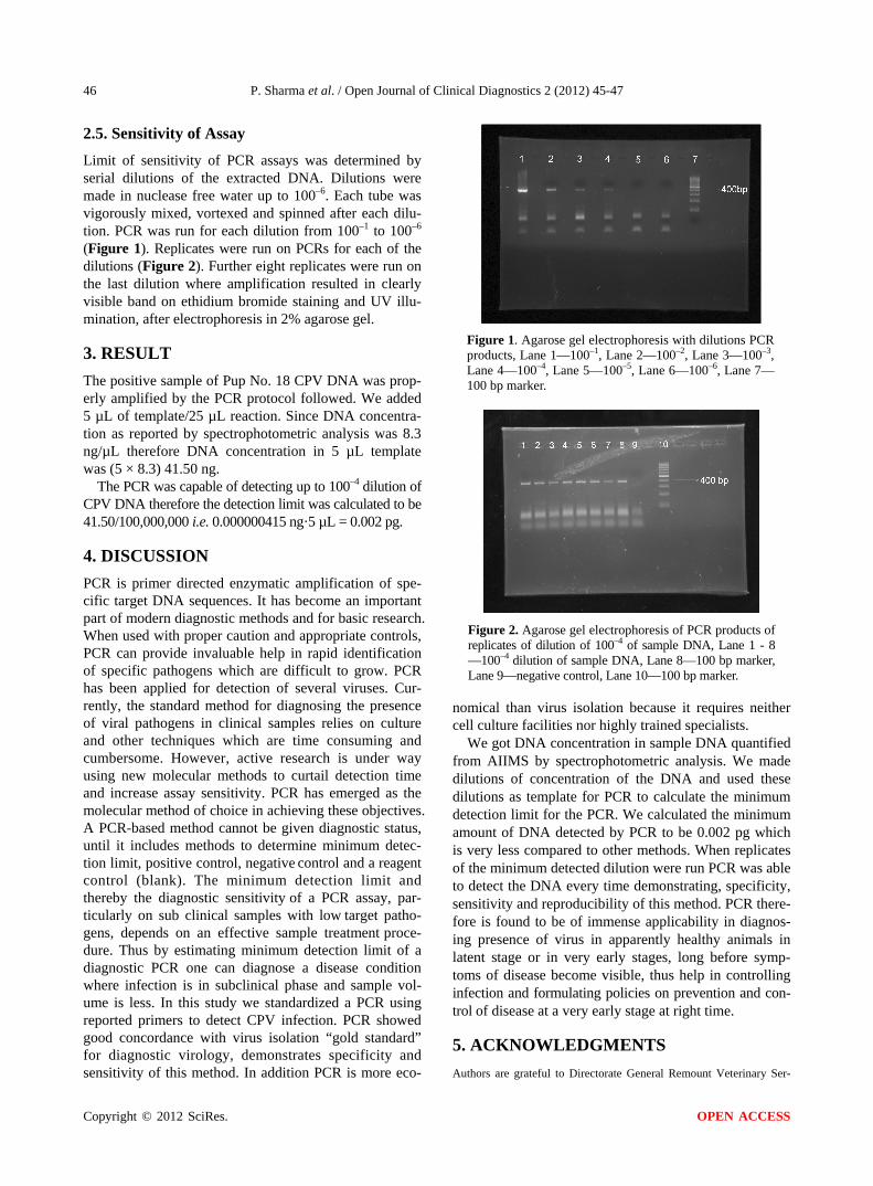
Limit of sensitivity of PCR assays was determined by serial dilutions of the extracted DNA. Dilutions were made in nuclease free water up to 100–6. Each tube was vigorously mixed, vortexed and spinned after each dilu-tion. PCR was run for each dilution from 100–1 to 100–6 (Figure 1). Replicates were run on PCRs for each of the dilutions (Figure 2). Further eight replicates were run on the last dilution where amplification resulted in clearly visible band on ethidium bromide staining and UV illu-mination, after electrophoresis in 2% agarose gel.
3. RESULT
The positive sample of Pup No. 18 CPV DNA was prop-erly amplified by the PCR protocol followed. We added 5 µL of template/25 µL reaction. Since DNA concentra-tion as reported by spectrophotometric analysis was 8.3 ng/µL therefore DNA concentration in 5 µL template was (5 × 8.3) 41.50 ng.
The PCR was capable of detecting up to 100–4 dilution of CPV DNA therefore the detection limit was calculated to be 41.50/100,000,000 i.e. 0.000000415 ng·5 µL = 0.002 pg.
4. DISCUSSION
PCR is primer directed enzymatic amplification of spe-cific target DNA sequences. It has become an important part of modern diagnostic methods and for basic research. When used with proper caution and appropriate controls, PCR can provide invaluable help in rapid identification of specific pathogens which are difficult to grow. PCR has been applied for detection of several viruses. Cur-rently, the standard method for diagnosing the presence of viral pathogens in clinical samples relies on culture and other techniques which are time consuming and cumbersome. However, active research is under way using new molecular methods to curtail detection time and increase assay sensitivity. PCR has emerged as the molecular method of choice in achieving these objectives. A PCR-based method cannot be given diagnostic status, until it includes methods to determine minimum detec-tion limit, positive control, negative control and a reagent control (blank). The minimum detection limit and thereby the diagnostic sensitivity of a PCR assay, par-ticularly on sub clinical samples with low target patho-gens, depends on an effective sample treatment proce-dure. Thus by estimating minimum detection limit of a diagnostic PCR one can diagnose a disease condition where infection is in subclinical phase and sample vol-ume is less. In this study we standardized a PCR using reported primers to detect CPV infection. PCR showed good concordance with virus isolation “gold standard” for diagnostic virology, demonstrates specificity and sensitivity of this method. In addition PCR is more eco-
Figure 1. Agarose gel electrophoresis with dilutions PCR products, Lane 1—100–1, Lane 2—100–2, Lane 3—100–3, Lane 4—100–4, Lane 5—100–5, Lane 6—100–6, Lane 7— 100 bp marker.
Figure 2. Agarose gel electrophoresis of PCR products of replicates of dilution of 100–4 of sample DNA, Lane 1 - 8 —100–4 dilution of sample DNA, Lane 8—100 bp marker, Lane 9—negative control, Lane 10—100 bp marker.
nomical than virus isolation because it requires neither cell culture facilities nor highly trained specialists.
We got DNA concentration in sample DNA quantified from AIIMS by spectrophotometric analysis. We made dilutions of concentration of the DNA and used these dilutions as template for PCR to calculate the minimum detection limit for the PCR. We calculated the minimum amount of DNA detected by PCR to be 0.002 pg which is very less compared to other methods. When replicates of the minimum detected dilution were run PCR was able to detect the DNA every time demonstrating, specificity, sensitivity and reproducibility of this method. PCR there- fore is found to be of immense applicability in diagnos- ing presence of virus in apparently healthy animals in latent stage or in very early stages, long before symp- toms of disease become visible, thus help in controlling infection and formulating policies on prevention and con- trol of disease at a very early stage at right time.
5. ACKNOWLEDGMENTS
Authors are grateful to Directorate General Remount Veterinary Ser-
Copyright © 2012 SciRes. OPEN ACCESS

P. Sharma et al. / Open Journal of Clinical Diagnostics 2 (2012) 45-47
Copyright © 2012 SciRes. OPEN ACCESS
47
[2] Ramadass, P. and Khader, T.G.A. (1982) Diagnosis of canine parvovirus infection by agar gel precipitation test and fluorescent antibody techniques. Cherion, 11, 323-328.
vices for providing facilities for conduct of this work and to Dr. H. K.
Prasad, Professor of Department of Biotechnology AIIMS, for assis-
tance provided. [3] Kumar and Dharmadheeran (2008) Detection of CPV
from fecal samples by PCR. Journal of Remount and Vet-erinary Corps, 47, 197-201.
REFERENCES [4] Narayanan, S., Amit, K., Chug, P.K., et al. (2001) Strain
characterization of Indian isolates of Canine Parvo Virus by PCR RFLP. Journal of Remount and Veterinary Corps, 40, 67-74.
[1] Sagazio, P., Tempesta, M., Buonavoglia, D., Cirone, F. and Bounavoglia, C. (1998) Antigenic characterization of canine parvovirus strains isolated in Italy. Journal of Vi-rological Methods, 73, 197-200. doi:10.1016/S0166-0934(98)00055-X