Embed Size (px)
Citation preview
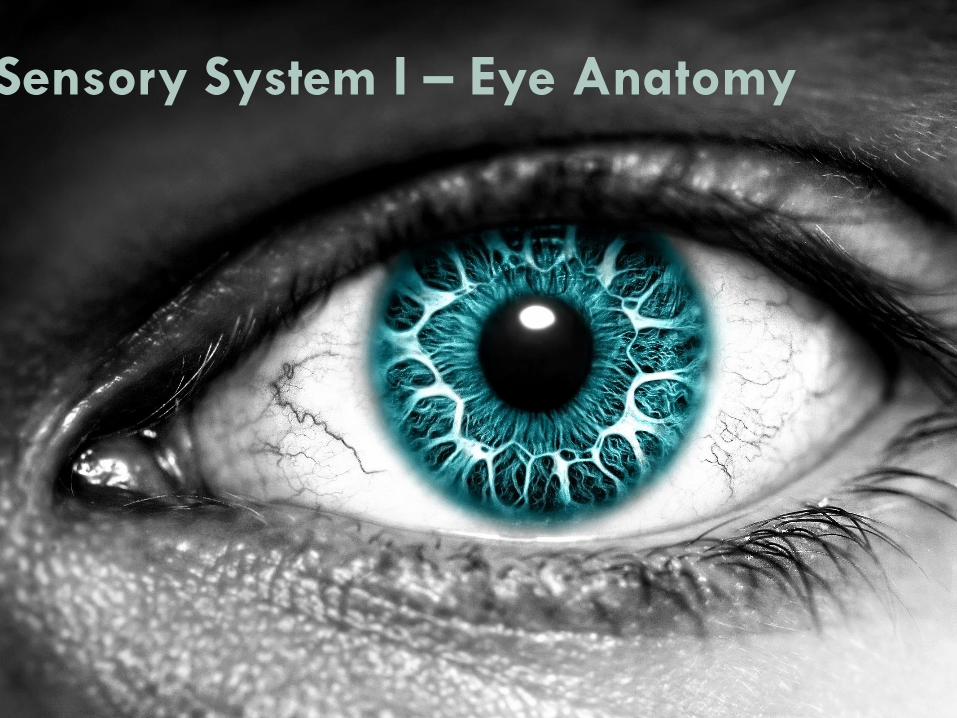
Sensory System I – Eye Anatomy
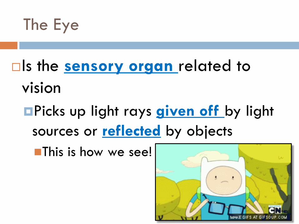
The Eye
Is the sensory organ related to
vision
Picks up light rays given off by light
sources or reflected by objects
This is how we see!
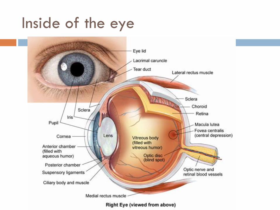
Inside of the eye
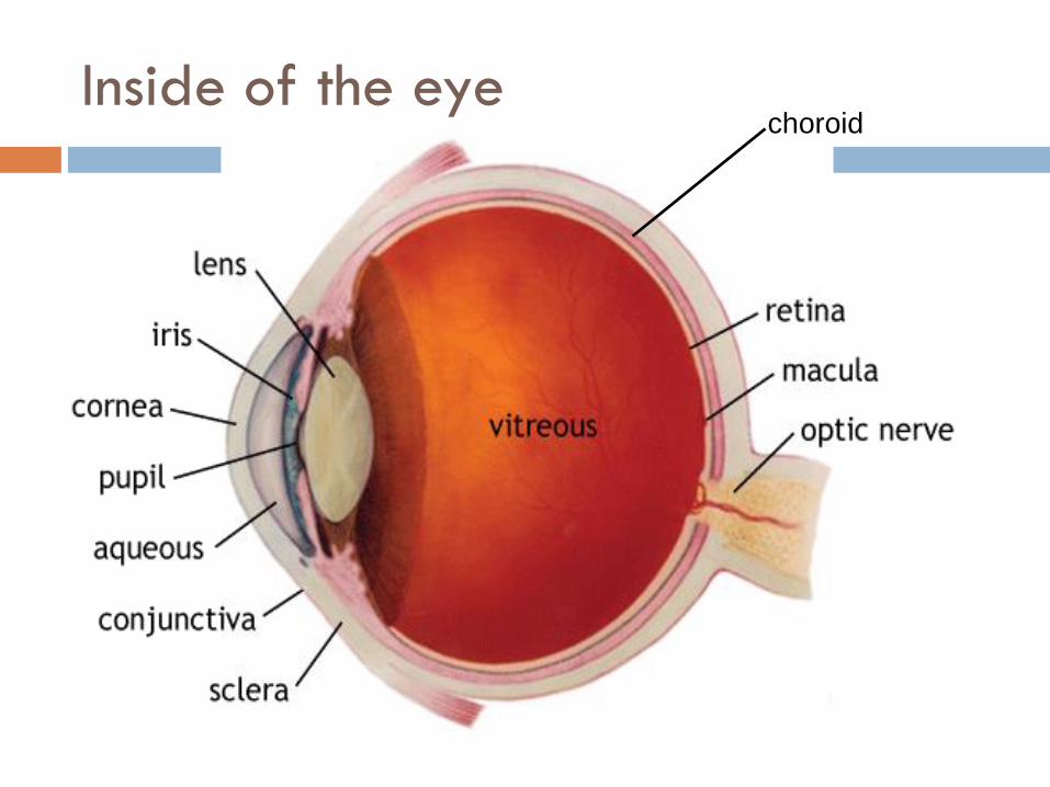
Inside of the eyechoroid
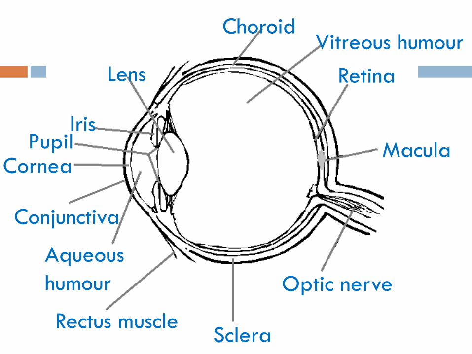
Lens
IrisPupil
Cornea
Aqueous
humour
Rectus muscleSclera
Optic nerve
ChoroidVitreous humour
Retina
Macula
Conjunctiva
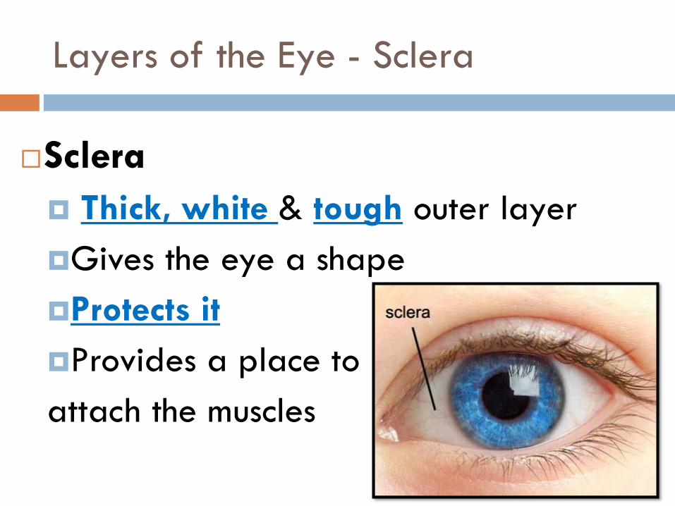
Layers of the Eye - Sclera
Sclera
Thick, white & tough outer layer
Gives the eye a shape
Protects it
Provides a place to
attach the muscles
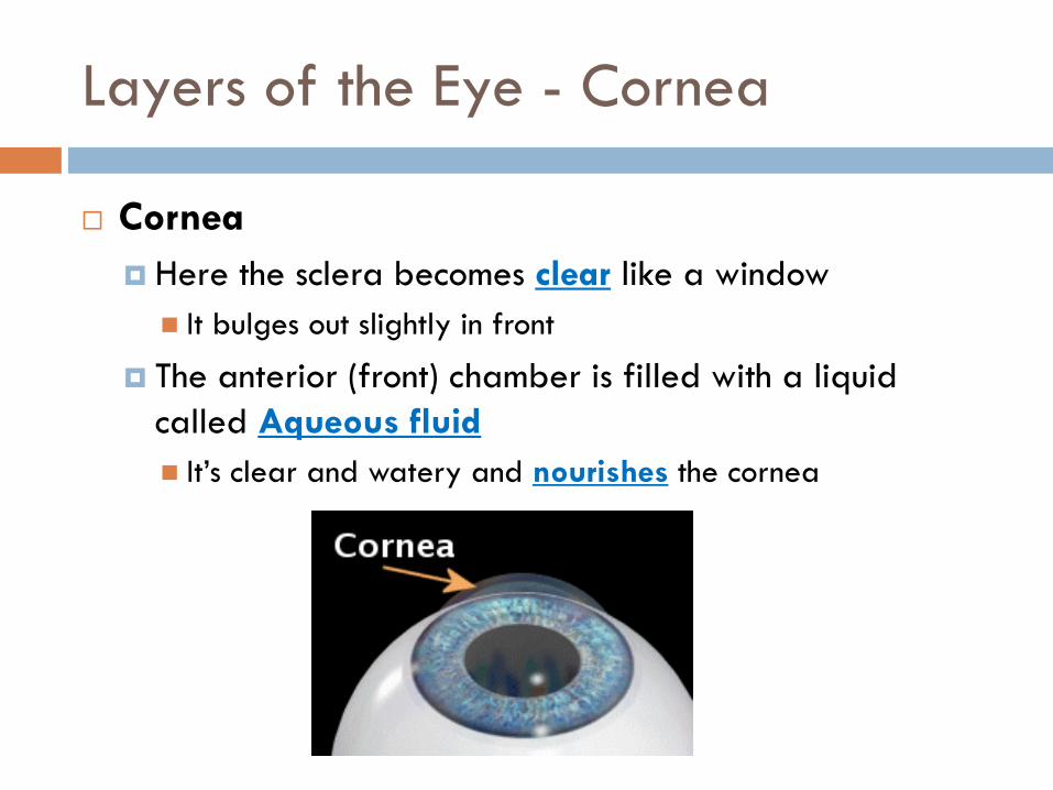
Layers of the Eye - Cornea
Cornea
Here the sclera becomes clear like a window
It bulges out slightly in front
The anterior (front) chamber is filled with a liquid
called Aqueous fluid
It’s clear and watery and nourishes the cornea
Layers of the Eye - Cornea
Choroid
Dark and pigmented middle layer of the eye
Very thin
Contains lots of blood vessels that nourish the eye
Prevents light from reflecting inside the eye
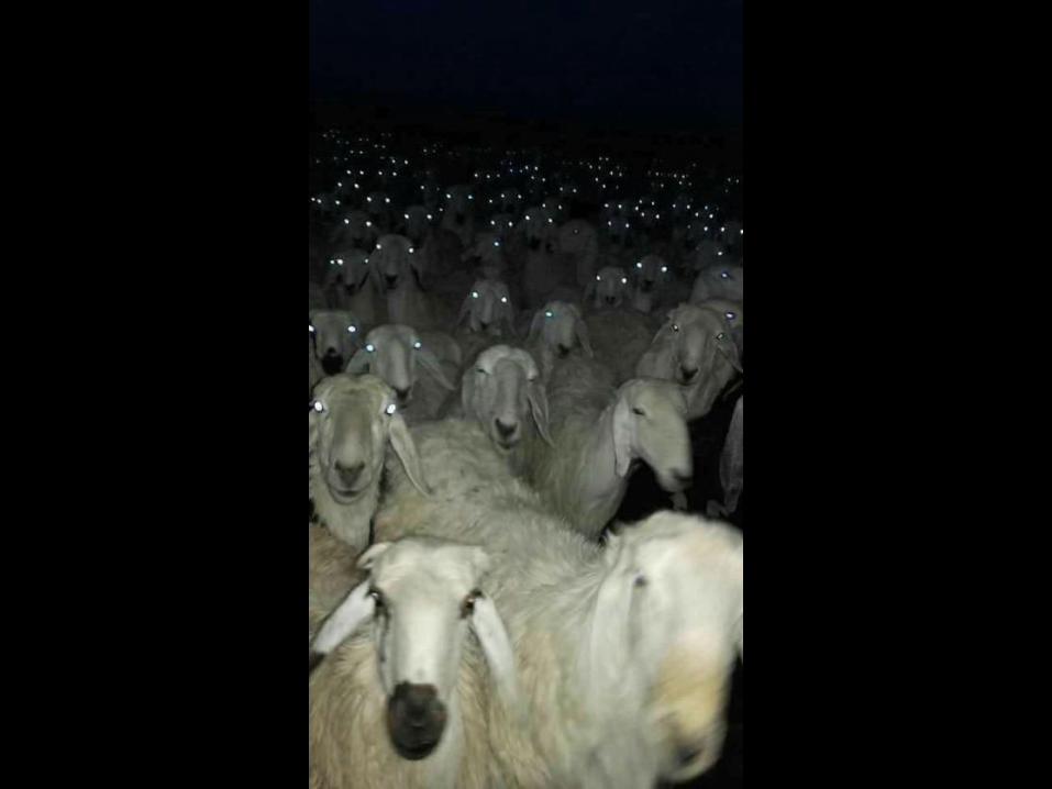
In some animals there is a bright strip called the
tapetum.
It’s found in nocturnal animals. It catches the light and shines
it back through the retina.
That’s why animals eyes glow in your headlights
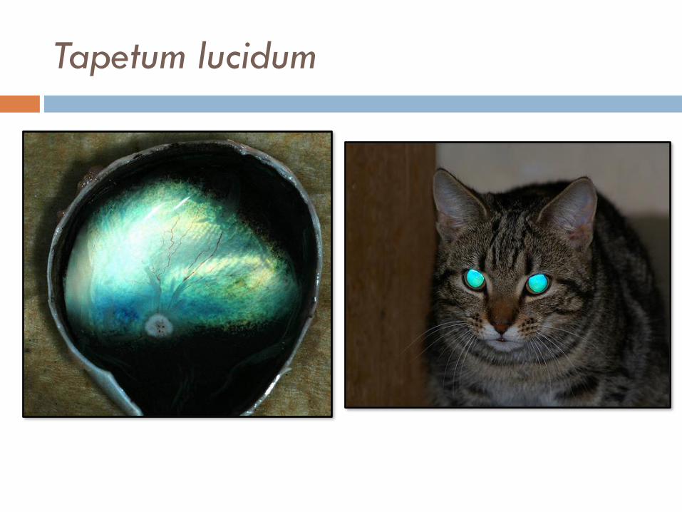
Tapetum lucidum
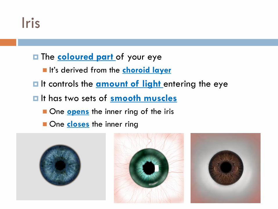
Iris
The coloured part of your eye
It’s derived from the choroid layer
It controls the amount of light entering the eye
It has two sets of smooth muscles
One opens the inner ring of the iris
One closes the inner ring
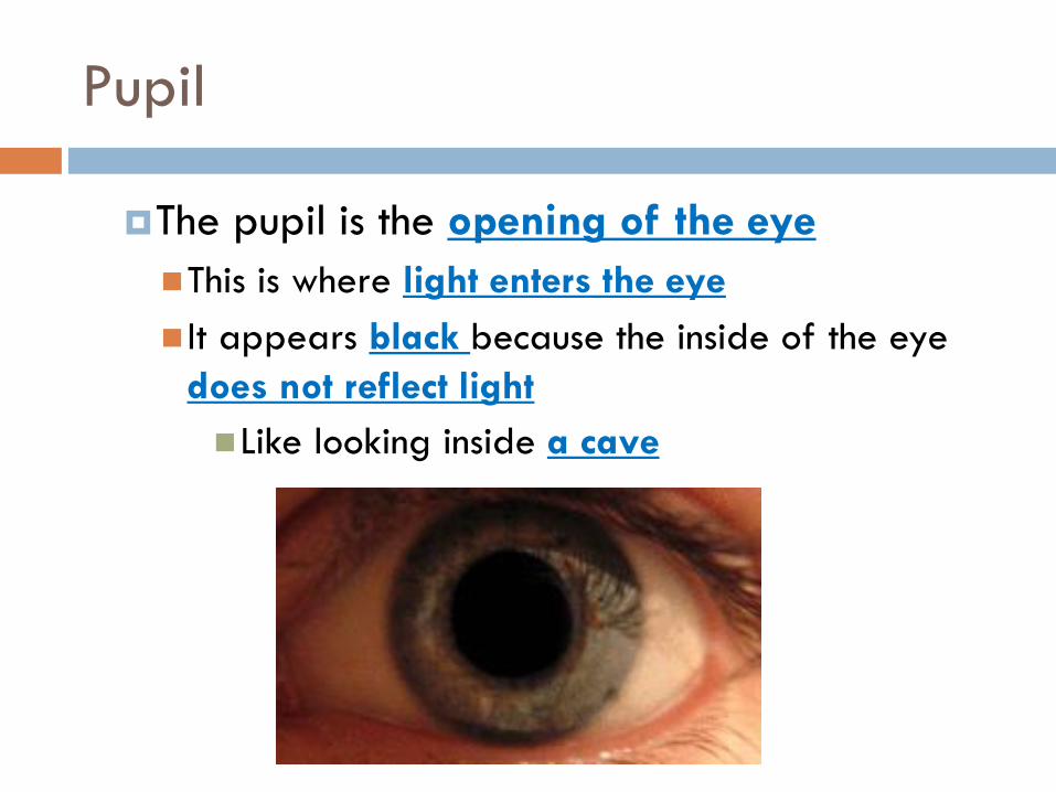
Pupil
The pupil is the opening of the eye
This is where light enters the eye
It appears black because the inside of the eye
does not reflect light
Like looking inside a cave
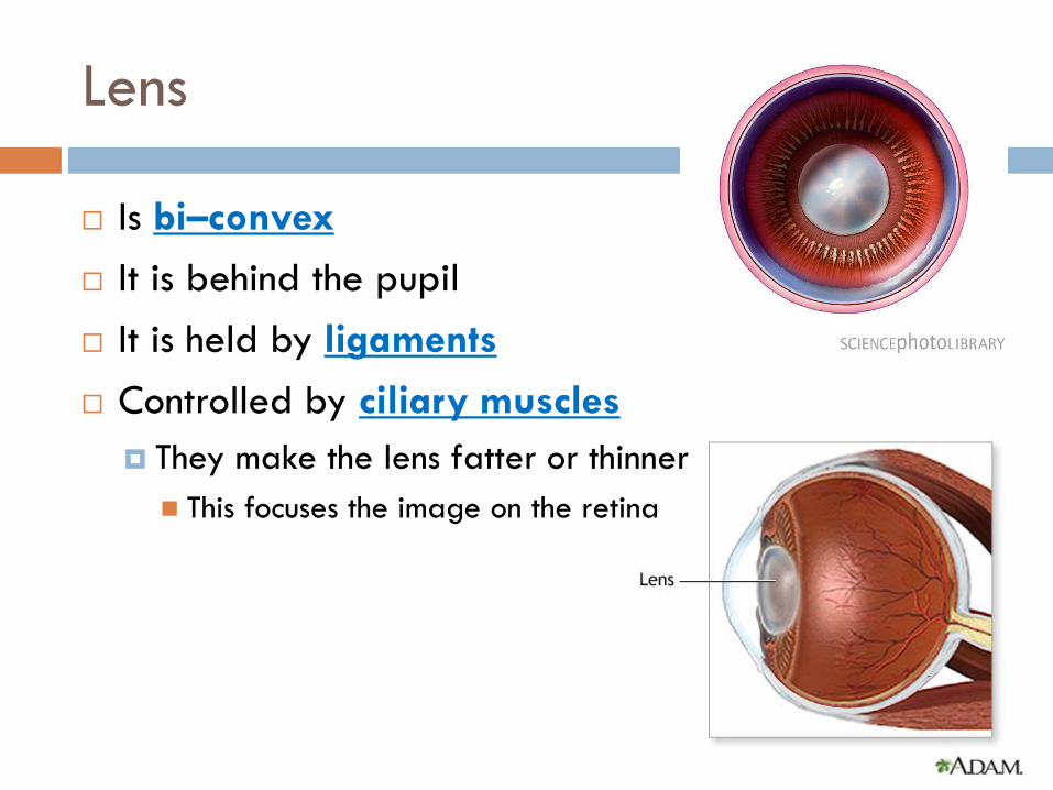
Lens
Is bi–convex
It is behind the pupil
It is held by ligaments
Controlled by ciliary muscles
They make the lens fatter or thinner
This focuses the image on the retina
Retina
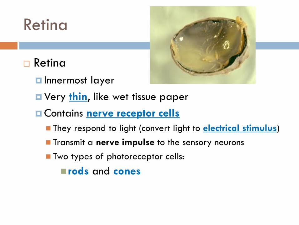
Retina
Innermost layer
Very thin, like wet tissue paper
Contains nerve receptor cells
They respond to light (convert light to electrical stimulus)
Transmit a nerve impulse to the sensory neurons
Two types of photoreceptor cells:
rods and cones
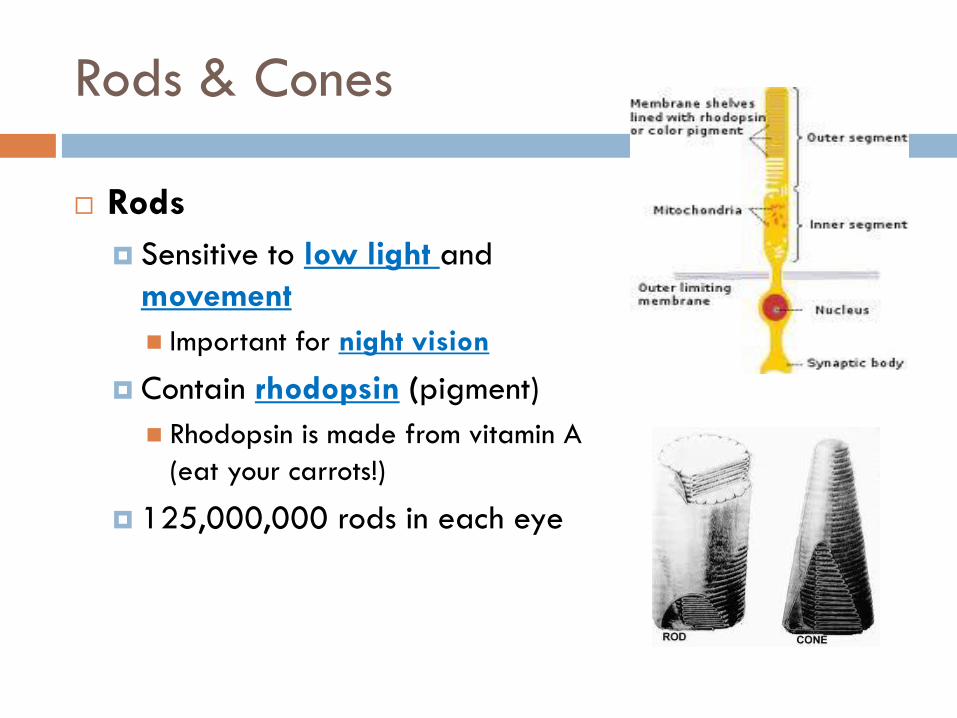
Rods & Cones
Rods
Sensitive to low light and
movement
Important for night vision
Contain rhodopsin (pigment)
Rhodopsin is made from vitamin A
(eat your carrots!)
125,000,000 rods in each eye
Rods & Cones
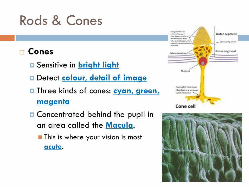
Cones
Sensitive in bright light
Detect colour, detail of image
Three kinds of cones: cyan, green,
magenta
Concentrated behind the pupil in
an area called the Macula.
This is where your vision is most
acute.
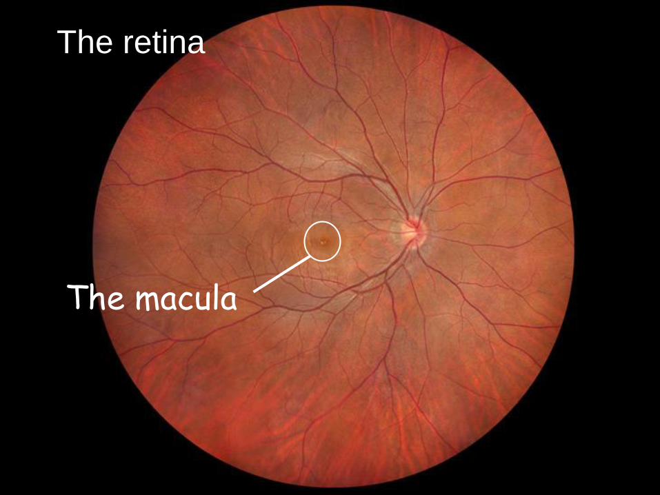
The retina
The macula
Rods & Cones
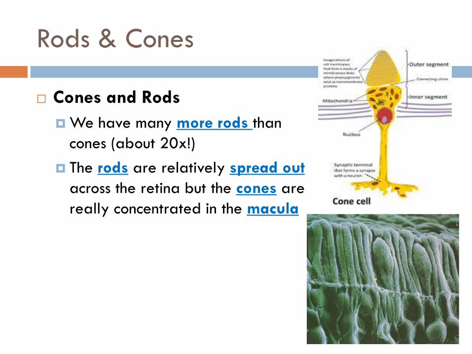
Cones and Rods
We have many more rods than
cones (about 20x!)
The rods are relatively spread out
across the retina but the cones are
really concentrated in the macula
Rods & Cones
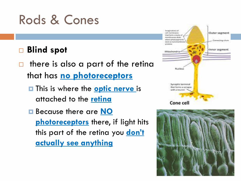
Blind spot
there is also a part of the retina
that has no photoreceptors
This is where the optic nerve is
attached to the retina
Because there are NO
photoreceptors there, if light hits
this part of the retina you don’t
actually see anything
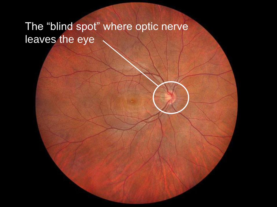
The “blind spot” where optic nerve
leaves the eye