Embed Size (px)
Citation preview
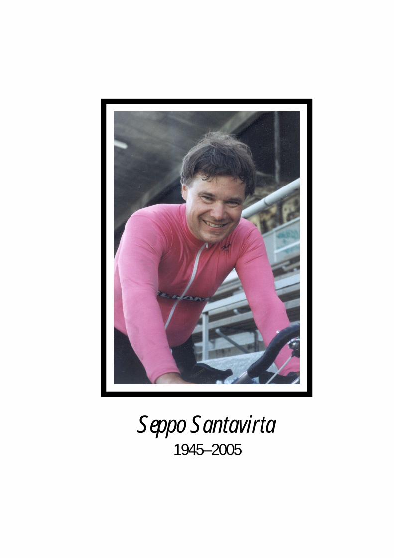
Seppo Santavirta1945–2005

Universities of Helsinki • Tampere • Kuopio • Oulu

Yleensä suomalaismies harjoittelee jopa yl-tiöpäisesti nuoruudessaan urheilun sittenjäädessä varhaisessa keski-iässä. Seppo jat-koi urheiluharrastustaan koko ikänsä, tree-naten elämänsä aikana lähes joka päivä.Oman pikaluistelijan aktiiviuransa jälkeenhän toimi Suomen Luisteluliitto ry:n pu-heenjohtajana 1978–1979 ja vuodesta 2001alkaen Suomen Pyöräilyunioni ry:n ratapyö-räilyjaoksen puheenjohtajana. Erinomaisenkuntonsa ansioista Seppo voitti suomen-mestaruuskisoissa pyöräilymitaleita Mas-ters-sarjassa, jollaiseksi sarjan nimi oli Se-pon aloitteesta ja positiivisesti asioita tar-kastelevan luonteen mukaan muutettu ai-emman ikämiessarjan sijaan. UrheilijanaSeppoa voidaan kuvata parhaiten sanoilla”vahva ja nopea”. Toisaalta hän oli kiinnos-tunut taiteen harrastaja ja hankki merkittä-vää asiantuntemusta suomalaisesta taitees-ta. Hän oli särmikäs oman tiensä kulkija, jo-ka poikkesi tavanomaisesta parempaan. Par-haiten hän rentoutui kesäpaikallaan Högså-rassa perheensä parissa. Sepon ennenaikainen menehtyminen on
iso menetys suomalaiselle ortopediyhteisöl-le. Seppoa jäävät kaipaamaan ystävät ja var-sinkin hänen rinnallaan uskollisesti kokoelämän ajan taivaltanut vaimo Nina ja hei-dän rakkaat poikansa Torsten ja Robin.Omnia conando docilis sollertia vincit
(tutkimusryhmämme motto).
Yrjö T. Konttinen
4175S U O M E N L Ä Ä K Ä R I L E H T I 4 1 / 2 0 0 5 V S K 6 0
JÄ
SE
NIS
TÖ
Ortopedian ja traumatologian professoriSeppo Sakari Santavirta menehtyi äkilliseensairauskohtaukseen 59-vuotiaana22.6.2005. Hän syntyi lääkäriperheeseen,mutta menetti molemmat vanhempansa jokouluiässä. Siksi Seppo lähtikin suoritta-maan asevelvollisuuttaan jo 17-vuotiaana janimitettiin aikoinaan Suomen nuorimmaksivänrikiksi 18-vuotiaana. Tämä tinkimättö-myys ja tavoitteellisuus leimasi jatkossakinhänen ammatillisia, urheilullisia ja kulttu-relleja pyrkimyksiään. Asevelvollisuutensa suoritettuaan hän seu-
rasi isänsä jalanjälkiä ja lähti Zürichin yli-opistoon, josta hän valmistui 1972 lääkärik-si ja jossa hän suoritti ensimmäisen tohto-rin tutkintonsa 1973. Palattuaan Suomeenhän teki Hyksissä kirurgiaan erikoistumisenohella toisen väitöskirjansa väitellen 1979Helsingin yliopistosta ja valmistuen samanavuonna kirurgian ja seuraavana vuonna or-topedian ja traumatologian erikoislääkärik-si. Hän oli pitkään Hyksin palveluksessa,mutta sai arvokasta koulutusta toimiessaan1986–1996 Invalidisäätiön sairaala OR-TONissa, jonka itsenäisyydestä noussuttalaatutyötä hän aina arvosti ja jonka säilyttä-misen hän näki tärkeänä. Seppo Santavirtanimitettiin Helsingin yliopiston ortopedianja traumatologian professoriksi 1996 kaik-kien asiantuntijoiden asetettua hänet avoi-messa kilvassa ensimmäiselle sijalle. Seppo perusti allekirjoittaneen kollegansa
kanssa kansainvälisen monitieteellisen TU-LES-tutkimusryhmän, joka saavutti myösSuomen Akatemian huippuyksikön ja ope-tusministerin tohtorikoulun aseman. Moni-tieteellisen yhteistyön polttopisteessä ovatbiomateriaalit ja niiden käyttö tekonivel-kirurgiassa, ihmisen varaosina ja kudostek-nologisissa sovelluksissa. Merkittäviin tut-kimussaavutuksiin kuuluvat timanttipin-noitteiden kehittäminen ja biomateriaalienkudosyhteensopivuuden selvittäminen. Innuce, todettiin että ihan oikeasti,”Diamonds are girl’s best friend”. Tästä tut-kimusalueesta Seppo Santavirta teki kol-mannen väitöskirjansa väitellen filosofiantohtoriksi biomateriaaliteknologian ja fysii-kan professorin Reijo Lappalaisen ohjauk-sessa Kuopion yliopistosta vuonna 2003.Tämä oli Sepolle oikea nuorennuskuuri,
kun hän intoa puhkuen palasi nuoruudenvoimien lähteelle sinne, mistä se hänelleparhaiten pulppusi: luovasta ja kovasta tie-teellisestä työstä ja rehellisestä kilvoittelus-ta. TULES-ryhmän avoimet ovet ja hyvä hen-
ki houkutteli yli 50 tutkijaa 21 eri maastaryhmään tieteen viljelyyn, Sepon auran vai-kutuspiiriin. Monet ryhmästä väitelleet ovatedenneet professoreiksi, dosenteiksi tai toi-mineet post-doceina ulkomailla. Kaikenkaikkiaan laajat kansainväliset yhteydet oli-vat ominaisia Seppo Santavirran työlle. Hänkuului useisiin toimituskuntiin kansain-välisissä ortopedisissa kliinisissä ja tieteelli-sissä sarjoissa. Seppo Santavirran ystävät,kirjaimellisesti ympäri maailmaa, ovat jul-kaisseet hänestä yksityiskohtaisemmannekrologin Acta Orthopaedica -lehdessä.Seppo Santavirta piti luovan tutkimustyön
lisäksi myös kliinisestä potilastyöstä. Huoli-matta kirurgisesta spesialiteetistaan SeppoSantavirta muisti ja korosti aina potilaan in-tegriteettiä ja oikeuksia, muistuttaen toi-menpidevaltaisen spesialiteetin tunnustet-tuna mestarina toimenpiteisiin fiksoituneitalääketieteen kandidaattikisällejä siitä, ettäpotilas on ihminen ja myös sellaisena koh-dattava ja kohdeltava, kaiken tutkimuksenja hoidon keskushahmona. Valtaenemistöopiskelijoista arvostikin suuresti riviopetta-jasta poikkeavaa elämäntaiteilijaa ja profes-soria, jonka opetuksessa innostuksen jakiinnostuksen herättäminen aina ohitti pas-siivisen tiedon syötön. Seppo Santavirta te-kikin syvälle luotaavaa tutkimustyötä co-pingista ja potilaiden elämänlaadusta vai-monsa, vs. professori Nina Santavirran jamatemaatikko, FT Svetlana A. Solovievankanssa.Henkilönä Seppo oli lahjakas ja monipuo-
linen. Tämä heijastui hänen vapaa-ajan har-rastuksissaan. Hän oli innokas urheilija.
In memoriamSEPPO SANTAVIRTA 5.12.1945–22.6.2005

Acta Orthopaedica 2005; 76 (5): 611–612 611
Seppo Santavirta1945-2005
Copyright© Taylor & Francis 2005. ISSN 1745–3674. Printed in Sweden – all rights reserved.DOI
Picture1998, Photograph: Eero Roine Seppo Sakari Santavirta, Professor of Orthopedics and Traumatology at the University of Helsinki, died after a sudden heart attack on June 22, 2005 at the age of 59. He was born into a physician’s family but lost both of his parents when he was still at school. After military service, he followed in his father’s footsteps and left for Switzerland to study medicine at the University of Zürich, from which he graduated in 1972. His first doctoral thesis, “Hyperplastische Scleimhautveränderungen im Dünn- und Dickdarm bei Morbus Menetrier” came from the same University in 1973. After returning to Finland, he started his specialization in surgery at the Helsinki University Central Hospital and at the same time he prepared his second Ph.D. thesis “Tourniquet ischaemia”, which he defended suc-cessfully in 1979. He became a specialist in Ortho-pedics and Traumatology in 1980. During most of his active time, he worked for the Helsinki Uni-versity Central Hospital, but he always valued his education and work at the Invalid Foundation ORTON (from1986 through 1996). He was nomi-nated Professor of Orthopedics and Traumatology at the University of Helsinki in 1996, after all four international experts had placed him in first place.
Together with Yrjö T. Konttinen, Professor of Medicine, Seppo founded an international and cross-scientific musculoskeletal diseases and inflammation research group (TULES). This group
achieved National Center of Excellence status from the Academy of Finland, and national Ph.D. Grad-uate School status from the Ministry of Education. The focus of the multidisciplinary research has been biomaterials and their use in arthroplasty sur-gery, as spare parts for humans and in tissue-engi-neering applications. The most important research achievements have been diamond coating of joint prostheses and studies on biocompatibility of bio-materials. Based on this research, Seppo Santavirta prepared and successfully defended his third Ph.D. thesis, “Compatibility of the totally replaced hip. Reduction of wear by amorphous diamond coat-ing”, and became a Doctor of Philosophy from the University of Kuopio in 2003 under the supervi-sion of Reijo Lappalainen, Professor of Biomateri-als Technology. Within the TULES group, Seppo hosted 50 researchers from 20 countries and many students of the group have advanced—becoming Professors, or working as postdoctoral fellows abroad.
Seppo Santavirta was a very flexible and cre-ative scientist. As an athlete, he favored individual sports, but he was a good team worker, encouraging young people to make the most of their talents. He was also loyal to his senior co-workers, a man true to his word. His interest in sports led him to work with sports medicine and sports injuries, and later with traffic accidents and musculoskeletal diseases in general. Early on, he made substantial contribu-tions to the understanding of occipito-atlanto-axial diseases. He described cervical spine involvement in trauma, in adult and juvenile rheumatoid arthri-

612 Acta Orthopaedica 2005; 76 (5): 611–612
tis, spondylarthropathies, psoriasis and Down’s syndrome, including the surgical treatment and out-come. This work often took him to Japan, where he had especially many good friends. Later, he started to work with biomaterials and joint replacements. Together with Kaj Tallroth, he observed a particu-larly aggressive form of aseptic loosening, which led to a beautiful series of publications on loosen-ing of hip arthroplasties, covering pathobiological processes from foreign body reactions and immune responses to surgical treatment and long-term out-come. He recognized the role of foreign body reac-tion and delayed-type hypersensitivity early on, and sought co-operation with physicists working on plasma acceleration methods to produce high-quality diamond coating of artificial joint compo-nents with no wear and no corrosion. A strong link via a trusted friend, Doc. Mika Hukkanen, to the laboratory of Professor Julia M. Polak in London, led to several excellent publications on neuropep-tides and innervation of the skeleton.
Seppo Santavirta liked research and clinical work equally and never forgot that patients are human beings, with feelings and lives of their own when away from the hospital bed, and he per-formed a great deal of coping and quality of life research together with his wife Docent Nina Santa-virta and the mathematician Dr. Svetlana A. Solo-vieva, originally from Russia. Most of his research was characterized by strong international links and networking, and he was invited to the editorial boards of many international journals in his field. He devoted his most important work for scientific journals to Acta Orthopaedica, being a co-editor since 1989 until his death. During this time, he
edited hundreds of manuscripts and raised enthusi-asm among many international researchers to write articles and reviews for Acta.
The untimely death of Seppo Santavirta ended a long orthopedics research career which showed no signs of waning; during his last year, Seppo pub-lished 15 scientific articles. Some of the last ones are published in this issue of Acta Orthopaedica: one Editorial on biotribology (page 613) and one review article on the medical treatment of rheuma-toid arthritis, together with Yrjö T. Konttinen and coworkers (page 614).
Seppo was a talented all-rounder, which was reflected in his spare-time hobbies. He was an eager sportsman. After his active sports career in speed skating, he was the chairman of the Finnish Skating Association 1978–1979, and from 2001 he was chairman of the Track Racing Division of the Finnish Bicycling Union. He was a connoisseur of the Arts, and attained considerable expertise in the Finnish Arts in particular. He had a charis-matic personality, and would do things in his own way—preferring to take the better course rather than the regular and mediocre one. He relaxed and enjoyed life best with his family at their summer cottage on Högsåra Island in the Finn-ish Archipelago. His early death is a huge loss to the Finnish and international orthopedics commu-nity, to his friends and co-workers, and in particu-lar to his wife Nina and sons Torsten and Robin.
Yrjö T. Konttinen, Professor of Medicine, Biomedicum Helsinki, Hel-sinki University, Finland
Per Aspenberg, Linköping, Sweden
Klaus D. Draenert, München, Germany
Enrique Gomez-Barrena, Madrid, Spain
Stuart B. Goodman, Stanford, CA, USA
William Jiranek, Richmond, VA, USA
Reijo Lappalainen, Kuopio, Finland
Lars Nordsletten, Oslo, Norway
Toshihigo Ogino, Yamagata, Japan
Dominique P. Pioletti, Lausanne, Switzerland
Claude Rieker, Winterthur, Switzerland
Anders Rydholm, Lund, Sweden
Jari Salo, Helsinki, Finland
Michael Silbermann, Haifa, Israel
Michiaki Takagi, Yamagata, Japan
Rihard Trebse, Ankaran, Slovenia
Timothy M. Wright, New York, NY, USA

NOVEL DIAMOND-LIKE CARBON – POLYMER –HYBRID COATINGS PREPARED WITH THE FILTERED PULSED ARC DISCHARGE METHOD
Alakoski E1,2, Tiainen V-M1, Kiuru M1, Soininen A1, Anttila A1
1ORTON Research Institute, the Invalid Foundation, Helsinki 2Department of Medicine, Invärtes Medicin, Helsinki University Hospital
Introduction and aims: The development of the Diamond-like Carbon – polymer –hybrid (DLC-p-h) coatings is an example of the role of chance in materials development. In a failed DLC experiment, PDMS (polydimetylsiloxane) used as an electrical insulator was accidentally vaporised amidst the carbon plasma and a film of interesting properties was formed. The film could not be marked by any marker pens and neither stickers nor tapes could be attached to it. This led us to modify the FPAD system to produce similar coatings in a controlled manner. We introduced a specially constructed carbon-polymer cathode. By controlling the amount of the polymer component vaporised and sputtered by the carbon plasma (in effect controlling the pulse frequency of the system) coatings with a wide range of mechanical and wetting properties can be deposited. The method and the novel coatings are currently in the process of being patented. Materials and methods: The FPAD unit is in a vacuum at a pressure of about 100 µPa. Carbon plasma arc similar to lightning is generated from the surface of the graphite cathode by discharging the ignition capacitor bank (C=10-20 pF). The plasma generated in the arc discharge then encounters a ring shaped graphite anode, which is at a higher potential (U=500-6000 V). As the plasma reaches the anode, the main capacitors are discharged. The main RCL-circuits parameters are typically the following: the tuning resistor R≈0.1 Ω, main capacitor capacitance C≈10-30 µF, filtering solenoid inductance L ≈ 3 µH. The current (in the order of several kA) from the main capacitors is lead through a curved solenoid and a synchronized magnetic field (~1 T) is created to steer the plasma towards the sample and to filter out the unwanted neutrals and larger particles. The deposited DLC-p-h coatings combine the exceptional mechanical properties of the DLC coatings and the non-wetting properties of the “parent” polymer. Table. Properties of PDMS and PTFE
Contact and sliding angles. The property usually referred to when describing the non-stick properties of materials is the contact angle of distilled water droplet. Low surface energy materials show high contact angles and vice versa. However, the sliding behaviour can be measured directly by measuring the critical tilt angle at which the droplet starts to slide down an inclined plane called the sliding
angle of the droplet. It is important to notice that the contact angle and sliding angle are independent of each other. So, to evaluate how a surface truly repels liquids one has to study the behaviour of droplets on near horizontal planar tilt angles. Results and conclusions: The Vickers hardness values of the DLC-PDMS-h coatings varied from 1 GPa to 70 GPa according to deposition speed [1]. The sp3 fraction of 70% measured with ESCA corresponds well with the high hardness [1]. Higher deposition speed means higher amount of the polymer component and better hydrophobicity and lower hardness. It must be noted that the coatings are not isotropic as the DLC-polymer cathode heats up during the deposition process and the evaporation and sputtering of the polymer is enhanced towards the end of the deposition. Both types of hybrid films showed high contact angles for water but DLC-PDMS-h coatings showed as a rule much lower sliding angles than DLC-PTFE-h. An oil drop slides easily down on DLC-PDMS-h coating and leaves no observable trace. However, because of the higher chemical resistance of PTFE the DLC-PTFE coating might be more suitable for some applications. Extremely low sliding angle 0.15 ± 0.03° was measured on the surface of DLC-PDMS-h coating with a 20 ml distilled water droplet [2]. This is thus far the lowest sliding angle measured from a solid surface.
Property PDMS PTFE Density (g/cm3) 1.1-1.6 2.17
Resistivity (Ωm) 1013 1017
Useful Temp. range/ °C -115 to 315 -240 to 205
Chemical resistance Good Excellent
A carbon-polymer arc discharge. The ring shaped anode can be seen on the right, the sample is on the left. References:[1]Preparation of diamond-like carbon - polymer -hybrid films with filtered pulsed arc discharge method, A. Anttila, V-M. Tiainen, M. Kiuru, E. Alakoski, K. Arstila, Surface Engineering, 19, 2003, 425-428.[2]Low sliding angles in hydrophobic and oleophobic coatings prepared with plasma discharge method, Mirjami Kiuru and Esa Alakoski, Materials Letters, 58, 2004, p. 2213-2216.

CURRICULUM VITAE Esa Artturi ALAKOSKI born 12.08.1968 in Kuusankoski, Finland
Current position(s): Researcher, Department of Medical Sciences, University of Helsinki, ORTON Research Institute, Tenholantie 10 FIN-00280 Helsinki, Finland.
Tel +358-(0)9-47482655, GSM +358-50-360 1968. e-mail [email protected]
Master's degree Doctorate 1999 Physics early 2006 Physics (estimated)
Sub-speciality Thin films, coatings and surface treatments&techn., amorphous diamond coatings, biomaterials. Relevant other experience relating to the application: Research Scientist, Accelerator laboratory Department of Physical sciences, University of Helsinki; 1999-2004 (autumn). Research Scientist, Deparment of Medical Sciences, Biomedicum (autumn) 2004-. Alakoski is a member of the National Centre of Excellence studying biomaterials and a member of Biomaterial and Tissue Engineering Graduate School. Ten selected representative publications or patents:
1. Energies and charge-state fractions of carbon plasma ions measured with Doppler-shifts in the pulsed arc-discharge system, V-M. Tiainen, E. Alakoski, M. Kiuru, A. Soininen, A. Anttila, The European Physical Journal, Applied Physics, vol. 22, 2003, p.111-114. 2.Tantalum as a buffer layer in the diamond like carbon coated artificial hip joints, M. Kiuru, E. Alakoski, V-M. Tiainen, R. Lappalainen, A. Anttila, Journal of Biomedical Materials Research (Applied Biomaterials), vol. 66B, 2003, p.425-428.3.Adhesion and quality test for tetrahedral amorphous carbon coating process, E. Alakoski, M. Kiuru, V-M. Tiainen, A. Anttila Diam. Relat. Mater., vol. 12, 2003, p.2115-2118. 4.Preparation of diamond-like carbon -polymer hybrid films with filtered pulsed arc discharge method, A. Anttila, V-M. Tiainen, M. Kiuru, E. Alakoski, K. Arstila, Surface Engineering, vol. 19 (2003) 425-428. 5.Low sliding angles in hydrophobic and oleophobic coatings prepared with plasma discharge method, Mirjami Kiuru and Esa Alakoski, Materials Letters, vol 58, 2004, 2213-2216. 6. Energy dependence of carbon plasma beam on the arc voltage and the anode-cathode distance in the pulsed arc-discharge method, Esa Alakoski and Veli-Matti Tiainen, Diamond and Related Materials, 14, 2005, p. 1451-1454. 7. A simplified arc-discharge set-up for high adhesion of DLC coatings, Esa Alakoski, Mirjami Kiuru, Veli-Matti Tiainen, Diamond and related materials, in press. 8. Effect of continuous in situ cathode polishing on plasma yield and energy in filtered pulsed arc discharge system, Esa Alakoski, Veli-Matti Tiainen, Mirjami Kiuru, submitted to Diamond and related materials. 9.Studies on diamond-like carbon and novel diamond-like carbon - polymer -hybrid coatings deposited with the filtered pulsed arc discharge method, Esa Alakoski, Summary for Thesis, manuscript. 10.Timanttisidoksiset hiili-polymeerimateriaalit, E. Alakoski, M. Kiuru, A. Soininen, V-M. Tiainen, A. Anttila, R. Lappalainen, Patent Applications 12.3.2002, Application number 20020462.
72

EVOLUTION IN TRANSLATIONAL RESEARCH ON BIOMATERIALS: FROM REPAIR TO REGENERATION AND FROM BIOSTABLE TO
BIOABSORBABLE MULTIFUNCTIONAL BIOMATERIALS
Ashammakhi N, Waris T, Kellomäki M, and Törmälä P
Department of Surgery, Oulu University Hospital, Oulu, Finland, and Institute of Biomaterials, Tampere University of Technology, Tampere, Finland
Tissue engineering is a new concept to develop tissue grafts that can be used to treat lost tissues due to disease or trauma. To obtain successful tissue reconstruction, it is necessary to use proper biomaterial, cell-source and provide adequate vascularity. Thus, our work has focused on evaluating bioabsorbable materials both in vitro and in vivo and on developing vascularization models. First, we have continued translational research taking bioabsorbable tissue repair implants to the clinic. First stage comprised the use of both bioabsorbable and metal implants in CMF surgery. The second stage comprised all-absorbable system and in a third phase fine-tuning work focused on reducing the time of operation by using bioabsorbable tacks. Because of fibro-inflammatory tissue reactions induced by biodegradable implants, this was exploited to develop fibrous tissue joints to replace diseased joints in rheumatoid and osteoarthritic joints. A new animal model was developed. In other occasions, where inflammatory reaction was regarded as unwanted effect, anti-inflammatory releasing implants were developed and evaluated in vitro and in vivo. Because of osteolysis that may to accompany the degradation of implants, bisphosphonate releasing implants were developed and being evaluated. To enhance the replacement of bioabsorbable devices with bone, osteoconductive element was added to the implants, e.g. screws and scaffolds that are evaluated in vitro and in vivo. To achieve proper graft or tissue engineered construct survival and functionality, vascularity is essential. Two models for microvascular tissue transfer were developed employing sheep and rabbits. Our studies have shown that it is possible to use bioabsorbable plates, screws and tacks to repair CMF bones. We have conducted an EU multicenter study on rare CMF syndromes and collected 165 cases [1]. Tacks were shown to reduce operation time which is important especially in infants. Adding bioactive glass as an osteoconductive component is being evaluated in animals and results are awaited. Preliminary results were encouraging [2] as showed even with genetically modified cells and cells derived from
syndromic (Crouzon) patients. Antibiotic releasing implants showed superiority to plain bioabsorbable and to titanium implants in significantly reducing bacterial attachment and biofilm formation [3]. Other multifunctional drug releasing implants have shown promise in vitro, and in vivo results are awaited. Tissue transfer models were successful [4] but cells may need to be combined with tissue flaps to enhance cartilage and bone formation. In conclusion, evolution of bioabsorbable implants has moved to the era or use of bioactive and multifunctional implants towards controlled tissue reactions and tailored clinical indications. Concepts of surgical therapy have also evolved from repair, to organ transplantation to arrive at reconstructive surgery employing tissue-engineered constructs. ACKNOWLEDGEMENTS Financial support from the Academy of Finland, TEKES and European Commission (Project “Spare Parts” and “Expertissues”) are greatly appreciated. REFERENCES [1] Ashammakhi et al. Successful Use of Biosorb Osteofixation Devices in 165 Cranial and Maxillofacial Cases: A Multicenter Report. J Craniofac Surg 2004; 15(4):692-701. [2] Dabernig et al. PLDLA 70/30 Plates and Bioactive Glass 13-93 Fibre-Based Scaffolds for Healing of Large Bone Defects. 4th Annual Meeting of the European Tissue Engineering Society (ETES), Munich, Germany, 31.8.-3.9.2005. O-156. [3] Niemelä et al. Ciprofloxacin-Releasing Bioabsorbable Polymer is Superior to Titanium in Preventing Staphylococcus Epidermidis Attachment and Biofilm Formation In Vitro. Key Engineering Materials 2005; 284-286:427–430. [4] Penttilä et al. Combining prefabricated microvascularied perichondrial flaps and bioabsorbable polylactide nonwoven scaffolds to tissue engineered cartilage. Joint Meeting of the Tissue Engineering Society International (TESI) and the European Tissue Engineering Society (ETES), Lausanne, Switzerland, 10-13.10.2004, P020.

CURRICULUM VITAE Nureddin AShammakhi born 10.6.1962 Current position(s): Professor
Vice Chairman, Institute of Biomaterials, Tampere University of Technology, POB 589, FIN-33101 Tampere, Finland. Tel: +358-40-8490980, +358-3-31152356 (office) Fax: +358-3-31152250, Email: [email protected]
Bachelor’s degree: Doctorate:1987 Tripoli 1996 Helsinki Fellowship of the Royal College of Surgeons: 2000 Edinburgh Relevant other experience related to the application: 1987-88 house officer, Alfateh University Hospitals; 1989-90 senior house officer, Salahuddin University Hospital; 1991 researcher and assistant / attachment, Helsinki University Hospital and Oulu University Hospital; 1994-98 basic surgical training including senior house officer, Birmingham, UK, clinical attachment Birmingham, Stoke-on-Trent, FRCS courses Birmingham, Glasgow, Dublin, Edinburgh & Stoke-on-Trent; 1998-2001 researcher, University of Oulu; 2001 visiting professor (May & July), Innsbruck University Clinic, Innsbruck, Austria; 2001 visiting professor (Aug), Providence Hospital, Southfield, MI, USA; 2002 docent, University of Oulu; 2002 docent, Tampere University of Technology. Evaluator (EO), Editorial board member (Eur J Plast Surg, J Craniofac Surg), Reviewer (Tissue Engineering, biomaterials, Biomacromolecules, J Biomed Mater Res). Sample Representative publications: 1. Ashammakhi N, Törmälä P. Editorial. From past to present and future is today. From inert to
multifunctional biomaterials. J Craniofac Surg 2004; 15(6):897. 2. Ashammakhi N, Suuronen R, Tiainen J, Törmälä P, Waris T. Spotlights on Naturally
Absorbable Osteofixation Devices. A Review. J Craniofac Surg 2003; 14(2):247-259 3. Länsman S, Karttunen A, Hirvelä H, Palosaari T, Kellomäki M, Ellä V, Ohtonen P, Törmälä
P, Waris T, Ashammakhi N. Persistence of indentation with bioabsorbable poly-l/d-lactide vs. silicone sponge scleral buckling implants. RETINA 2005; 25(5):581-586.
4. Gaissmaier C, Fritz J, Krackhardt T, Flesch I, Aicher W, Ashammakhi N. Effect of human platelet supernatant on proliferation and matrix synthesis of human articular chondrocytes in monolayer (2D) and three-dimensional (3D) alginate cultures. Biomaterials 2005; 26:1953-1960.
5. Ashammakhi N, Renier D, Arnaud E, Marchac D, Ninkovic M, Donaway D, Jones B, Serlo W, Laurikainen K, Törmälä P and Waris T. Successful Use of Biosorb Osteofixation Devices in 165 Cranial and Maxillofacial Cases: A Multicenter Report. J Craniofac Surg 2004; 15:692-701.
6. Tiainen J, Soini Y, Törmälä P, Waris T, Ashammakhi N. Self-Reinforced Polylactide-Polyglycolide 80/20 Screws take more than 1½ years to Resorb in Rabbit Cranial Bone. J Biomed Mater Res (Appl Biomat) 2004; 70B(1):49-55.
7. Penttilä H, Huttunen P, von Smitten K, Ashammakhi N, Waris T. Reinnervation of arterial grafts by noradrenergic nerves occurs in rats as indicated by increased levels of noradrenaline. Plast Reconstr Surg 2004; 113(7):2057-2060.
8. Waris E, Ninkovic M, Harpf C, Ashammakhi N. Self-Reinforced bioabsorbable miniplates for skeletal fixation in complex hand injury. J Hand Surg [Am] 2004; 29(3):452-457.
9. Serlo W, Ashammakhi N, Länsman S, Törmälä P, Waris T. A New Technique for Correction of Trigonocephaly Using New Bioabsorbable Osteofixation Tacks and Plates and a Novel Tack-shooter. J Craniofac Surg 2003; 14(1):92-96
10. Niemelä S-M, Ikäheimo I, Koskela M, Veiranto M, Suokas E, Törmälä P, Waris T, Ashammakhi N, Syrjälä H. Ciprofloxacin-releasing bioabsorbable polymer is superior to titanium in preventing Staphylococcus epidermis attachment and biofilm formation in vitro. J Biomed Mater Res [Appl Biomat], in press

PRO-INFLAMMATORY CYTOKINE AND PROTEINASE CASCADES IN HOST RESPONSE AGAINST IMPLANTS
Beklen A1, Konttinen YT1
1Department of Medicine, Invärtes Medicin, Helsinki University Hospital
INTRODUCTION Periodontal diseases, particularly periodontitis is a chronic tissue destructive disease leading to loosening and loss of tooth in any age. Dental implants can be used to replace missing natural teeth with natural-looking and natural-feeling implants. Such implants, however, can loosen. Mechanisms responsible for periodontitis and peri-implantitis are somewhat similar. Loosening is thought to be due to mainly chronic infection with pocket bacteria/dental plaque-induced host inflammation and excessive/falsely directed mechanical loading. We hypothesize that, cytokine induced neutrophil derived proteinases in gingival crevicular fluid and peri-implant sulcus fluid are important for the destruction of tissues surrounding both tooth and implant. MATERIAL AND METHODS Fluids and tissue samples were collected from five healthy controls (mean age 31) and seven patients with tissue destruction (mean age 46). Immunohistochemistry was used for the localization of cytokine and proteases in tissue specimens. Immunofluorometric assay was used to analyze matrix metalloproteinase-8 or MMP-8 and MMP-9 levels in peri-implant sulcus fluid. Western blots and gelatin zymography were used for the detection of pro and active forms of MMP-3,-8,-9. Modified prourokinase substrate method was used to assess MMP activities in cultured gingival fibroblasts stimulated with 10 ng/ml of tumour necrosis factor alpha. RESULTS Analysis of peri-implant sulcus fluid revealed that both MMP-8 and MMP-9 levels were high in patients who had loosening implants. Western blots showed that gingival crevicular fluid from adult periodontitis contained
partially activated form of MMP-3. In healthy controls MMP-3 was found in latent or complexed form. Western blots also disclosed and zymography confirmed the presence of both latent and active forms of MMP-8 or collagenase-2) and MMP-9 or gelatinase B in adult periodontitis. For MMP-8 and MMP-9 species both latent pro-MMP and active form were observed in periodontitis; whereas only its latent proform was found and only in low amounts in controls. Matrix metalloproteinase activity in tumor necrosis factor-α stimulated and non-stimulated gingival fibroblasts revealed a significant increase of proform of MMP-3 but not in the active form of MMP-3. CONCLUSION Investigation of the levels of matrix metalloproteinases in peri-implant sulcus fluid and gingival crevicular fluid suggests that MMP-8 with MMP-9 may in part be responsible for the irreversible destruction of bone and soft tissues around implant and tooth. In addition to that the results suggest that fibroblast- and neutrophil-derived proteases co-operate to increase the matrix metalloproteinase burden in the gingival crevicular fluid of subjects with adult periodontitis or implantitis. Resident gingival fibroblasts produce proMMP-3 into gingival crevicular fluid, where it becomes activated, probably as a result of the action of the neutrophil-derived cathepsin G and elastase. Active MMP-3 then activates proMMP-8 and proMMP-9 leading to tissue degradation and loose of implants. REFERENCES [1] Birkedal-Hansen H, Moore WG, Bodden MK, Windsor LJ, et al. (1993). Matrix metalloproteinases: a review. Crit Rev Oral Biol Med 4:197-250.

CURRICULUM VITAE Arzu BEKLEN born 20.03.1973 in Ankara, Turkey Current position: PhD student,
(2002-), Department of Anatomy, Biomedicum Helsinki, P.O. Box 63 (Haartmaninkatu 8), FIN-00014 University of Helsinki, Finland. Tel: +358-(0)9-191-25212, Fax +358-(0)9-191 25218 E-mail: [email protected]
Master’s degree: Doctorate:2002 Istanbul, Turkey 2007 Helsinki (estimated) Relevant other experience relating to the application: Research Fellow 2002-, TULES-group, Helsinki University, Department of Anatomy, Helsinki Finland Representative publications:1. Beklen A: M.Sc. Thesis Comparison of surface structures of tooth prepared by high speed rotary burs and air abrasion using ESEM/FEG. Istanbul: Bogazici University, 2002 2. Konttinen YT, Zhao D, Beklen A, Ma A, Takagi M, Kivelä-Rajamäki M, Ashammakhi N: The microenvironment around total hip replacement prosthesis. Clin Orthop Rel Res , (430):28-38, 2005 3. Beklen A, Laine M, Ventä I, Hyrkäs T, Konttinen YT: Role of TNF-α and Its Receptors in Pericoronitis. Journal of Dental Research, in press 4. Beklen A, Tuter G, Sorsa T, Hanemaaijer R, Virtanen I, Tervahartiala T, Konttinen YT: Gingival Tissue and Crevicular Fluid Co-Operation in Adult Periodontitis Journal of Dental Research, in press 5. Safkan-Seppälä B, Sorsa T, Tervahartiala T, Beklen A, Konttinen YT: Collagenases in gingival crevicular fluid in insulin-dependent diabetes Mellitus. Journal of Periodontology, in press 6. Konttinen YT, Sillat T, Saat R, Ma G, Zhao D, Beklen A: Biomaterials In: Topics in Tissue Engineering, Edited by: N.Ashammaki, P.Ferreti, in press 7. Beklen A, Pihakari A, Hietanen J, Konttinen YT: Chronic Sinusitis Associated with the Use of Unrecognized BoneSubstitute: Case Report, submitted

00,010,020,030,040,050,060,070,080,090,1
0,11
0 40 1 000 2 000 3 000 4 000 5 000 6 000 7 000 8 000 9 000 10 000Number of Loading Cycles
Am
plitu
de o
f Ver
tical
Mov
emen
t (m
m)
Fig. 1. Subsidence up to 5 millions (A) and the amplitude of vertical movement (micromotion) up to 10,000 cycles (B).
EFFECT OF AMORPHOUS DIAMOND COATING ON SUBSIDENCE OF METALLIC PINS SIMULATING CEMENTED FEMORAL STEM IN CYCLIC FATIGUE TESTING
H. Kawaji1,2, A. Koistinen1, M. Takagi2, S.S. Santavirta3, R. Lappalainen1
1Dept. of Applied Physics, University of Kuopio, P.O.Box 1627, FIN-70211 Kuopio, Finland 2Dept. of Orthopaedic Surgery, Yamagata University School of Medicine, 990-9585 Yamagata, Japan
3Dept. of Orthopaedics and Traumatology, University Central Hospital, P.O.Box 266, FIN-00029 HUS, Finland INTRODUCTION Bone cement is widely used for total hip replacement (THR). Its elasticity can distribute the point stresses between the bone and the stem. However, micromotion at the stem/bone cement interface and excessive subsidence of femoral stem cause wear particles and damage of bone cement which lead to periprosthetic osteolysis [1]. One of the theoretical benefits of titanium (Ti) alloy is a lower modulus of elasticity to reduce proximal stress shielding and bone resorption. However, Ti alloy is not recommended for cemented THR because Ti alloy stems may be severely damaged due to wear and crevice corrosion. However, advantages of bone cement and Ti alloy have a great potential for THR. Furthermore, reduction of excessive subsidence and micromotion is an important factor concerning to the longevity of the cemented femoral stem of THR. Amorphous diamond (AD) coating has turned out to be a promising biomaterial for artificial joint, due to excellent wear and corrosion resistance, biocompatibility, longevity and versatility [2,3]. Our hypothesis was that AD coating could improve stability at the stem/bone cement interface. The purpose of this study was to investigate the effect of AD coating on subsidence and micromotion of metallic pins in the model system for the cemented femoral stems. METHODS Widely used metallic biomaterials, Ti alloy, TiAl6V4 (N=7) and stainless steel (SS), AISI 316L (N=12) were used. The tapered pins (length 70 mm, angle 2.3°) had proximal and distal diameters of 10 and 8 mm, respectively. Different surface roughness values of the pins were achieved by grinding and polishing (Ra = 50 or 100 nm for Ti alloy pins and Ra = 10 or 100 nm for SS pins). Half of the SS pin set was coated with AD coating, 600-800 nm thick, by the filtered pulsed plasma arc discharge method [4]. The pins were cemented in nylon blocks (height 50 mm) with bone cement Refobacin®- Palacos® R (Biomet Europe, Dordrecht, The Netherlands). The minimum thickness of the cement mantle was 3 mm.
Cyclic fatigue testing (5 million cycles/pin) was carried out by using a servohydraulic tester (Instron, Canton, MA, USA). The vertical load profile was a scaled Paul’s gait curve with a peak load of 0.850 kN at 10 Hz (ISO 14242-1). The torsional load with ±3.750 Nm at 0.5 Hz (ISO 7206) was applied simultaneously. Testing was carried out in diluted bovine serum (total protein content 35 mg/ml). The vertical position was monitored through- out the tests by the software of the testing equipment. RESULTS The cumulative subsidence of the pins is shown in Figure 1A. Following tendencies of the subsidence were found:
with respect to coating: AD-coated < non AD-coated; with respect to surface roughness: rough < smooth; and with respect to material: Ti < SS.
The amplitude of vertical movement for a single loading cycle (micromotion at the interface) is shown in Figure 1B. For non AD-coated pins, the amplitude was high within 1,000 cycles and then reached a plateau, while the amplitude of AD-coated pins was more stable throughout the tests. DISCUSSION AND CONCLUSION Low subsidence of the AD-coated pins is most probably due to the surface characteristics of AD and higher stability of the interface for AD. Evidently, the rough pins are more stable due to difference in the shear forces at the interface. Lower subsidence of the Ti alloy pins is probably due to difference in the elastic modulus and the surface chemistry.
Based on the results of the micromotion within 1,000 cycles, debonding at the interface already occurred at the early stage in all specimens. Micromotion was least in the group of AD-coated SS pins. Thus, the interface is the most stable in this group. In conclusion, AD coating seems to be a potential material for surface treatment of femoral stem for clinical cemented fixation. REFERENCES [1] Santavirta S et al. (1998) Curr Orthop, 12, 51-57. [2] Lappalainen R et al. (1998) Clin Orthop, 352, 118-127. [3] Lappalainen R and Santavirta S (2005) Clin Orthop, 430, 72-79. [4] Anttila A et al. (1997) Adv Mater, 9, 1161-1164.
-1,3-1,2-1,1
-1-0,9-0,8-0,7-0,6-0,5-0,4-0,3-0,2-0,1
0
0 0,5 1 1,5 2 2,5 3 3,5 4 4,5 5Number of Loading Cycles (millions)
Subs
iden
ce (m
m)
Ti alloy, Ra=50nm (N=3) Ti alloy, Ra=100nm (N=4)SS, Ra=10nm (N=3) SS, Ra=100nm(N=4)SS, Ra=10nm, AD-coated (N=2) SS, Ra=100nm, AD-coated (N=3)
A
B
CURRICULUM VITAE Hiroyuki Kawaji born 21.3.1963 in Japan Current position(s): Visiting researcher
Department of Applied Physics, University of Kuopio, POB 1627, FIN-70211 Kuopio, Finland. Tel: +358-17-162278, Fax: +358-17-162585, Email: [email protected] Lecturer Department of Orthopaedic Surgery, Yamagata University School of Medicine, 2-2-2 Iida-Nishi, Yamagata 990-9585, Japan. Tel: +81-23-628-5355, Fax: +81-23-628-5357
Bachelor´s degree Licentiate degree 1989 Yamagata, Japan (Medicine) 1989 Japan (Medical doctor lisence)
1996 Approved orthopaedic surgeon by the Japanese Orthopaedic Association
Relevant other experience relating to the application: Resident 1989, 5m, Yamagata University School of Medicine; Acting Hospital Orthopaedic Surgeon 1990-1991, 12m, Tsubame Rosai Hospital; 1991, 6m, Yamagata Rehabilitation Center for children; 1991-1992, 12m, Sagae City Hospital; 1992-1994, 18m, Yokote Municipal Hospital; Acting Senior Orthopaedic Surgeon, 1994-1996, 30m, Municipal Sakata Hospital; 1996-1997, 6m, Yamagata Prefectural Nihonkai Hospital; 1998-2001, 36m, Yamagata Saisei Hospital; 2001-2003, 30m, Sanyudo Hospital; Assistant 1997-1998, 12m, Yamagata University School of Medicine; Lecturer 2003-, Department of Orthopaedic Surgery, Yamagata University School of Medicine Representative publications:
1. Ida H, Takagi M, Yamakawa M, Kawaji H, Takei I, Ogino T: Periprosthetic and synovial connective tisuue response to HDP particles in rats. Transaction, 3rd Combined Orthopaedic Research Societies Meeting, USA, Canada, Europe and Japan 3: 178, 1998.
2. Takagi M, Santavirta S, Ida H, Ishii M, Yamakawa M, Sato T, Kawaji H, Takei I, Hamasaki M, Ogino T, Niissalo S, Konttinen YT: Biological role of matrix metalloproteinases in loose artificial hip joints. Journal of Japanese Orthopaedic Association 72: S1747, 1998.
3. Ishii M, Kawaji H, Nitta H, Hamasaki M, Ida H, Takagi M: Impaction allograft technique for revision THA. Transaction, International Orthopaedic Symposium in Yokohama, 72nd Annual Meeting of the Japanese Orthopaedic Association, 59, 1999.
4. Sato T, Takagi M, Ida H, Ishii M, Yamakwa M, Kawaji H, Takei I: Analysis of periprosthetic bone remodeling in loose hip joints. Transaction, Societe Internationale de Recherche Orthopedique et de Traumatologie (SIROT) 99, 431-433, 1999.
5. Ida H, Takagi M, Yamakwa M, Sato T, Kawaji H, Takei H, Ogino T: Periprosthetic bone remodeling in rat femur model. Transaction, Societe Internationale de Recherche Orthopedique et de Traumatologie (SIROT) 99: 54, 1999.
6. Kawaji H, Nishimura G, Watanabe S, Mabuchi A, Ikeda T,, Ohashi H, Sasaki A, Sano T, Ikegawa S: Autosomal dominant precocious osteoarthropathy due to a mutation of the cartilage oligomeric matrix protein (COMP) gene: further expansion of the phenotypic variations of COMP defects. Skeletal Radiol 31: 730-737, 2002.
7. Mabuchi A, Manabe N, Haga N, Kitoh H, Ikeda T, Kawaji H, Tamai K, Hamada J, Nakamura S et al.: Novel types of COMP mutations and genotype-phenotype association in pseudoachondroplasia and multiple epiphyseal dysplasia. Hum Genet 112: 84-90, 2003.
8. Kawaji H, Kostinen M, Takagi M, Lappalainen R, Santavirta S: Effect of surface condition of metallic pins simulating cemented femoral stem on subsidence in cyclic fatigue testing. Finn J Orthop Traumatol 28: 161-166, 2005.

BIODEGRADABLE TISSUE ENGINEERING AND INTERPOSITION ARTHROPLASTY SCAFFOLDS
Minna Kellomäki, Ville Ellä, Elina Talvitie, Anna-Maija Haltia, Mikko Huttunen, Laura Sippola, Pertti
Törmälä Institute of Biomaterials, Tampere University of Technology, Tampere, Finland
INTRODUCTION Highly porous, three-dimensional biodegradable scaffolds with interconnective pores are essential for tissue engineering “living” implant to replace extracellular matrix for specific period of either cell culture or tissue healing or both. Several methods to manufacture scaffolds have been reported in the literature, for example solvent-casting followed by salt-leaching, freeze-drying and methods to prepare non-woven to 3-D structures. However, clear comparisons between the methods have not been published. The aim of the studies has been to compare the important properties of the scaffolds manufactured by different methods from synthetic bioabsorbable polymers. An overview of the current situation of interposition arthroplasty project is given. METHODS In all cases poly-L-D-lactide 96/4 stereocopolymer is reported as a model polymer. Inherent viscosities varied from 1.8 to 5.5 dl/g and its influence to the scaffold properties was examined. Five production methods were studied. As a standard, commonly used solvent-casting salt-leaching was performed using chloroform as a solvent. Melt-spinning hot-drawing followed by non-woven technique, braiding or knitting was the main interest in the studies. Also freeze-drying was experimented. Several scaffold properties were followed, but in this presentation degree of porosity, interconnectivity of the pores and mechanical properties are reported from the scaffolds prepared. All the structures, including plain fibers, were tested in vitro (37C, pH 7.4, PBS).
RESULTS Structurally the methods gave different scaffolds. Solvent-casting salt-leaching needed 90 wt-% of salt to obtain interconnective porosity for the scaffold and pores had shapes and dimensions of the salt particles. Textile techniques gave interconnectivity in all cases and pores had large range of sizes. Braided and knitted scaffolds had organized structure while others had randomly oriented pores. Studied scaffolds had degrees of porosity from 40-50% (braided) to up to 90% (other methods). Mechanical strength of the scaffolds was difficult to compare but relative strength retention in vitro described more the behavior. Solvent-cast salt-leached and non-woven scaffolds lost their mechanical strength completely in 20 weeks while others retained 50% of the initial strength for 24 weeks in vitro. DISCUSSION Manufacturing methods to produce scaffolds for tissue engineering can be chosen to meet the requirements of the application. Initial strength as well as strength retention either in vitro or in vivo can be tailored by changing the polymer used and also by using different production methods. Important advantages of textile technologies lie in their high throughput speed and large production capacity. The knitted joint scaffolds are at the multicenter clinical studies for hand and feet small joints. These studies are giving awaited information of functionality of the first knitted scaffold structures used in humans. ACKNOWLEDGEMENTS Financial support from the Finnish Academy, TEKES and European Union is acknowledged.

CURRICULUM VITAE Minna Auli Eliisa KELLOMÄKI born 17.11.1964 in
Rauma, Finland
Current position(s): Professor of Biomaterials and Tissue Engineering, Head of Institute, Institute of Biomaterials, Tampere University of Technology (TUT), PO Box 589, FIN-33101 Tampere, Finland.
Tel +358-(0)3-3115 2615, GSM +358-(0)40-706 6312, Fax +358-(0)3-3115 2250, e-mail [email protected] Master's degree Licentiate degree Doctorate 1993 Mechanical Eng N/A 2000 Materials Engineering
Docentship Professorship Sub-speciality N/A 2002 Biomaterials Bioabsorbable Material Engineering Relevant other experience relating to the application: Research Student 1989-1991, Institute of Plastics, Tampere University of Technology; Material design and manufacturing 1990-1991, Hollming Material's Technology, Tampere; Research student 1992, Institute of Plastics, Tampere University of Technology, Research Assistant 1993-1994, Institute of Plastics, Tampere University of Technology; Visiting Research Scientist 1994-1995, IRC in Biomedical Materials, QMW College, University of London, U.K., Research Assistant 1995, Institute of Plastics, Tampere University of Technology, Research Assistant 1996-1998, Institute of Biomaterials, Tampere University of Technology; Senior Research Scientist 1999-2001, Institute of Biomaterials, Tampere University of Technology, Tampere, Finland. Representative publications or patents (Minna Kellomäki): 1. Kellomäki M, Niiranen H, Puumanen K, Ashammakhi N, Waris T, Törmälä P. Bioabsorbable scaffolds for guided bone regeneration and generation. Biomaterials 2000;21:2495-2505. 2. Kellomäki M, Törmälä P. Processing of Resorbable Poly-alpha-Hydroxy Acids for Use as Tissue-Engineering Scaffolds. In: Hollander, A.P. et al. (Eds.). Methods in Molecular Biology, Biopolymer Methods in Tissue Engineering. 238. Totowa, NJ. Humana Press. 2003, s. 1-10. 3. Kellomäki M., Pohjonen T, Törmälä P. Self Reinforced Polylactides, Optimization of Degradation and Mechanical Properties. In: Arshady, R. (Ed.). Polymeric Biomaterials, The PBM series, Volume 2: Biodegradble Polymers. 2003, s. 211 - 235. 4. Honkanen PB, Kellomäki M, Lehtimäki MY, Mäkelä S, Törmälä P, Lehto MUK. Bioreconstructive joint scaffold implant arthroplasty in metacarpophalangeal joints: Short-term results of a new treatment concept in rheumatoid arthritis patients. Tissue Engineering 2003; 9 (5): 957-965. 5. Törmälä, P., Lehtimäki, M., Lehto, M., Kellomäki, M. Honkanen, P. 2003. Joint prosthesis. WO 03/043546. 14 s. 6. Niiranen, H., Törmälä, P., Kellomäki, M. Carlozzi, G. 2004. Bioabsorbable, osteopromoting fixation plate. US 6692498. 22 s. 7. Kellomäki M, Paasimaa S, Törmälä P. Pliable polylactide plates for guided bone regeneration: manufacturing and in vitro. Proceedings of the Institution of Mechanical Engineers. Engineering in Medicine, Part H 2000;214-H6:615-629. 8. Puumanen K, Kellomäki M, Ritsilä V, Böhling T, Pihlajamäki H, Törmälä P, Waris T, Repair of maxillary alveolar cleft defects with two different bioabsorbable implants: An experimental study in growing rabbits. European Journal of Plastic Surgery 2001;24:66-76. 9. Niiranen, H., Pyhältö, T., Rokkanen, P., Kellomäki, M. Törmälä, P. In vitro and in vivo behavior of self-reinforced bioabsorbable polymer and self-reinforced bioabsorbable polymer/bioactive glass composites. Journal of Biomedical Materials Research A 2004, 69A, s. 699-708. 10. Kellomäki M, Puumanen K, Ashammakhi N, Waris T, Paasimaa S, Törmälä P. Bioabsorbable laminated membranes for guided bone regeneration. Technological Health Care Related Articles 2002;10 (3, 4): 165-172.

DIAMOND COATED SCREWS – VALIDATION OF TEST EQUIPMENT, EXPERIMENTS WITH HUMAN BONE AND MICROSCOPIC ANALYSIS OF MICROFRACTURES
Koistinen A1, Santavirta SS2, Kröger H3, I. Kiviranta4, Lappalainen R1 1 Department of Applied Physics and the BioMater Centre, University of Kuopio, Kuopio, Finland
2 Department of Orthopaedics and Traumatology, University of Helsinki, Helsinki, Finland 3 Department of Surgery, Kuopio University Hospital, Kuopio, Finland
4Jyväskylä Central Hospital, Jyväskylä, Finland
INTRODUCTION Internal fracture fixation devices, such as screws, cause problems by failures during insertion and removal and by microfracture formation in bone tissue. Several factors including the surface finish of the screws affect the forces experienced by the screws. Smooth amorphous diamond (AD) coatings could prevent the problems related to the screws by low friction and inertness [1]. This study describes the methods and results for analyzing insertion properties of cortical bone screws and microfracture formation in cadaver human bone specimens. METHODS The equipment. Based on the ASTM standard F543-00 requirements a custom-made equipment for testing insertion torque was firstly manufactured and validated with homogenous test specimens [2]. Testing with human bone. Testing with cadaver human bone specimens was performed with stainless steel alloy screws [3]. Prior to testing, half of the screws was coated at the University of Kuopio with AD coating. Bone mineral density (BMD) of the test specimens was determined using a pQCT equipment. Microscopic analysis. Cylindrical bone blocks were prepared through dehydrating in ethanol and embedding into 2-hydroxyethylmethacrylate (HEMA). The blocks were then sectioned with a microsaw, ground and polished with a grinding system. Finally, the thin sections (about 25 µm) were stained with toluidine blue and examined with a photomicroscope using normal transmitted light [4]. RESULTS The equipment. The results with low variation showed reliability of the equipment (see Table 1). The material and the rate of rotation had a clear effect on the torque values. Testing with human bone. Both the BMD and the screw diameter had an effect on the torque values. AD coating reduced the torque needed for screw insertion up to 50%. Interestingly, the thinner screws needed relatively higher torque in the middle phase of the insertion than the thicker screws (Fig. 1). Microscopic analysis. It was found that the AD-coated screws resulted in lower amount of fractures in the bone tissue, especially with the thinner screws (diameter 2.7 mm), see Fig 2.
Table 1. Maximum insertion torque in pine wood and Teflon. The screw diameter was 3.5 mm. Values are presented as the mean±SD (N=6). The range of values is also presented in the brackets.
Speed (1/min) t wood (Nm) t Teflon (Nm)
2.5 0.17 ± 0.03 (0.14–0.22) 0.13 ± 0.02 (0.10–0.15) 5 0.26 ± 0.05 (0.21–0.34) 0.18 ± 0.02 (0.16–0.21)
7.5 0.28 ± 0.04 (0.20–0.32) 0.17 ± 0.04 (0.11–0.22) 10 0.30 ± 0.05 (0.23–0.36) 0.20 ± 0.03 (0.14–0.23)
0 200 400
0
0.1
0.2
0.3
0.4
0.5
0.6
0.7
0.8
TOR
QU
E (N
m)
TIME (s)
3.5 mm screw
0 200 400
0
0.05
0.1
0.15
0.2
TOR
QU
E (N
m)
TIME (s)
2.7 mm screw
MEAN OF THE SIGNALS (N=4)MEAN ± SD
Fig. 1. Mean insertion torque indicated by the solid line and the range of the standard deviation by the dashed line (N = 4). Left; screw diameter 2.7 mm and right; screw diameter 3.5 mm. Note different scaling. A B Fig. 2. Microscopic photographs of cortical bone after insertion and removal of bone screws (diameter 2.7 mm). A) Screw with the AD coating; B) Screw without the AD coating. Magnification 10x DISCUSSION AND CONCLUSION The custom-made equipment for testing of insertion properties of the screws proved repeatable results with low variation. Testing with human cadaver bone specimens showed that the insertion torque was significantly affected by the BMD and that the AD coatings provided reduced friction and torque. Thus, lower amount of energy is absorbed by the bone when using the AD-coated screws. Furthermore, microscopic analysis of stained thin sections is a useful tool for analysing microfractures in bone. In conclusion, AD coating seems to effectively reduce the damage formation, especially with thin screws. REFERENCES [1] Lappalainen et al. (1998) Clin Orthop 352: 118-127. [2] Koistinen et al.(2003) P Inst Mech Eng H217:503-508. [3] Koistinen et al. (2005) Biomaterials 26: 5687-5694. [4] Koistinen et al. (Draft)

CURRICULUM VITAE Arto Pekka Koistinen born 10.6.1977 in Finland Current position(s): Project coordinator
Department of Applied Physics and the BioMater centre, University of Kuopio, POB 1627, FIN-70211 Kuopio, Finland Tel: +358-40-5177691, +358-50-3497737 (home) Fax: +358-17-162585, Email: [email protected]
Master’s degree: Doctorate:2002 Kuopio, Finland 2006 Kuopio (estimated) Relevant other experience relating to the application: 2001 assistant physicist, 2 months, Kuopio University Hospital; 2001 research assistant, 2 months, University of Kuopio; 2002 research assistant, 5.5 months, University of Helsinki; 2002 project researcher, 4 months, University of Kuopio; 2003 project researcher, 12 months, University of Kuopio; 2004 project researcher, 12 months, University of Kuopio; 2005 project researcher, 7 months, University of Kuopio; 2005- project coordinator, University of Kuopio Representative publications: 1. Koistinen A, Santavirta SS, Lappalainen R. Apparatus to test insertion torque of bone screws.
P Inst Mech Eng H, 2003; 217: 503-8 2. Koistinen A, Santavirta SS, Kröger H, Lappalainen R. Effect of bone mineral density and
amorphous diamond coatings on insertion torque of bone screws. Biomaterials, 2005; 26: 5687-5694.
3. Korhonen RK, Koistinen A, Konttinen YT, Santavirta SS, Lappalainen R. The effect of geometry and abduction angle on the stresses in cemented UHMWPE acetabular cups - finite element simulations and experimental tests. Biomed Eng Online, 2005; 4: 32.
4. Väänänen P, Koistinen A, Santavirta SS, Lappalainen R. Abduktiokulman vaikutus lonkkatekonivelen sementoidun UHMWPE-kupin toimivuuteen. SOT, 2005; 28: 168-173.
5. Kawaji H, Koistinen A, Tagaki M, Lappalainen R, Santavirta SS. Effect of Surface Conditions of Metallic Pins Simulating Cemented Femoral Stem on Subsidence in Cyclic Fatigue Testing. SOT, 2005; 28: 161-166.
6. Koistinen A, Santavirta SS, Lappalainen R. Improvement of bone screw insertion torque by amorphous diamond coatings. Oral presentation, 6th EFORT Congress 2003, Helsinki, Finland
7. Koistinen A, Santavirta SS, Korhonen H, Lappalainen R. Improvement of cortical bone screw insertion torque by amorphous diamond coatings: experiments with human femoral bone. Poster presentation, 18th European Conference on Biomaterials 2003, Stuttgart, Germany.
8. Koistinen A, Santavirta SS, Kröger H, Töyräs J, Lappalainen R. Effect of bone mineral density on bone screw insertion torque - a study with peripheral quantitative computed tomography (pQCT) and human femoral bone. Poster presentation, 7th World Biomaterials Congress 2004, Sydney, Australia.
9. Koistinen A, Santavirta SS, Kröger H, Lappalainen R. Major improvement in insertion torque of self-tapping cortical bone screws in human cadaveric bone using amorphous diamond coatings. Poster presentation, ORS Combined Meeting 2004, Banff, Canada.
10. Väänänen P, Koistinen A, Santavirta SS, Lappalainen R. Simulator study on the effect of abduction angle on the performance of cemented UHMWPE acetabular cup of THR. Poster presentation, 5th ORS Combined Meeting 2004, Banff, Canada.
11. Kawaji H, Koistinen A, Takagi M, Santavirta SS, Lappalainen R. Subsidence of cemented femoral stem-shaped metallic pins with different surface conditions in cyclic fatigue testing. Oral presentation, 19th European Conference on Biomaterials, Sorrento, Italy.

FOREIGN BODY INFLAMMATION Yrjö T. Konttinen
Department of Medicine, Invärtes Medicin, Helsinki University Hospital, ORTON Orthopaedic Hospital of the Invalid Foundation, Helsinki, and COXA Hospital for Joint
Replacement, Tampere
Control siRNA
The concept of “polyethylene disease” and then “particle disease” related to wear and material fatigue emerged concomitantly with the idea of aseptic loosening. Particle disease is considered to be caused by chronic foreign body inflammation. When the monocytes/ macrophages phagocytose very small particles of implant-derived synthetic materials and attempt to destroy them, the result is recruitment of more cells, local activation of phagocytes and resident cells, release of pro-inflammatory cytokines and formation of foreign body giant cells, osteoclasts and granulomas. If the phagocytes sense danger, they take up a fight they can not win. The biological machinery, which can degrade and destroy all components of the extracellular matrix, including the tough collagen triple helix, is powerless in its fight against covalent and metallic bonds. The host defense system simply puts more effort into the process, leading to foreign body inflammation with all of its consequences. One inadvertent consequence is local production of tumor necrosis factor-alpha, interleukin-1 beta and, in particular, macrophage-colony stimulating factor and receptor activator of nuclear factor kappa B ligand. This stimulates osteoclast formation, shifting the delicate osteoclast-osteoblast balance in a negative direction so that linear and/or polycyclic aggressive granulomatosis arises. This is also an excellent model to study to study osteoclast biology. The strategy for avoiding particle disease has been to minimize particle production through development of hard-to-hard bearings (ceramic-to-ceramic, metal-to-metal), the use of non-wearable surface coatings, such as diamond coating, and the use of highly cross-linked ultrahigh molecular weight polyethylene.

CURRICULUM VITAE Yrjö Tapio KONTTINEN born 28.09.1952 in Finland
Current position(s): Professor in Medicine Institute of Clinical Medicine, University of Helsinki 2003; Chief Physician Medicine, Meilahti Hospital, Helsinki University Central Hospital 2000-; Research Director ORTON Research Institute, Invalid Foundation 1994-; Research Director COXA Hospital for Joint Replacement, 2004-; Director Center of Excellence Guided tissue regeneration and medical, dental and veterinary biomaterials 2000-2005, Academy of Finland; Director Graduate School in Biomaterials and Tissue Engineering 2003-2006, Ministry of Education. Biomedicum Helsinki, P.O. Box 63 (Haartmaninkatu 8), 00014 University of Helsinki, Finland. Tel: +358-(0)9-19125210 or +358-(0)40-5350396, Fax: +358-(0)9-19125218; E-mail: [email protected]; home page http://www.helsinki.fi/science/bgs; skype name ykonttin; IP number for point-to-point videoconferences 128.214.178.5
Master’s degree Licentiate degree Doctorate1973 Helsinki, Finland 1977 Helsinki, Finland & 1981 Helsinki, Finland
Uppsala, Sweden Docentship Professorship Sub-speciality1986 Experimental Medicine 1999-2003 Oral Medicine 1984 Medicine 1989 Internal Medicine 2003- Medicine 1986 Rheumatology 2001 Administration
Relevant other experience and studies abroadHouse officer 1974-1981, 47 m, HYKS; Umeå U. Hospital, Sweden; Göttingen U. Hospital, Germany; Åland Central Hospital, Åland; Rigshospitalet, Copenhagen, Denmark; General Practitioner 1977-1978, 7 m, Östra Nylands Muncipal Health Centre; Acting House Officer/ Specialist 1981-1984, 6 m, HYKS and Jorvi; Rheumatology Fellow 1985-1986, 13 m, HYKS; Acting Hospital Physician 1981-1982, Internal Medicine and Rheumatology, HYKS; Acting Senior Physician 1986-1987, 9 1/2 m, Consultant Rheumatologist HYKS 1989-1999 International Research Fellow, University of California San Diego, funded by NIH Fogarty Center (Professor Nathan J. Zvaifler and Professor Harry G. Bluestein, UCSD, 225 Dickinson Str, San Diego, California) total 19 months, 1982-1984 Visiting Professor, Institute of Molecular Immunology, Hospital for Joint Diseases, NYU Medical School, 301 East 17 Street, New York (Professor Robert J. Winchester) total 22 months, 1989-1991 Visiting Professor, Division of Autoimmune and Molecular Diseases, Department of Pediatrics, College of Physicians & Surgeons, 630 W. 168th Street, New York, 3 months, 1991 Visiting Professor, Rheumatology / Clinical Immunology, 562 Heritage Medical Research Center, University of Alberta, Edmonton, Alberta T6G 2S2, Canada (Prof Anthony S. Russell, Prof Paul Davis) 3 months, 1993 Visiting Scientist, Laboratoire d'Histodynamique Osseuse, Université Claude-Bernard, Faculté de Médecine Alexis Carrel, Rue Guillaume Paradin, 69372 Lyon Cedex 8 and Hôpital Edouard Herriot, Pavillon F, 69437 Lyon Cedex 3, France (Prof Pierre J. Meunier) 1 month, 1995
3 patents and 507 original papers in international peer-review journals (13 first papers in PubMed under Konttinen Y are papers published by my father Yrjö Paul Herman). According to a survey from 2001 on citations, when I had 411 papers, I had been cited 7066 times, which gives a mean citation index of 17.2 citations per paper. 112 invited lectures abroad Sweden 14, Norway 9, Denmark 8, Iceland 2, Estonia 4, Russia 5, Germany 7, France 2, Italy 8, Spain 2, Switzerland 3, Hungary 1, Czech Republic 1, Portugal 5, Greece 3, Slovenia 1, England & Scotland 10, Israel 4, USA 9 (New York 5, Florida 2, Texas 1, New Hampshire 1), Mexico 1, Canada 3, Japan 8, China 1, India 1. This does not include abstract and poster presentations 29 already supervised PhD thesis works 50 past or present foreign visiting scientists from 21 different countries Member in 10 Editorial Boards of International Peer Review Journals Peer reviews for 52 journals Grant evaluations for 21 international organizations in, e.g. UK, Ireland, USA, France, Switzerland, the Netherlands, France, Czech Republic, Sweden, Norway and Singapore.

CONVENTIONAL AMORPHOUS DIAMOND COATINGS
Lappalainen R Department of Applied Physics, BioMater Centre, University of Kuopio, Kuopio, Finland
INTRODUCTION Most of the biomedical applications can be divided into two characteristic components: bulk material, which is mainly responsible for the mechanical and structural properties; and surface layer material, which interacts with the biological environment. The idea of surface modification or coating is to retain the desired bulk properties while modifying only the outermost surface. Because high quality amorphous diamond (AD) coatings deposited using ion or plasma beams are biocompatible, chemically inert, extremely hard and wear resistant, they are very potential to improve surface characteristics. However, biomedical applications are normally in very severe and demanding environment especially in long-term use. Therefore, the coating should last even a human lifetime without significant wear or delamination. This paper will show that this is a challenging task, but it is possible to achieve very good results, too. METHODS Experimental samples were made of commercial grade AISI316L, Ti6Al4V and CoCrMo alloys (Goodfellow, Cambridge, England) in the form of rods (8-10 mm in diameter), hip joint balls and cups or they were commercial bone screws. Before deposition of the AD coating, the rod samples were first mechanically ground with SiC paper (Mirka, Jepua, Finland) and polished progressively with diamond paste (Struers, Rodovre, Denmark) to give a typical surface roughness of (center line average) Ra=10 nm or 100 nm. Then, the samples were cleaned with acetone and ethanol using an ultrasonic washer. The vacuum chamber was pumped down to a vacuum of about 100 µPa and the samples were cleaned with an Ar+ sputtering ion gun. Proper intermediate layers were deposited on samples using a filtered pulsed plasma arc discharge unit or sputtering to improve adhesion between the substrate and AD films. By starting the AD deposition with high ion energy (140 eV) extremely strong adhesion was achieved. AD films were smooth (e.g., compared to coatings deposited with chemical vapor deposition methods) and the coatings were prepared at room temperature. The films were very pure; for example, the hydrogen content measured by the forward recoil spectroscopy method is < 0.1 at.% and the overall purity better than 99 at.% measured by the backscattering method. According to the measurements using the electron scattering for chemical analysis method, the relative amount of the sp3 diamond bonding in the films at the optimum deposition energy is around 80-85 %, which is a typical maximum value obtained with the plasma ion beams and with ion beams. Relevant properties of AD coatings were studied using customized corrosion and wear tests and equipment as well as with simulators such as a commercial hip joint
simulator with six rotating stations and six soak-controls (ShoreWestern, Monrovia, CA, USA) or an Instron 8874 dynamic tester available at the University of Kuopio. RESULTS The main results of our extensive development and testing for AD coatings can be summarized as
1. AD-AD pair on articulating surfaces: an improvement in wear and corrosion resistance even by a factor of a million in long-term simulator testing [1,2]
2. AD-coated bone screws: reduction in friction and torque even by 50 %, less bone microfractures [3]
3. AD-coated metals against bone cement: major improvement in the stability and reduced wear and corrosion [4]
4. AD-coated Ti pins in a rat femur: significantly improved bone growth even compared to Ti [5]
DISCUSSION In order to succeed in demanding environment of a human body, AD coatings must fulfill several requirements: high adhesion of the coating to the substrate, good corrosion resistance in biological fluids and high quality and surface finish. To achieve these goals, typically special procedures such as combination of different techniques, high deposition energies, intermediate layers or laminated structures are needed. Due to extreme properties of diamond, the coatings can improve corrosion and wear resistance even by a factor of million compared to conventional materials. This is a major advantage, e.g. in articulation surfaces of artificial joints, in bone screws or in other implant fixation systems with straight contact to living tissues or with bone cement. On the other hand, inertness and biocompatibility are important features in contact with body fluids like blood or saliva, e.g. in heart valves and teeth implants. Furthermore, surface properties of AD coatings can be optimized by proper dopants like Ca, P, F, metals, functional groups or monomers. REFERENCES [1] A. Anttila, S. S. Santavirta, R. Lappalainen, Y. T. Konttinen, FI-Patent 115029 B, 2005. [2] R. Lappalainen, M. Selenius, A. Anttila, Y. T. Konttinen and S. Santavirta, Reduction of wear in total hip replacement prostheses by amorphous diamond coatings. J Biomed Mater Res. 66B(2003)410-413. [3] A. Koistinen, S. Santavirta, H. Kröger, R. Lappalainen, Effect of bone mineral density and amorphous diamond coatings on insertion torque of bone screws. Biomaterials 26(2005)5687-5694. [4] H. Kawaji, A. Koistinen, M. Takagi, R. Lappalainen, S. S. Santavirta, Effect of surface condition of metallic pins simulating cemented femoral stem on subsidence in cyclic fatigue testing. Finnish Journal Orthopaedics and Traumatology 28(2): 161-166, 2005. [5] JJP Jaatinen, RK Korhonen, A Pelttari, HJ Helminen, H Korhonen, R Lappalainen, H Kröger, Amorphous diamond coating enhances osseointegration of titanium implant in the rat femur – a pilot study. BRG Symposium, 2004.

CURRICULUM VITAE Reijo Tapani LAPPALAINEN born 05.11.1959 in Varpaisjärvi, Finland
Current position(s): Professor of Biomaterials Technology, Department of Applied Physics, University of Kuopio, Melania. Savilahdentie 9, FIN-70210 Kuopio, Finland. Tel +358-(0)17-162 564, GSM +358-44-353 2966, Fax +358-(0)17-162 585.
e-mail [email protected], home page:http://venda.uku.fi/research/biomaterials/ and www.uku.fi/biomater/
Master's degree Licentiate degree Doctorate 1982 Physics 1985 Physics 1986 Physics
Docentship Professorship Sub-speciality 1990 Helsinki 1999 Kuopio Thin films, coatings and surface treatments&techn. Biomedical Technology Relevant other experience relating to the application: Postdoctoral Research Associate 1990-1992, Dept Materials Science and Engineering, Cornell University, New York, USA; Senior Research Fellow 1992-1993, the Academy of Finland; Acting Associate Professor 1993-1995, Department of Physics, University of Helsinki; Senior Research Fellow 1996-1999 by the Academy of Finland. Project leader in several projects related to research and teaching. Prof. R. Lappalainen is a member of the National Centre of Excellence studying biomaterials and tissue engineering and local coordinator for Biomaterial Garduate School. Ten selected representative publications or patents during the last 10 years (total number of scientific publications including patents and extended abstracts about 150)
1. Aspenberg P, Anttila A, Konttinen YT, Lappalainen R, Goodman SB, Nordsletten L, Santavirta
S. Bening response to particles of diamond and SiC: bone chamber studies of new joint replacement coating materials in rabbits. Biomaterials 1996;17:807-812.
2. Lappalainen R, Helle P, Siren S, Salmi T. Laser marking of a metal surface. US-patent 1997;#5,632,916.
3. Anttila A, Lappalainen R, Tiainen V-M, Hakovirta M. Superior attachment of high-quality hydrogen-free amorphous diamond films to solid materials. Advanced Materials 1997;9:1161-1164.
4. Lappalainen R, Anttila A, Heinonen H. Diamond coated total hip replacements, Clin Orthop 1998;352:118-127.
5. Sierpowska J, Töyräs J, Hakulinen MA, Saarakkala S, Jurvelin JS and Lappalainen R, Electrical and dielectric properties of bovine trabecular bone – relations with mechanical properties and mineral density, Phys. Med. Biol. 2003;48:775-786.
6. Lappalainen R, Selenius M, Anttila A, Konttinen Y T, Santavirta S S, Reduction of wear in total hip replacement prostheses by amorphous diamond coatings. J Biomed Mater Res. 2003;66B:410-3.
7. Anttila A, Santavirta S, Lappalainen R, Konttinen YT. Tekonivel, FI-patent 2005;#115029 B. 8. Lappalainen R, Santavirta S. Potential of coatings in total hip replacements. Clin Orthop 2005;
430:72-79. 9. Koistinen A, Santavirta S, Kröger H, Lappalainen R, Effect of bone mineral density and
amorphous diamond coatings on insertion torque of bone screws. Biomaterials 2005;26:5687-5694.
10. Korhonen RK, Koistinen A, Konttinen YT, Santavirta SS, Lappalainen R. The effect of geometry and abduction angle on the stresses in cemented UHMWPE acetabular cups - finite element simulations and experimental tests. BioMedical Engineering OnLine 2005;4:32.
72

ADAMs in the formation of foreign body giant cells and osteoclasts
GUOFENG MA1, MARI AINOLA3, MIKKO LILJESTRÖM3, MIKA HUKKANEN3, JARI SALO2, YRJÖ T. KONTTINEN1
1Department of Medicine, 2Department of Orthopedics and Traumatology, Helsinki University Central Hospital, 3Department of Anatomy, Biomedicum, University of Helsinki, Helsinki, Finland
INTRODUCTION The formation of multinucleated cells such as myotubes, macrophage-derived giant cells and osteoclasts is the result of cell-cell fusion of mononuclear precursors. The ADAMs (an acronym for A Disintegrin And Metalloproteinase) is a family of multifunctional proteins that exhibit a significant similarity with snake venom metalloproteases and are involved in cell-cell fusion processes [1]. As fusion molecules, ADAM12 and ADAM9 are involved in cell-cell fusion processes and participate in myoblast fusion and, also in osteoclast fusion. METHODS Immunohistochemical staining Cryostat sections are fixed in acetone and incubated with ADAMs antibody. Western Blot SDS-PAGE was run. The gels were blotted onto nitrocellulose membrane. Then incubated with ADAMs antibody. In Situ Hybridization Sections were hybridized with Dig-labeled RNA probe. Monocytes isolation and osteoclasts induction Monocytes were isolated from buffy coat of healthy blood donors and cultured with M-CSF and RANKL. Flow cytometry Peripheral blood mononuclear cells were incubated ADAMs antibody and were analyzed by flow cytometry. ACKNOWLEDGEMENTS Financial support from the Finnish Academy, Finska Läkaresällskapet, Orion Pharma grant. REFERENCES 1. Seals (2003) Genes Dev 17:7-30. 2. Naus (2005) J Rheumatol 32:1870-2. 3. Ma GF (2005) J Rheumatol 32: 1943-50.
RESULTS
Immunohistochemical staining of ADAM 12 in synovial membrane-like interface tissue from revision total hip replacement operation showing macrophage-like cells (A) and multinuclear foreign body giant cells (B) are ADAM 12 positive. In situ hybridization of ADAM 12 of with antisense probe. ADAM 12 mRNA was detected in mono- and multinuclear cells (C). Immunofluorescence staining of M-CSFand RANKL stimulated human monocytes at culture day 14. ADAM 12 staining is shown together with nuclear counterstaining of the same field (D). ADAM 9 (E) Western blotting of human peripheral blood monocytes stimulated with M-CSF and RANKL for 1 day, 3 days, 7 days and 14 days (lane1-4). Lane 5 demonstrates in non-conditioned medium and lane 6 demonstrates standards. A 60 kD band is seen corresponding fusion active form of ADAM 12.
DISCUSSION Upon M-CSF and RANKL costimulation of human monocytes the proportion of ADAM 12 positive cells increased and they formed multinuclear giant cells and osteoclasts at day 14. In situ hybridisation disclosed ADAM 12 and ADAM 9 mRNA containing in mononuclear cells, often in a close spatial relationship with multinuclear cells present in interface tissue around loosening implants [2, 3]. In conclusion, ADAMs are involved in cell-cell fusions resulting in multinuclear giant cell formation.

CURRICULUM VITAE Guofeng Ma born 07.01.1971 in P. R. China Current position(s): Researcher (2002-), Department of Medicine, Institute of Biomedicine,
University of Helsinki, P.O.Box 63, Haartmaninkatu 8, 00014. Telephone number office: 09-19125212. e-mail: [email protected]
Master’s degree Licentiate degree Doctorate1998 Tianjin, P.R.China 2000-, Helsinki Docentship Professorship Sub-speciality Relevant other experience relating to the application: Researcher (Qiaofu Medical Incorporation) June, 1998 - September, 2000, Beijing, P.R.China; Researcher (Biochemistry) September, 2000 - October, 2002, Biomembrane Group In the Department of Biochemistry of Institute of Biomedicine in Helsinki University Representative publications:
1. Ma J, Chen T, Mandelin J, Čeponis A, Miller NE, Hukkanen M, Ma GF, Konttinen YT: Regulation of macrophage activation. Cell Mol Life Sci 60: 2334-46, 2003
2. Stenman M, Ainola M, Valmu L, Luukkainen R, Bjartell A, Ma GF, Stenman UH, Sorsa T, Konttinen YT: Activated trypsin-2 degrades human type II collagen in mesenchymally transformed rheumatoid arthritis synovitis tissue, Am J Pathol. 167:1119-24, 2005
3. Konttinen YT, Sillat T, Saat R, Ma GF, Zhao D, Beklen A: Biomaterials in implants and tissue engineered constructs. In: Topics in Tissue Engineering. In press
4. Konttinen YT, Beklen A, Ma GF, Zhao D, Kivelä-Rajamäki M, Ashammakhi N: The microenviroment around THR. Clin Orthop Rel Res, 430:28-38, 2005
5. Ma GF, Ainola M, Liljeström M, Zhao DS, Niissalo S, Santavirta S, Konttinen YT: Increased expression and activation of ADAM12 in aseptic loosening of total hip replacement implants. J Rheumatol, 32:1943-50, 2005
6. Ma GF, Liljeström M, Ainola M, Reijo Lappalainen, Konttinen YT, Jari Salo: Expression of a Fusion Molecular, ADAM9 (Meltrin-γ) in aseptic loosening of the total hip replacement. Submitted
7. Ma GF, Ali A, Verzijl N, Hanemaaijer R, van de Koppele J, Konttinen YT, Jari Salo: Increased collagen degradation around loosened total hip replacement implants, Submitted
8. Abstract in 4th ANNUAL SEMINAR OF “TISSUE ENGINEERING AND MEDICAL, DENTAL AND VETERINARY BIOMATERIAL RESEARCH GROUP (BRG)” Helsinki, June, 2003
9. Poster in Science Fair, Biomedicum, Helsinki, November, 2003 10. Oral presentation in 5th ANNUAL SEMINAR OF “TISSUE ENGINEERING AND
MEDICAL, DENTAL AND VETERINARY BIOMATERIAL RESEARCH GROUP (BRG)” Helsinki, June, 2004
11. Abstract in 5th ANNUAL SEMINAR OF “TISSUE ENGINEERING AND MEDICAL, DENTAL AND VETERINARY BIOMATERIAL RESEARCH GROUP (BRG)” Helsinki, June, 2004
12. Poster in The 2nd Annual Meeting of the National Biomaterial and Tissue Engineering Graduate School, Kuopio, June, 2004
13. Oral presentation in TURKU INTERNATIONAL BIOMATERIALS DAYS, Turku,October 2004

COLLAGENOLYTIC ENZYMES IN OSTEOBLASTS AND UNDIFFERENTIATED MESENCHYMAL CELLS DURING OSTEOGENIC
DIFFERENTIATION
Mandelin J1,2,3, Sillat T1,2, , Li TF1, Liljeström M1,2,3, Korhonen M1, Hukkanen M1,2,3, Konttinen YT2,3,4
1 Institute of Biomedicine/Anatomy, Biomedicum Helsinki, University of Helsinki 2 Department of Medicine/ Invärtes medicin, Helsinki University Central Hospital
3 ORTON Research Institute (Invalid Foundation), Helsinki, Finland 4 COXA, The Joint Replacement Hospital, Tampere University Central Hospital
OBJECTIVE To study the presence and role of collagenolytic enzymes in bone forming cells in order to better understand the mechanisms of formation and maintenance of bone extracellular matrix. DESIGN Immunohistochemical stainings were performed for decalcified trabecular bone collected from patients operated due to the fracture of femoral neck. In vitro studies were performed using osteoblast-like cells and human bone marrow mesenchymal stem cells (MSCs) that were differentiated in vitro towards osteoblast phenotype with dexamethasone, ascorbic acid and β-glycerolphosphate. RESULTS Immunohistochemistry with antibodies against cathepsin K, the principal
Fig. Immunostaining for cathepsin K in osteoblast-like cells.
collagenolytic proteinase in osteoclasts, showed cathepsin K immunoreactivity in bone lining cells, cuboidal osteoblasts and some osteocytes. Isolated osteoblast-like cells produced cathepsin K in vitro shown by the presence of both 42 kDa pro- and 27 kDa processed cathepsin K in cell culture medium. Quantitative RT-PCR showed relatively high cathepsin K mRNA production in isolated osteoblasts (382 ± 124 copies per one PBGD mRNA copy, in comparison to 5 ± 2 copies of MMP-13). Cathepsin K mRNA level was significantly reduced, when the cells were cultured under conditions of osteogenic differentiation (after 28 days 17 ± 7 cathepsin K mRNA copies per PBGD copy). Bone marrow derived hMSCs, after 28 day of osteogenic differentiation, showed similar levels of cathepsin K mRNA expression. DISCUSSION The cathepsin K expression by osteoblasts may contribute to organic bone matrix remodeling and recycling of improperly processed collagen I. The importance of osteoblastic cathepsin K to pathologies characterized by abnormal bone matrix turnover remains to be studied. ACKNOWLEDGEMENTS Financial support from the Center of Excellence scheme of the Academy of Finland and TEKES is acknowledged.

CURRICULUM VITAE Jami Antero Mandelin born 12.12.1972 in Lahti, Finland Current position(s): Postdoctoral associate (2005-…) Department of Molecular and Human Genetics, Baylor College of Medicine, Houston, Texas 77030 Fax +1-713-798-1530, E-Mail [email protected] Master’s degree Licentiate degree Doctorate study 1999 Helsinki (Biochemistry) N/A 2005 Helsinki (Medicine) Reviewer for the following journals: Acta Orthopaedica Scandinavica, Journal of Biomedical Materials Research (Part B), Journal of Dental Research, Arthritis & Rheumatism Representative publications: 1. Mandelin J, Li T-F, Hukkanen M, Liljeström M, Salo J, Santavirta S, Konttinen YT. Interface
tissue fibroblasts from loose total hip replacement prosthesis produce receptor activator of NF-kappaB ligand, osteoprotegerin and cathepsin K. J Rheumatol 2005;32:713-20.
2. Mandelin J, Liljestrom M, Li TF, Ainola M, Hukkanen M, Salo J, Santavirta S, Konttinen YT.
Pseudosynovial fluid from loosened total hip prosthesis induces osteoclast formation. J Biomed Mater Res B Appl Biomater 2005;74:582-588.
3. Mandelin J, Li T-F, Liljeström M, Kroon ME, Hanemaaijer R, Santavirta S, Konttinen YT.
Imbalance of RANKL/RANK/OPG system in interface tissue in loosening of total hip replacement. J Bone Joint Surg (Br) 2003;85-B:1196-1201.
4. Mandelin J, Li TF, Hukkanen MV, Liljestrom M, Chen ZK, Santavirta S, Kitti U, Konttinen YT.
Increased expression of a novel osteoclast-stimulating factor, ADAM8, in interface tissue around loosened hip prostheses. J Rheumatol 2003;30:2033-8.
5. Mandelin J, Hukkanen M, Li T-F, Korhonen M, Liljeström M, Sillat T, Montonen M,
Hanemaaijer R, Salo J, Santavirta S, Konttinen YT. Cathepsin K expression in human osteoblasts. Bone In press.
6. *Paloneva J, *Mandelin J, Kiialainen A, Böhling T, Prudlo J, Hakola P, Haltia M, Konttinen YT,
Peltonen L. DAP12/TREM2 deficiency results in impaired osteoclast differentiation and osteoporotic features. J Exp Med. 2003;198:669-75. *Equal contribution
7. Konttinen YT, Mandelin J, Li TF, Salo J, Lassus J, Liljestrom M, Hukkanen M, Takagi M,
Virtanen I, Santavirta S. Acidic cysteine endoproteinase cathepsin K in the degeneration of the superficial articular hyaline cartilage in osteoarthritis. Arthritis Rheum 2002;46:953-960.
8. Konttinen YT, Takagi M, Mandelin J, Lassus J, Salo J, Ainola M, Li TF, Virtanen I, Liljeström
M, Sakai H, Kobayashi Y, Sorsa T, Lappalainen R, Demulder A, Santavirta S. Acid attack and cathepsin K in bone resorption around total hip replacement prosthesis. J Bone Miner Res 2001;16:1780-86.
9. Paloneva J, Manninen T, Christman G, Hovanes K, Mandelin J, Adolfsson R, Bianchin M, Bird
T, Miranda R, Salmaggi A, Tranebjaerg L, Konttinen Y, Peltonen L: Mutations in Two Genes Encoding Different Subunits of a Receptor Signaling Complex Result in an Identical Disease Phenotype. Am J Hum Genet. 71:656-662, 2002.
10. Nystedt JM, Brandt BM, Mandelin J, Vilim FS, Ziff EB, Panula P: Analysis of human
neuropeptide FF gene expression. J Neurochem. 82:1330-1342, 2002.

0.5 1 1.5 2 2.5 3 3.5 4 4.5 5
00.5
11.5
22.5
3
3.54
4.5
Cycles (million)
Wear rate (mg)
AAS
Grav.
add debris(1 0 mg/ml)
no debrisno debris add debris
(1 mg/ml)
EFFECT OF THIRD-BODY PARTICLES ON THE WEAR OF METAL-ON-METAL, CERAMIC-ON-CERAMIC AND DIAMOND-ON-DIAMOND THRs
M. Selenius1, S.S. Santavirta2, H. Kawaji1,3, R. Lappalainen1
1Dept. of Applied Physics, University of Kuopio, P.O.Box 1627, FIN-70211 Kuopio, Finland 2Dept. of Orthopaedics and Traumatology, University Central Hospital, P.O.Box 266, FIN-00029 HUS, Finland
3Dept. of Orthopaedic Surgery, Yamagata University School of Medicine, 990-9585 Yamagata, Japan
INTRODUCTION Wear debris induced periprosthetic bone loss and aseptic loosening is regarded as the main long-term problem of prostheses and contributes to the reduced longevity of total hip replacements (THRs) [1-2]. Due to high hardness and durability ceramic-ceramic bearings in THRs have potential to provide an osteolysis free solution for hip arthroplasty.
Bone cement assists in distributing stresses between bone and metal stem. Unfortunately, micromotion at the interface between implant and bone may lead to the release of a large amount of bone cement particles [3-4]. In addition, stress concentrations at the implant/bone cement interface may lead to microfracture of bone cement. Metal, polymer, and bone cement debris can generate third body wear of the metal and polymer prosthesis components and finally lead to aseptic loosening [5-6]. Especially, bone cements contain typically about 10 wt.% ceramic (BaSO4 or ZrO2) particles as a X-ray contrast agent. There have been several studies on the wear of UHMWPE with third body particles in hip joint simulators, but no reports of similar systematic studies for hard sliding pairs: metal-on-metal (MOM) and ceramic-on-ceramic (COC). However, ceramic particles can severely damage metal surfaces and coatings.
The aim of this study was to compare the wear volumes and wear debris generated from MOM, COC and AD coating-on-AD coating hip replacements under third body wear conditions in the same hip joint simulator. MATERIALS AND METHODS Different types of THRs tested are illustrated in Fig. 1. They were all modular systems including a ball, a cup insert and a shell for non-cemented fixation. Amorphous diamond (AD) coating (20 microns thick) was deposited using a filtered pulsed plasma arc discharge technique.
Fig.1. MOM (Zimmer Protasul-21WF), COC (Zimmer Cerasul), and AD-on-AD (custom made) test pairs.
Wear testing of modular implants was performed with a commercial hip joint simulator with six rotating and dynamically loaded stations and six soak-controls (ShoreWestern Manufacturing Inc., Monrovia, CA, USA). Cyclic loading of 5 million cycles corresponded to a Paul gait curve with a peak load of 3kN and a frequency of 1 Hz. Diluted bovine serum with standard additives of EDTA and antibacterial agents were used as a lubricant. The serum was filtered through a 0.2 micron filter and had a total protein content of 35 mg/ml after dilution. The first 1.5
million cycles was tested without third body particles. Bone cement particles (containing 10 wt.% ZrO2) were added in concentration of 1mg/ml after 1.5 million cycles and 10 mg/ml after 2.5 million cycles. Then the last 1.5 million cycles were tested without third body particles. Volume weighted mean size of bone cement particles was 110 µm. The serum with/without bone cement particles was replaced every 500 000 cycles. The oscillatory cup motion was ±23o about a horizontal axis and a cup and a ball were mounted in anatomic position. Load and friction were monitored throughout the tests. RESULTS The wear of MOM pairs increased significantly when debris was added in the lubricant (Fig. 2). The wear of COC and AD-on-AD was below <0.2 mm3/million cycles. Overall wear of the MOM pairs was 13.5 mg after 5 million cycles. Fig. 2. Wear rate of MOM pairs determined by AAS or gravimetry. DISCUSSION AND CONCLUSION MOM: third body particles can significantly increase wear and release of ions. COC: wear rate is very low even with bone cement particles. Tolerances in fixation are critical and can lead to fracture. This study shows that AD coatings do not delaminate or wear even with ceramics particles when the coatings are thick enough to withstand high stresses in point contacts. Furthermore, they are effective against corrosion [7]. REFERENCES [1] Malchau H, Herberts P. Prognosis of total hip replacement. The Swedish National Hip Arthroplasty Registry, 1998;1-16. [2] Witt JD, Swann M. J. Bone J. Surg 1991;73B:559-563.[3] Santavirta SS, Lappalainen R, Pekko P, Anttila A, Konttinen YT. Clin.Orthop. 1999;369:92. [4] Rae T. J. Bone Jt. Surg. 1981;63:435. [5] Williams JM, Buchanan RA, Lee IS. Nucl. Instr. Meth. Phys. Res. 1989;37:676. [6] Lappalainen R, Heinonen H, Anttila A, Santavirta S. Diamond Relat. Mater. 1998; 7:482. [7] Lappalainen R and Santavirta S. Clin. Orthop. 2005;430:72-79.

CURRICULUM VITAE Mikko SELENIUS born 24.10.1968 in Finland Current Position(s): Researcher University of Kuopio, Department of Applied Physics Researcher BioMater Centre (Material testing and surface analysis) POB 1627, FIN-70211 Kuopio, Finland. Tel: +358-17-162580 Fax: +358-17-162585, E-mail: [email protected] Master’s degree: 1998 Joensuu, Finland: Heterogenous hydrogenation of d-xylonic acid, using ruthenium and rhodium-molybdenum catalyst Doctorate: 2005 Kuopio (estimation) Relevant work experience: Assistant researcher, 1997-1998, 11 months, University of Joensuu; Assistant researcher, 1999-2000, 14 months, University of Helsinki; Researcher 2000-2001, 8 months, University of Kuopio; Project researcher 2001- University of Kuopio; Project researcher (Material testing and surface analysis) 2002- BioMater Centre; Project coordinator (Gas phase coatings) 2003- BioMater Centre. Publications: 1. IC. Clarke, FW. Chan, A. Essner, V. Good, C. Kaddick, R. Lappalainen , M. Laurent, H.
McKellop, W. McGarry, D. Schroeder, M. Selenius , M.C. Shen , M. Ueno, A. Wang, J. Yao, Multi-laboratory simulator studies on effects of serum proteins on PTFE cup wear, Wear, 250(2001)188-198.
2. R. Lappalainen, M. Selenius, A. Anttila, Y.T. Konttinen, S. Santavirta, Hip simulator study of amorphous diamond – amorphous diamond THR couples, ESB 2002 conference (extended abstract).
3. R. Lappalainen, M. Selenius, A. Anttila, Y.T. Konttinen, S. Santavirta, Prediction of clinical wear in total hip replacements using hip simulator studies, Physics Days 2003, conference abstract.
4. R. Lappalainen, P. Pekko, M. Selenius, Y.T. Konttinen, S. Santavirta, Amorphous diamond coating as a counterface material for bone cement in total joint prostheses, submitted for publication.
5. R. Lappalainen, M. Selenius, A. Anttila, Y.T. Konttinen, S. Santavirta, Reduction of wear in total hip replacement prostheses by amorphous diamond coatings. J. Biomed. Mat. Res., 66B (1)(2003)410-413.
6. H. Kuopanportti, E. Hotti, M. Selenius, R. Lappalainen, New environmentally friendly method to coat metal with wood distillate, ICECFOP Conf. Proc., Porto, Portugal, 2004, 1-7.
7. M. Selenius, S. Santavirta , R. Lappalainen , Simulation studies of the five most commonly used THR implants in Finland, 7th World Biomaterials Congress, Sydney 2004 (extended abstract).
8. M. Selenius, S. Santavirta, R. Lappalainen, Wear simulation of five most commonly used THR implants in Finland: Characterization of wear debris and surface changes of THRs, Combined ORS, Banff, Canada, 2004.
9. M. Selenius, S. S. Santavirta, R. Lappalainen, Wear simulation of five most commonly used THR implants in Finland. Finnish Journal Orthopaedics and Traumatology, 28(2)(2005)156-160.

THE LONG-TERM EFFECT OF PLA BANDS (version II )ON THE HEALING OF STERNOTOMY IN THE SHEEP.
, HARRI HEINO & PERTTI TORMALASILBERMANNMICHAEL Technion, Haifa, ISRAEL and Tampere Technical University, FINLAND
INTRODUCTIONThe current project involves the clinical problems of management of bone healing. The patients undergoing open heart surgery often develop deep wound infection in the sternal bone. Today more older people than before (75 years), with all their potential medical complications, are undergoing cardiac surgery. There is a need for an in-viv
63
REPAIR CHARACTERISITICS :7 WEEKS POST - OP
140.3355733656BENDING MOMENT[Nmm/mm]
65.368.3212177.6COMPRESSION LOAD[N/mm]
0.017(9 days)
0.03320.06590.0128BONE SCAN(% dose/cc)
0.1530.3560.6520.954BMD(g/cm2)
LIMA & RIMA
# 1225
8 WIRES
# 1115
7 BANDS
# 1072
INTACT (CONTROL)
# 8141
Macroscopic appearance of the healed bone, no sign of the sternotomy-11months
Middle Body, Under Band #3
o experimental model, that will be as close as ossible to the clinical state in humans. Because of omechanical reasons we have elected to use the sheep
METHODSOn the bases of various tests, the sheep appears as an appropriate model for studies on bone healing. We have developed an in vivo procedure for standardization of surgically induced bone injury. Hence, we succeeded to check the effects of biodegradable PLA bands on the healing of sternotomy in sheep. The surgical procedure : cutting the skin, exposing the sternal bone, cutting the sternal bone by using an electrical saw and thereafter, closure of sternotomy via biodegradable (fig 1) PLA bands (fig 2) following by closing the overlying tissues. Control sternotomies were closed using stainless steel wires (fig 3).
The pattern of repair under the bands :
New blood vessel
Fibrous tissue
Original bone
Upper body - under band #2
New bone
Bone marrow
Fibrous connective tissuein the sternotomy line
Original bone
Original bone
Cartilage
The pattern of repair between the bands :
BoneCartilage in the cut line
pbias our experimental model.
DISCUSSIONBased upon our findings in vivo we can sum up:1. By 6 months post op. the CT images revealed
the restoration of the bone architecture to its original form.
2. By11 months post op. the microscopic findings indicated neovascularization, which facilitated the formation of new bone.
3. From the clinical point of view, the bands in their present form enabled the healing of the bone throughout the osteotomy line.
4. The bands are biocompatible ( after 11 months in vivo).
Virtual illustration of our sheep sternalModel
Sternum
BandCut line
fig 1
fig 3fig 2RECOMMENDATIONS1. Make the bands more flexible.
2. Self-locking bands will shorten the surgery.
3. Improve the quality of the needle & its
attachment to the band.
RESULTS
Note the big gap between the bone fragments
3 weeks post op. - CT image 6 months post op. - CT image
A complete healing is noticed
ACKNOWLEDGEMENTSFunded by the Institute of Biomaterials,Tampere Technical University, Tampere.

Professor Michael Silbermann Professor Michael Silbermann (D.M.D.; Ph.D., and certified specialist in oral and maxillo-facial surgery) is head of the laboratory for musculoskeletal research and the Dr. Irving and Jeannette Benveniste Chair in Medicine at the Faculty of Medicine in the Technion – Israel Institute of Technology, Haifa, Israel. He has gained international reputation through multinational research projects related to bone and cartilage development, metabolism and senescence. These have been funded by Israel, U.S. and German research agencies (Israel National Council for Research and Development, Ministry of Science; Chief Scientist Office in the Ministries of Health and Commerce and Industry); NIH, USAID, and BSF in the United States; BMBF, GBF and GSF in Germany). Large financial support was obtained also from industries: Laser Industries, Israel; Teva Pharmaceutical Industry, Israel; General Biotechnologies, Israel; Diagnostic Technologies, USA; Nordisc Gentofte, Denmark; Surgical Biopolymer Materials, France; Medical Bracing Systems, Israel. He has recently been awarded a European Commission Grants #70786, 71329, and 71395. He has been awarded research fellowship grants by the American Association of Dental Research, The Fogarty Foundation (Bethesda, MD), EMBO, Max Planck Society, and Japan Society for the Promotion of Sciences. He has undertaken a number of senior national and international administrative duties especially in the field of biomedical research:
1974-1994 Chairman of the Department of Anatomy and Cell Biology, Faculty of Medicine, Technion, Haifa, Israel
1989-1992 Vice Dean for Research and Development and Dean of the Faculty of Medicine, Technion, Haifa, Israel
1993-1996 Chief Scientist, Ministry of Health, Israel
1994-1997 Member and Chairman of the Board of Governors, the United States-Israel Binational Science Foundation
1995-1997 Member of the National Council for Research and Development, State of Israel
1995-1997 Member of the Committee for the Promotion of Infrastructure in Biomedical Research, Ministry of Science, Israel
1995-2001 Member of the Foulkes Foundation (United Kingdom) Committee for Doctoral Fellowships. The Israel Academy of Sciences and Humanities
1996-1997 Member of the National Steering Committee for FDA Regulations for GLP, State of Israel
1996-1997 Member of the National Committee for Ethics in Science and Intellectual Property, Israel
1996- Executive Director, the Middle East Cancer Consortium
1996-1997 Member, BIOMED2, Program Committee, DGXII, EC, Brussels, FP4
1997-2000 Member, Scientific Committee of Medicinal Products and Medical Devices, DGXXIV, EC, Brussels, FP5
1998-2000 Member, Risk Assessment Coordination Group, DGXXIV, Brussels, FP5
1998-2001 Chairman, National Committee for Examining Academic Specialty Training in Health, Social and Education Professionals, The Council for Higher Education, State of Israel
1999-2002 Evaluator, Directorate for Health Research, EC, Brussels, FP5
2000-2002 External Reviewer, Centers for Excellence in Biomaterials and Tissue Engineering, The Finnish Academy of Sciences, Helsinki
2002- Editorial Board European Journal of Cells & Materials
2002- Member, review Committee of Expression of Interest, EU, Brussels, FP6
Bibliography 251 original articles on in vitro systems of cartilage and bone, age-related changes in articular cartilages, osteoarthritis, and osteoporosis. In addition, research was conducted with respect to endocrinological involvement in the growth, development, and aging of skeletal tissues. Over 400 presentations in international and national scientific meetings. Organized 26 international workshops and conferences.
Tutoring 32 graduate students for MSc. and DSc degrees, 4 for MD degree, and 33 physicians (basic sciences as part of their specialty training).

FROM BASIC RESEARCH TO INNOVATIONS
Pertti Törmälä TUT, Institute of Biomaterials
(on leave) Bioretec Ltd., CSO
INTRODUCTION This abstract comprises a short summary of surgical product innovations of bioabsorbable materials related to basic research of BRG-Center of Excellence between 2000-2005. STUDIES OF BIOPOLYMERS Melt extrusion of polymers and copolymers. Solid state deformation-orientation. Microstructure and properties of processed materials. PRODUCTS MADE OF ORIENTED BIOPOLYMERS Bioabsorbable screws, tacks and plates for fixation of craniomaxillofacial fractures and osteotomies (Bionx Implants Ltd.). Autocompression fixation implants for orthopaedics and traumatology (Bioretec Ltd.). Self-locking bands for closing of sternotomy after open heart surgery (Bioretec, Ltd.) Instruments for installation of bioabsorbable fixation implants (KI-Technology, Ltd., EM-Technology, Ltd. and TH-Tools, Ltd.). STUDIES OF BIOPOLYMER-CERAMIC COMPOSITES Melt extrusion and molding of lactide copolymer-bioceramic (TCP or bioactive glass) composites: - Structural studies - Material technical properties - In vitro and in vivo behaviour.
COMPOSITE PRODUCTS Interference screws for fixation of ACL-transplants (Linvatec Biomaterials, Ltd.). Osteoconductive fusion implants for vertebroplasty (Bioretec, Ltd.). TISSUE ENGINEERING STUDIES Regeneration of small joints with bioreplaceable joint prostheses. PRODUCTS Bioreplaceable small joint scaffolds (Linvatec Biomaterials, Ltd.). DRUG-RELEASE STUDIES Studies on antibiotic- and anti-inflammatory drug-releasing bioabsorbable polymers and composites. PRODUCTS Antibiotic releasing bone fracture fixation screws (Bioretec, Ltd.). Osteoconductive, antibiotic releasing bone defect fillers (Bioretec, Ltd.). FUTURE PRODUCT TRENDS Bioactive and drug-releasing tissue management implants. Reconstruction of damaged tissues with hybrid implants comprising bioabsorbable scaffolds and patients own cells (e.g. osteoblasts, fibrocytes, fat cells, chondrocytes or stem cells).

1
TAMPERE UNIVERSITY OF TECHNOLOGY Institute of Biomaterials
CURRICULUM VITAE FOR PERTTI TÖRMÄLÄ 24.10.2005 PROFESSIONAL PROFILE
Over 25 years of innovative, scientific and entrepreneurial experience in science and technology of bioabsorbable composite materials for surgical applications. Founded the first successful international company specializing in the development, production and marketing of bioabsorbable implants for management of musculosceletal and cranio-maxillofacial traumas.
EDUCATION
University of Helsinki (Helsinki, Finland) Ph.D. in Polymer Science, 1974
University of Helsinki (Helsinki, Finland) B.S. Medicine, 1973
PROFESSIONAL AFFILIATIONS
Biomedical Engineering Society EC Committee for Surgical Implants European Society for Biomaterials International Society for Artificial Organs IUPAC Macromolecular Division Committee
POSTAL ADDRESS STREET ADDRESS TELEPHONE TELEFAX P.O.Box 589 Hermiankatu 12 B +358-3-365 2230 +358-3-365 2250 FIN-33101 TAMPERE FIN-33720 TAMPERE FINLAND FINLAND

2 EMPLOYMENT HISTORY 2005-2006 Sabbathial leave from TUT; Position: Chief Scientific Officer, Bioretec, Ltd.
1995-2005 Tampere University of Technology, (TUT) Academy Professor, Head of Institute
Research, development and consulting to the pharmaceutical and medical device industries in processing, structure, properties and medical applications of bioabsorbable synthetic polymers and composites.These materials are used to manufacture bioabsorbable surgical implants for orthopaedics and traumatology, maxillofacial surgery, plastic surgery, thoracic surgery and urology. Bioabsorbable implants are digested by living tissue, so they do not need a removal operation after tissue healing.
1996-2003 Bionx Implants Inc.,
Executive Vice President R&D, Board Member
Science and technology of synthetic bioabsorbable polymeric materials and ceramic materials, development of bioabsorbable implants for surgical applications
Accomplishments:
*Participated in founding Bionx Implants, Inc. with the mission of commercializing the self-reinforced, bioabsorbable polymer technologies developed in TUT. The company was sold in 2003 to Conmed Corp. and continues with the name Linvatec Biomaterials R&D and manufacturing at Tampere.
1986-1991 TUT Research Professor of Academy of Finland, Institute Chairman
Research and development in polymer science and technology and in biomaterials.
1985-2005 TUT
Professor, Institute Chairman
Research, development, education and industrial consulting in polymer science and technology and in biomaterials.
Supervised inventor group, which managed as first in the world to develop ultra-high strength self-reinforced bioabsorbable polymeric composite materials for medical applications.
1984-1992 Bionx Implants Ltd.
Chairman

3
1983-1985 TUT Professor of Textile Technology , Institute Chairman
Research, development, education and industrial consulting in fiber science and technology.
1975-1983 TUT
Associate Professor of Non-metallic Materials
Research, development, education and industrial consulting in science and technology of polymeric and ceramic materials. AWARDS AND HONOURS
Inventor Award of Ministry of Trade and Industry 1987 Finnish Engineering Work Award 1988 Technology Award of Nordic Council 1988 Award of Tampere University of Technology 1993 Innovation Award (the third prize) of the President of Finland 1997 Award of Technical Creativity of Tampere City 1998 Reward of Foundation of New Technology (Turku, Finland) 1999 M.D. h.c. of University of Helsinki 2000 Technology Award of Technological Foundation 2005.
PATENTS Main inventor or co-inventor in more than 200 international patents describing processing, structures, properties and medical or technical applications of synthetic polymeric, ceramic or composite materials. PUBLICATIONS More than 1000 international referee publications and printed conference abstracts of polymer science and technology, especially describing biopolymers and the medical applications.

BIOABSORBABLE OSTEOSYNTHESIS IN THE HAND: BIOMECHANICAL ANALYSIS
Waris E, Ashammakhi N, Raatikainen T, Kaarela O, Happonen H, Törmälä P, Konttinen YT
Institute of Biomedicine/ Anatomy, University of Helsinki, Helsinki
INTRODUCTION In unstable fractures and those associated with a complex hand injury, operative intervention and stable internal fixation of a bone are required. However, osteosynthesis devices become unnecessary or even harmful after consolidation. Bioabsorbable devices may offer clinical advantages in hand fracture fixation, but they must be of a composition and design to provide reliable and stable osteofixation. This study was undertaken to investigate the primary fixation stabilities provided by self-reinforced (SR) bioabsorbable hand bone fixation systems, in cases of transverse and oblique metacarpal osteotomies, using a cadaver human and pig metacarpal osteotomy models. These results were compared with those of standard metallic osteofixation. MATERIALS & METHODS, RESULTS Transverse metacarpal osteotomy (ref 1). One hundred twelve fresh-frozen metacarpals from humans had three-point bending and torsional loading after transverse osteotomy followed by fixation using seven methods: dorsal and dorsolateral 2-mm self-reinforced polylactide-polyglycolide (PLGA) 80/20 plating, dorsal and dorsolateral 2-mm SR poly-L/DL-lactide (PL(DL)LA) 70/30 plating, dorsal 1.7-mm titanium plating, dorsal 2.3-mm titanium plating, and crossed 1.25-mm Kirschner wires. In apex dorsal and palmar bending, dorsal SR-PLGA and P(L/DL)LA plates provided stability comparable with dorsal titanium 1.7-mm plating. When the bioabsorbable plates were applied dorsolaterally, apex palmar rigidity was increased and apex dorsal rigidity was decreased. Bioabsorbable platings resulted in higher torsional rigidity than 1.7-mm titanium plating and in failure torque comparable with 2.3-mm titanium plating.
In the oblique osteotomy study (ref 2), 160 fresh second metacarpal bones of domestic pigs were osteotomized and then fixed using bioabsorbable pins, screws or a plate. 1.5 mm SR poly-L-lactide (PLLA) pins provided fixation rigidity comparable with 1.5 mm Kirschner wires in dorsal and palmar apex bending, whereas in lateral apex bending and in torsion the rigidity was equal to that of 1.25 mm Kirschner wires. 2.0 mm SR-P(L/DL)LA 70/30 screws provided rigidity comparable with that of 1.5 mm Kirschner wires in all testing modes. The bioabsorbable plate considerably enhanced the bending stabilities of the fixation system, but a single interfragmentary screw provided only limited rotational rigidity. DISCUSSION The biomechanical results demonstrate that using SR implants, adequate fixation stability for hand fracture fixation can be achieved. We are in the early stages of treating fractures of the hand with SR-P(L/DL)LA 70/30 miniplates and screws. In preliminary three patients, with complex hand injuries, the SR-P(L/DL)LA 70/30 miniplates combined with 1.5 mm or 2.0 mm screws gave adequate stability for bone fixation and allowed union. The final conclusions await a larger series of fractures and long-term follow-up. ACKNOWLEDGEMENTS Financial support from the Finnish Academy, the National Technology Agency, the European Commission, and the Ministry of Education are acknowledged. REFERENCES [1] Waris E et al. (2003). Clin Orthop 410:310-319. [2] Waris E et al. (2002). J Hand Surg 27A:902–909. [3] Waris E et al (2004). J Hand Surg 29A:452-457. [4] Waris E et al (2004). J Hand Surg 29B(6):590-598.

CURRICULUM VITAE Eero Mikko Waris born 26.6.1976 in Finland Current position(s): PhD student
Biomedicum Helsinki, Institute of Biomedicine/ Anatomy P.O. Box 63 (Haartmaninkatu 8) FIN-00014 University of Helsinki, Finland, Tel: +358 40 7379376, Fax: +358 9 191 25261, Email: [email protected] Specialising physician in SurgeryPeijas Hospital, Vantaa
Master’s degree: Doctorate:2001 Kuopio, Finland 2006 Helsinki (estimated) Relevant other experience relating to the application: Assistant od Anatomy, Institute of Biomedicine/ Anatomy, Helsinki University, 12 mo during the years 1998-2001, Visiting Fellow, Institute for Craniofacial and Reconstructive Surgery, Province Hospital, Southfield Michigan, USA, Dr. Ian T. Jackson, 2 mo 1999; PhD student, National Graduate School of Clinical Investigation, January 2003 – Representative publications: 1. Konttinen YT, Kemppinen P, Li TF, Waris E, Pihlajamäki H, Sorsa T, Takagi M, Santavirta S,
Schultz GS, Humphreys-Beher MG. Transforming and epidermal growth factors in degenerated intervertebral discs. J Bone Joint Surg 81B(6):1058-63, 1999
2. Xu J-W, Ma J, Li TF, Waris E, Alberty A, Santavirta S, Konttinen YT. Expression of epidermal growth factor and transforming growth factor alpha in interfacial membranes retrieved at revision total hip arthroplasty. Ann Rheuma Dis 59(10):822-827, 2000.
3. Ceponis A, Waris E, Mönkkönen J, Laasonen L, Hyttinen M, Solovieva SA, Hanemaaijer R, Bitsch A, Konttinen YT. Low-dose, non-cytotoxic, intra-articular liposomal clodronate delays development of erosions and prevents proteoglycan loss in established antigen-induced arthritis (AIA) in rabbits. Arthritis Rheum 44(8): 1908-16, 2001.
4. Konttinen YT, Xu JW, Waris E, Li TF, Gomez-Barrena E, Nordsletten L, Santavirta S. Interleukin-6 in aseptic loosening total hip replacement prostheses. Clin Exp Rheumatol 20, 485-490, 2002
5. Wei H, Jyväsjärvi E, Niissalo S, Hukkanen M, Waris E, Konttinen YT, Pertovaara A. The influence of chemical sympathectomy on pain responsivity and K adrenergic antinociception in neuropathic animals. Neuroscience 114(3) 655-668, 2002.
6. Waris E, Ashammakhi N, Raatikainen T, Törmälä P, Santavirta S, Konttinen YT. Self-reinforced bioabsorbable versus metallic fixation systems for metacarpal and phalangeal fractures. A biomechanical study. J Hand Surg 27A:902–909, 2002.
7. Waris E, Ashammakhi N, Happonen H, Raatikainen T, Kaarela O, Törmälä P, Santavirta S, Konttinen YT. Bioabsorbable miniplating versus metallic fixation for metacarpal fractures. Clin Orthop 410:310-319, 2003.
8. Waris E, Pakkanen M, Lassila K, Törmälä P, Konttinen YT, Suuronen R, Ashammakhi N. Alloplastic injectable biomaterials for soft tissue augmentation: report on two cases with complications associated with a new material (DermaLive®) and a review of the literature. Eur J Plast Surg 26:350-355, 2003.
9. Waris E, Ninkovic M, Harpf C, Ninkovic M, Ashammakhi N. Self-reinforced bioabsorbable miniplates for skeletal fixation in complex hand injury. Three case reports. J Hand Surg 29A:452-457, 2004.
10. Waris E, Ashammakhi N, Kelly C, Andrus L, Waris T, Jackson IT. Transphyseal bioabsorbable screws cause temporary growth retardation in rabbit femur. J Pediatr Orthop 25(3):342-345, 2005


![Johann Sebastian Bach (1685–1750)Rondeau-CD-booklet].pdf2 3 „Denn Silbermann wird aus dem Werk erkennt“ Die Silbermann-Orgel Crostau / The Silbermann organ Crostau Johann Sebastian](https://img.pdfslide.net/doc/110x75/60c84d42158b6924932c9343/johann-sebastian-bach-1685a1750-rondeau-cd-bookletpdf-2-3-adenn-silbermann.jpg)