Embed Size (px)
Citation preview
www.ajicjournal.orgSeptember 2016 • Vol. 44 • No. 9 • Supplement
COMMENTARY
Indoor air as a vehicle for human pathogens: Introduction, objectives, and expectation of outcome
S95
SA Sattar
GLOBAL PERSPECTIVE ARTICLE
Airborne spread of infectious agents in the indoor environment S102J Wei and Y Li
STATE OF THE SCIENCE REVIEW
Generic aspects of the airborne spread of human pathogens indoors andemerging air decontamination technologies
S109
MK Ijaz, B Zargar, KE Wright, JR Rubino, and SA Sattar
Assessing microbial decontamination of indoor air with particular focus on human pathogenic viruses
S121
C Duchaine
MAJOR ARTICLES
Mathematical modeling and simulation of bacterial distribution in an aerobiology chamber using computational fluid dynamics
S127
B Zargar, FM Kashkooli, M Soltani, KE Wright, MK Ijaz, and SA Sattar
Aerobiology of the built environment: Synergy between Legionella and fungi S138A Alum and GZ Isaacs
COMMENTARIES
The Role of Indoor Air as a Vehicle for Human Pathogens: Summary of Presentations, Knowledge Gaps, and Directions for the Future
S144
SA Sattar and MK Ijaz
Workshop on “The Role of Indoor Air as a Vehicle for Human Pathogens”: A Panel Discussion
S147
SA Sattar
Publication of this supplement is primarily supported by RB, Montvale, New Jersey, with additional support from MicroBioTest, a division of Microbac Laboratories, Inc., Sterling, Virginia. The City University of New York (CUNY) and the University of Ottawa, Ottawa, Canada, are academic sponsors. Editorial support was provided by Ashely O’Dunne, PhD; Shannon O’Sullivan, ELS; and Alanna Franchetti, ELS of Medergy (Yardley, PA), and funded by RB.
Information for authors is available online at www.ajicjournal.org. Submit manuscripts for consideration at http://ees.elsevier.com/ajic.
NOTES
Commentary
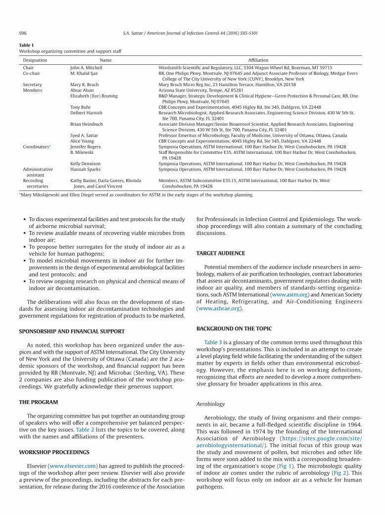
Indoor air as a vehicle for human pathogens: Introduction, objectives,and expectation of outcome
Syed A. Sattar MSc, Dip Bact, MS, PhD *Professor Emeritus of Microbiology, Faculty of Medicine, University of Ottawa, Ottawa, ON, Canada
Key Words:Aerobiologyindoor airairborne pathogensair decontaminationairborne pollutants
Airborne spread of pathogens can be rapid, widespread, and difficult to prevent. In this international work-shop, a panel of 6 experts will expound on the following: (1) the potential for indoor air to spread a widerange of human pathogens, plus engineering controls to reduce the risk for exposure to airborne infec-tious agents; (2) the behavior of aerosolized infectious agents indoors and the use of emerging airdecontamination technologies; (3) a survey of quantitative methods to recover infectious agents and theirsurrogates from indoor air with regard to survival and inactivation of airborne pathogens; (4) mathe-matical models to predict the movement of pathogens indoors and the use of such information to optimizethe benefits of air decontamination technologies; and (5) synergy between different infectious agents,such as legionellae and fungi, in the built environment predisposing to possible transmission-related healthimpacts of aerosolized biofilm-based opportunistic pathogens. After the presentations, the panel will addressa set of preformulated questions on selection criteria for surrogate microbes to study the survival andinactivation of airborne human pathogens, desirable features of technologies for microbial decontami-nation of indoor air, knowledge gaps, and research needs. It is anticipated that the deliberations of theworkshop will provide the attendees with an update on the significance of indoor air as a vehicle for trans-mitting human pathogens with a brief on what is currently being done to mitigate the risks from airborneinfectious agents.© 2016 Association for Professionals in Infection Control and Epidemiology, Inc. Published by Elsevier
Inc. All rights reserved.
“Clean air is a basic requirement of life. The quality of air insidehomes, offices, schools, day care centres, public buildings, healthcare facilities or other private and public buildings where peoplespend a large part of their life is an essential determinant ofhealthy life and people’s well-being. . .” –World Health Organi-zation, 2010
I welcome you all to this multinational workshop! This work-shop was conceived over a year ago, and the organizing committee(Table 1) formally requested that ASTM International (www.astm.org/)hold the event under its auspices. ASTM’s Committee E35, which deals
with pesticides, antimicrobials, and alternative control agents, ap-proved the proposal in April 2015.
Mounting recognition of indoor air as a vehicle for infectiousagents is leading government regulators, such as the U.S. Environ-mental Protection Agency, to refine and update their guidelines,1
researchers to develop bettermeans of studying airborne pathogens,2
and civil engineers and architects to find innovativemeans of makingindoor air safer while keeping energy conservation in mind.3
Although comprehensive guidelines and standardized means areavailable to study chemical pollutants in indoor air,4 there remainsa general lack of suitable experimental facilities and standardized pro-tocols to quantitatively assess the survival of pathogens in indoor airand to document their removal and inactivation by physical and chem-ical means. This workshop will address these issues, among others.
SPECIFIC OBJECTIVES
The workshop’s specific objectives, therefore, are as follows:
• To provide a forum for the exchange of ideas on current re-search on the role of the indoor environment in general andindoor air in particular in the spread of human pathogens;
* Address correspondence to Syed A. Sattar MSc, Dip Bact, MS, PhD, ProfessorEmeritus of Microbiology, Faculty of Medicine, University of Ottawa, 451 Smyth Rd,Ottawa, ON, K1H 8M5, Canada.
E-mail address: [email protected] of this supplement is primarily supported by RB,Montvale, New Jersey,
with additional support fromMicroBioTest, a division of Microbac Laboratories, Inc.,Sterling, Virginia. The City University of New York (CUNY) and the University of Ottawa,Ottawa, Canada, are academic sponsors. Editorial support was provided by AshleyO’Dunne, PhD; Shannon O’Sullivan, ELS; and Alanna Franchetti, ELS of Medergy(Yardley, PA), and funded by RB.
Conflicts of Interest: None to report.
0196-6553/© 2016 Association for Professionals in Infection Control and Epidemiology, Inc. Published by Elsevier Inc. All rights reserved.http://dx.doi.org/10.1016/j.ajic.2016.06.010
American Journal of Infection Control 44 (2016) S95-S101
Contents lists available at ScienceDirect
American Journal of Infection Control
journal homepage: www.aj ic journal .org
American Journal of Infection Control
• To discuss experimental facilities and test protocols for the studyof airborne microbial survival;
• To review available means of recovering viable microbes fromindoor air;
• To propose better surrogates for the study of indoor air as avehicle for human pathogens;
• To model microbial movements in indoor air for further im-provements in the design of experimental aerobiological facilitiesand test protocols; and
• To review ongoing research on physical and chemical means ofindoor air decontamination.
The deliberations will also focus on the development of stan-dards for assessing indoor air decontamination technologies andgovernment regulations for registration of products to be marketed.
SPONSORSHIP AND FINANCIAL SUPPORT
As noted, this workshop has been organized under the aus-pices andwith the support of ASTM International. The City Universityof New York and the University of Ottawa (Canada) are the 2 aca-demic sponsors of the workshop, and financial support has beenprovided by RB (Montvale, NJ) and Microbac (Sterling, VA). These2 companies are also funding publication of the workshop pro-ceedings. We gratefully acknowledge their generous support.
THE PROGRAM
The organizing committee has put together an outstanding groupof speakers who will offer a comprehensive yet balanced perspec-tive on the key issues. Table 2 lists the topics to be covered, alongwith the names and affiliations of the presenters.
WORKSHOP PROCEEDINGS
Elsevier (www.elsevier.com) has agreed to publish the proceed-ings of the workshop after peer review. Elsevier will also providea preview of the proceedings, including the abstracts for each pre-sentation, for release during the 2016 conference of the Association
for Professionals in Infection Control and Epidemiology. The work-shop proceedings will also contain a summary of the concludingdiscussions.
TARGET AUDIENCE
Potential members of the audience include researchers in aero-biology, makers of air purification technologies, contract laboratoriesthat assess air decontaminants, government regulators dealing withindoor air quality, and members of standards-setting organiza-tions, such ASTM International (www.astm.org) and American Societyof Heating, Refrigerating, and Air-Conditioning Engineers(www.ashrae.org).
BACKGROUND ON THE TOPIC
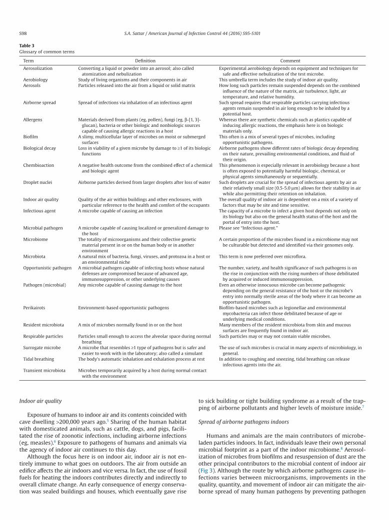
Table 3 is a glossary of the common terms used throughout thisworkshop’s presentations. This is included in an attempt to createa level playing fieldwhile facilitating the understanding of the subjectmatter by experts in fields other than environmental microbiol-ogy. However, the emphasis here is on working definitions,recognizing that efforts are needed to develop a more comprehen-sive glossary for broader applications in this area.
Aerobiology
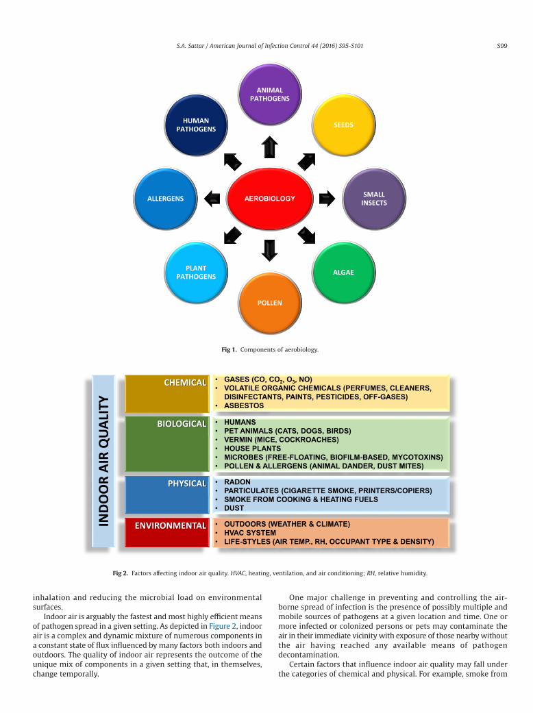
Aerobiology, the study of living organisms and their compo-nents in air, became a full-fledged scientific discipline in 1964.This was followed in 1974 by the founding of the InternationalAssociation of Aerobiology (https://sites.google.com/site/aerobiologyinternational/). The initial focus of this group wasthe study and movement of pollen, but microbes and other lifeforms were soon added to the mix with a corresponding broaden-ing of the organization’s scope (Fig 1). The microbiologic qualityof indoor air comes under the rubric of aerobiology (Fig 2). Thisworkshop will focus only on indoor air as a vehicle for humanpathogens.
Table 1Workshop organizing committee and support staff
Designation Name Affiliation
Chair John A. Mitchell Wordsmith Scientific and Regulatory, LLC, 3304 Wagon Wheel Rd, Bozeman, MT 59715Co-chair M. Khalid Ijaz RB, One Philips Pkwy, Montvale, NJ 07645 and Adjunct Associate Professor of Biology, Medgar Evers
College of The City University of New York (CUNY), Brooklyn, New YorkSecretary Mary K. Bruch Mary Bruch Micro Reg Inc, 23 Hamilton Terrace, Hamilton, VA 20158Members Absar Alum Arizona State University, Tempe, AZ 85281
Elizabeth (Ilze) Bruning R&D Manager, Strategic Development & Clinical Hygiene—Germ Protection & Personal Care, RB, OnePhilips Pkwy, Montvale, NJ 07645
Tony Buhr CBR Concepts and Experimentation, 4045 Higley Rd, Ste 345, Dahlgren, VA 22448Delbert Harnish Research Microbiologist, Applied Research Associates, Engineering Science Division, 430 W 5th St,
Ste 700, Panama City, FL 32401Brian Heimbuch Associate Division Manager/Senior Bioaerosol Scientist, Applied Research Associates, Engineering
Science Division, 430 W 5th St, Ste 700, Panama City, FL 32401Syed A. Sattar Professor Emeritus of Microbiology, Faculty of Medicine, University of Ottawa, Ottawa, CanadaAlice Young CBR Concepts and Experimentation, 4045 Higley Rd, Ste 345, Dahlgren, VA 22448
Coordinators* Jennifer Rogers Symposia Operations, ASTM International, 100 Barr Harbor Dr, West Conshohocken, PA 19428B. Milewski Staff Responsible for Committee E35, ASTM International, 100 Barr Harbor Dr, West Conshohocken,
PA 19428Kelly Dennison Symposia Operations, ASTM International, 100 Barr Harbor Dr, West Conshohocken, PA 19428
Administrativeassistant
Hannah Sparks Symposia Operations, ASTM International, 100 Barr Harbor Dr, West Conshohocken, PA 19428
Recordingsecretaries
Kathy Baxter, Darla Goeres, RhondaJones, and Carol Vincent
Members, ASTM Subcommittee E35.15, ASTM International, 100 Barr Harbor Dr, WestConshohocken, PA 19428
*Mary Mikolajewski and Ellen Diegel served as coordinators for ASTM in the early stages of the workshop planning.
S96 S.A. Sattar / American Journal of Infection Control 44 (2016) S95-S101
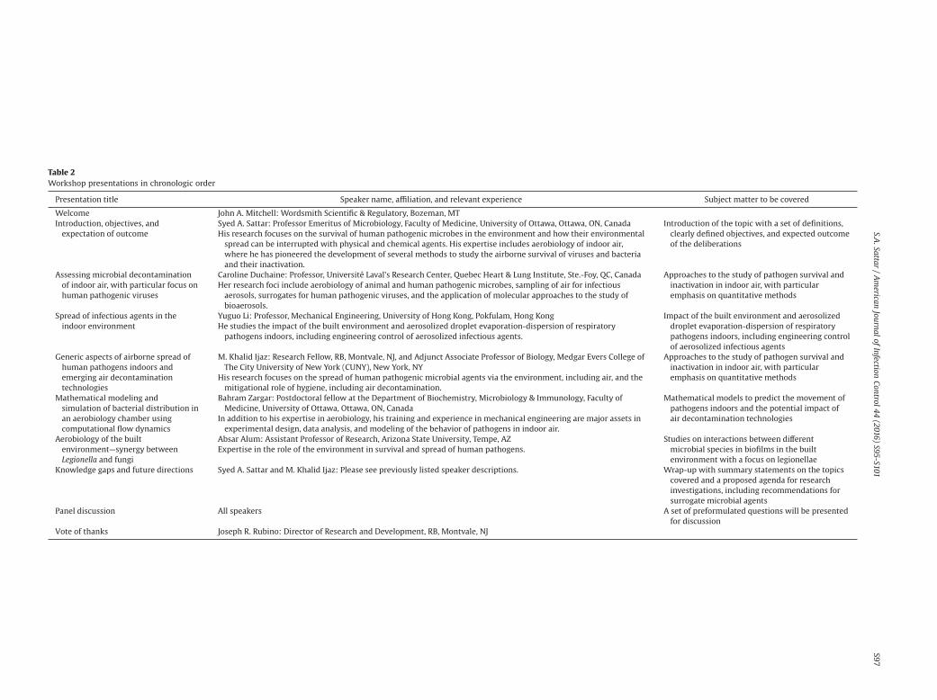
Table 2Workshop presentations in chronologic order
Presentation title Speaker name, affiliation, and relevant experience Subject matter to be covered
Welcome John A. Mitchell: Wordsmith Scientific & Regulatory, Bozeman, MTIntroduction, objectives, andexpectation of outcome
Syed A. Sattar: Professor Emeritus of Microbiology, Faculty of Medicine, University of Ottawa, Ottawa, ON, CanadaHis research focuses on the survival of human pathogenic microbes in the environment and how their environmentalspread can be interrupted with physical and chemical agents. His expertise includes aerobiology of indoor air,where he has pioneered the development of several methods to study the airborne survival of viruses and bacteriaand their inactivation.
Introduction of the topic with a set of definitions,clearly defined objectives, and expected outcomeof the deliberations
Assessing microbial decontaminationof indoor air, with particular focus onhuman pathogenic viruses
Caroline Duchaine: Professor, Université Laval’s Research Center, Quebec Heart & Lung Institute, Ste.-Foy, QC, CanadaHer research foci include aerobiology of animal and human pathogenic microbes, sampling of air for infectiousaerosols, surrogates for human pathogenic viruses, and the application of molecular approaches to the study ofbioaerosols.
Approaches to the study of pathogen survival andinactivation in indoor air, with particularemphasis on quantitative methods
Spread of infectious agents in theindoor environment
Yuguo Li: Professor, Mechanical Engineering, University of Hong Kong, Pokfulam, Hong KongHe studies the impact of the built environment and aerosolized droplet evaporation-dispersion of respiratorypathogens indoors, including engineering control of aerosolized infectious agents.
Impact of the built environment and aerosolizeddroplet evaporation-dispersion of respiratorypathogens indoors, including engineering controlof aerosolized infectious agents
Generic aspects of airborne spread ofhuman pathogens indoors andemerging air decontaminationtechnologies
M. Khalid Ijaz: Research Fellow, RB, Montvale, NJ, and Adjunct Associate Professor of Biology, Medgar Evers College ofThe City University of New York (CUNY), New York, NY
His research focuses on the spread of human pathogenic microbial agents via the environment, including air, and themitigational role of hygiene, including air decontamination.
Approaches to the study of pathogen survival andinactivation in indoor air, with particularemphasis on quantitative methods
Mathematical modeling andsimulation of bacterial distribution inan aerobiology chamber usingcomputational flow dynamics
Bahram Zargar: Postdoctoral fellow at the Department of Biochemistry, Microbiology & Immunology, Faculty ofMedicine, University of Ottawa, Ottawa, ON, Canada
In addition to his expertise in aerobiology, his training and experience in mechanical engineering are major assets inexperimental design, data analysis, and modeling of the behavior of pathogens in indoor air.
Mathematical models to predict the movement ofpathogens indoors and the potential impact ofair decontamination technologies
Aerobiology of the builtenvironment—synergy betweenLegionella and fungi
Absar Alum: Assistant Professor of Research, Arizona State University, Tempe, AZExpertise in the role of the environment in survival and spread of human pathogens.
Studies on interactions between differentmicrobial species in biofilms in the builtenvironment with a focus on legionellae
Knowledge gaps and future directions Syed A. Sattar and M. Khalid Ijaz: Please see previously listed speaker descriptions. Wrap-up with summary statements on the topicscovered and a proposed agenda for researchinvestigations, including recommendations forsurrogate microbial agents
Panel discussion All speakers A set of preformulated questions will be presentedfor discussion
Vote of thanks Joseph R. Rubino: Director of Research and Development, RB, Montvale, NJ
S97S.A
.Sattar/Am
ericanJournalofInfection
Control44(2016)
S95-S101
Indoor air quality
Exposure of humans to indoor air and its contents coincided withcave dwelling >200,000 years ago.5 Sharing of the human habitatwith domesticated animals, such as cattle, dogs, and pigs, facili-tated the rise of zoonotic infections, including airborne infections(eg, measles).6 Exposure to pathogens of humans and animals viathe agency of indoor air continues to this day.
Although the focus here is on indoor air, indoor air is not en-tirely immune to what goes on outdoors. The air from outside anedifice affects the air indoors and vice versa. In fact, the use of fossilfuels for heating the indoors contributes directly and indirectly tooverall climate change. An early consequence of energy conserva-tion was sealed buildings and houses, which eventually gave rise
to sick building or tight building syndrome as a result of the trap-ping of airborne pollutants and higher levels of moisture inside.7
Spread of airborne pathogens indoors
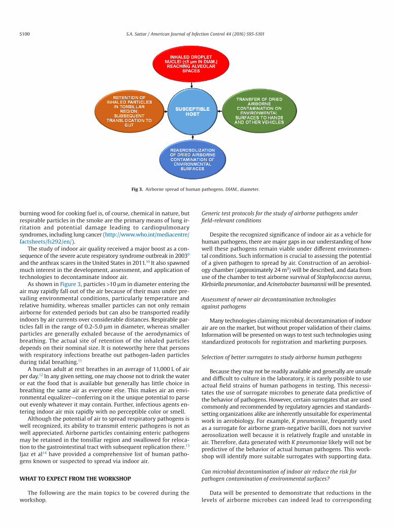
Humans and animals are the main contributors of microbe-laden particles indoors. In fact, individuals leave their own personalmicrobial footprint as a part of the indoor microbiome.8 Aerosol-ization of microbes from biofilms and resuspension of dust are theother principal contributors to the microbial content of indoor air(Fig 3). Although the route by which airborne pathogens cause in-fections varies between microorganisms, improvements in thequality, quantity, and movement of indoor air can mitigate the air-borne spread of many human pathogens by preventing pathogen
Table 3Glossary of common terms
Term Definition Comment
Aerosolization Converting a liquid or powder into an aerosol; also calledatomization and nebulization
Experimental aerobiology depends on equipment and techniques forsafe and effective nebulization of the test microbe.
Aerobiology Study of living organisms and their components in air This umbrella term includes the study of indoor air quality.Aerosols Particles released into the air from a liquid or solid matrix How long such particles remain suspended depends on the combined
influence of the nature of the matrix, air turbulence, light, airtemperature, and relative humidity.
Airborne spread Spread of infections via inhalation of an infectious agent Such spread requires that respirable particles carrying infectiousagents remain suspended in air long enough to be inhaled by apotential host.
Allergens Materials derived from plants (eg, pollen), fungi (eg, β-[1, 3]-glucan), bacteria or other biologic and nonbiologic sourcescapable of causing allergic reactions in a host
Whereas there are synthetic chemicals such as plastics capable ofinducing allergic reactions, the emphasis here is on biologicmaterials only.
Biofilm A slimy, multicellular layer of microbes on moist or submergedsurfaces
This often is a mix of several types of microbes, includingopportunistic pathogens.
Biological decay Loss in viability of a given microbe by damage to ≥1 of its biologicfunctions
Airborne pathogens show different rates of biologic decay dependingon their nature, prevailing environmental conditions, and fluid oftheir origin.
Chembioaction A negative health outcome from the combined effect of a chemicaland biologic agent
This phenomenon is especially relevant in aerobiology because a hostis often exposed to potentially harmful biologic, chemical, orphysical agents simultaneously or sequentially.
Droplet nuclei Airborne particles derived from larger droplets after loss of water Such droplets are crucial for the spread of infectious agents by air astheir relatively small size (0.5-5.0 μm) allows for their stability in airwhile also permitting their retention on inhalation.
Indoor air quality Quality of the air within buildings and other enclosures, withparticular reference to the health and comfort of the occupants
The overall quality of indoor air is dependent on a mix of a variety offactors that may be site and time sensitive.
Infectious agent A microbe capable of causing an infection The capacity of a microbe to infect a given host depends not only onits biology but also on the general health status of the host and theportal of entry into the host.
Microbial pathogen A microbe capable of causing localized or generalized damage tothe host
Please see “Infectious agent.”
Microbiome The totality of microorganisms and their collective geneticmaterial present in or on the human body or in anotherenvironment
A certain proportion of the microbes found in a microbiome may notbe culturable but detected and identified via their genomes only.
Microbiota A natural mix of bacteria, fungi, viruses, and protozoa in a host oran environmental niche
This term is now preferred over microflora.
Opportunistic pathogen A microbial pathogen capable of infecting hosts whose naturaldefenses are compromised because of advanced age,immunosuppression, or other underlying causes
The number, variety, and health significance of such pathogens is onthe rise in conjunction with the rising numbers of those debilitatedby acquired or induced immunosuppression.
Pathogen (microbial) Any microbe capable of causing damage to the host Even an otherwise innocuous microbe can become pathogenicdepending on the general resistance of the host or the microbe’sentry into normally sterile areas of the body where it can become anopportunistic pathogen.
Perikairots Environment-based opportunistic pathogens Biofilm-based microbes such as legionellae and environmentalmycobacteria can infect those debilitated because of age orunderlying medical conditions.
Resident microbiota A mix of microbes normally found in or on the host Many members of the resident microbiota from skin and mucoussurfaces are frequently found in indoor air.
Respirable particles Particles small enough to access the alveolar space during normalbreathing
Such particles may or may not contain viable microbes.
Surrogate microbe A microbe that resembles ≥1 type of pathogens but is safer andeasier to work with in the laboratory; also called a simulant
The use of such microbes is crucial in many aspects of microbiology, ingeneral.
Tidal breathing The body’s automatic inhalation and exhalation process at rest In addition to coughing and sneezing, tidal breathing can releaseinfectious agents into the air.
Transient microbiota Microbes temporarily acquired by a host during normal contactwith the environment
S98 S.A. Sattar / American Journal of Infection Control 44 (2016) S95-S101
inhalation and reducing the microbial load on environmentalsurfaces.
Indoor air is arguably the fastest andmost highly efficient meansof pathogen spread in a given setting. As depicted in Figure 2, indoorair is a complex and dynamic mixture of numerous components ina constant state of flux influenced by many factors both indoors andoutdoors. The quality of indoor air represents the outcome of theunique mix of components in a given setting that, in themselves,change temporally.
One major challenge in preventing and controlling the air-borne spread of infection is the presence of possibly multiple andmobile sources of pathogens at a given location and time. One ormore infected or colonized persons or pets may contaminate theair in their immediate vicinity with exposure of those nearbywithoutthe air having reached any available means of pathogendecontamination.
Certain factors that influence indoor air quality may fall underthe categories of chemical and physical. For example, smoke from
AEROBIOLOGY
ANIMAL PATHOGENS
SEEDS
SMALL INSECTS
ALGAE
POLLEN
PLANT PATHOGENS
ALLERGENS
HUMAN PATHOGENS
Fig 1. Components of aerobiology.
IND
OO
R A
IR Q
UA
LITY
CHEMICAL
BIOLOGICAL
PHYSICAL
ENVIRONMENTAL
• GASES (CO, CO2, O3, NO)• VOLATILE ORGANIC CHEMICALS (PERFUMES, CLEANERS,
DISINFECTANTS, PAINTS, PESTICIDES, OFF-GASES)• ASBESTOS
• HUMANS• PET ANIMALS (CATS, DOGS, BIRDS)• VERMIN (MICE, COCKROACHES)• HOUSE PLANTS• MICROBES (FREE-FLOATING, BIOFILM-BASED, MYCOTOXINS)• POLLEN & ALLERGENS (ANIMAL DANDER, DUST MITES)
• RADON• PARTICULATES (CIGARETTE SMOKE, PRINTERS/COPIERS)• SMOKE FROM COOKING & HEATING FUELS• DUST
• OUTDOORS (WEATHER & CLIMATE)• HVAC SYSTEM• LIFE-STYLES (AIR TEMP., RH, OCCUPANT TYPE & DENSITY)
Fig 2. Factors affecting indoor air quality. HVAC, heating, ventilation, and air conditioning; RH, relative humidity.
S99S.A. Sattar / American Journal of Infection Control 44 (2016) S95-S101
burning wood for cooking fuel is, of course, chemical in nature, butrespirable particles in the smoke are the primary means of lung ir-ritation and potential damage leading to cardiopulmonarysyndromes, including lung cancer (http://www.who.int/mediacentre/factsheets/fs292/en/).
The study of indoor air quality received a major boost as a con-sequence of the severe acute respiratory syndrome outbreak in 20039
and the anthrax scares in the United States in 2011.10 It also spawnedmuch interest in the development, assessment, and application oftechnologies to decontaminate indoor air.
As shown in Figure 3, particles >10 μm in diameter entering theair may rapidly fall out of the air because of their mass under pre-vailing environmental conditions, particularly temperature andrelative humidity, whereas smaller particles can not only remainairborne for extended periods but can also be transported readilyindoors by air currents over considerable distances. Respirable par-ticles fall in the range of 0.2-5.0 μm in diameter, whereas smallerparticles are generally exhaled because of the aerodynamics ofbreathing. The actual site of retention of the inhaled particlesdepends on their nominal size. It is noteworthy here that personswith respiratory infections breathe out pathogen-laden particlesduring tidal breathing.11
A human adult at rest breathes in an average of 11,000 L of airper day.12 In any given setting, onemay choose not to drink the wateror eat the food that is available but generally has little choice inbreathing the same air as everyone else. This makes air an envi-ronmental equalizer—conferring on it the unique potential to parseout evenly whatever it may contain. Further, infectious agents en-tering indoor air mix rapidly with no perceptible color or smell.
Although the potential of air to spread respiratory pathogens iswell recognized, its ability to transmit enteric pathogens is not aswell appreciated. Airborne particles containing enteric pathogensmay be retained in the tonsillar region and swallowed for reloca-tion to the gastrointestinal tract with subsequent replication there.13
Ijaz et al14 have provided a comprehensive list of human patho-gens known or suspected to spread via indoor air.
WHAT TO EXPECT FROM THE WORKSHOP
The following are the main topics to be covered during theworkshop.
Generic test protocols for the study of airborne pathogens underfield-relevant conditions
Despite the recognized significance of indoor air as a vehicle forhuman pathogens, there aremajor gaps in our understanding of howwell these pathogens remain viable under different environmen-tal conditions. Such information is crucial to assessing the potentialof a given pathogen to spread by air. Construction of an aerobiol-ogy chamber (approximately 24m3) will be described, and data fromuse of the chamber to test airborne survival of Staphylococcus aureus,Klebsiella pneumoniae, and Acinetobacter baumanniiwill be presented.
Assessment of newer air decontamination technologiesagainst pathogens
Many technologies claimingmicrobial decontamination of indoorair are on the market, but without proper validation of their claims.Informationwill be presented onways to test such technologies usingstandardized protocols for registration and marketing purposes.
Selection of better surrogates to study airborne human pathogens
Because theymay not be readily available and generally are unsafeand difficult to culture in the laboratory, it is rarely possible to useactual field strains of human pathogens in testing. This necessi-tates the use of surrogate microbes to generate data predictive ofthe behavior of pathogens. However, certain surrogates that are usedcommonly and recommended by regulatory agencies and standards-setting organizations alike are inherently unsuitable for experimentalwork in aerobiology. For example, K pneumoniae, frequently usedas a surrogate for airborne gram-negative bacilli, does not surviveaerosolization well because it is relatively fragile and unstable inair. Therefore, data generated with K pneumoniae likely will not bepredictive of the behavior of actual human pathogens. This work-shop will identify more suitable surrogates with supporting data.
Can microbial decontamination of indoor air reduce the risk forpathogen contamination of environmental surfaces?
Data will be presented to demonstrate that reductions in thelevels of airborne microbes can indeed lead to corresponding
Fig 3. Airborne spread of human pathogens. DIAM., diameter.
S100 S.A. Sattar / American Journal of Infection Control 44 (2016) S95-S101
reductions in the microbial contamination of environmental sur-faces in a given setting. The use of integrated models could helpanalyze outbreaks, evaluate the relative importance of hygiene andinfection prevention and control for policymakers, and provide guid-ance in environmental design for greater occupant safety andcomfort. These will be illustrated using some recent examples, in-cluding severe acute respiratory syndrome, influenza, and MiddleEast respiratory syndrome.
Update on the recovery and quantitation of viable microbes inindoor air
Quantitative recovery of viable microbes from air is vital inaerobiologic studies. An update will be given on available methods,including their strengths and limitations.
Mathematical modeling to help better design experimental chambersfor work in aerobiology
Experimentation with airborne microbes is generally quite laborintensive and costly and requires special biosafety precautions. Math-ematical models can assist greatly in optimizing aerobiology chamberdesign and in predicting the influence of furniture and other objectson the movement of microbes. Data will be presented with specif-ic reference to a chamber that fully conforms to guidance from theU.S. Environmental Protection Agency.
Significance of biofilms as sources of pathogens in thebuilt environment
Biofilms are not only common in the built environment, but theycan be common sources of airborne pathogens. Such biofilms oftencontain several microbial species having complex interactions
between them. This will be illustratedwith the example of how fungiand legionellae coexist with potential risks to human health.
References
1. U.S. Environmental Protection Agency. Product performance test guidelines:OCSPP 810.2500: air sanitizers—efficacy data recommendations. 2012. Availablefrom: http://www.regulations.gov/#!documentDetail;D=EPA-HQ-OPPT-2009-0150-0025. Accessed May 2, 2016.
2. Sattar SA, Kibbee RJ, Zargar B, Wright KE, Rubino RJ, Ijaz MK. Decontaminationof indoor air to reduce the risk of airborne infections: studies on survival andinactivation of airborne pathogens using an aerobiology chamber. Am J InfectControl 2016; Jun 30. [Epub ahead of print].
3. Wei J, Li Y. Airborne spread of infectious agents in the indoor environment. AmJ Infect Control 2016. doi:10.1016/j.ajic.2016.06.003.
4. World Health Organization. WHO guidelines for indoor air quality: selectedpollutants. 2010. Available from: http://www.who.int/indoorair/publications/9789289002134/en/. Accessed May 2, 2016.
5. Wolfe ND, Dunavan CP, Diamond J. Origins of major human infectious diseases.Nature 2007;447:279-83.
6. Morand S, McIntyre KM, Baylis M. Domesticated animals and human infectiousdiseases of zoonotic origins: domestication time matters. Infect Genet Evol2014;24:76-81.
7. Sundell J, Levin H, Nazaroff WW, CainWS, FiskWJ, Grimsrud DT, et al. Ventilationrates and health: multidisciplinary review of the scientific literature. Indoor Air2011;21:191-204.
8. Meadow JF, Altrichter AE, Bateman AC. Humans differ in their personal microbialcloud. PeerJ 2015;3:1258.
9. Wong G, Liu W, Liu Y, Zhou B, Bi Y, Gao GF. MERS, SARS, and Ebola: the role ofsuper-spreaders in infectious disease. Cell Host Microbe 2015;18:398-401.
10. Weis CP, Intrepido AJ, Miller AK, Cowin PG, Durno MA, Gebhardt JS, et al.Secondary aerosolization of viable Bacillus anthracis spores in a contaminatedUS Senate Office. JAMA 2002;288:2853-8.
11. Fabian P, McDevitt JJ, DeHaanWH, Fung RO, Cowling BJ, Chan KH, et al. Influenzavirus in human exhaled breath: an observational study. PLoS ONE 2008;3:e2691.
12. California Environmental Protection Agency. Howmuch air do we breathe? 1994.Available from: http://www.arb.ca.gov/research/resnotes/notes/94-11.htm.Accessed June 6, 2016.
13. Alonso C, Raynor PC, Davies PR, Torremorell M. Concentration, size distribution,and infectivity of airborne particles carrying swine viruses. PLoS ONE2015;10:e0135675.
14. Ijaz MK, Zargar B, Wright KE, Rubino JR, Sattar SA. Generic aspects of airbornespread of human pathogens indoors and emerging air-decontaminationtechnologies. Am J Infect Control 2016. doi:10.1016/j.ajic.2016.06.008.
S101S.A. Sattar / American Journal of Infection Control 44 (2016) S95-S101
Global Perspective Article
Airborne spread of infectious agents in the indoor environment
Jianjian Wei PhD, Yuguo Li PhD *Department of Mechanical Engineering, The University of Hong Kong, Hong Kong
Key Words:Respiratory dropletindoor aircoughingdroplet dispersioninfection controlenvironmental ventilation
Background: Since the 2003 severe acute respiratory syndrome epidemic, scientific exploration of in-fection control is no longer restricted to microbiologists or medical scientists. Many studies have reportedon the release, transport, and exposure of expiratory droplets because of respiratory activities. This reviewfocuses on the airborne spread of infectious agents from mucus to mucus in the indoor environment andtheir spread as governed by airflows in the respiratory system, around people, and in buildings at dif-ferent transport stages.Methods: We critically review the literature on the release of respiratory droplets, their transport and dis-persion in the indoor environment, and the ultimate exposure of a susceptible host, as influencedby airflows.Results: These droplets or droplet nuclei are transported by expired airflows, which are sometimes af-fected by the human body plume and use of a face mask, as well as room airflow. Room airflow is affectedby human activities such as walking and door opening, and some droplets are eventually captured by asusceptible individual because of his or her inspired flows; such exposure can eventually lead to long-range spread of airborne pathogens. Direct exposure to the expired fine droplets or droplet nuclei resultsin short-range airborne transmission. Deposition of droplets and direct personal exposure to expired largedroplets can lead to the fomite route and the droplet-borne route, respectively.Conclusions: We have shown the opportunities for infection control at different stages of the spread. Wepropose that the short-range airborne route may be important in close contact, and its control may beachieved by face masks for the source patients and use of personalized ventilation. Our discussion of theeffect of thermal stratification and expiratory delivery of droplets leads to the suggestion that displace-ment ventilation may not be applicable to hospital rooms where respiratory infection is a concern.© 2016 Association for Professionals in Infection Control and Epidemiology, Inc. Published by Elsevier
Inc. All rights reserved.
Since the 2003 severe acute respiratory syndrome epidemic, the2009 H1N1 influenza pandemic, and the 2014 Middle East respi-ratory syndrome epidemic, scientific exploration of infection controlis no longer restricted to microbiologists or medical scientists. Fluidmechanics has played a role in understanding the mechanism oftransmission and in developing engineering interventions; forexample, the studies of airflow dynamics by Yu et al1 provided
plausible evidence of airborne transmission of severe acute respi-ratory syndrome. Airborne spread of infectious agents is directlyrelevant to the airborne route, and indirectly to the droplet-borneand fomite routes. Breathing, talking, sneezing, and coughing aremajor sources of some respiratory pathogens. Up to 40,000 drop-lets are expelled at a velocity of 100 m/s during a sneeze,2 and acough can generate approximately 3,000 droplet nuclei.3 We nowunderstand to some degree where and how respiratory droplets areformed and the pathogen content in each size of droplet. Turbu-lence and coherent structures in the airflow, mostly invisible,transport respiratory droplets between people. For example, vortexstructures in coughing probably carry particles over long distances.4
Our body’s thermal plumes can bring fine droplet nuclei upward,and vortices generated during door opening and wakes behindwalking individuals can transport contaminated air out of an iso-lation room. Turbulence generated by supply air jets causes mixingand dilution of room air. Understanding these airflows is crucial tominimizing spread of infectious agents and infection transmission.
Here, we review the release of respiratory droplets, their trans-port and dispersion in the indoor environment, and the ultimate
* Address correspondence to Yuguo Li, PhD, Department of Mechanical Engineering,The University of Hong Kong, Pokfulam Rd, Hong Kong.
E-mail address: [email protected] (Y. Li).Funding/Support: This research was funded by the Research Grants Council
General Research Fund (HKU7142/12) and the Natural Science Foundation of China(51278440). Publication of this supplement is primarily supported by RB, Montvale,New Jersey, with additional support fromMicroBioTest, a division of Microbac Labo-ratories, Inc., Sterling, Virginia. The City University of New York (CUNY) and theUniversity of Ottawa, Ottawa, Canada, are academic sponsors. Editorial support wasprovided by Ashley O’Dunne, PhD; Shannon O’Sullivan, ELS; and Alanna Franchetti,ELS of Medergy (Yardley, PA), and funded by RB.
Conflicts of Interest: None to report.
0196-6553/© 2016 Association for Professionals in Infection Control and Epidemiology, Inc. Published by Elsevier Inc. All rights reserved.http://dx.doi.org/10.1016/j.ajic.2016.06.003
American Journal of Infection Control 44 (2016) S102-S108
Contents lists available at ScienceDirect
American Journal of Infection Control
journal homepage: www.aj ic journal .org
American Journal of Infection Control
exposure of a susceptible host, as influenced by airflows. Microbi-al survival in the environment is beyond the scope of this article.
RELEASE, TRANSPORT, AND EXPOSURE
Release of droplets from mucus to mouth
If we understand the mechanism of where and how respirato-ry droplets are generated, we may have opportunities to suppressthem at the source. Knowing the number and size of respiratorydroplets is also crucial.
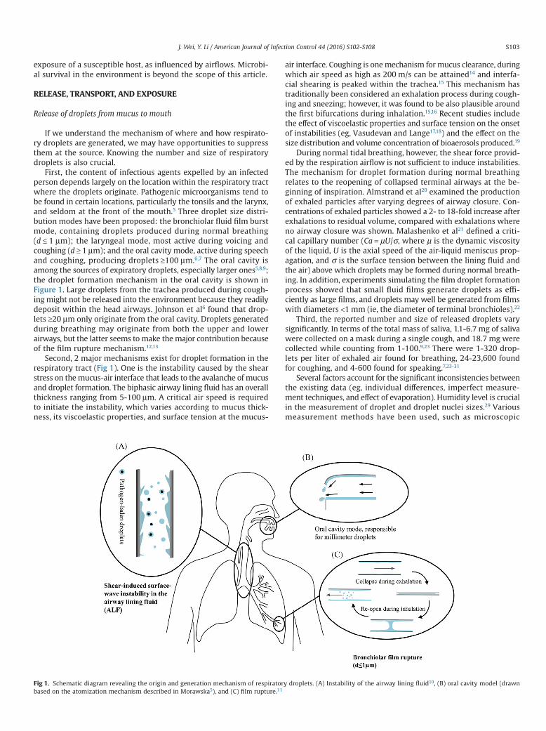
First, the content of infectious agents expelled by an infectedperson depends largely on the location within the respiratory tractwhere the droplets originate. Pathogenic microorganisms tend tobe found in certain locations, particularly the tonsils and the larynx,and seldom at the front of the mouth.5 Three droplet size distri-bution modes have been proposed: the bronchiolar fluid film burstmode, containing droplets produced during normal breathing(d ≤ 1 μm); the laryngeal mode, most active during voicing andcoughing (d ≥ 1 μm); and the oral cavity mode, active during speechand coughing, producing droplets ≥100 μm.6,7 The oral cavity isamong the sources of expiratory droplets, especially larger ones5,8,9;the droplet formation mechanism in the oral cavity is shown inFigure 1. Large droplets from the trachea produced during cough-ing might not be released into the environment because they readilydeposit within the head airways. Johnson et al6 found that drop-lets ≥20 μm only originate from the oral cavity. Droplets generatedduring breathing may originate from both the upper and lowerairways, but the latter seems tomake themajor contribution becauseof the film rupture mechanism.12,13
Second, 2 major mechanisms exist for droplet formation in therespiratory tract (Fig 1). One is the instability caused by the shearstress on themucus-air interface that leads to the avalanche of mucusand droplet formation. The biphasic airway lining fluid has an overallthickness ranging from 5-100 μm. A critical air speed is requiredto initiate the instability, which varies according to mucus thick-ness, its viscoelastic properties, and surface tension at the mucus-
air interface. Coughing is onemechanism formucus clearance, duringwhich air speed as high as 200 m/s can be attained14 and interfa-cial shearing is peaked within the trachea.15 This mechanism hastraditionally been considered an exhalation process during cough-ing and sneezing; however, it was found to be also plausible aroundthe first bifurcations during inhalation.15,16 Recent studies includethe effect of viscoelastic properties and surface tension on the onsetof instabilities (eg, Vasudevan and Lange17,18) and the effect on thesize distribution and volume concentration of bioaerosols produced.19
During normal tidal breathing, however, the shear force provid-ed by the respiration airflow is not sufficient to induce instabilities.The mechanism for droplet formation during normal breathingrelates to the reopening of collapsed terminal airways at the be-ginning of inspiration. Almstrand et al20 examined the productionof exhaled particles after varying degrees of airway closure. Con-centrations of exhaled particles showed a 2- to 18-fold increase afterexhalations to residual volume, compared with exhalations whereno airway closure was shown. Malashenko et al21 defined a criti-cal capillary number (Ca = μU/σ, where μ is the dynamic viscosityof the liquid, U is the axial speed of the air-liquid meniscus prop-agation, and σ is the surface tension between the lining fluid andthe air) above which droplets may be formed during normal breath-ing. In addition, experiments simulating the film droplet formationprocess showed that small fluid films generate droplets as effi-ciently as large films, and droplets may well be generated from filmswith diameters <1 mm (ie, the diameter of terminal bronchioles).22
Third, the reported number and size of released droplets varysignificantly. In terms of the total mass of saliva, 1.1-6.7 mg of salivawere collected on a mask during a single cough, and 18.7 mg werecollected while counting from 1-100.9,23 There were 1-320 drop-lets per liter of exhaled air found for breathing, 24-23,600 foundfor coughing, and 4-600 found for speaking.7,23-31
Several factors account for the significant inconsistencies betweenthe existing data (eg, individual differences, imperfect measure-ment techniques, and effect of evaporation). Humidity level is crucialin the measurement of droplet and droplet nuclei sizes.29 Variousmeasurement methods have been used, such as microscopic
Fig 1. Schematic diagram revealing the origin and generation mechanism of respiratory droplets. (A) Instability of the airway lining fluid10, (B) oral cavity model (drawnbased on the atomization mechanism described in Morawska5), and (C) film rupture.11
S103J. Wei, Y. Li / American Journal of Infection Control 44 (2016) S102-S108
observations,8 optical particle counting,25 aerodynamic particle count-ing and scanning mobility particle sizing,29 interferometric Mieimaging,30 and laser aerosol particle spectrometry.31 Lindsley et al31
found that individuals infected with influenza virus produce a sig-nificantly greater volume of aerosol during clinical illness comparedwith during the asymptomatic stage (P = .0143). This enhance-ment in aerosol generation during illness may play an importantrole in influenza virus transmission.
Finally, we are interested in the quantity of pathogens in eachsize category of aerosols. The size of viruses varies from 0.02-0.3 μm, and the size of bacteria varies from0.5-10 μm in their nakedform. It is anticipated that small viral pathogens travel readilywithinthe lungs and between individuals and their environment in smalldroplet nuclei. The influenza virus RNAdetectedbyquantitative poly-merase chain reaction in human exhaled breath suggests that itmaybe contained in fine particles generated during tidal breathing.32,33
Lindsley et al34 measured the content of influenza virus in aerosolparticles from human coughs. Thirty-five percent of the influenzaRNA detectedwere contained in particles >4 μm in aerodynamic di-ameter, whereas 23% were contained in particles 1-4 μm and 42%were in particles <1 μm, suggesting that much of the viral RNA iscontained within particles in the respirable size range.Mycobacte-rium tuberculosis, the causative agent of tuberculosis, has beenreported in small dropletnuclei, andpatientshavegeneratedbacteria-laden aerosols in a diameter range of 0.65-4.7 μmduring coughing.35
Spread of droplets from the mouth and nose to theindoor environment
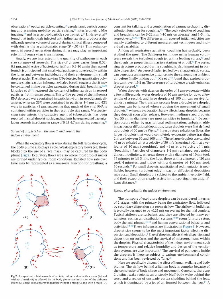
When the expiratory flow is weak during the full respiratory cycle,the body plume also plays a role. Weak expiratory flows (eg, thoseblocked by the use of a face mask) may be captured by the bodyplume (Fig 2). Expiratory flows are also where most droplet nucleiare formed under typical room conditions. Exhaled flow rate overtime may be represented as a sinusoidal function for breathing, a
constant for talking, and a combination of gamma probability dis-tribution functions for coughing.36,37 The peak velocities of coughingand breathing can be 6-22 m/s (>10 m/s on average) and 1-5 m/s,respectively.30,36,38 The differences in reported initial velocities aremainly attributed to different measurement techniques and indi-vidual variability.
Among all respiratory activities, coughing has probably beenstudied the most. The Schlieren technique using human volun-teers reveals the turbulent cough jet with a leading vortex,39 andthe cough has properties similar to a starting jet or puff.40 The vortexring structure produced during coughing is also important in par-ticle dispersion.4 An aerosol-laden jet, led by a characteristic vortex,can penetrate an impressive distance into the surrounding ambientair before finally mixing out.41 Xie et al42 found that expired drop-lets can travel 1.5-2m. The presence of turbulence greatly enhancesdroplet spread.43
Water droplets with sizes on the order of 1 μm evaporate withina few milliseconds, water droplets of 10 μm survive for up to a fewtenths of a second, and large droplets of 100 μm can survive foralmost a minute. The transient process from a droplet to a dropletnucleus can be ignored when studying the movement of smalldroplets,44 whereas evaporation barely affects large droplets becausethey deposit soon after release. However, medium-sized droplets(eg, 50 μm in diameter) are most sensitive to humidity.43 Deposi-tion occurs either by gravitational sedimentation, turbulent eddyimpaction, or diffusional deposition. Large droplets were first definedas droplets >100 μm byWells.45 In respiratory exhalation flows, thelargest droplets that would completely evaporate before traveling2 m are between 60 and 100 μm.42 These large droplets are carried>6m by exhaled air at a velocity of 50 m/s (sneezing), >2m at a ve-locity of 10 m/s (coughing), and <1 m at a velocity of 1 m/s(breathing). Particles of diameters 1-3 μm remained suspendedalmost indefinitely, whereas those with a diameter of 10 μm took17 minutes to fall 3 m to the floor, those with a diameter of 20 μmtook 4 minutes, and those with a diameter of 100 μm took10 seconds.46 For small droplets, gravitational sedimentation is neg-ligible; however, turbulent eddy impact or diffusional depositionmay occur. Small droplets are subject to the ambient velocity field,and their evaporation clearly assists in transporting them a signif-icant distance.47
Spread of droplets in the indoor environment
The transport of expiratory droplets can be considered in termsof 2 stages, with the primary being the expiratory flow, followedby secondary dispersion via room airflow. The airflow in buildingsis typically designed to be <0.25m/s on average for thermal comfort.Typical airflows are turbulent, and they are affected by many pa-rameters, such as air distribution systems,48-50 room furniture setup,body thermal plumes,51-53 and human conversational behavior andactivities.54-56 These influences are illustrated in Figure 3. However,droplet size seems to be the most important factor affecting dis-persion and deposition.5 Size of droplets affects their dispersion anddeposition on surfaces and the survival of microorganisms withinthe droplets. Physical characteristics of the indoor environment, suchas temperature and relative humidity and design of the ventila-tion system, are also important.5 The survival of pathogens insidethe droplets is likewise subject to various environmental condi-tions and has been reviewed by Tang.58
Herewe specifically discuss the effect of humanwalking and bodyplumes. The airflow behind a human body is complex because ofthe complexity of body shape and movement. Generally, there are2 distinct wake regions: an unsteady bluff-body wake behind thetorso and a region of unsteady vortex shedding behind the legs,which is dominated by a jet of air formed between the legs.54 A
Fig 2. Escaped microbial aerosols of an infected individual with a mask (A) andwithout a mask (B) as affected by the body plume and inhalation of the airborneinfectious agent(s) of a nearby individual without a mask (C) and with a mask (D).
S104 J. Wei, Y. Li / American Journal of Infection Control 44 (2016) S102-S108
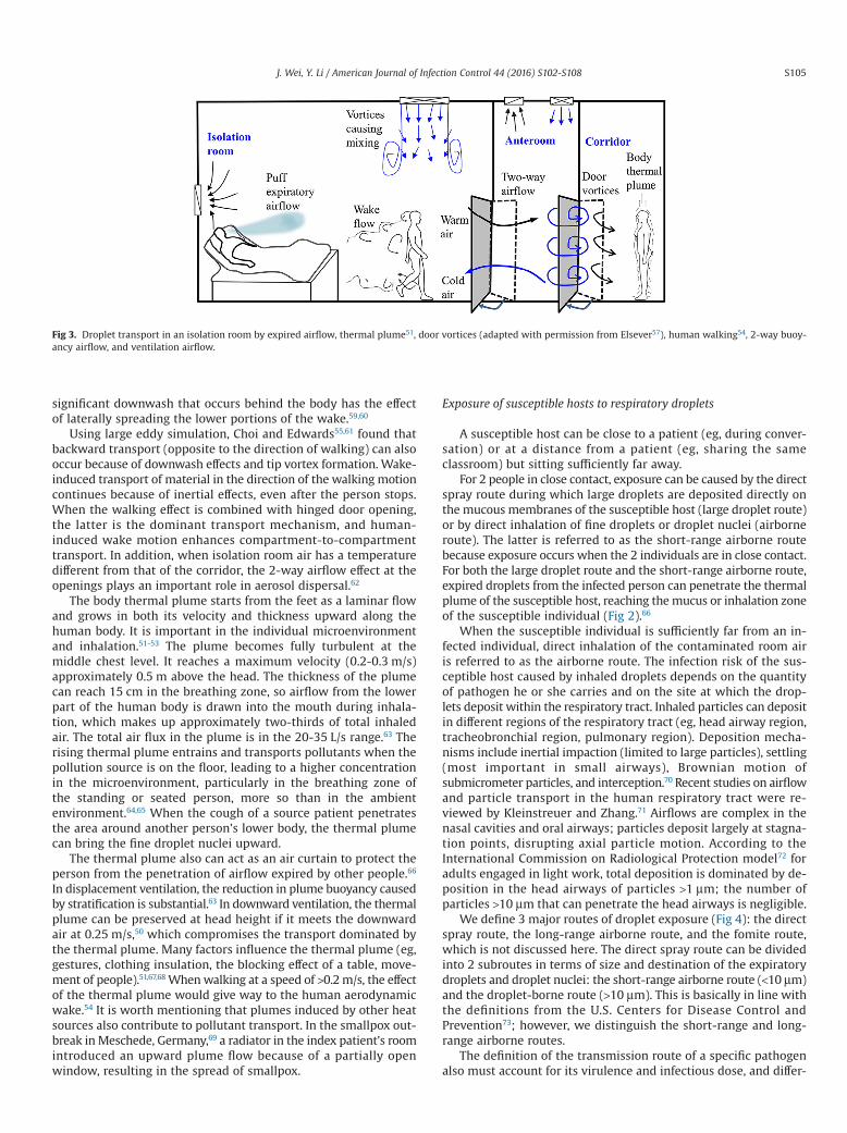
significant downwash that occurs behind the body has the effectof laterally spreading the lower portions of the wake.59,60
Using large eddy simulation, Choi and Edwards55,61 found thatbackward transport (opposite to the direction of walking) can alsooccur because of downwash effects and tip vortex formation. Wake-induced transport of material in the direction of the walking motioncontinues because of inertial effects, even after the person stops.When the walking effect is combined with hinged door opening,the latter is the dominant transport mechanism, and human-induced wake motion enhances compartment-to-compartmenttransport. In addition, when isolation room air has a temperaturedifferent from that of the corridor, the 2-way airflow effect at theopenings plays an important role in aerosol dispersal.62
The body thermal plume starts from the feet as a laminar flowand grows in both its velocity and thickness upward along thehuman body. It is important in the individual microenvironmentand inhalation.51-53 The plume becomes fully turbulent at themiddle chest level. It reaches a maximum velocity (0.2-0.3 m/s)approximately 0.5 m above the head. The thickness of the plumecan reach 15 cm in the breathing zone, so airflow from the lowerpart of the human body is drawn into the mouth during inhala-tion, which makes up approximately two-thirds of total inhaledair. The total air flux in the plume is in the 20-35 L/s range.63 Therising thermal plume entrains and transports pollutants when thepollution source is on the floor, leading to a higher concentrationin the microenvironment, particularly in the breathing zone ofthe standing or seated person, more so than in the ambientenvironment.64,65 When the cough of a source patient penetratesthe area around another person’s lower body, the thermal plumecan bring the fine droplet nuclei upward.
The thermal plume also can act as an air curtain to protect theperson from the penetration of airflow expired by other people.66
In displacement ventilation, the reduction in plume buoyancy causedby stratification is substantial.63 In downward ventilation, the thermalplume can be preserved at head height if it meets the downwardair at 0.25 m/s,50 which compromises the transport dominated bythe thermal plume. Many factors influence the thermal plume (eg,gestures, clothing insulation, the blocking effect of a table, move-ment of people).51,67,68 Whenwalking at a speed of >0.2m/s, the effectof the thermal plume would give way to the human aerodynamicwake.54 It is worth mentioning that plumes induced by other heatsources also contribute to pollutant transport. In the smallpox out-break inMeschede, Germany,69 a radiator in the index patient’s roomintroduced an upward plume flow because of a partially openwindow, resulting in the spread of smallpox.
Exposure of susceptible hosts to respiratory droplets
A susceptible host can be close to a patient (eg, during conver-sation) or at a distance from a patient (eg, sharing the sameclassroom) but sitting sufficiently far away.
For 2 people in close contact, exposure can be caused by the directspray route during which large droplets are deposited directly onthe mucous membranes of the susceptible host (large droplet route)or by direct inhalation of fine droplets or droplet nuclei (airborneroute). The latter is referred to as the short-range airborne routebecause exposure occurs when the 2 individuals are in close contact.For both the large droplet route and the short-range airborne route,expired droplets from the infected person can penetrate the thermalplume of the susceptible host, reaching themucus or inhalation zoneof the susceptible individual (Fig 2).66
When the susceptible individual is sufficiently far from an in-fected individual, direct inhalation of the contaminated room airis referred to as the airborne route. The infection risk of the sus-ceptible host caused by inhaled droplets depends on the quantityof pathogen he or she carries and on the site at which the drop-lets deposit within the respiratory tract. Inhaled particles can depositin different regions of the respiratory tract (eg, head airway region,tracheobronchial region, pulmonary region). Deposition mecha-nisms include inertial impaction (limited to large particles), settling(most important in small airways), Brownian motion ofsubmicrometer particles, and interception.70 Recent studies on airflowand particle transport in the human respiratory tract were re-viewed by Kleinstreuer and Zhang.71 Airflows are complex in thenasal cavities and oral airways; particles deposit largely at stagna-tion points, disrupting axial particle motion. According to theInternational Commission on Radiological Protection model72 foradults engaged in light work, total deposition is dominated by de-position in the head airways of particles >1 μm; the number ofparticles >10 μm that can penetrate the head airways is negligible.
We define 3 major routes of droplet exposure (Fig 4): the directspray route, the long-range airborne route, and the fomite route,which is not discussed here. The direct spray route can be dividedinto 2 subroutes in terms of size and destination of the expiratorydroplets and droplet nuclei: the short-range airborne route (<10 μm)and the droplet-borne route (>10 μm). This is basically in line withthe definitions from the U.S. Centers for Disease Control andPrevention73; however, we distinguish the short-range and long-range airborne routes.
The definition of the transmission route of a specific pathogenalso must account for its virulence and infectious dose, and differ-
Fig 3. Droplet transport in an isolation room by expired airflow, thermal plume51, door vortices (adapted with permission from Elsever57), human walking54, 2-way buoy-ancy airflow, and ventilation airflow.
S105J. Wei, Y. Li / American Journal of Infection Control 44 (2016) S102-S108
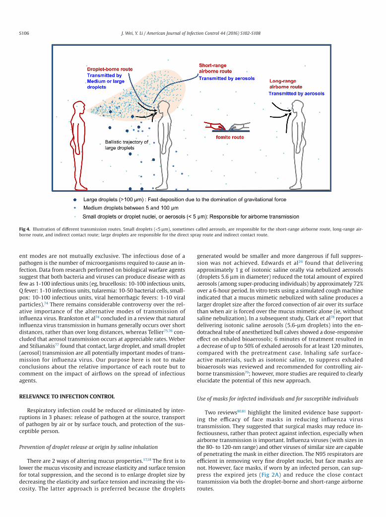
ent modes are not mutually exclusive. The infectious dose of apathogen is the number of microorganisms required to cause an in-fection. Data from research performed on biological warfare agentssuggest that both bacteria and viruses can produce disease with asfew as 1-100 infectious units (eg, brucellosis: 10-100 infectious units,Q fever: 1-10 infectious units, tularemia: 10-50 bacterial cells, small-pox: 10-100 infectious units, viral hemorrhagic fevers: 1-10 viralparticles).74 There remains considerable controversy over the rel-ative importance of the alternative modes of transmission ofinfluenza virus. Brankston et al74 concluded in a review that naturalinfluenza virus transmission in humans generally occurs over shortdistances, rather than over long distances, whereas Tellier75,76 con-cluded that aerosol transmission occurs at appreciable rates. Weberand Stilianakis77 found that contact, large droplet, and small droplet(aerosol) transmission are all potentially important modes of trans-mission for influenza virus. Our purpose here is not to makeconclusions about the relative importance of each route but tocomment on the impact of airflows on the spread of infectiousagents.
RELEVANCE TO INFECTION CONTROL
Respiratory infection could be reduced or eliminated by inter-ruptions in 3 phases: release of pathogen at the source, transportof pathogen by air or by surface touch, and protection of the sus-ceptible person.
Prevention of droplet release at origin by saline inhalation
There are 2 ways of altering mucus properties.17,18 The first is tolower themucus viscosity and increase elasticity and surface tensionfor total suppression, and the second is to enlarge droplet size bydecreasing the elasticity and surface tension and increasing the vis-cosity. The latter approach is preferred because the droplets
generated would be smaller and more dangerous if full suppres-sion was not achieved. Edwards et al26 found that deliveringapproximately 1 g of isotonic saline orally via nebulized aerosols(droplets 5.6 μm in diameter) reduced the total amount of expiredaerosols (among super-producing individuals) by approximately 72%over a 6-hour period. In vitro tests using a simulated coughmachineindicated that a mucus mimetic nebulized with saline produces alarger droplet size after the forced convection of air over its surfacethan when air is forced over the mucus mimetic alone (ie, withoutsaline nebulization). In a subsequent study, Clark et al78 report thatdelivering isotonic saline aerosols (5.6-μm droplets) into the en-dotracheal tube of anesthetized bull calves showed a dose-responsiveeffect on exhaled bioaerosols; 6 minutes of treatment resulted ina decrease of up to 50% of exhaled aerosols for at least 120 minutes,compared with the pretreatment case. Inhaling safe surface-active materials, such as isotonic saline, to suppress exhaledbioaerosols was reviewed and recommended for controlling air-borne transmission79; however, more studies are required to clearlyelucidate the potential of this new approach.
Use of masks for infected individuals and for susceptible individuals
Two reviews80,81 highlight the limited evidence base support-ing the efficacy of face masks in reducing influenza virustransmission. They suggested that surgical masks may reduce in-fectiousness, rather than protect against infection, especially whenairborne transmission is important. Influenza viruses (with sizes inthe 80- to 120-nm range) and other viruses of similar size are capableof penetrating the mask in either direction. The N95 respirators areefficient in removing very fine droplet nuclei, but face masks arenot. However, face masks, if worn by an infected person, can sup-press the expired jets (Fig 2A) and reduce the close contacttransmission via both the droplet-borne and short-range airborneroutes.
Fig 4. Illustration of different transmission routes. Small droplets (<5 μm), sometimes called aerosols, are responsible for the short-range airborne route, long-range air-borne route, and indirect contact route; large droplets are responsible for the direct spray route and indirect contact route.
S106 J. Wei, Y. Li / American Journal of Infection Control 44 (2016) S102-S108
Environmental ventilation for the long-range airborne route
A multidisciplinary systematic review49 suggested that ventila-tion rate and airflow patterns contribute directly to the airbornespread of infectious agents; however, the minimum ventilation ratefor effective airborne transmission control is unknown at present.The current minimum requirement is 12 air changes per hour fornegative-pressure airborne isolation rooms.82,83 Natural ventila-tion may offer a low-cost alternative.83,84 The current negative-pressure isolation rooms with a ceiling supply and bottom returnsystem are recommended, but gaseous and fine particles were foundto be removed more efficiently by ceiling-level exhausts, and largeparticles were removed mainly by deposition, rather than byventilation.85 Displacement ventilation has been recommended asa more energy-efficient approach in nonhospital settings. However,in the case of the isolation room, the stable thermal stratificationzone may cause the lock-up phenomenon to occur86 if the exhaledpollutant is not caught by the thermal plume penetrating into theupper zone, resulting in a longer residence time of pollutants.87 Dis-placement ventilation can create what might be referred to asinversion clouds in rooms. Because deposition is the main mech-anism for removing large droplets,85 floor cleaning in hospitals isabsolutely necessary.
Personalized ventilation for the short-range airborne route
This may be a less well-known technology in the infection controlcommunity. Its principle is based on detectable jets of air with ahigh momentum directed at a person’s face.88,89 It may not be ef-fective when the mobility of the subject is considered. An air supplypillow was suggested for hospital use.90 The personalized ventila-tion (PV) system can be supplemented with a general ventilationsystem in the room. Experiments with PV, together with verticalventilation from ceiling-mounted terminals, show increased effi-ciency of personal protection by a factor of up to 35.90 A combinationof PV and the personalized exhaust method was suggested.91
CONCLUSIONS
By reviewing the airborne spread of infectious agents frommucusto mucus in the indoor environment, we have shown the oppor-tunities for infection control at different stages of the spread. Wepropose that the short-range airborne route may be important inclose contact, and its control may be achieved by face masks for thesource patients and the use of PV. Our discussion of the effect ofthermal stratification and expiratory delivery of droplets leads tothe suggestion that displacement ventilation may not be applica-ble to hospital rooms where respiratory infection is a concern. Thesaline inhalation method was discussed after a discussion of themechanisms of droplet formation and origin.
References
1. Yu ITS, Li Y, Wong TW, Tam W, Chan AT, Lee JH, et al. Evidence of airbornetransmission of the severe acute respiratory syndrome virus. N Engl J Med2004;350:1731-9.
2. Cole EC, Cook CE. Characterization of infectious aerosols in health care facilities:an aid to effective engineering controls and preventive strategies. Am J InfectControl 1998;26:453-64.
3. Fitzgerald D, Haas D. Mycobacterium tuberculosis. In: Mandell GL, Bennett JE,Dolin R, editors. Principles and practice of infectious diseases. 6th ed. Philadelphia(PA): Churchill Livingstone; 2005:2852-86.
4. Hunt JCR, Delfos R, Eames I, Perkins RJ. Vortices, complex flows and inertialparticles. Flow Turbul Combust 2007;79:207-34.
5. Morawska L. Droplet fate in indoor environments, or can we prevent the spreadof infection? Indoor Air 2006;16:335-47.
6. Johnson GR, Morawska L, Ristovski ZD, Hargreaves M, Mengersen K, Chao CYH,et al. Modality of human expired aerosol size distributions. J Aerosol Sci2011;42:839-51.
7. Morawska L, Johnson GR, Ristovski ZD, Hargreaves M, Mengersen K, Corbett S,et al. Size distribution and sites of origin of droplets expelled from the humanrespiratory tract during expiratory activities. J Aerosol Sci 2009;40:256-69.
8. Duguid JP. The numbers and the sites of origin of the droplets expelled duringexpiratory activities. Edinburgh Med J 1945;52:385-401.
9. Xie X, Li Y, Sun H, Liu L. Exhaled droplets due to talking and coughing. J R SocInterface 2009;6(Suppl 6):S703-14.
10. Edwards D, Fiegel J, Dehaan W, Brande M, Man J, Clarke R. Novel inhalants forcontrol and protection against airborne infections. Respir Drug Deliv 2006;1:41-8.
11. Johnson GR, Morawska L. The mechanism of breath aerosol formation. J AerosolMed 2009;22:229-37.
12. Almstrand AC, Ljungström E, Lausmaa J, Bake B, Sjövall P, Olin AC. Airwaymonitoring by collection and mass spectrometric analysis of exhaled particles.Anal Chem 2008;81:662-8.
13. Fabian P, Brain J, Houseman EA, Gern J, Milton DK. Origin of exhaled breathparticles from healthy and human rhinovirus-infected subjects. J Aerosol MedPulm Drug Deliv 2011;24:137-47.
14. Ross BB, Gramiak R, Rahn H. Physical dynamics of the cough mechanism. J ApplPhysiol 1955;8:264-8.
15. Chowdhary R, Singh V, Tattersfield AE, Sharma SD, Kar S, Gupta AB. Relationshipof flow and cross-sectional area to frictional stress in airway models of asthma.J Asthma 1999;36:419-26.
16. Wang Y, Liu YX, Sun XZ, Yu S, Gao F. Numerical analysis of respiratory flowpatterns within human upper airway. Acta Mech Sin 2009;25:737-46.
17. VasudevanM, Lange CF. Property dependence of onset of instability in viscoelasticrespiratory fluids. Int J Eng Sci 2005;43:1292-8.
18. Vasudevan M, Lange CF. Surface tension effects on instability in viscoelasticrespiratory fluids. Math Biosci 2007;205:180-94.
19. Anwarul Hasan MD, Lange CF, King ML. Effect of artificial mucus properties onthe characteristics of airborne bioaerosol droplets generated during simulatedcoughing. J Nonnewton Fluid Mech 2010;165:1431-41.
20. Almstrand AC, Bake B, Ljungström E, Larsson P, Bredberg A, Mirgorodskaya E,et al. Effect of airway opening on production of exhaled particles. J Appl Phys2010;108:584-8.
21. Malashenko A, Tsuda A, Haber S. Propagation and breakup of liquid menisci andaerosol generation in small airways. J Aerosol Med 2009;22:341-53.
22. Holmgren H, Ljungstrom E. Influence of film dimensions on film dropletformation. J Aerosol Med 2012;25:47-53.
23. Zhu S, Kato S, Yang J-H. Study on transport characteristics of saliva dropletsproduced by coughing in a calm indoor environment. Build Environ2006;41:1691-702.
24. Fairchild CI, Stampfer JF. Particle concentration in exhaled breath. Am Ind HygAssoc J 1987;48:948-9.
25. Papineni RS, Rosenthal FS. The size distribution of droplets in the exhaled breathof healthy human subjects. J Aerosol Med 1997;10:105-16.
26. Edwards DA, Man JC, Brand P, Katstra JP, Sommerer K, Stone HA, et al. Inhalingto mitigate exhaled bioaerosols. Proc Natl Acad Sci USA 2004;101:17383-8.
27. Duguid JP. The size and the duration of air-carriage of respiratory droplets anddroplet-nuclei. J Hyg (Lond) 1946;44:471-9.
28. Loudon RG, Roberts RM. Cough frequency in patients with respiratory disease.Am Rev Respir Dis 1967;96:1137-43.
29. Yang SH, Lee GWM, Chen CM, Wu CC, Yu KP. The size and concentration ofdroplets generated by coughing in human subjects. J Aerosol Med 2007;20:484-94.
30. Chao CYH, Wan MP, Morawska L, Johnson GR, Ristovski ZD, Hargreaves M, et al.Characterization of expiration air jets and droplet size distributions immediatelyat the mouth opening. J Aerosol Sci 2009;40:122-33.
31. LindsleyWG, Pearce TA, Hudnall JB, Davis KA, Davis SM, Fisher MA, et al. Quantityand size distribution of cough-generated aerosol particles produced by influenzapatients during and after illness. J Occup Environ Hyg 2012;9:443-9.
32. Fabian P, McDevitt JJ, DehaanWH, Fung ROP, Cowling BJ, Chan KH, et al. Influenzavirus in human exhaled breath: an observational study. PLoS ONE 2008;3:e2691.
33. Milton DK, Fabian MP, Cowling BJ, Grantham ML, McDevitt JJ. Influenza virusaerosols in human exhaled breath: particle size, culturability, and effect of surgicalmasks. PLoS Pathog 2013;9:e1003205.
34. Lindsley WG, Blachere FM, Thewlis RE, Vishnu A, Davis KA, Cao G, et al.Measurements of airborne influenza virus in aerosol particles from humancoughs. PLoS ONE 2010;5:e15100.
35. Fennelly KP, Martyny JW, Fulton KE, Orme IM, Cave DM, Heifets LB. Cough-generated aerosols of Mycobacterium tuberculosis: a new method to studyinfectiousness. Am J Respir Crit Care Med 2004;169:604-9.
36. Gupta JK, Lin CH, Chen Q. Flow dynamics and characterization of a cough. IndoorAir 2009;19:517-25.
37. Gupta JK, Lin CH, Chen QY. Characterizing exhaled airflow from breathing andtalking. Indoor Air 2010;20:31-9.
38. Kwon S-B, Park J, Jang J, Cho Y, Park D-S, Kim C, et al. Study on the initial velocitydistribution of exhaled air from coughing and speaking. Chemosphere2012;87:1260-4.
39. Tang JW, Liebner TJ, Craven BA, Settles GS. A schlieren optical study of the humancough with and without wearing masks for aerosol infection control. J R SocInterface 2009;6(Suppl 6):S727-36.
S107J. Wei, Y. Li / American Journal of Infection Control 44 (2016) S102-S108
40. Vansciver M, Miller S, Hertzberg J. Particle image velocimetry of human cough.Aerosol Sci Technol 2011;45:415-22.
41. Settles GS. Fluid mechanics and homeland security. Annu Rev Fluid Mech2006;38:87-110.
42. Xie X, Li Y, Chwang ATY, Ho PL, Seto WH. How far droplets can move in indoorenvironments—revisiting the Wells evaporation–falling curve. Indoor Air2007;17:211-25.
43. Wei J, Li Y. Enhanced spread of expiratory droplets by turbulence in a coughjet. Build Environ 2015;93:86-96.
44. Chen C, Zhao B. Some questions on dispersion of human exhaled droplets inventilation room: answers from numerical investigation. Indoor Air 2010;20:95-111.
45. Wells WF. On air-borne infection study II. Droplets and droplet nuclei. Am JEpidemiol 1934;20:611-8.
46. Knight V. Viruses as agents of airborne contagion. Ann NY Acad Sci1980;353:147-56.
47. Eames I, Tang JW, Hunt JCR, Li Y. Murder, death and disease. Math Today2009;Feb:234-7.
48. Qian H, Li Y, Nielsen PV, Hyldgaard CE, Wong TW, Chwang AT. Dispersion ofexhaled droplet nuclei in a two-bed hospital ward with three different ventilationsystems. Indoor Air 2006;16:111-28.
49. Li Y, Leung GM, Tang JW, Yang X, Chao CY, Lin JZ, et al. Role of ventilation inairborne transmission of infectious agents in the built environment—amultidisciplinary systematic review. Indoor Air 2007;17:2-18.
50. Nielsen PV. Control of airborne infectious diseases in ventilated spaces. J R SocInterface 2009;6(Suppl 6):S747-55.
51. Clark RP, Edholm OG. Man and his thermal environment. London, England:Arnold; 1985.
52. Murakami S. Analysis and design of micro-climate around the human body withrespiration by CFD. Indoor Air 2004;14:144-56.
53. Gao NP, Niu JL. CFD study of the thermal environment around a human body:a review. Indoor Built Environ 2005;14:5-16.
54. Edge BA, Paterson EG, Settles GS. Computational study of the wake andcontaminant transport of a walking human. J Fluid Eng 2005;127:967-77.
55. Choi JI, Edwards JR. Large-eddy simulation of human-induced contaminanttransport in room compartments. Indoor Air 2012;22:77-87.
56. Tang JW, Eames I, Li Y, Taha YA,Wilson P, Bellingan G, et al. Door-openingmotioncan potentially lead to a transient breakdown in negative-pressure isolationconditions: the importance of vorticity and buoyancy airflows. J Hosp Infect2005;61:283-6.
57. Tang JW, Li Y, Eames I, Chan PKS, Ridgway GL. Factors involved in the aerosoltransmission of infection and control of ventilation in healthcare premises. J HospInfect 2006;64:100-14.
58. Tang JW. The effect of environmental parameters on the survival of airborneinfectious agents. J R Soc Interface 2009;6(Suppl 6):S737-46.
59. Poussou SB, Mazumdar S, Plesniak MW, Sojka PE, Chen Q. Flow and contaminanttransport in an airliner cabin induced by a moving body: model experimentsand CFD predictions. Atmos Environ 2010;44:2830-9.
60. Hang J, Li Y, Ching WH, Wei J, Jin R, Liu L, et al. Potential airborne transmissionbetween two isolation cubicles through a shared anteroom. Build Environ2015;89:264-78.
61. Choi JI, Edwards JR. Large eddy simulation and zonal modeling of human-inducedcontaminant transport. Indoor Air 2008;18:233-49.
62. Chen C, Zhao B, Yang X, Li Y. Role of two-way airflow owing to temperaturedifference in severe acute respiratory syndrome transmission: revisiting thelargest nosocomial severe acute respiratory syndrome outbreak in Hong Kong.J R Soc Interface 2011;8:699-710.
63. Craven BA, Settles GS. A computational and experimental investigation of thehuman thermal plume. J Fluid Eng 2006;128:1251-8.
64. Lewis HE, Foster AR, Mullan BJ, Cox RN, Clark RP. Aerodynamics of humanmicroenvironment. Lancet 1969;293:1273-7.
65. Rim D, Novoselac A. Transport of particulate and gaseous pollutants in the vicinityof a human body. Build Environ 2009;44:1840-9.
66. Liu L. Expiratory droplet exposure between individuals in a ventilated room [PhDthesis]. Hong Kong: The University of Hong Kong; 2011.
67. Zukowska D, Melikov A, Popiolek Z. Impact of personal factors and furniturearrangement on the thermal plume above a sitting occupant. Build Environ2012;49:104-16.
68. Spitzer IM, Marr DR, Glauser MN. Impact of manikin motion on particle transportin the breathing zone. J Aerosol Sci 2010;41:373-83.
69. Wehrle PF, Posch J, Richter KH, Henderson DA. An airborne outbreak of smallpoxin a German hospital and its significance with respect to other recent outbreaksin Europe. Bull World Health Organ 1970;43:669-79.
70. Hinds WC. Aerosol technology: properties, behavior, and measurement ofairborne particles. 2nd ed. New York (NY): Wiley; 1999.
71. Kleinstreuer C, Zhang Z. Airflow and particle transport in the human respiratorysystem. Annual review of fluid mechanics. Palo Alto (CA): Annual Reviews; 2010.
72. International Commission on Radiological Protection (ICRP). Human respiratorytract model for radiological protection, annals of the ICRP, publication 66.Tarrytown (NY): Elsevier Science; 1994.
73. Garner JS. Hospital Infection Control Practices Advisory Committee. Guidelinefor isolation precautions in hospitals. Infect Control Hosp Epidemiol 1996;17:53-80.
74. Brankston G, Gitterman L, Hirji Z, Lemieux C, Gardam M. Transmission ofinfluenza A in human beings. Lancet Infect Dis 2007;7:257-65.
75. Tellier R. Review of aerosol transmission of influenza A virus. Emerg Infect Dis2006;12:1657-62.
76. Tellier R. Aerosol transmission of influenza A virus: a review of new studies. JR Soc Interface 2009;6(Suppl 6):S783-90.
77. Weber TP, Stilianakis NI. Inactivation of influenza A viruses in the environmentand modes of transmission: a critical review. J Infect 2008;57:361-73.
78. Clark REA, Katstra J, Man JC, Dehaan W. Pulmonary delivery of anti-contagionaerosol to diminish exhaled bioaerosols and airborne infectious disease. Am JInfect Control 2005;33:e85.
79. Fiegel J, Clarke R, Edwards DA. Airborne infectious disease and the suppressionof pulmonary bioaerosols. Drug Discov Today 2006;11:51-7.
80. Cowling BJ, Zhou Y, Ip DKM, Leung GM, Aiello AE. Face masks to preventtransmission of influenza virus: a systematic review. Epidemiol Infect2010;138:449-56.
81. Bin-Reza F, Lopez Chavarrias V, Nicoll A, Chamberland ME. The use of masksand respirators to prevent transmission of influenza: a systematic review of thescientific evidence. Influenza Other Respir Viruses 2012;6:257-67.
82. Centers for Disease Control and Prevention (CDC). Guidelines for environmentalinfection control in health-care facilities. Atlanta (GA): U.S. Department of Healthand Human Services; 2003.
83. World Health Organization (WHO). Infection prevention and control of epidemic-and pandemic-prone acute respiratory diseases in health care: WHO interimguidelines. Geneva, Switzerland: World Health Organization; 2007.
84. Escombe AR, Oeser CC, Gilman RH, Navincopa M, Ticona E, Pan W, et al. Naturalventilation for the prevention of airborne contagion. PLoS Med 2007;4:309-17.
85. Qian H, Li Y. Removal of exhaled particles by ventilation and deposition in amultibed airborne infection isolation room. Indoor Air 2010;20:284-97.
86. Stymne H, Sandberg M, Mattsson M. Dispersion pattern of contaminants in adisplacement ventilated room – implications for demand control. The12th AIVCConference “Air Movement and Ventilation Control within Buildings”, Ottawa,Canada, September 24-27, 1991:173-89.
87. Li Y, Nielsen PV, Sandberg M. Displacement ventilation in hospital environments.ASHRAE J 2011;53:86-8.
88. Pantelic J, Sze-To GN, Tham KW, Chao CYH, Khoo YCM. Personalized ventilationas a control measure for airborne transmissible disease spread. J R Soc Interface2009;6(Suppl 6):S715-26.
89. Alain M, Kamel G, Nesreen G. A simplified combined displacement andpersonalized ventilation model. HVAC&R Res 2012;18:737-49.
90. Nielsen PV, Jiang H, Polak M. Bed with integrated personalized ventilation forminimizing cross infection. The 10th International Conference on Air Distributionin Rooms, Helsinki, Finland, 13-15 June, 2007.
91. Yang J, Sekhar SC, Cheong KWD, Raphael B. Performance evaluation of a novelpersonalized ventilation–personalized exhaust system for airborne infectioncontrol. Indoor Air 2015;25:176-87.
S108 J. Wei, Y. Li / American Journal of Infection Control 44 (2016) S102-S108
State of the Science Review
Generic aspects of the airborne spread of human pathogens indoorsand emerging air decontamination technologies
M. Khalid Ijaz DVM, MSc(Honors), PhD a,b,*, Bahram Zargar BSc Engg, MSc Engg, PhD c,Kathryn E. Wright MA, MSc, PhD c, Joseph R. Rubino BA, MA a,Syed A. Sattar MSc, Dip Bact, MS, PhD d
a RB, Montvale, NJb Department of Biology, Medgar Evers College of the City University of New York (CUNY), Brooklyn, NYc Department of Biochemistry, Microbiology, and Immunology, University of Ottawa, Ottawa, ON, Canadad Professor Emeritus of Microbiology, Faculty of Medicine, University of Ottawa, Ottawa, ON, Canada
Key Words:Indoor air microbiomeAirborne pathogensMicrobial aerosolsAerosolized pathogensAir-surface-air nexusAerobiologic testing chamberAir decontamination technologies
Indoor air can be an important vehicle for a variety of human pathogens. This review provides examplesof airborne transmission of infectious agents from experimental and field studies and discusses how air-borne pathogens can contaminate other parts of the environment to give rise to secondary vehicles leadingair-surface-air nexus with possible transmission to susceptible hosts. The following groups of human patho-gens are covered because of their known or potential airborne spread: vegetative bacteria (staphylococciand legionellae), fungi (Aspergillus, Penicillium, and Cladosporium spp and Stachybotrys chartarum), entericviruses (noro- and rotaviruses), respiratory viruses (influenza and coronaviruses), mycobacteria (tuber-culous and nontuberculous), and bacterial spore formers (Clostridium difficile and Bacillus anthracis). Anoverview of methods for experimentally generating and recovering airborne human pathogens is in-cluded, along with a discussion of factors that influencemicrobial survival in indoor air. Available guidelinesfrom the U.S. Environmental Protection Agency and other global regulatory bodies for the study of air-borne pathogens are critically reviewed with particular reference to microbial surrogates that arerecommended. Recent developments in experimental facilities to contaminate indoor air with microbialaerosols are presented, along with emerging technologies to decontaminate indoor air under field-relevant conditions. Furthermore, the role that air decontaminationmay play in reducing the contaminationof environmental surfaces and its combined impact on interrupting the risk of pathogen spread in bothdomestic and institutional settings is discussed.
© 2016 Published by Elsevier Inc. on behalf of Association for Professionals in Infection Control andEpidemiology, Inc.
Air, a universal environmental equalizer, affects all living and non-living forms on planet earth. For humans, it has profound healthimplications in all indoor environments where we normally spendmost of our time.1-3 Air quality is also forever changing because of
the influence of many controllable and uncontrollable factors thatare virtually everywhere. Indoor air, in particular, can expose us tonoxious chemicals, particulates, and a variety of infectious agents,as well as pollen and other allergens.4,5
Emerging pathogens, such as noroviruses6 and Clostridiumdifficile,7 have also been detected in indoor air, with a strong po-tential for airborne dissemination. Pathogens discharged into theair may settle on environmental surfaces, which could then becomesecondary vehicles for the spread of infectious agents indoors.8 Thepossible transmission of drug-resistant bacteria by indoor air addsanother cause for concern.9 A combination of on-going societalchanges is adding further to the potential of air as a vehicle for in-fectious agents.10-12 The quality of indoor air is therefore a prominentpublic health concern13,14 that requires a clear understanding of thetransmission processes for the development and implementationof targeted infection prevention and control measures.15
* Address correspondence to M. Khalid Ijaz, DVM, MSc(Honors), PhD, RB, OnePhilips Parkway, Montvale, NJ 07645.
E-mail address: [email protected] (M.K. Ijaz).Funding/Support: This paper was presented at a workshop organized under the
auspices of ASTM International’s biannual meeting held in April 2016. Publicationof this supplement is primarily supported by RB, Montvale, New Jersey, with addi-tional support from MicroBioTest, a division of Microbac Laboratories, Inc., Sterling,Virginia. The City University of New York (CUNY) and the University of Ottawa, Ottawa,Canada, are academic sponsors. Editorial support was provided by Ashely O’Dunne,PhD; Shannon O’Sullivan, ELS; and Alanna Franchetti, ELS of Medergy (Yardley, PA),and funded by RB.
Conflicts of Interest: None to report.
0196-6553/© 2016 Published by Elsevier Inc. on behalf of Association for Professionals in Infection Control and Epidemiology, Inc.http://dx.doi.org/10.1016/j.ajic.2016.06.008
American Journal of Infection Control 44 (2016) S109-S120
Contents lists available at ScienceDirect
American Journal of Infection Control
journal homepage: www.aj ic journal .org
American Journal of Infection Control

Although direct and indirect exposure to pathogens in the aircan occur by other means, infections from the inhalation andretention, including translocation and ingestion after inhalationof droplet nuclei, are generally regarded as true airborne spread.Aerosols of various sizes that contain infectious agents can beemitted from a variety of sources, such as infected or colonizedindividuals16 or flushing toilets, andmay expose susceptible personseither directly (droplet transmission) or by remaining suspendedin the air for inhalation (airborne transmission).17,18 Contrary tothe conventionally held belief, modeling work has redefined theWells evaporation-falling curve,19,20 revealing that expelled largedroplets could be carried >6 m away by exhaled air at a velocity of50 m/s (sneezing), >2 m away at a velocity of 10 m/s (coughing),and <1 m away at a velocity of 1 m/s (breathing), leading topotential transmission of short-range infectious agents that containaerosols.21
Airborne transmission requires that pathogens survive the processof aerosolization and persist in the air long enough to be transmit-ted to a susceptible host.22 Aerosolized pathogens may settle ontoenvironmental surfaces in the immediate vicinity, leading to genesisof secondary vehicles (Fig 1).23 This review provides current infor-mation on the spread of human pathogens by indoor air, with a focuson the major classes of human pathogens from experimental andfield studies, and on emerging air decontamination technologies,including test protocols developed to assess their performance underfield-relevant conditions.
METHODS FOR STUDYING AIRBORNE HUMAN PATHOGENS
The study of aerosolized human pathogens requires the abilityto produce them experimentally at the appropriate size, storethem, and sample them for residual infectious content over apredetermined time period.13 The equipment must also simulatenaturally occurring environmental conditions and the duration ofexposure to accurately assess aerosol survivability.24 Various
analytical methods and air samplers have been used to character-ize airborne pathogens and overcome the challenges of collectingand analyzing them. Relevant studies have been reviewed indetail elsewhere.13,25,26
ENVIRONMENTAL FACTORS THAT INFLUENCE AIRBORNEMICROBIAL SURVIVAL
Aerosolized microbes must survive the prevailing environmen-tal conditions to potentially infect a susceptible host.22 Multiplefactors affect airborne survival of microbes indoors (Table 1).13,31 Theeffect of these factors on different types of microbes varies, and gen-eralizations can be difficult because of differences in the experimentalmethodologies used.27 Air temperature, relative humidity (RH), andturbulence are among the more important factors affecting the fateand spread of infectious agents indoors.
The analysis of air samples for microbes now includes methodsthat are based on the polymerase chain reaction (PCR). However,PCR-based methods typically cannot differentiate between viableand nonviable microbes.32 A recent study found that PCR substan-tially overestimated the quantity of infectious airborne influenzavirus, but the differences in infectious versus noninfectious virusover time were similar to data from quantification by plaque-forming units, which determined that virus losses were evidentwithin 30-60 minutes postaerosolization.32 Generally, envelopedviruses survive better at lower RH, but there are many exceptions.28
Other factors that affect aerosol activation in relation to RH includeevaporative activity (ie, dehydration, rehydration), surface areas ofparticles, and pH.28
AIRBORNE SPREAD OF MAJOR CLASSES OF HUMAN PATHOGENS
Although studies with experimental animals have determinedthe susceptibility to airborne pathogens and the minimal infec-tive inhalation dose of a given pathogen,25 there are wide variations
Fig 1. Sources of airborne pathogens indoors and potential for environmental surface contamination. These sources may include humans; pets; plants; plumbing systems,such as operational toilets and shower heads; heating, ventilation, vacuuming, mopping, and air-conditioning systems; resuspension of settled dust; and outdoor air. Theyellow and red dots represent human pathogens or harmless microorganisms. Adapted with permission from BioMed Central.23
S110 M.K. Ijaz et al. / American Journal of Infection Control 44 (2016) S109-S120
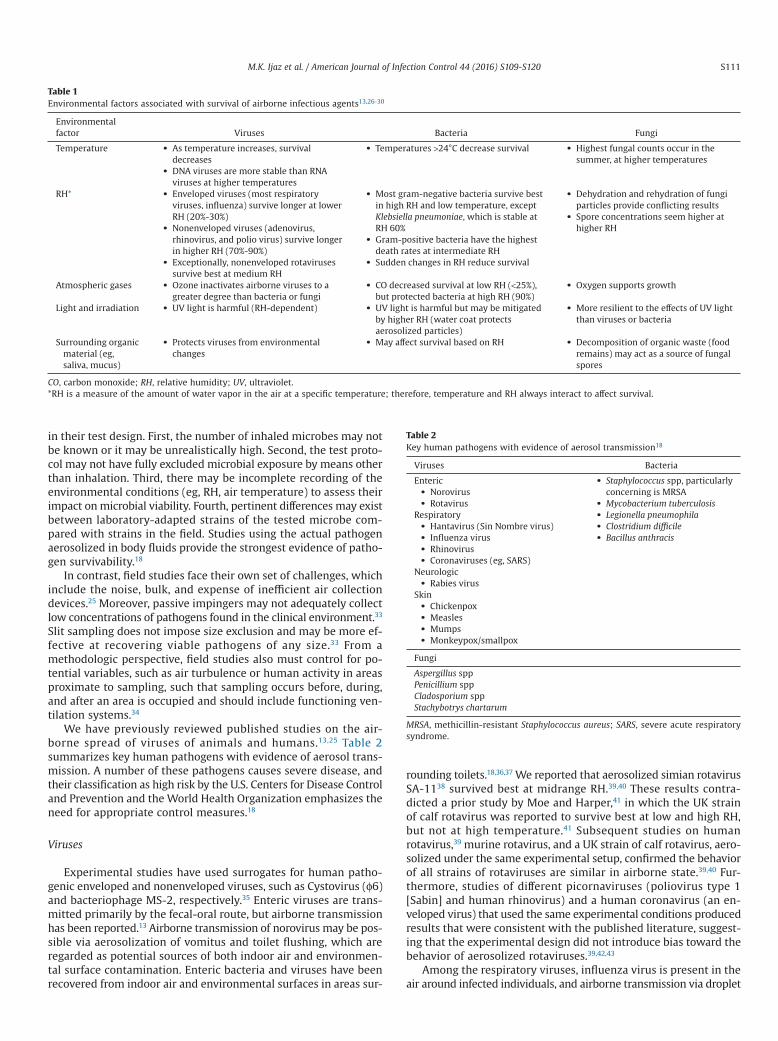
in their test design. First, the number of inhaled microbes may notbe known or it may be unrealistically high. Second, the test proto-col may not have fully excluded microbial exposure by means otherthan inhalation. Third, there may be incomplete recording of theenvironmental conditions (eg, RH, air temperature) to assess theirimpact onmicrobial viability. Fourth, pertinent differences may existbetween laboratory-adapted strains of the tested microbe com-pared with strains in the field. Studies using the actual pathogenaerosolized in body fluids provide the strongest evidence of patho-gen survivability.18
In contrast, field studies face their own set of challenges, whichinclude the noise, bulk, and expense of inefficient air collectiondevices.25 Moreover, passive impingers may not adequately collectlow concentrations of pathogens found in the clinical environment.33
Slit sampling does not impose size exclusion and may be more ef-fective at recovering viable pathogens of any size.33 From amethodologic perspective, field studies also must control for po-tential variables, such as air turbulence or human activity in areasproximate to sampling, such that sampling occurs before, during,and after an area is occupied and should include functioning ven-tilation systems.34
We have previously reviewed published studies on the air-borne spread of viruses of animals and humans.13,25 Table 2summarizes key human pathogens with evidence of aerosol trans-mission. A number of these pathogens causes severe disease, andtheir classification as high risk by the U.S. Centers for Disease Controland Prevention and the World Health Organization emphasizes theneed for appropriate control measures.18
Viruses
Experimental studies have used surrogates for human patho-genic enveloped and nonenveloped viruses, such as Cystovirus (ϕ6)and bacteriophage MS-2, respectively.35 Enteric viruses are trans-mitted primarily by the fecal-oral route, but airborne transmissionhas been reported.13 Airborne transmission of norovirus may be pos-sible via aerosolization of vomitus and toilet flushing, which areregarded as potential sources of both indoor air and environmen-tal surface contamination. Enteric bacteria and viruses have beenrecovered from indoor air and environmental surfaces in areas sur-
rounding toilets.18,36,37 We reported that aerosolized simian rotavirusSA-1138 survived best at midrange RH.39,40 These results contra-dicted a prior study by Moe and Harper,41 in which the UK strainof calf rotavirus was reported to survive best at low and high RH,but not at high temperature.41 Subsequent studies on humanrotavirus,39 murine rotavirus, and a UK strain of calf rotavirus, aero-solized under the same experimental setup, confirmed the behaviorof all strains of rotaviruses are similar in airborne state.39,40 Fur-thermore, studies of different picornaviruses (poliovirus type 1[Sabin] and human rhinovirus) and a human coronavirus (an en-veloped virus) that used the same experimental conditions producedresults that were consistent with the published literature, suggest-ing that the experimental design did not introduce bias toward thebehavior of aerosolized rotaviruses.39,42,43
Among the respiratory viruses, influenza virus is present in theair around infected individuals, and airborne transmission via droplet
Table 1Environmental factors associated with survival of airborne infectious agents13,26-30
Environmentalfactor Viruses Bacteria Fungi
Temperature • As temperature increases, survivaldecreases
• DNA viruses are more stable than RNAviruses at higher temperatures
• Temperatures >24°C decrease survival • Highest fungal counts occur in thesummer, at higher temperatures
RH* • Enveloped viruses (most respiratoryviruses, influenza) survive longer at lowerRH (20%-30%)
• Nonenveloped viruses (adenovirus,rhinovirus, and polio virus) survive longerin higher RH (70%-90%)
• Exceptionally, nonenveloped rotavirusessurvive best at medium RH
• Most gram-negative bacteria survive bestin high RH and low temperature, exceptKlebsiella pneumoniae, which is stable atRH 60%
• Gram-positive bacteria have the highestdeath rates at intermediate RH
• Sudden changes in RH reduce survival
• Dehydration and rehydration of fungiparticles provide conflicting results
• Spore concentrations seem higher athigher RH
Atmospheric gases • Ozone inactivates airborne viruses to agreater degree than bacteria or fungi
• CO decreased survival at low RH (<25%),but protected bacteria at high RH (90%)
• Oxygen supports growth
Light and irradiation • UV light is harmful (RH-dependent) • UV light is harmful but may be mitigatedby higher RH (water coat protectsaerosolized particles)
• More resilient to the effects of UV lightthan viruses or bacteria
Surrounding organicmaterial (eg,saliva, mucus)
• Protects viruses from environmentalchanges
• May affect survival based on RH • Decomposition of organic waste (foodremains) may act as a source of fungalspores
CO, carbon monoxide; RH, relative humidity; UV, ultraviolet.*RH is a measure of the amount of water vapor in the air at a specific temperature; therefore, temperature and RH always interact to affect survival.
Table 2Key human pathogens with evidence of aerosol transmission18
Viruses Bacteria
Enteric• Norovirus• Rotavirus
Respiratory• Hantavirus (Sin Nombre virus)• Influenza virus• Rhinovirus• Coronaviruses (eg, SARS)
Neurologic• Rabies virus
Skin• Chickenpox• Measles• Mumps• Monkeypox/smallpox
• Staphylococcus spp, particularlyconcerning is MRSA
• Mycobacterium tuberculosis• Legionella pneumophila• Clostridium difficile• Bacillus anthracis
Fungi
Aspergillus sppPenicillium sppCladosporium sppStachybotrys chartarum
MRSA, methicillin-resistant Staphylococcus aureus; SARS, severe acute respiratorysyndrome.
S111M.K. Ijaz et al. / American Journal of Infection Control 44 (2016) S109-S120
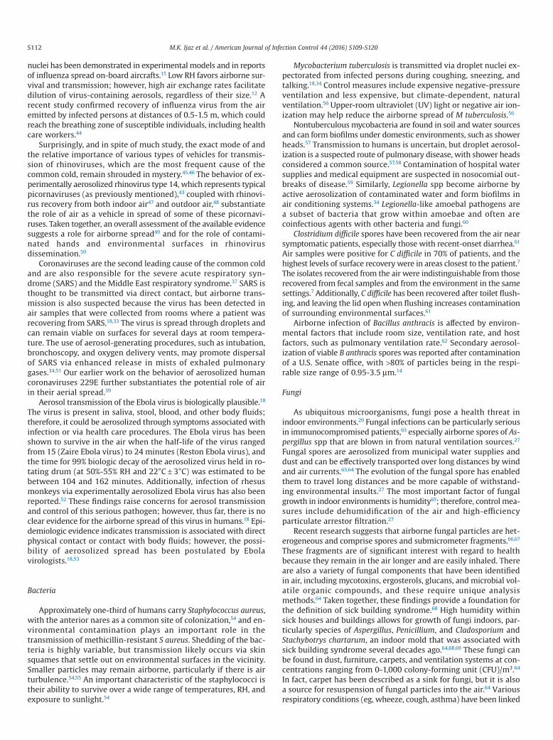
nuclei has been demonstrated in experimental models and in reportsof influenza spread on-board aircrafts.15 Low RH favors airborne sur-vival and transmission; however, high air exchange rates facilitatedilution of virus-containing aerosols, regardless of their size.12 Arecent study confirmed recovery of influenza virus from the airemitted by infected persons at distances of 0.5-1.5 m, which couldreach the breathing zone of susceptible individuals, including healthcare workers.44
Surprisingly, and in spite of much study, the exact mode of andthe relative importance of various types of vehicles for transmis-sion of rhinoviruses, which are the most frequent cause of thecommon cold, remain shrouded in mystery.45,46 The behavior of ex-perimentally aerosolized rhinovirus type 14, which represents typicalpicornaviruses (as previously mentioned),43 coupled with rhinovi-rus recovery from both indoor air47 and outdoor air,48 substantiatethe role of air as a vehicle in spread of some of these picornavi-ruses. Taken together, an overall assessment of the available evidencesuggests a role for airborne spread49 and for the role of contami-nated hands and environmental surfaces in rhinovirusdissemination.50
Coronaviruses are the second leading cause of the common coldand are also responsible for the severe acute respiratory syn-drome (SARS) and the Middle East respiratory syndrome.37 SARS isthought to be transmitted via direct contact, but airborne trans-mission is also suspected because the virus has been detected inair samples that were collected from rooms where a patient wasrecovering from SARS.18,33 The virus is spread through droplets andcan remain viable on surfaces for several days at room tempera-ture. The use of aerosol-generating procedures, such as intubation,bronchoscopy, and oxygen delivery vents, may promote dispersalof SARS via enhanced release in mists of exhaled pulmonarygases.34,51 Our earlier work on the behavior of aerosolized humancoronaviruses 229E further substantiates the potential role of airin their aerial spread.39
Aerosol transmission of the Ebola virus is biologically plausible.18
The virus is present in saliva, stool, blood, and other body fluids;therefore, it could be aerosolized through symptoms associated withinfection or via health care procedures. The Ebola virus has beenshown to survive in the air when the half-life of the virus rangedfrom 15 (Zaire Ebola virus) to 24 minutes (Reston Ebola virus), andthe time for 99% biologic decay of the aerosolized virus held in ro-tating drum (at 50%-55% RH and 22°C ± 3°C) was estimated to bebetween 104 and 162 minutes. Additionally, infection of rhesusmonkeys via experimentally aerosolized Ebola virus has also beenreported.52 These findings raise concerns for aerosol transmissionand control of this serious pathogen; however, thus far, there is noclear evidence for the airborne spread of this virus in humans.18 Epi-demiologic evidence indicates transmission is associated with directphysical contact or contact with body fluids; however, the possi-bility of aerosolized spread has been postulated by Ebolavirologists.18,53
Bacteria
Approximately one-third of humans carry Staphylococcus aureus,with the anterior nares as a common site of colonization,54 and en-vironmental contamination plays an important role in thetransmission of methicillin-resistant S aureus. Shedding of the bac-teria is highly variable, but transmission likely occurs via skinsquames that settle out on environmental surfaces in the vicinity.Smaller particles may remain airborne, particularly if there is airturbulence.54,55 An important characteristic of the staphylococci istheir ability to survive over a wide range of temperatures, RH, andexposure to sunlight.54
Mycobacterium tuberculosis is transmitted via droplet nuclei ex-pectorated from infected persons during coughing, sneezing, andtalking.18,34 Control measures include expensive negative-pressureventilation and less expensive, but climate-dependent, naturalventilation.56 Upper-room ultraviolet (UV) light or negative air ion-ization may help reduce the airborne spread of M tuberculosis.56
Nontuberculousmycobacteria are found in soil and water sourcesand can form biofilms under domestic environments, such as showerheads.57 Transmission to humans is uncertain, but droplet aerosol-ization is a suspected route of pulmonary disease, with shower headsconsidered a common source.57,58 Contamination of hospital watersupplies and medical equipment are suspected in nosocomial out-breaks of disease.59 Similarly, Legionella spp become airborne byactive aerosolization of contaminated water and form biofilms inair conditioning systems.34 Legionella-like amoebal pathogens area subset of bacteria that grow within amoebae and often arecoinfectious agents with other bacteria and fungi.60
Clostridium difficile spores have been recovered from the air nearsymptomatic patients, especially those with recent-onset diarrhea.61
Air samples were positive for C difficile in 70% of patients, and thehighest levels of surface recoverywere in areas closest to the patient.7
The isolates recovered from the air were indistinguishable from thoserecovered from fecal samples and from the environment in the samesettings.7 Additionally, C difficile has been recovered after toilet flush-ing, and leaving the lid open when flushing increases contaminationof surrounding environmental surfaces.61
Airborne infection of Bacillus anthracis is affected by environ-mental factors that include room size, ventilation rate, and hostfactors, such as pulmonary ventilation rate.62 Secondary aerosol-ization of viable B anthracis spores was reported after contaminationof a U.S. Senate office, with >80% of particles being in the respi-rable size range of 0.95-3.5 μm.14
Fungi
As ubiquitous microorganisms, fungi pose a health threat inindoor environments.29 Fungal infections can be particularly seriousin immunocompromised patients,63 especially airborne spores of As-pergillus spp that are blown in from natural ventilation sources.27
Fungal spores are aerosolized from municipal water supplies anddust and can be effectively transported over long distances by windand air currents.63,64 The evolution of the fungal spore has enabledthem to travel long distances and be more capable of withstand-ing environmental insults.27 The most important factor of fungalgrowth in indoor environments is humidity65; therefore, control mea-sures include dehumidification of the air and high-efficiencyparticulate arrestor filtration.27
Recent research suggests that airborne fungal particles are het-erogeneous and comprise spores and submicrometer fragments.66,67
These fragments are of significant interest with regard to healthbecause they remain in the air longer and are easily inhaled. Thereare also a variety of fungal components that have been identifiedin air, including mycotoxins, ergosterols, glucans, and microbial vol-atile organic compounds, and these require unique analysismethods.64 Taken together, these findings provide a foundation forthe definition of sick building syndrome.68 High humidity withinsick houses and buildings allows for growth of fungi indoors, par-ticularly species of Aspergillus, Penicillium, and Cladosporium andStachybotrys chartarum, an indoor mold that was associated withsick building syndrome several decades ago.64,68,69 These fungi canbe found in dust, furniture, carpets, and ventilation systems at con-centrations ranging from 0-1,000 colony-forming unit (CFU)/m3.64
In fact, carpet has been described as a sink for fungi, but it is alsoa source for resuspension of fungal particles into the air.64 Variousrespiratory conditions (eg, wheeze, cough, asthma) have been linked
S112 M.K. Ijaz et al. / American Journal of Infection Control 44 (2016) S109-S120
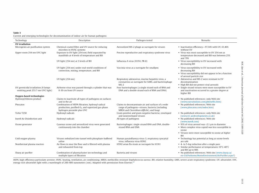
Table 3Current and emerging technologies for decontamination of indoor air for human pathogens
Technology Description Pathogen tested Remarks
UV irradiationMicrogenix air purification system Chemical-coated filter and UV source for reducing
microbes in HVAC systemsAerosolized MS-2 phage as surrogate for viruses • Inactivation efficiency = 97.34% with UV, 61.46%
without UVUpper-room 254 nm UVC light Exposure to UV light (254 nm) field separated by
manifolds at 4 levels of temperature and RHPorcine reproductive and respiratory syndrome virus • Virus was most susceptible to UV 254 nm as
temperature decreased and RH was between 25%and 79%
UV light (254 nm) at 3 levels of RH Influenza A virus (H1N1, PR-8) • Virus susceptibility to UV increased withdecreasing RH
UV light (254 nm) under real-world conditions ofconvection, mixing, temperature, and RH
Vaccinia virus as a surrogate for smallpox • Virus susceptibility to UV increased withdecreasing RH
• Virus susceptibility did not appear to be a functionof aerosol particle size
UV light (254 nm) Respiratory adenovirus, murine hepatitis virus, acoronavirus as surrogate for SARS, and bacteriophageMS-2
• Adenovirus and MS-2 were resistant to UVdecontamination
• High RH did not protect viral aerosolsUV germicidal irradiation (8 lampsemitting peak 253.7 nm UVC light)
Airborne virus was passed through a cylinder that was0-30 cm from UV source
Four bacteriophages (a single strand each of RNA andDNA and a double strand each of RNA and DNA)
• Single-strand viruses were more susceptible to UVand inactivation occurred to a greater degree athigher RH
Oxygen-based technologiesHydroxyl/Odorox product Claims to inactivate all types of pathogens on surfaces
and in the air• No published references; only Web site(www.eairsolutions.com/pbenefits.htm)
Phocatox Combination of HEPA filtration, hydroxyl radicalproduction, purified O3, and vaporized gas-phasehydrogen peroxide plus UVC
Claims to decontaminate air and surfaces of a widerange of pathogens—viruses, bacteria (includingMRSA and Clostridium difficile), and fungi
• No published references; Web site(www.phocatox.com/index.htm)
TriAir T250 Hydroxyl radicals Gram-positive and gram-negative bacteria; envelopedand nonenveloped viruses
• No published references; only Web site(www.tri-airdevelopments.co.uk)
Inov8 Air Disinfection unit Hydroxyl radicals All types of pathogens • No published references; Web site(www.inov8.com)
Ozone generator Gaseous ozone and aerosolized virus were generatedcontinuously into the chamber
Bacteriophages: single-strand RNA and DNA, double-strand RNA and DNA
• 95% of virus aerosol was <2.1 μm in diameter• More complex virus capsid was less susceptible toozone
• Viruses were more susceptible to ozone at higherRH
Cold oxygen plasma Viruses nebulized into tunnel with phosphate-bufferedsaline
Human parainfluenza virus-3, respiratory syncytialvirus, influenza virus H5N2
• Technology has potential as long as ozone levelsare safe
Nonthermal plasma reactors Air flows in near the floor and is filtered with plasmaand exhausted from top
H5N2 avian flu strain as surrogate for H1N1 • 4- to 5-log reduction after a single pass• Similar performance at temperatures 10°C-40°Cand RH up to 98%
Sharp air purifier Combination of plasmacluster ion technology andmultiple layers of filtration
Bacteria and viruses • No published references; Web site (www.sharp.ca/en-CA/Forhome/HomeEnvironment/AirPurifier.aspx?)
HEPA, high-efficiency particulate arrestor; HVAC, heating, ventilation, air conditioning;MRSA, methicillin-resistant Staphylococcus aureus; RH, relative humidity; SARS, severe acute respiratory syndrome; UV, ultraviolet; UVC,energy-rich ultraviolet light with a wavelength of 200-400 nanometers (nm). Adapted with permission from Elsevier.25
S113M.K.Ijaz
etal./Am
ericanJournalofInfection
Control44(2016)
S109-S120
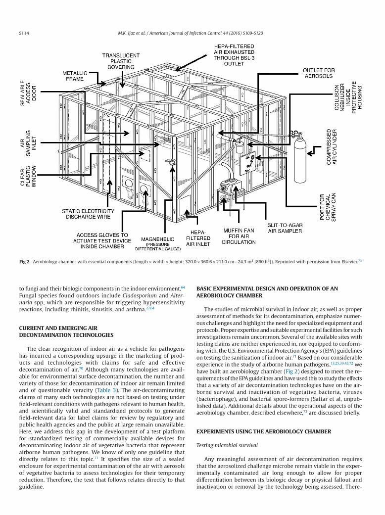
to fungi and their biologic components in the indoor environment.64
Fungal species found outdoors include Cladosporium and Alter-naria spp, which are responsible for triggering hypersensitivityreactions, including rhinitis, sinusitis, and asthma.27,64
CURRENT AND EMERGING AIRDECONTAMINATION TECHNOLOGIES
The clear recognition of indoor air as a vehicle for pathogenshas incurred a corresponding upsurge in the marketing of prod-ucts and technologies with claims for safe and effectivedecontamination of air.70 Although many technologies are avail-able for environmental surface decontamination, the number andvariety of those for decontamination of indoor air remain limitedand of questionable veracity (Table 3). The air-decontaminatingclaims of many such technologies are not based on testing underfield-relevant conditions with pathogens relevant to human health,and scientifically valid and standardized protocols to generatefield-relevant data for label claims for review by regulatory andpublic health agencies and the public at large remain unavailable.Here, we address this gap in the development of a test platformfor standardized testing of commercially available devices fordecontaminating indoor air of vegetative bacteria that representairborne human pathogens. We know of only one guideline thatdirectly relates to this topic.71 It specifies the size of a sealedenclosure for experimental contamination of the air with aerosolsof vegetative bacteria to assess technologies for their temporaryreduction. Therefore, the text that follows relates directly to thatguideline.
BASIC EXPERIMENTAL DESIGN AND OPERATION OF ANAEROBIOLOGY CHAMBER
The studies of microbial survival in indoor air, as well as properassessment of methods for its decontamination, emphasize numer-ous challenges and highlight the need for specialized equipment andprotocols. Properexpertise andsuitable experimental facilities for suchinvestigations remain uncommon. Several of the available siteswithtesting claims are neither experienced in, nor equipped to conform-ingwith, theU.S. Environmental ProtectionAgency’s (EPA) guidelineson testing the sanitization of indoor air.71 Based on our considerableexperience in the study of airborne human pathogens,13,25,39,43,72 wehave built an aerobiology chamber (Fig 2) designed to meet the re-quirementsof theEPAguidelinesandhaveused this to study theeffectsthat a variety of air decontamination technologies have on the air-borne survival and inactivation of vegetative bacteria, viruses(bacteriophage), and bacterial spore-formers (Sattar et al, unpub-lished data). Additional details about the operational aspects of theaerobiology chamber, described elsewhere,73 are discussed briefly.
EXPERIMENTS USING THE AEROBIOLOGY CHAMBER
Testing microbial survival
Any meaningful assessment of air decontamination requiresthat the aerosolized challenge microbe remain viable in the exper-imentally contaminated air long enough to allow for properdifferentiation between its biologic decay or physical fallout andinactivation or removal by the technology being assessed. There-
Fig 2. Aerobiology chamber with essential components (length × width × height: 320.0 × 360.6 × 211.0 cm—24.3 m3 [860 ft3]). Reprinted with permission from Elsevier.73
S114 M.K. Ijaz et al. / American Journal of Infection Control 44 (2016) S109-S120
fore, initial testing is required to determine the rate of biologicdecay of the test microorganism(s) under the experimental condi-tions to be used for testing potential air decontaminationtechnologies. For this, the test microorganism(s) was aerosolizedinto the chamber, and 2-minute air samples were collected atdifferent intervals using a slit-to-agar (STA) sampler over an8-hour period. The culture plates were incubated at 36°C ± 1°C,the CFU on them was recorded, and the data were analyzed todetermine the rate of biologic decay.
The results of the tests on the airborne survival of 3 types of veg-etative bacteria are shown in Figure 3. Acinetobacter baumannii (ATCC19606; ATCC, Manassas, VA) proved to be the most stable in air, fol-lowed by S aureus (ATCC 6538; ATCC) and Klebsiella pneumoniae(ATCC 4352; ATCC).
Testing of 3 types of indoor air decontamination devices
Three types of commercially available indoor air decontamina-tion devices that were based on UV light and high-efficiencyparticulate arrestor filtration were tested for their ability to reduce
the levels of viable bacteria in the air of the chamber (Table 4). Theair within the chamber was first experimentally contaminated withaerosolized test bacterium suspended in a soil load. The test device,placed inside the chamber, was remotely operated, and samples ofthe chamber air were collected directly onto Petri plates using STAand were incubated for CFU determinations.
As shown in Figures 4A and 4B, the air decontamination devicesthat were tested could achieve a 3-log10 reduction in viability of Saureus and K pneumoniae in 38-45 minutes (Table 5). So far, suchtesting has been conducted only once with A baumannii using device1, and as the data presented in Figure 5 show, it reduced the via-bility of A baumannii by 3 log10 in 38 minutes (Table 5).
Testing with repeated microbial challenge
In this experiment, device 1 was tested for its ability to manageongoing fluctuations in the microbiologic quality of indoor air. Asuspension of S aureus was nebulized into the chamber at 3 sepa-rate time points, while the device operated continuously. As shownin Figure 6, the device’s efficacy after the 3 challenges with
Fig 3. Comparative rates of biologic decay of aerosolized A. baumannii, S. aureus, and K. pneumoniae held within the aerobiology chamber. A. baumannii, Acinetobacter baumannii;cfu, colony forming units; K. pneumoniae, Klebsiella pneumoniae; S. aureus, Staphylococcus aureus.
Table 4Specifications of 3 devices that were tested for their ability to decontaminate experimentally aerosolized microbial challenge within the aerobiology chamber
Device no.Flow rate, ft3/min
(m3/min)Time to expose entire
contents of the chamber onceTheoretical no. of exposuresof an aerosol particle in 8 h
UV light bulbwattage
1 100 (2.831) 0.143 h (8.594 min) 55.94 5 (LB 4000)2 120 (3.398) 0.12 h (7.16 min) 66.67 8 (LB 5000)3 60 (1.699) 0.239 h (14.32 min) 33.47 9 (ZW6S12W)
UV, ultraviolet.
S115M.K. Ijaz et al. / American Journal of Infection Control 44 (2016) S109-S120

aerosolized bacteria was almost the same. The times at which thedevice demonstrated 3-log10 reductions after each nebulization werefound to be 40, 39.5, and 40.9 minutes. The mean of the 3-log10 re-duction times was 40.13 ± 0.71 minutes, giving an average biologicdecay rate of aerosolized bacteria of 0.0753 ± 0.0024 CFU/m3/minafter the 3 nebulizations.
Reducing microbial contamination of environmental surfaces byinactivation of airborne vegetative bacteria
As previously mentioned, larger particles of aerosolized patho-gens often settle onto environmental surfaces in the immediatevicinity, leading to contamination as a secondary vehicle of trans-
A
B
Fig 4. (A) Comparative inactivation rates of airborne Staphylococcus aureus during the operation of 3 indoor air decontamination devices. cfu, colony forming units. Re-printed with permission from Elsevier.73 (B) Comparative inactivation rates of airborne Klebsiella pneumoniae during the operation of 3 indoor air decontamination devices.cfu, colony forming units. Reprinted with permission from Elsevier.73
S116 M.K. Ijaz et al. / American Journal of Infection Control 44 (2016) S109-S120
mission, or reaerosolize through human or mechanical activity, suchas vacuuming. To determine if targeting aerosolized microorgan-isms could reduce the contamination of environmental surfaces,device 1, with proven efficacy against airborne vegetative bacte-
ria, was tested to see if it could also reduce the level of contaminationof environmental surfaces in the same setting. S aureus (ATCC 6538;ATCC) was used as the challengemicrobial aerosol, and surfaces weredisposable plastic Petri plates (100 mm in diameter).
Fig 5. Inactivation of aerosolized Acinetobacter baumannii during operation of an indoor air decontamination device (device 1). cfu, colony forming units.
Fig 6. Repeated microbial challenge with aerosolized S. aureus during operation of device 1. cfu, colony forming units; LR, log10 reduction; S. aureus, Staphylococcus aureus.
S117M.K. Ijaz et al. / American Journal of Infection Control 44 (2016) S109-S120
Fifteen sterile plastic plates were placed in groups of 3 on thefloor of the aerobiology chamber, with one set in each of the 4corners and one in the center. The lids of the plates were removed.A suspension of S aureus in a soil load was nebulized into thechamber with the muffin fan operating for 5 minutes to evenly dis-tribute the airborne bacterial particles. A 2-minute air sample wasthen collected from the chamber using an STA sampler to deter-mine the initial level of airborne contamination. Ten minutes wereallowed to elapse for circulation of the airborne bacteria in thechamber. The muffin fan was then turned off and the airborne bac-teria were allowed to settle for 30minutes. At the end of this period,the Petri plates were retrieved and eluted for CFU to determine thetiter of microbial contamination deposited on each one. Such testingallowed us to determine the levels of airborne bacteria that couldsettle on the plates without air decontamination procedures.
The experiment was repeated in exactly the same manner, butwith the test device in the chamber activated and allowed to workfor 45 minutes. At the end of this period, the Petri plates were re-trieved and eluted for CFU to determine the titer of microbialcontamination deposited on each plate.
The results indicated that the nebulization of the microbial sus-pension for 10 minutes produced 4.7 log10 CFU/m3 of air in thechamber. The average level of CFU on the control and test Petri platesheld in the chamber was 200 ± 110 and 8.3 ± 8.9, respectively. Thedevice could reduce the contamination of the plates from air-borne bacteria by 95% as compared with the controls.
DISCUSSION AND CONCLUDING REMARKS
Recognition that human pathogens can be transmitted via indoorair emphasizes the need for the development of control proce-dures that limit exposure and reduce the risk of infection insusceptible individuals. This need is heightened by an increase inthe aging population and numbers of the immunosuppressed. Wemust also be prepared for an intentional or accidental release of in-fectious aerosols. Standardization of sampling and analytical methodsis crucial to developing an understanding of airborne pathogens26
and technologies for their effective control.We have described the creation and application of an aerobiol-
ogy test chamber that complies with the relevant guideline of theEPA.71 The chamber was successfully used (1) to study the air-borne survival of 3 types of vegetative bacteria under ambientconditions; (2) to test the ability of 3 commercial indoor air de-contamination devices to abate experimentally generated aerosolsof 3 types of vegetative bacteria; (3) to test one of the devices forits ability to deal with repeated microbial challenge with vegeta-tive bacteria in simulation of situations in which indoor air iscontaminated on an on-going basis; and (4) to test one of the airdecontamination devices for its effectiveness in reducing the levelof microbial contamination of environmental surfaces as a func-tion of reducing airborne bacteria.
Each of these experiments was completed successfully, therebydemonstrating the suitability of the aerobiology chamber and theprotocols for aerosol generation and sampling. The use of the STAsampler proved particularly effective for providing event-related in-formation on the levels of viable bacteria in the air of the chamber.
The testing with A baumannii clearly demonstrated that it is moresuitable than K pneumoniae as a surrogate for gram-negative bac-teria. A baumannii is not only a relevant airborne pathogen that ismore resistant to aerosolization, but it also is more stable in the air-borne state.74 Therefore, it is recommended that it be consideredas an alternative for K pneumoniae by regulatory agencies, such asthe EPA, for testing and registration of air decontaminationtechnologies.Ta
ble
5Regression
coeffi
cients,P
values
comparing
decayratesof
efficacy
testswithstability
inair,andtimes
requ
ired
toachieve3log 1
0redu
ctions
Device
Slop
eIntercept
Pvalue
≥3log 1
0redu
ction(m
in)
Sau
reus
Kpn
eumon
iae
Abaum
annii
Sau
reus
Kpn
eumon
iae
Abaum
annii
Sau
reus
Kpn
eumon
iae
Abaum
annii
Sau
reus
Kpn
eumon
iae
Abaum
annii
Stability
inair
−0.0064
−0.024
4−0.0037
4.64
714.46
574.88
29NA
NA
NA
NA
NA
NA
Efficacy
testdevice
1−0.075
2−0.099
−0.079
4.69
774.68
714.78
213.82
×10
−10
2.93
×10
−5NA
4545
38Effi
cacy
testdevice
2−0.076
6−0.098
3ND
4.82
434.57
63ND
1.39
×10
−92.00
×10
−4NA
4545
ND
Efficacy
testdevice
3−0.022
4−0.036
9ND
4.93
014.44
49ND
2.13
×10
−10
2.93
×10
−5NA
215
215
ND
Abaum
annii,Acinetobacterbaum
annii;Kpn
eumon
iae,Klebsiella
pneumon
iae;
NA,no
tapplicable;ND,not
done;Sau
reus,Staph
ylococcusau
reus.
S118 M.K. Ijaz et al. / American Journal of Infection Control 44 (2016) S109-S120
The experimental facility and test protocols described here aresuitable for work with other types of airborne human pathogens,such as viruses, fungi, and bacterial spore formers. The aerobiol-ogy chamber also could be readily adapted to assess emergingtechnologies of indoor air decontamination. Although the work re-ported here was performed in a sealed and empty chamber, asspecified in the EPA guidelines, the aerobiology chamber can bemodified to represent air exchanges, and furniture can be intro-duced to simulate a typical room under both domestic andinstitutional settings.
Air, in general, is crucial to the establishment and maintenanceof the indoor microbiome, and the continual redistribution of mi-crobes indoors occurs at the air-surface-air nexus. Although classicairborne spread of pathogens occurs via droplet nuclei, droplets canpotentially contaminate environmental surfaces, depending on theirsize and prevailing environmental conditions, thereby creating sec-ondary vehicles for pathogens. Therefore, targeting airbornepathogens could potentially provide an additional advantage by re-ducing environmental surface contamination. Our preliminaryfindings indicate that a reduction in the level of viable airborne bac-teria using active air decontamination can also reduce bacterialcontamination on environmental surfaces in the same setting. There-fore, targeting airborne pathogens could entail additional benefits,such as preventing or reducing the deposition of harmful mi-crobes on secondary vehicles that include frequently touchedenvironmental surfaces and also preventing or reducing theirresuspension from these surfaces back into the air via a variety ofindoor activities (Fig 1).8,12,17,75,76 Further studies should investi-gate the role air decontamination may play in reducing thecontamination of environmental surfaces and its combined impacton interrupting the risk of pathogen spread in both domestic andinstitutional settings.
Acknowledgments
We thank Dr John A. Mitchell (Wordsmith Scientific and Regu-latory, LLC, Bozeman, MT) and Elizabeth Bruning (RB, Montvale, NJ)for their critical review and feedback.
References
1. Fernstrom A, Goldblatt M. Aerobiology and its role in the transmission ofinfectious diseases. J Pathog 2013;2013:493960.
2. KowalskiW. Hospital airborne infection control. Boca Raton (FL): CRC Press; 2012.3. Traistaru E, Moldovan R, Menelaou A, Kakourou P, Georgescu C. A comparative
study on the quality of air in offices and homes. J Environ Sci Health A Tox HazardSubst Environ Eng 2013;48:1806-14.
4. Mandal J, Brandl H. Bioaerosols in indoor environment–a review with specialreference to residential and occupational locations. Open Environ Biol Monit J2011;4:83-96.
5. Mandin C, Derbez M, Kitchner S. Schools, office buildings, leisure settings:diversity of indoor air quality issues. Global review of indoor air quality in thesesettings. Ann Pharm Fr 2012;70:204-12.
6. Nenonen NP, Hannoun C, Svensson L, Toren K, Andersson LM, Westin J, et al.Norovirus GII.4 detection in environmental samples from patient rooms duringnosocomial outbreaks. J Clin Microbiol 2014;52:2352-8.
7. Best EL, Fawley WN, Parnell P, Wilcox MH. The potential for airborne dispersalof Clostridium difficile from symptomatic patients. Clin Infect Dis 2010;50:1450-7.
8. Gralton J, Tovey E, McLaws ML, Rawlinson WD. The role of particle size inpathogen transmission: a review. J Infect 2011;62:1-13.
9. Muzlay M, Moore G, Turton JF, Wilson AP. Dissemination of antibiotic-resistantenterococci within the ward environment: the role of airborne bacteria and therisk posed by unrecognized carriers. Am J Infect Control 2013;41:57-60.
10. Eames I, Tang JW, Li Y, Wilson P. Airborne transmission of disease in hospitals.J R Soc Interface 2009;6(Suppl 6):S697-702.
11. Sattar SA, Tetro J, Springthorpe VS. Impact of changing societal trends on thespread of infectious diseases in American and Canadian homes. Am J InfectControl 1999;27:S4-21.
12. Yang W, Marr LC. Dynamics of airborne influenza A viruses indoors anddependence on humidity. PLoS ONE 2011;6:e21481.
13. Sattar SA, Ijaz MK. Spread of viral infections by aerosols. Crit Rev Environ Control1987;17:89-131.
14. Weis CP, Intrepido AJ, Miller AK, Cowin PG, Durno MA, Gebhardt JS, et al.Secondary aerosolization of viable Bacillus anthracis spores in a contaminatedUS Senate Office. JAMA 2002;288:2853-8.
15. Goldmann DA. Transmission of viral respiratory infections in the home. PediatrInfect Dis J 2000;19(Suppl):S97-102.
16. Meadow JF, Altrichter AE, Bateman AC, Stenson J, Brown GZ, Green JL, et al.Humans differ in their personal microbial cloud. PeerJ 2015;3:e1258.
17. Li Y, Tang J, Noakes CJ, Hoddson M. Engineering control of respiratory infectionand low-energy design of healthcare facilities. Sci Technol Built Environ2015;21:25-34.
18. Jones RM, Brosseau LM. Aerosol transmission of infectious disease. J OccupEnviron Med 2015;57:501-8.
19. Wells WF. On airborne infection. Study II. Droplets and droplet nuclei. Am JHygeine 1934;20:611-8.
20. Wells WF. Airborne contagion and air hygiene. Cambridge (MA): HarvardUniversity Press; 1955.
21. Xie X, Li Y, Chwang AT, Ho PL, Seto WH. How far droplets can move in indoorenvironments–revisiting the Wells evaporation-falling curve. Indoor Air2007;17:211-25.
22. Cox CS. Airborne bacteria and viruses. Sci Prog 1989;73:469-99.23. Prussin AJ, Marr LC. Sources of airbornemicroorganisms in the built environment.
Microbiome 2015;3:78.24. Verreault D, Duchaine C, Marcoux-Voiselle M, Turgeon N, Roy CJ. Design of an
environmentally controlled rotating chamber for bioaerosol aging studies. InhalToxicol 2014;26:554-8.
25. Yates M, Nakatsu C, Miller R, Pillai S, editors. Manual of environmentalmicrobiology. 4th ed. Washington (DC): ASM Press; 2015 p. 3.2.7-1-24.
26. Verreault D, Moineau S, Duchaine C. Methods for sampling of airborne viruses.Microbiol Mol Biol Rev 2008;72:413-44.
27. Tang JW. The effect of environmental parameters on the survival of airborneinfectious agents. J R Soc Interface 2009;6(Suppl 6):S737-46.
28. Yang W, Marr LC. Mechanisms by which ambient humidity may affect virusesin aerosols. Appl Environ Microbiol 2012;78:6781-8.
29. Haleem Khan AA, Karuppayil SM. Fungal pollution of indoor environments andits management. Saudi J Biol Sci 2012;19:405-26.
30. Wang Z, Reponen T, Grinshpun SA, Gorny RL, Willeke K. Effect of sampling timeand air humidity on the bioefficiency of filter samplers for bioaerosol collection.J Aerosol Sci 2001;32:661-74.
31. Roth Y, Chapnik JS, Cole P. Feasibility of aerosol vaccination in humans. Ann OtolRhinol Laryngol 2003;112:264-70.
32. Brown JR, Tang JW, Pankhurst L, Klein N, Gant V, Lai KM, et al. Influenza virussurvival in aerosols and estimates of viable virus loss resulting from aerosolizationand air-sampling. J Hosp Infect 2015;91:278-81.
33. Booth TF, Kournikakis B, Bastien N, Ho J, Kobasa D, Stadnyk L, et al. Detectionof airborne severe acute respiratory syndrome (SARS) coronavirus andenvironmental contamination in SARS outbreak units. J Infect Dis 2005;191:1472-7.
34. Srikanth P, Sudharsanam S, Steinberg R. Bio-aerosols in indoor environment:composition, health effects and analysis. Indian J Med Microbiol 2008;26:302-12.
35. Aranha-Creado H, Brandwein H. Application of bacteriophages as surrogates formammalian viruses: a case for use in filter validation based on precedents andcurrent practices in medical and environmental virology. Available from:http://www.pall.com/ps/PDFGenerator?URL=http://www.pall.com/main/oem-materials-and-devices/literature-library-details-print-pdf.page?id=3624.Accessed March 24, 2016.
36. Verani M, Bigazzi R, Carducci A. Viral contamination of aerosol and surfacesthrough toilet use in health care and other settings. Am J Infect Control2014;42:758-62.
37. La RG, Fratini M, Della LS, Iaconelli M, Muscillo M. Viral infections acquiredindoors through airborne, droplet or contact transmission. Ann Ist Super Sanita2013;49:124-32.
38. Sattar SA, Ijaz MK, Johnson-Lussenburg CM, Springthorpe VS. Effect of relativehumidity on the airborne survival of rotavirus SA11. Appl Environ Microbiol1984;47:879-81.
39. Ijaz MK, Sattar SA, Johnson-Lussenburg CM, Springthorpe VS. Comparison of theairborne survival of calf rotavirus and poliovirus type 1 (Sabin) aerosolized asa mixture. Appl Environ Microbiol 1985;49:289-93.
40. Ijaz MK, Sattar SA, Alkarmi T, Dar FK, Bhatti AR, Elhag KM. Studies on the survivalof aerosolized bovine rotavirus (UK) and a murine rotavirus. Comp ImmunolMicrobiol Infect Dis 1994;17:91-8.
41. Moe K, Harper GJ. The effect of relative humidity and temperature on the survivalof bovine rotavirus in aerosol. Arch Virol 1983;76:211-6.
42. Ijaz MK, Brunner AH, Sattar SA, Nair RC, Johnson-Lussenburg CM. Survivalcharacteristics of airborne human coronavirus 229E. J Gen Virol 1985;66:2743-8.
43. Karim YG, Ijaz MK, Sattar SA, Johnson-Lussenburg CM. Effect of relative humidityon the airborne survival of rhinovirus-14. Can J Microbiol 1985;31:1058-61.
44. Mubareka S, Granados A, Naik U, Darwish I, Cutts TA, Astrakianakis G, et al.Influenza virus emitted by naturally-infected hosts in a healthcare setting. J ClinVirol 2015;73:105-7.
45. Dick EC, Jennings LC, Mink KA, Wartgow CD, Inhorn SL. Aerosol transmissionof rhinovirus colds. J Infect Dis 1987;156:442-8.
S119M.K. Ijaz et al. / American Journal of Infection Control 44 (2016) S109-S120
46. Gwaltney JM. Rhinoviruses. In: Evans AS, Kaslow RA, editors. Viral Infections ofhumans: epidemiology and control. 4th ed. New York (NY): Plenum Medical;1997. p. 815-38.
47. Myatt TA, Johnston SL, Rudnick S, Milton DK. Airborne rhinovirus detection andeffect of ultraviolet irradiation on detection by a semi-nested RT-PCR assay. BMCPublic Health 2003;3:5.
48. Myatt TA, Johnston SL, Zuo Z, Wand W, Kebadze T, Rudnick S, et al. Detectionof airborne rhinovirus and its relation to outdoor air supply in officeenvironments. Am J Respir Crit Care Med 2004;169:1187-90.
49. Sun Y, Wang Z, Zhang Y, Sundell J. In China, students in crowded dormitorieswith a low ventilation rate have more common colds: evidence for airbornetransmission. PLoS ONE 2011;6:e27140.
50. Kramer A, Schwebke I, Kampf G. How long do nosocomial pathogens persist oninanimate surfaces? A systematic review. BMC Infect Dis 2006;6:130.
51. Somogyi R, Vesely AE, Azami T, Preiss D, Fisher J, Correia J, et al. Dispersal ofrespiratory droplets with open vs closed oxygen delivery masks: implicationsfor the transmission of severe acute respiratory syndrome. Chest 2004;125:1155-7.
52. Johnson E, Jaax N, White J, Jarhling P. Lethal experimental infections of rhesusmonkeys by aerosolized Ebola virus. Int J Exp Pathol 1995;76:227-36.
53. Osterholm MT, Moore KA, Kelley NS, Brosseau LM, Wong G, Murphy FA, et al.Transmission of Ebola viruses: what we know and what we do not know. MBio2015;6:e00137.
54. Dancer SJ. Importance of the environment in meticillin-resistant Staphylococcusaureus acquisition: the case for hospital cleaning. Lancet Infect Dis 2008;8:101-13.
55. Shiomori T, Miyamoto H, Makishima K. Significance of airborne transmissionof methicillin-resistant Staphylococcus aureus in an otolaryngology-head and neck surgery unit. Arch Otolaryngol Head Neck Surg 2001;127:644-8.
56. Escombe AR, Moore DA, Gilman RH, Navincopa M, Ticona E, Mitchell B, et al.Upper-room ultraviolet light and negative air ionization to prevent tuberculosistransmission. PLoS Med 2009;6:e43.
57. Johnson MM, Odell JA. Nontuberculous mycobacterial pulmonary infections. JThorac Dis 2014;6:210-20.
58. Falkinham JO III. Mycobacterial aerosols and respiratory disease. Emerg InfectDis 2003;9:763-7.
59. Phillips MS, von Reyn CF. Nosocomial infections due to nontuberculousmycobacteria. Clin Infect Dis 2001;33:1363-74.
60. Roig J, Rello J. Legionnaires’ disease: a rational approach to therapy. J AntimicrobChemother 2003;51:1119-29.
61. Best EL, Sandoe JA, WilcoxMH. Potential for aerosolization of Clostridium difficileafter flushing toilets: the role of toilet lids in reducing environmentalcontamination risk. J Hosp Infect 2012;80:1-5.
62. Fennelly KP, Davidow AL, Miller SL, Connell N, Ellner JJ. Airborne infection withBacillus anthracis–from mills to mail. Emerg Infect Dis 2004;10:996-1002.
63. Anaissie EJ, Stratton SL, Dignani MC, Lee CK, Summerbell RC, Rex JH, et al.Pathogenic molds (including Aspergillus species) in hospital water distributionsystems: a 3-year prospective study and clinical implications for patients withhematologic malignancies. Blood 2003;101:2542-6.
64. Nevalainen A, Taubel M, Hyvarinen A. Indoor fungi: companions andcontaminants. Indoor Air 2015;25:125-56.
65. Ayanbimpe GM, Danjuma WS, Okolo MO. Relationship between fungalcontamination of indoor air and health problems of some residents in Jos.Available from: http://www.intechopen.com/books/air-quality-monitoring-and-modeling/-relationship-between-fungal-contamination-of-indoor-air-and-health-problems-of-some-residents-in-jo. Accessed February 18, 2016.
66. Mensah-Attipoe J, Saari S, Veijalainen AM, Pasanen P, Keskinen J, Leskinen JT,et al. Release and characteristics of fungal fragments in various conditions. SciTotal Environ 2016;547:234-43.
67. Afanou KA, Straumfors A, Skogstad A, Skaar I, Hjeljord L, Skare O, et al. Profileand morphology of fungal aerosols characterized by field emission scanningelectron microscopy (FESEM). Aerosol Sci Technol 2015;49:423-35.
68. Cabral JP. Can we use indoor fungi as bioindicators of indoor air quality?Historical perspectives and open questions. Sci Total Environ 2010;408:4285-95.
69. Kuhn DM, Ghannoum MA. Indoor mold, toxigenic fungi, and Stachybotryschartarum: infectious disease perspective. Clin Microbiol Rev 2003;16:144-72.
70. Kowalski WJ, Bahnfleth WP, Striebig BA, Whittam TS. Demonstration of ahermetic airborne ozone disinfection system: studies on E. coli. AIHA J (Fairfax,Va) 2003;64:222-7.
71. Environmental Protection Agency. Air sanitizers–efficacy data recommendations.Test Guideline No. #OCSPP 810.2500-Air Sanitizers-2013-03-12 [EPA 730-C-11-003]. Available from: http://www.noticeandcomment.com/-ocspp-810-2500-air-sanitizers-2013-03-12-epa-730-c-11-003-fn-24288.aspx. Accessed December4, 2015.
72. Ijaz MK, Karim YG, Sattar SA, Johnson-Lussenburg CM. Development of methodsto study the survival of airborne viruses. J Virol Methods 1987;18:87-106.
73. Sattar SA, Kibbee RJ, Zargar B, Wright KE, Rubino JR, Ijaz MK. Decontaminationof indoor air to reduce the risk of airborne infections: studies on survival andinactivation of airborne pathogens using an aerobiology chamber. Am J InfectControl 2016;[Epub ahead of print].
74. Spellberg B, Bonomo RA. “Airborne assault”: a new dimension in Acinetobacterbaumannii transmission*. Crit Care Med 2013;41:2042-4.
75. Hospodsky D, Yamamoto N, Nazaroff WW, Miller D, Gorthala S, Peccia J.Characterizing airborne fungal and bacterial concentrations and emission ratesin six occupied children’s classrooms. Indoor Air 2015;25:641-52.
76. Liu L, Wei J, Li Y, Ooi A. Evaporation and dispersion of respiratory droplets fromcoughing. Indoor Air 2016;doi:10.1111/ina.12297.
S120 M.K. Ijaz et al. / American Journal of Infection Control 44 (2016) S109-S120
State of the Science Review
Assessing microbial decontamination of indoor air with particularfocus on human pathogenic viruses
Caroline Duchaine PhD *Centre de recherche de l’Institut universitaire de cardiologie et de pneumologie de Québec, Québec City, QC, Canada
Key Words:Airborne transmission of pathogensaerosol aging chamberbacteriophagesair decontamination
Transmission of bacterial, fungal, and viral pathogens is of primary importance in public and occupationalhealth and infection control. Although several standardized protocols have been proposed to target mi-crobes on fomites through surface decontamination, use ofmicrobicidal agents, and cleaning processes, onlylimited guidance is available onmicrobial decontamination of indoor air to reduce the risk of pathogen trans-mission between individuals. This article reviews the salient aspects of airborne transmission of infectiousagents, exposure assessment, in vitro assessment of microbicidal agents, and processes for air decontam-ination for infection prevention and control. Laboratory-scale testing (eg, rotating chambers, wind tunnels)and promising field-scale methodologies to decontaminate indoor air are also presented. The potential ofbacteriophages as potential surrogates for the study of airborne humanpathogenic viruses is also discussed.© 2016 Association for Professionals in Infection Control and Epidemiology, Inc. Published by Elsevier
Inc. All rights reserved.
Althoughthemicrobialworld is rich indiversity,onlyasmallportionof microbes represent a risk to human and animal health. However,the socioeconomic impact of suchharmfulmicrobes is enormous andrepresents an important worldwide challenge in public and occu-pational health and in veterinary medicine.1 Among the vehicles formicrobial spread, indoor air is perhaps the least understood, likelybecause of a general lack of standardized protocols to study the sur-vival and removal or inactivation of airbornemicrobes. This is a briefreview of airborne transmission of infectious agents, along with anassessmentof available technologies for thedecontaminationof indoorair, with particular reference to human pathogenic viruses.
According to Roy and Milton,2 certain types of pathogens are ob-ligated to spread by air only; pulmonary tuberculosis is a goodexample of this.3 Others may do so preferentially (eg, measles,
varicella), and still others may be opportunistic with regard totheir airborne spread (eg, smallpox, influenza, noroviruses). Thereare still others that may be carried by air to multiply in their host.For example, methicillin-resistant Staphylococcus aureus (MRSA) nasalcarriage has been linked to exposure to contaminated air.4
For some airborne infectious agents, the respiratory systemmaynot be the ultimate target. For example, epidemiologic evidence5,6
suggests that airborne particles of human norovirus, a major causeof acute gastroenteritis, may first be retained in the tonsillar region,with subsequent translocation to the gastrointestinal tract. Re-cently,molecular analysis of air foundevidenceof norovirus in severalareas of health care facilities.7 The pandemic potential of human in-fluenza viruses is related to their ability to spread by air.8,9 In light ofthis evidence, safe and effective decontamination of indoor airwouldbe an important adjunct to infection prevention and control.10
For most viral infections of humans, epidemiologic profiles cor-respond to direct-contact transmission through coughing, sneezing,or speaking-related emissions of pathogen-containing droplets andsubsequent contact with the mouth or nose of a susceptible host.Droplets emitted by an infected person vary in size between 0.3 and2,000 μm.11-14 Although the general size range of pathogen-ladendroplet nuclei is 0.5-5.0 μm, it is hypothesized that themicrobe itselfhas little influence in this regard. The size of such particles is drivenmainly by their solute content.15
The water content of air will influence the rate at which drop-lets will evaporate to become droplet nuclei (Fig 1). Droplet nucleiare preferentially formed at low relative humidity (RH), whereashigh RH may favor maintenance and settling of droplets.14
* Address correspondence to Caroline Duchaine, PhD, Centre de recherche del’Institut universitaire de cardiologie et de pneumologie de Québec, 2725 CheminSte-Foy, Québec City, QC, G1V4G5, Canada.
E-mail address: [email protected]/Support: Supported by a Natural Sciences and Engineering Research
Council of Canada Grant (RGPIN-2014-05900) and the Ministère de l’écologie, dudéveloppement durable et de l’énergie-France Programme (190-0190-THUR-BSAF). Publication of this supplement is primarily supported by RB, Montvale, NewJersey, with additional support from MicroBioTest, a division of Microbac Labora-tories, Inc., Sterling, Virginia. The City University of New York (CUNY) and theUniversity of Ottawa, Ottawa, Canada, are academic sponsors. Editorial support wasprovided by Ashely O’Dunne, PhD; Shannon O’Sullivan, ELS; and Alanna Franchetti,ELS of Medergy (Yardley, PA), and funded by RB.
Conflicts of Interest: None to report.
0196-6553/© 2016 Association for Professionals in Infection Control and Epidemiology, Inc. Published by Elsevier Inc. All rights reserved.http://dx.doi.org/10.1016/j.ajic.2016.06.009
American Journal of Infection Control 44 (2016) S121-S126
Contents lists available at ScienceDirect
American Journal of Infection Control
journal homepage: www.aj ic journal .org
American Journal of Infection Control
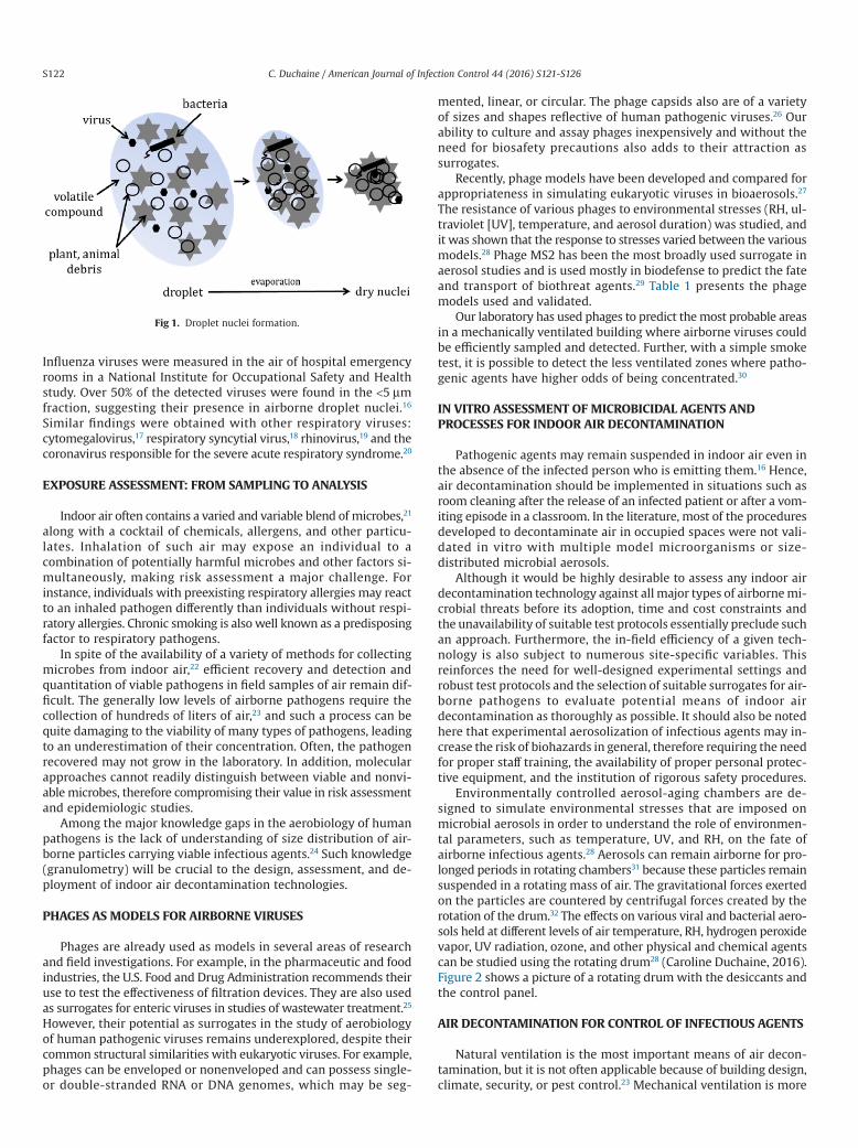
Influenza viruses were measured in the air of hospital emergencyrooms in a National Institute for Occupational Safety and Healthstudy. Over 50% of the detected viruses were found in the <5 μmfraction, suggesting their presence in airborne droplet nuclei.16
Similar findings were obtained with other respiratory viruses:cytomegalovirus,17 respiratory syncytial virus,18 rhinovirus,19 and thecoronavirus responsible for the severe acute respiratory syndrome.20
EXPOSURE ASSESSMENT: FROM SAMPLING TO ANALYSIS
Indoor air often contains a varied and variable blend of microbes,21
along with a cocktail of chemicals, allergens, and other particu-lates. Inhalation of such air may expose an individual to acombination of potentially harmful microbes and other factors si-multaneously, making risk assessment a major challenge. Forinstance, individuals with preexisting respiratory allergies may reactto an inhaled pathogen differently than individuals without respi-ratory allergies. Chronic smoking is alsowell known as a predisposingfactor to respiratory pathogens.
In spite of the availability of a variety of methods for collectingmicrobes from indoor air,22 efficient recovery and detection andquantitation of viable pathogens in field samples of air remain dif-ficult. The generally low levels of airborne pathogens require thecollection of hundreds of liters of air,23 and such a process can bequite damaging to the viability of many types of pathogens, leadingto an underestimation of their concentration. Often, the pathogenrecovered may not grow in the laboratory. In addition, molecularapproaches cannot readily distinguish between viable and nonvi-ablemicrobes, therefore compromising their value in risk assessmentand epidemiologic studies.
Among the major knowledge gaps in the aerobiology of humanpathogens is the lack of understanding of size distribution of air-borne particles carrying viable infectious agents.24 Such knowledge(granulometry) will be crucial to the design, assessment, and de-ployment of indoor air decontamination technologies.
PHAGES AS MODELS FOR AIRBORNE VIRUSES
Phages are already used as models in several areas of researchand field investigations. For example, in the pharmaceutic and foodindustries, the U.S. Food and Drug Administration recommends theiruse to test the effectiveness of filtration devices. They are also usedas surrogates for enteric viruses in studies of wastewater treatment.25
However, their potential as surrogates in the study of aerobiologyof human pathogenic viruses remains underexplored, despite theircommon structural similarities with eukaryotic viruses. For example,phages can be enveloped or nonenveloped and can possess single-or double-stranded RNA or DNA genomes, which may be seg-
mented, linear, or circular. The phage capsids also are of a varietyof sizes and shapes reflective of human pathogenic viruses.26 Ourability to culture and assay phages inexpensively and without theneed for biosafety precautions also adds to their attraction assurrogates.
Recently, phage models have been developed and compared forappropriateness in simulating eukaryotic viruses in bioaerosols.27
The resistance of various phages to environmental stresses (RH, ul-traviolet [UV], temperature, and aerosol duration) was studied, andit was shown that the response to stresses varied between the variousmodels.28 Phage MS2 has been the most broadly used surrogate inaerosol studies and is used mostly in biodefense to predict the fateand transport of biothreat agents.29 Table 1 presents the phagemodels used and validated.
Our laboratory has used phages to predict themost probable areasin a mechanically ventilated building where airborne viruses couldbe efficiently sampled and detected. Further, with a simple smoketest, it is possible to detect the less ventilated zones where patho-genic agents have higher odds of being concentrated.30
IN VITRO ASSESSMENT OF MICROBICIDAL AGENTS ANDPROCESSES FOR INDOOR AIR DECONTAMINATION
Pathogenic agents may remain suspended in indoor air even inthe absence of the infected person who is emitting them.16 Hence,air decontamination should be implemented in situations such asroom cleaning after the release of an infected patient or after a vom-iting episode in a classroom. In the literature, most of the proceduresdeveloped to decontaminate air in occupied spaces were not vali-dated in vitro with multiple model microorganisms or size-distributed microbial aerosols.
Although it would be highly desirable to assess any indoor airdecontamination technology against allmajor types of airbornemi-crobial threats before its adoption, time and cost constraints andthe unavailability of suitable test protocols essentially preclude suchan approach. Furthermore, the in-field efficiency of a given tech-nology is also subject to numerous site-specific variables. Thisreinforces the need for well-designed experimental settings androbust test protocols and the selection of suitable surrogates for air-borne pathogens to evaluate potential means of indoor airdecontamination as thoroughly as possible. It should also be notedhere that experimental aerosolization of infectious agents may in-crease the risk of biohazards in general, therefore requiring the needfor proper staff training, the availability of proper personal protec-tive equipment, and the institution of rigorous safety procedures.
Environmentally controlled aerosol-aging chambers are de-signed to simulate environmental stresses that are imposed onmicrobial aerosols in order to understand the role of environmen-tal parameters, such as temperature, UV, and RH, on the fate ofairborne infectious agents.28 Aerosols can remain airborne for pro-longed periods in rotating chambers31 because these particles remainsuspended in a rotating mass of air. The gravitational forces exertedon the particles are countered by centrifugal forces created by therotation of the drum.32 The effects on various viral and bacterial aero-sols held at different levels of air temperature, RH, hydrogen peroxidevapor, UV radiation, ozone, and other physical and chemical agentscan be studied using the rotating drum28 (Caroline Duchaine, 2016).Figure 2 shows a picture of a rotating drumwith the desiccants andthe control panel.
AIR DECONTAMINATION FOR CONTROL OF INFECTIOUS AGENTS
Natural ventilation is the most important means of air decon-tamination, but it is not often applicable because of building design,climate, security, or pest control.23 Mechanical ventilation is more
Fig 1. Droplet nuclei formation.
S122 C. Duchaine / American Journal of Infection Control 44 (2016) S121-S126
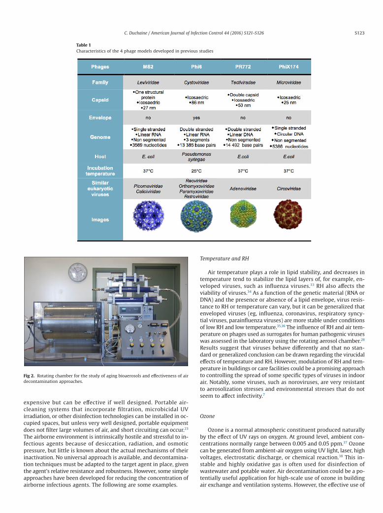
expensive but can be effective if well designed. Portable air-cleaning systems that incorporate filtration, microbicidal UVirradiation, or other disinfection technologies can be installed in oc-cupied spaces, but unless very well designed, portable equipmentdoes not filter large volumes of air, and short circuiting can occur.23
The airborne environment is intrinsically hostile and stressful to in-fectious agents because of desiccation, radiation, and osmoticpressure, but little is known about the actual mechanisms of theirinactivation. No universal approach is available, and decontamina-tion techniques must be adapted to the target agent in place, giventhe agent’s relative resistance and robustness. However, some simpleapproaches have been developed for reducing the concentration ofairborne infectious agents. The following are some examples.
Temperature and RH
Air temperature plays a role in lipid stability, and decreases intemperature tend to stabilize the lipid layers of, for example, en-veloped viruses, such as influenza viruses.33 RH also affects theviability of viruses.34 As a function of the genetic material (RNA orDNA) and the presence or absence of a lipid envelope, virus resis-tance to RH or temperature can vary, but it can be generalized thatenveloped viruses (eg, influenza, coronavirus, respiratory syncy-tial viruses, parainfluenza viruses) are more stable under conditionsof low RH and low temperature.35,36 The influence of RH and air tem-perature on phages used as surrogates for human pathogenic viruseswas assessed in the laboratory using the rotating aerosol chamber.28
Results suggest that viruses behave differently and that no stan-dard or generalized conclusion can be drawn regarding the virucidaleffects of temperature and RH. However, modulation of RH and tem-perature in buildings or care facilities could be a promising approachto controlling the spread of some specific types of viruses in indoorair. Notably, some viruses, such as noroviruses, are very resistantto aerosolization stresses and environmental stresses that do notseem to affect infectivity.7
Ozone
Ozone is a normal atmospheric constituent produced naturallyby the effect of UV rays on oxygen. At ground level, ambient con-centrations normally range between 0.005 and 0.05 ppm.37 Ozonecan be generated from ambient-air oxygen using UV light, laser, highvoltages, electrostatic discharge, or chemical reaction.38 This in-stable and highly oxidative gas is often used for disinfection ofwastewater and potable water. Air decontamination could be a po-tentially useful application for high-scale use of ozone in buildingair exchange and ventilation systems. However, the effective use of
Table 1Characteristics of the 4 phage models developed in previous studies
Fig 2. Rotating chamber for the study of aging bioaerosols and effectiveness of airdecontamination approaches.
S123C. Duchaine / American Journal of Infection Control 44 (2016) S121-S126
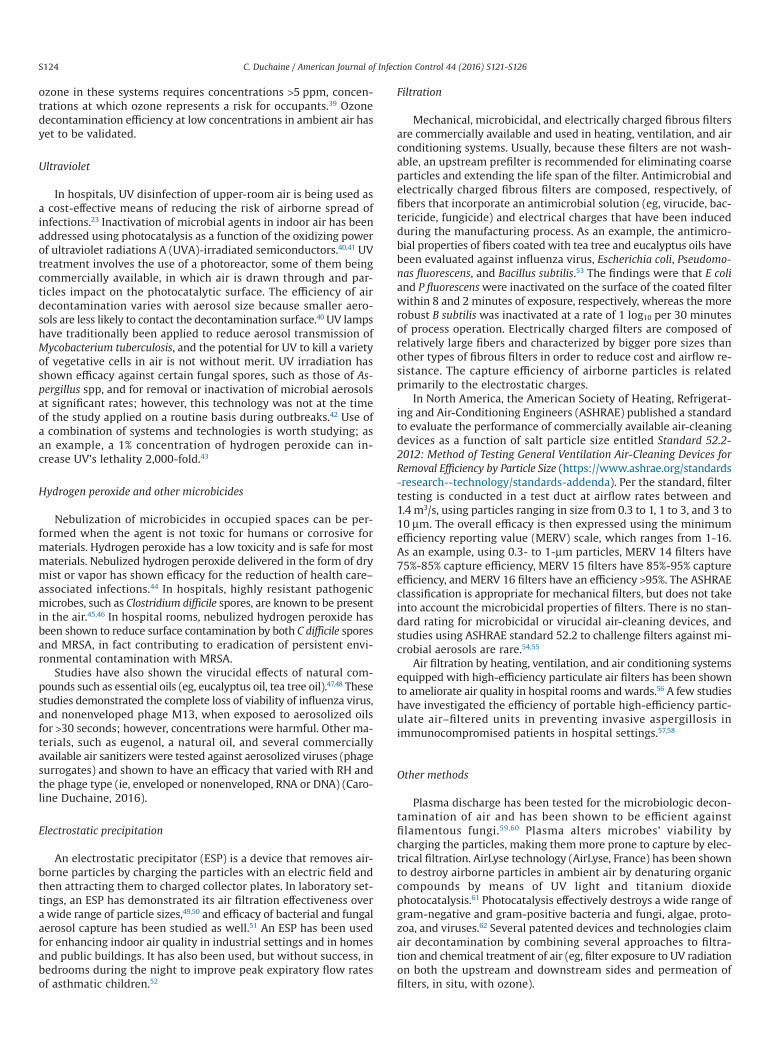
ozone in these systems requires concentrations >5 ppm, concen-trations at which ozone represents a risk for occupants.39 Ozonedecontamination efficiency at low concentrations in ambient air hasyet to be validated.
Ultraviolet
In hospitals, UV disinfection of upper-room air is being used asa cost-effective means of reducing the risk of airborne spread ofinfections.23 Inactivation of microbial agents in indoor air has beenaddressed using photocatalysis as a function of the oxidizing powerof ultraviolet radiations A (UVA)-irradiated semiconductors.40,41 UVtreatment involves the use of a photoreactor, some of them beingcommercially available, in which air is drawn through and par-ticles impact on the photocatalytic surface. The efficiency of airdecontamination varies with aerosol size because smaller aero-sols are less likely to contact the decontamination surface.40 UV lampshave traditionally been applied to reduce aerosol transmission ofMycobacterium tuberculosis, and the potential for UV to kill a varietyof vegetative cells in air is not without merit. UV irradiation hasshown efficacy against certain fungal spores, such as those of As-pergillus spp, and for removal or inactivation of microbial aerosolsat significant rates; however, this technology was not at the timeof the study applied on a routine basis during outbreaks.42 Use ofa combination of systems and technologies is worth studying; asan example, a 1% concentration of hydrogen peroxide can in-crease UV’s lethality 2,000-fold.43
Hydrogen peroxide and other microbicides
Nebulization of microbicides in occupied spaces can be per-formed when the agent is not toxic for humans or corrosive formaterials. Hydrogen peroxide has a low toxicity and is safe for mostmaterials. Nebulized hydrogen peroxide delivered in the form of drymist or vapor has shown efficacy for the reduction of health care–associated infections.44 In hospitals, highly resistant pathogenicmicrobes, such as Clostridium difficile spores, are known to be presentin the air.45,46 In hospital rooms, nebulized hydrogen peroxide hasbeen shown to reduce surface contamination by both C difficile sporesand MRSA, in fact contributing to eradication of persistent envi-ronmental contamination with MRSA.
Studies have also shown the virucidal effects of natural com-pounds such as essential oils (eg, eucalyptus oil, tea tree oil).47,48 Thesestudies demonstrated the complete loss of viability of influenza virus,and nonenveloped phage M13, when exposed to aerosolized oilsfor >30 seconds; however, concentrations were harmful. Other ma-terials, such as eugenol, a natural oil, and several commerciallyavailable air sanitizers were tested against aerosolized viruses (phagesurrogates) and shown to have an efficacy that varied with RH andthe phage type (ie, enveloped or nonenveloped, RNA or DNA) (Caro-line Duchaine, 2016).
Electrostatic precipitation
An electrostatic precipitator (ESP) is a device that removes air-borne particles by charging the particles with an electric field andthen attracting them to charged collector plates. In laboratory set-tings, an ESP has demonstrated its air filtration effectiveness overa wide range of particle sizes,49,50 and efficacy of bacterial and fungalaerosol capture has been studied as well.51 An ESP has been usedfor enhancing indoor air quality in industrial settings and in homesand public buildings. It has also been used, but without success, inbedrooms during the night to improve peak expiratory flow ratesof asthmatic children.52
Filtration
Mechanical, microbicidal, and electrically charged fibrous filtersare commercially available and used in heating, ventilation, and airconditioning systems. Usually, because these filters are not wash-able, an upstream prefilter is recommended for eliminating coarseparticles and extending the life span of the filter. Antimicrobial andelectrically charged fibrous filters are composed, respectively, offibers that incorporate an antimicrobial solution (eg, virucide, bac-tericide, fungicide) and electrical charges that have been inducedduring the manufacturing process. As an example, the antimicro-bial properties of fibers coated with tea tree and eucalyptus oils havebeen evaluated against influenza virus, Escherichia coli, Pseudomo-nas fluorescens, and Bacillus subtilis.53 The findings were that E coliand P fluorescenswere inactivated on the surface of the coated filterwithin 8 and 2 minutes of exposure, respectively, whereas the morerobust B subtilis was inactivated at a rate of 1 log10 per 30 minutesof process operation. Electrically charged filters are composed ofrelatively large fibers and characterized by bigger pore sizes thanother types of fibrous filters in order to reduce cost and airflow re-sistance. The capture efficiency of airborne particles is relatedprimarily to the electrostatic charges.
In North America, the American Society of Heating, Refrigerat-ing and Air-Conditioning Engineers (ASHRAE) published a standardto evaluate the performance of commercially available air-cleaningdevices as a function of salt particle size entitled Standard 52.2-2012: Method of Testing General Ventilation Air-Cleaning Devices forRemoval Efficiency by Particle Size (https://www.ashrae.org/standards-research--technology/standards-addenda). Per the standard, filtertesting is conducted in a test duct at airflow rates between and1.4 m3/s, using particles ranging in size from 0.3 to 1, 1 to 3, and 3 to10 μm. The overall efficacy is then expressed using the minimumefficiency reporting value (MERV) scale, which ranges from 1-16.As an example, using 0.3- to 1-μm particles, MERV 14 filters have75%-85% capture efficiency, MERV 15 filters have 85%-95% captureefficiency, andMERV 16 filters have an efficiency >95%. The ASHRAEclassification is appropriate for mechanical filters, but does not takeinto account the microbicidal properties of filters. There is no stan-dard rating for microbicidal or virucidal air-cleaning devices, andstudies using ASHRAE standard 52.2 to challenge filters against mi-crobial aerosols are rare.54,55
Air filtration by heating, ventilation, and air conditioning systemsequipped with high-efficiency particulate air filters has been shownto ameliorate air quality in hospital rooms andwards.56 A few studieshave investigated the efficiency of portable high-efficiency partic-ulate air–filtered units in preventing invasive aspergillosis inimmunocompromised patients in hospital settings.57,58
Other methods
Plasma discharge has been tested for the microbiologic decon-tamination of air and has been shown to be efficient againstfilamentous fungi.59,60 Plasma alters microbes’ viability bycharging the particles, making themmore prone to capture by elec-trical filtration. AirLyse technology (AirLyse, France) has been shownto destroy airborne particles in ambient air by denaturing organiccompounds by means of UV light and titanium dioxidephotocatalysis.61 Photocatalysis effectively destroys a wide range ofgram-negative and gram-positive bacteria and fungi, algae, proto-zoa, and viruses.62 Several patented devices and technologies claimair decontamination by combining several approaches to filtra-tion and chemical treatment of air (eg, filter exposure to UV radiationon both the upstream and downstream sides and permeation offilters, in situ, with ozone).
S124 C. Duchaine / American Journal of Infection Control 44 (2016) S121-S126
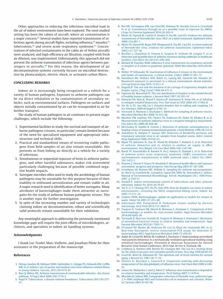
Other approaches to reducing the infectious microbial load inthe air of indoor environments have been explored. Themost studiedsetting has been the cabins of aircraft, where air contamination isa major concern.63 Several studies have reported transmission of in-fectious agents during aircraft flights, including influenza,64 measles,65
tuberculosis,66 and severe acute respiratory syndrome.67 Concen-trations of selected contaminants in the cabin air of Airbus aircraftswere analyzed, and high-efficiency air filtration, coupled with freshair dilution, was implemented. Unfortunately, this approach did notprevent the airborne transmission of infectious agents between pas-sengers in aircrafts.68 For this reason, the development of airdecontamination systems currently focuses on microbial destruc-tion by photocatalysis, electric shock, or activated carbon fibers.
CONCLUDING REMARKS
Indoor air is increasingly being recognized as a vehicle for avariety of human pathogens. Exposure to airborne pathogens canbe via direct inhalation or by contamination of secondary ve-hicles, such as environmental surfaces. Pathogens on surfaces andobjects initially contaminated by air can be resuspended in air forfurther transport.
The study of human pathogens in air continues to present majorchallenges, which include the following:
1. Experimental facilities to study the survival and transport of air-borne pathogens (viruses, in particular) remain limited becauseof the need for specialized equipment and appropriate infra-structure and technical skills.
2. Practical and standardized means of recovering viable patho-gens from field samples of air also remain unavailable; thisprevents us from linking air directly as a vehicle for a varietyof infections.
3. Simultaneous or sequential exposure of hosts to airborne patho-gens, and other harmful substances, makes risk assessmentparticularly challenging because of possible combined nega-tive health impacts.
4. Surrogatemicrobes often used to study the aerobiology of humanpathogens may be unsuitable for this purpose because of theirinability to withstand aerosolization and remain viable in air.Amajor research need is identification of better surrogates. Manyattributes of bacteriophages make them attractive as surro-gates for the study of airborne human pathogenic viruses. Thisis another topic for further investigation.
5. In spite of the increasing number and variety of technologiesclaiming indoor air decontamination, robust and scientificallyvalid protocols remain unavailable for their validation.
Anymeaningful approach to addressing the previouslymentionedknowledge gaps will require the joint efforts of microbiologists, ar-chitects, and specialists in indoor air handling systems.
Acknowledgments
I thank Luc Trudel, Marc Veillette, and Jonathan Pilote for theirassistance in the preparation of the manuscript.
References
1. Ortega-Sanchez IR, Molinari NAM, Fairbrother G, Szilagyi PG, Edwards KM, GriffinMR, et al. Indirect, out-of-pocket andmedical costs from influenza-related illnessin young children. Vaccine, 2012;30:4175-81
2. Roy CJ, Milton DK. Airborne transmission of communicable infection—the elusivepathway. N Engl J Med 2004;350:1710-2.
3. Fogel N. Tuberculosis: a disease without boundaries. Tuberculosis 2015;95:527-31.
4. Bos ME, Verstappen KM, van Cleef BA, DohmenW, Dorado-García A, GravelandH, et al. Transmission through air as a possible route of exposure for MRSA.J Expo Sci Environ Epidemiol 2016;26:263-9.
5. Marks PJ, Vipond IB, Carlisle D, Deakin D, Fey RE, Caul EO. Evidence for airbornetransmission of Norwalk-like virus (NLV) in a hotel restaurant. Epidemiol Infect2000;124:481-7.
6. Marks PJ, Vipond IB, Regan FM,Wedgwood K, Fey RE, Caul EO. A school outbreakof Norwalk-like virus: evidence for airborne transmission. Epidemiol Infect2003;131:727-36.
7. Bonifait L, Charlebois R, Vimont A, Turgeon N, Veillette M, Longtin Y, et al.Detection and quantification of airborne norovirus during outbreaks in healthcarefacilities. Clin Infect Dis 2015;61:299-304.
8. Richard M, Fouchier RAM. Influenza A virus transmission via respiratory aerosolsor droplets as it relates to pandemic potential. FEMS Microbiol Rev 2016;40:68-85.
9. Weber TP Stilianakis NI. Inactivation of influenza A viruses in the environmentand modes of transmission: a critical review. J Infect 2008;57:361-73.
10. Heimbuch BK, Wallace WH, Balzli CL, Laning ML, Harnish DA, Wander JD.Bioaerosol exposure to personnel in a clinical environment absent patients.J Occup Environ Hyg 2016;13:D11-5.
11. Duguid JP. The size and the duration of air-carriage of respiratory droplets anddroplet-nuclei. J Hyg (Lond) 1946;44:471-9.
12. Papineni RS, Rosenthal FS. The size distribution of droplets in the exhaled breathof healthy human subjects. J Aerosol Med 1997;10:105-16.
13. Edwards DA, Man JC, Brand P, Katstra JP, Sommerer K, Stone HA, et al. Inhalingto mitigate exhaled bioaerosols. Proc Natl Acad Sci USA 2004;101:17383-8.
14. Xie XJ, Li YG, Sun HQ, Liu L. Exhaled droplets due to talking and coughing. J RSoc Interface 2009;6(Suppl 6):S703-14.
15. Verreault D, Moineau S, Duchaine C. Methods for sampling of airborne viruses.Microbiol Mol Biol Rev 2008;72:413-44.
16. Blachere FM, Lindsley WG, Pearce TA, Anderson SE, Fisher M, Khakoo R, et al.Measurement of airborne influenza virus in a hospital emergency department.Clin Infect Dis 2009;48:438-40.
17. McCluskey R, Sandin R, Greene J. Detection of airborne cytomegalovirus inhospital rooms of immunocompromised patients. J Virol Methods 1996;56:115-8.
18. Aintablian N, Walpita P, Sawyer MH. Detection of Bordetella pertussis andrespiratory syncytial virus in air samples from hospital rooms. Infect ControlHosp Epidemiol 1998;19:918-23.
19. Myatt TA, Johnston SL, Zuo ZF, Wand M, Kebadze T, Rudnick S, et al. Detectionof airborne rhinovirus and its relation to outdoor air supply in officeenvironments. Am J Respir Crit Care Med 2004;169:1187-90.
20. Booth TF, Kournikakis B, Bastien N, Ho J, Kobasa D, Stadnyk L, et al. Detectionof airborne severe acute respiratory syndrome (SARS) coronavirus andenvironmental contamination in SARS outbreak units. J Infect Dis 2005;191:1472-7.
21. Douwes J, Thorne P, Pearce N, Heederik D. Bioaerosol health effects and exposureassessment: progress and prospects. Ann Occup Hyg 2003;47:187-200.
22. Grinshpun SA, Buttner MP, Willeke K. Sampling for airborne microorganisms.In: Hurst CJ, Crawford RL, Garland JL, Lipson DA, Mills AL, Stetzenbach L, editors.Manual of Environmental Microbiology. 3rd ed. Washington (DC): ASM Press;2007. p. 939-51.
23. Nardell E. Indoor environmental control of tuberculosis and other airborneinfections. Indoor Air 2016;26:79-87.
24. Xie X, Li Y, Chwang ATY, Ho PL, Seto WH. How far droplets can move in indoorenvironments—revisiting the Wells evaporation-falling curve. Indoor Air2007;17:211-25.
25. Grabow WOK. Bacteriophages: update on application as models for viruses inwater. Water SA 2001;27:251-68.
26. Ackermann HW, Prangishvili D. Prokaryote viruses studied by electronmicroscopy. Arch Virol 2012;157:1843-9.
27. Turgeon N, Toulouse MJ, Martel B, Moineau S, Duchaine C. Comparison of fivebacteriophages as models for viral aerosol studies. Appl Environ Microbiol2014;80:4242-50.
28. Verreault D, Marcoux-Voiselle M, Turgeon N, Moineau S, Duchaine C. Resistanceof aerosolized bacterial viruses to relative humidity and temperature. ApplEnviron Microbiol 2015;81:7305-11.
29. O’Connell KP, Bucher JR, Anderson PE, Cao CJ, Khan AS, Gostomski MV, et al.Real-time fluorogenic reverse transcription-PCR assays for detection ofbacteriophage MS2. Appl Environ Microbiol 2006;72:478-83.
30. Audet M, Girard M, Belzile M, Godbout S, Duchaine C Understanding aerosolizedviral particles behaviour in a mechanically ventilated agricultural building usingnebulized bacteriophages. Presented at American Association for AerosolResearch 32nd Annual Conference; 2013 Sept 30-Oct 4; Portland, OR.
31. Goldberg LJ, Watkins HM, Boerke EE, Chatigny MA. The use of a rotating drumfor the study of aerosols over extended periods of time. Am J Hyg 1958;68:85-93.
32. Gruel RL, Reid CR, Allemann RT. The optimum rate of drum rotation for aerosolaging. J Aerosol Sci 1987;18:17-22.
33. Polozov IV, Bezrukov L, Gawrisch K. Progressive ordering with decreasingtemperature of the phospholipids of influenza virus. Nat Chem Biol 2008;4:248-55.
34. Lowen AC, Mubareka S, Steel J, Palese P. Influenza virus transmission is dependenton relative humidity and temperature. PLoS Pathog 2007;3:1470-6.
35. Meschke JS, Sobsey MD. Comparative reduction of Norwalk virus, poliovirus type1, F+ RNA coliphage MS2 and Escherichia coli in miniature soil columns. WaterSci Technol 2003;47:85-90.
S125C. Duchaine / American Journal of Infection Control 44 (2016) S121-S126
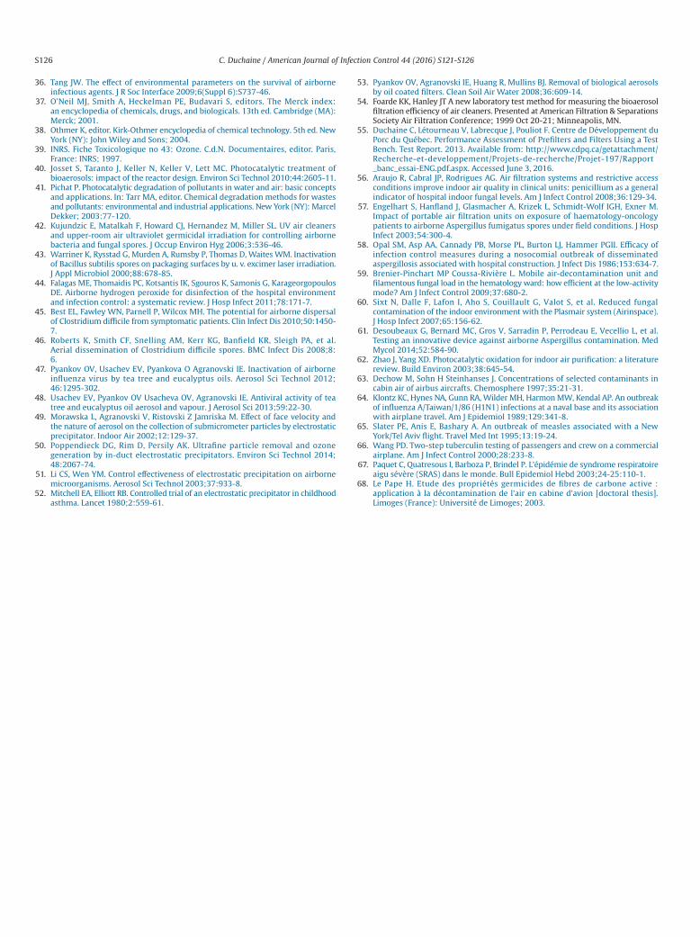
36. Tang JW. The effect of environmental parameters on the survival of airborneinfectious agents. J R Soc Interface 2009;6(Suppl 6):S737-46.
37. O’Neil MJ, Smith A, Heckelman PE, Budavari S, editors. The Merck index:an encyclopedia of chemicals, drugs, and biologicals. 13th ed. Cambridge (MA):Merck; 2001.
38. Othmer K, editor. Kirk-Othmer encyclopedia of chemical technology. 5th ed. NewYork (NY): John Wiley and Sons; 2004.
39. INRS. Fiche Toxicologique no 43: Ozone. C.d.N. Documentaires, editor. Paris,France: INRS; 1997.
40. Josset S, Taranto J, Keller N, Keller V, Lett MC. Photocatalytic treatment ofbioaerosols: impact of the reactor design. Environ Sci Technol 2010;44:2605-11.
41. Pichat P. Photocatalytic degradation of pollutants in water and air: basic conceptsand applications. In: Tarr MA, editor. Chemical degradation methods for wastesand pollutants: environmental and industrial applications. New York (NY): MarcelDekker; 2003:77-120.
42. Kujundzic E, Matalkah F, Howard CJ, Hernandez M, Miller SL. UV air cleanersand upper-room air ultraviolet germicidal irradiation for controlling airbornebacteria and fungal spores. J Occup Environ Hyg 2006;3:536-46.
43. Warriner K, Rysstad G, Murden A, Rumsby P, Thomas D, WaitesWM. Inactivationof Bacillus subtilis spores on packaging surfaces by u. v. excimer laser irradiation.J Appl Microbiol 2000;88:678-85.
44. Falagas ME, Thomaidis PC, Kotsantis IK, Sgouros K, Samonis G, KarageorgopoulosDE. Airborne hydrogen peroxide for disinfection of the hospital environmentand infection control: a systematic review. J Hosp Infect 2011;78:171-7.
45. Best EL, Fawley WN, Parnell P, Wilcox MH. The potential for airborne dispersalof Clostridium difficile from symptomatic patients. Clin Infect Dis 2010;50:1450-7.
46. Roberts K, Smith CF, Snelling AM, Kerr KG, Banfield KR, Sleigh PA, et al.Aerial dissemination of Clostridium difficile spores. BMC Infect Dis 2008;8:6.
47. Pyankov OV, Usachev EV, Pyankova O Agranovski IE. Inactivation of airborneinfluenza virus by tea tree and eucalyptus oils. Aerosol Sci Technol 2012;46:1295-302.
48. Usachev EV, Pyankov OV Usacheva OV, Agranovski IE. Antiviral activity of teatree and eucalyptus oil aerosol and vapour. J Aerosol Sci 2013;59:22-30.
49. Morawska L, Agranovski V, Ristovski Z Jamriska M. Effect of face velocity andthe nature of aerosol on the collection of submicrometer particles by electrostaticprecipitator. Indoor Air 2002;12:129-37.
50. Poppendieck DG, Rim D, Persily AK. Ultrafine particle removal and ozonegeneration by in-duct electrostatic precipitators. Environ Sci Technol 2014;48:2067-74.
51. Li CS, Wen YM. Control effectiveness of electrostatic precipitation on airbornemicroorganisms. Aerosol Sci Technol 2003;37:933-8.
52. Mitchell EA, Elliott RB. Controlled trial of an electrostatic precipitator in childhoodasthma. Lancet 1980;2:559-61.
53. Pyankov OV, Agranovski IE, Huang R, Mullins BJ. Removal of biological aerosolsby oil coated filters. Clean Soil Air Water 2008;36:609-14.
54. Foarde KK, Hanley JT A new laboratory test method for measuring the bioaerosolfiltration efficiency of air cleaners. Presented at American Filtration & SeparationsSociety Air Filtration Conference; 1999 Oct 20-21; Minneapolis, MN.
55. Duchaine C, Létourneau V, Labrecque J, Pouliot F. Centre de Développement duPorc du Québec. Performance Assessment of Prefilters and Filters Using a TestBench. Test Report. 2013. Available from: http://www.cdpq.ca/getattachment/Recherche-et-developpement/Projets-de-recherche/Projet-197/Rapport_banc_essai-ENG.pdf.aspx. Accessed June 3, 2016.
56. Araujo R, Cabral JP, Rodrigues AG. Air filtration systems and restrictive accessconditions improve indoor air quality in clinical units: penicillium as a generalindicator of hospital indoor fungal levels. Am J Infect Control 2008;36:129-34.
57. Engelhart S, Hanfland J, Glasmacher A, Krizek L, Schmidt-Wolf IGH, Exner M.Impact of portable air filtration units on exposure of haematology-oncologypatients to airborne Aspergillus fumigatus spores under field conditions. J HospInfect 2003;54:300-4.
58. Opal SM, Asp AA, Cannady PB, Morse PL, Burton LJ, Hammer PGII. Efficacy ofinfection control measures during a nosocomial outbreak of disseminatedaspergillosis associated with hospital construction. J Infect Dis 1986;153:634-7.
59. Brenier-Pinchart MP Coussa-Rivière L. Mobile air-decontamination unit andfilamentous fungal load in the hematology ward: how efficient at the low-activitymode? Am J Infect Control 2009;37:680-2.
60. Sixt N, Dalle F, Lafon I, Aho S, Couillault G, Valot S, et al. Reduced fungalcontamination of the indoor environment with the Plasmair system (Airinspace).J Hosp Infect 2007;65:156-62.
61. Desoubeaux G, Bernard MC, Gros V, Sarradin P, Perrodeau E, Vecellio L, et al.Testing an innovative device against airborne Aspergillus contamination. MedMycol 2014;52:584-90.
62. Zhao J, Yang XD. Photocatalytic oxidation for indoor air purification: a literaturereview. Build Environ 2003;38:645-54.
63. Dechow M, Sohn H Steinhanses J. Concentrations of selected contaminants incabin air of airbus aircrafts. Chemosphere 1997;35:21-31.
64. Klontz KC, Hynes NA, Gunn RA,Wilder MH, HarmonMW, Kendal AP. An outbreakof influenza A/Taiwan/1/86 (H1N1) infections at a naval base and its associationwith airplane travel. Am J Epidemiol 1989;129:341-8.
65. Slater PE, Anis E, Bashary A. An outbreak of measles associated with a NewYork/Tel Aviv flight. Travel Med Int 1995;13:19-24.
66. Wang PD. Two-step tuberculin testing of passengers and crew on a commercialairplane. Am J Infect Control 2000;28:233-8.
67. Paquet C, Quatresous I, Barboza P, Brindel P. L’épidémie de syndrome respiratoireaigu sévère (SRAS) dans le monde. Bull Epidemiol Hebd 2003;24-25:110-1.
68. Le Pape H. Etude des propriétés germicides de fibres de carbone active :application à la décontamination de l’air en cabine d’avion [doctoral thesis].Limoges (France): Université de Limoges; 2003.
S126 C. Duchaine / American Journal of Infection Control 44 (2016) S121-S126
Major Article
Mathematical modeling and simulation of bacterial distribution inan aerobiology chamber using computational fluid dynamics
Bahram Zargar BSc(Engg), MSc(Engg), PhD a, Farshad M. Kashkooli BSc(Engg), MSc(Engg) b,M. Soltani BSc(Engg), MSc(Engg), PhD b,c, Kathryn E. Wright MA, MSc, PhD a,M. Khalid Ijaz DVM, MSc(Honors), PhD d,e, Syed A. Sattar MSc, Dip Bact, MS, PhD f,*a Department of Biochemistry, Microbiology, and Immunology, University of Ottawa, Ottawa, Ontario, Canadab Department of Mechanical Engineering, K. N. T. University of Technology, Tehran, Iranc Division of Nuclear Medicine, Department of Radiology and Radiological Science, Johns Hopkins University, School of Medicine, Baltimore, MDd RB, Montvale, NJe Department of Biology, Medgar Evers College of the City University of New York (CUNY), Brooklyn, NYf Professor Emeritus of Microbiology, Faculty of Medicine, University of Ottawa, Ottawa, ON, Canada
Key Words:Airborne spread of infectious agentsDistribution of particles in indoor airSampling air for infectious agentsPredictive modeling of bacterialdistribution in an aerobiology chamber
Background: Computer-aided design and draft, along with computer-aided engineering software, are usedwidely in different fields to create, modify, analyze, and optimize designs.Methods: We used computer-aided design and draft software to create a 3-dimensional model of an aero-biology chamber built in accordance with the specifications of the 2012 guideline from the EnvironmentalProtection Agency for studies on survival and inactivation of microbial pathogens in indoor air. The modelwas used to optimize the chamber’s airflow design and the distribution of aerosolized bacteria inside it.Results: The findings led to the identification of an appropriate fan and its location inside the chamberfor uniform distribution of microbes introduced into the air, suitability of air sample collection from thecenter of the chamber alone as representative of its bacterial content, and determination of the influ-ence of room furnishings on airflow patterns inside the chamber.Conclusions: Theincorporationof thismodelingstudy’sfindingscouldfurther improvethedesignof thechamberand the predictive value of the experimental data using it. Further, it could make data generation faster andmore economical by eliminating the need for collecting air samples from multiple sites in the chamber.© 2016 Association for Professionals in Infection Control and Epidemiology, Inc. Published by Elsevier
Inc. All rights reserved.
INTRODUCTION
Prediction of particle transport in turbulent flow is essential indifferent fields, such as dispersion of passive or reactive particlesin turbulent media and in studying air pollution.1 For example, we
are exposed to airborne particulates in workplaces, homes, and otherindoor settings.2 The fate and deposition of such particulates indoorshave substantial implications for human and animal health, cleanrooms, and air decontamination.3-5 Therefore, a good understand-ing of the particle-laden turbulent flow is important in addressingindoor air quality issues and in controlling particle dispersion.
Mitigating the spread of microbial contaminants by indoor airis an essential design consideration for homes, biomedical and healthcare facilities, and other public settings. Once airborne, the move-ment of microbes is difficult to control because they may becomerapidly dispersed by air movement or adhere to other surfaces fortravel with them.6,7 Ventilation, either natural or mechanical, canprovide adequate air exchanges to reduce the risk for airborne mi-crobial spread; however, mechanical ventilation, particularly withconditioning, can be expensive.8 According to the Guidelines for Designand Construction of Hospital and Health Care Facilities,9 6-15 airchanges per hour are needed to maintain a healthful environmentwhile reducing exposure to harmful chemicals and microbes. This
* Address correspondence to Syed A. Sattar, MSc, Dip Bact, MS, PhD, ProfessorEmeritus of Microbiology, Faculty of Medicine, University of Ottawa, 451 Smyth Rd,Ottawa, ON K1H 8M5, Canada.
E-mail address: [email protected] (S.A. Sattar).Funding/Support: This paper was presented at a workshop organized under the
auspices of ASTM International’s biannual meeting held in April 2016. Publicationof this supplement is primarily supported by RB, Montvale, New Jersey, with addi-tional support from MicroBioTest, a division of Microbac Laboratories, Inc., Sterling,Virginia. The City University of New York (CUNY) and the University of Ottawa, Ottawa,Canada, are academic sponsors. Editorial support was provided by Ashley O’Dunne,PhD; Shannon O’Sullivan, ELS; and Alanna Franchetti, ELS of Medergy (Yardley, PA),and funded by RB.
0196-6553/© 2016 Association for Professionals in Infection Control and Epidemiology, Inc. Published by Elsevier Inc. All rights reserved.http://dx.doi.org/10.1016/j.ajic.2016.06.005
American Journal of Infection Control 44 (2016) S127-S137
Contents lists available at ScienceDirect
American Journal of Infection Control
journal homepage: www.aj ic journal .org
American Journal of Infection Control
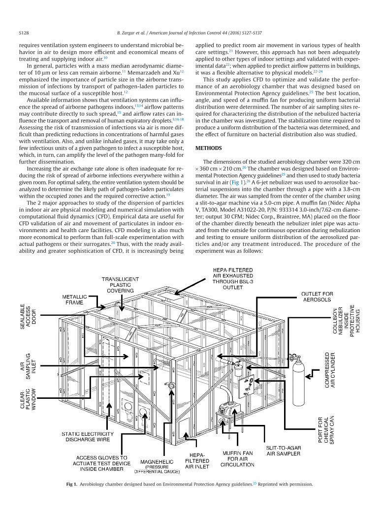
requires ventilation system engineers to understand microbial be-havior in air to design more efficient and economical means oftreating and supplying indoor air.10
In general, particles with a mass median aerodynamic diame-ter of 10 μm or less can remain airborne.11 Memarzadeh and Xu12
emphasized the importance of particle size in the airborne trans-mission of infections by transport of pathogen-laden particles tothe mucosal surface of a susceptible host.12
Available information shows that ventilation systems can influ-ence the spread of airborne pathogens indoors,13,14 airflow patternsmay contribute directly to such spread,15 and airflow rates can in-fluence the transport and removal of human expiratory droplets.5,16-18
Assessing the risk of transmission of infections via air is more dif-ficult than predicting reductions in concentrations of harmful gaseswith ventilation. Also, and unlike inhaled gases, it may take only afew infectious units of a given pathogen to infect a susceptible host,which, in turn, can amplify the level of the pathogen many-fold forfurther dissemination.
Increasing the air exchange rate alone is often inadequate for re-ducing the risk of spread of airborne infections everywhere within agiven room. For optimal safety, the entire ventilation system should beanalyzed to determine the likely path of pathogen-laden particulateswithin the occupied zones and the required corrective action.19
The 2 major approaches to study of the dispersion of particlesin indoor air are physical modeling and numerical simulation withcomputational fluid dynamics (CFD). Empirical data are useful forCFD validation of air and movement of particulates in indoor en-vironments and health care facilities. CFD modeling is also muchmore economical to perform than full-scale experimentation withactual pathogens or their surrogates.20 Thus, with the ready avail-ability and greater sophistication of CFD, it is increasingly being
applied to predict room air movement in various types of healthcare settings.21 However, this approach has not been adequatelyapplied to other types of indoor settings and validated with exper-imental data22; when applied to predict airflow patterns in buildings,it was a flexible alternative to physical models.22-24
This study applies CFD to optimize and validate the perfor-mance of an aerobiology chamber that was designed based onEnvironmental Protection Agency guidelines.25 The best location,angle, and speed of a muffin fan for producing uniform bacterialdistribution were determined. The number of air sampling sites re-quired for characterizing the distribution of the nebulized bacteriain the chamber was investigated. The stabilization time required toproduce a uniform distribution of the bacteria was determined, andthe effect of furniture on bacterial distribution also was studied.
METHODS
The dimensions of the studied aerobiology chamber were 320 cm× 360 cm × 210 cm.26 The chamber was designed based on Environ-mental Protection Agency guidelines25 and then used to study bacteriasurvival in air (Fig 1).26 A 6-jet nebulizer was used to aerosolize bac-terial suspensions into the chamber through a pipe with a 3.8-cmdiameter. The air was sampled from the center of the chamber usinga slit-to-agar machine via a 5.0-cm pipe. A muffin fan (Nidec AlphaV, TA300, Model A31022-20, P/N: 933314 3.0-inch/7.62-cm diame-ter; output 30 CFM; Nidec Corp., Braintree, MA) placed on the floorof the chamber directly beneath the nebulizer inlet pipe was actu-ated from the outside for continuous operation during nebulizationand testing to ensure uniform distribution of the aerosolized par-ticles and/or any treatment introduced. The procedure of theexperiment was as follows:
Fig 1. Aerobiology chamber designed based on Environmental Protection Agency guidelines.25 Reprinted with permission.
S128 B. Zargar et al. / American Journal of Infection Control 44 (2016) S127-S137
• The fan was activated at least 300 seconds before the experi-ment to circulate the air inside the chamber;
• The test bacterial suspension was nebulized into the chamberfor 10 minutes using a 6-jet collison nebulizer; and
• Before sampling, the air in the chamber was allowed to circu-late for 300 seconds following the nebulization process.
MATHEMATICAL MODELING, MATERIALS, ANDNUMERIC METHODOLOGIES
Theoretical background
The Eulerian-Lagrangian approach was implemented directlyusing the discrete phase model. In this approach, the fluid phaseis treated as a continuum material by solving the time-averagedNavier-Stokes equations, and the dispersed phase is solved by track-ing a large number of particles through the calculated flowfield.27 The governing equations are itemized in the followingsections.
Governing equations for the continuous phaseThe continuous gas-flow phase is governed by the following equa-
tions for unsteady compressible flow:4
• Continuity equation:
∂∂
+∇⋅( ) =ρ ρt
u!
0 (1)
• Momentum equation:
D uDt
pρ τ! !( ) = −∇ +∇⋅ (2)
• Energy equation:
∂( )∂
+∇⋅ +( )( ) = ∇⋅ ∇ + ( )( )⋅ρ ρ τEx
u E p k T ueff eff! ! ! (3)
Where ρ, t, u, r, p, τ, E, T, keff, and τeff are fluid density, time, fluidphase velocity, thermodynamic pressure, stress tensor, energy, tem-perature, effective conductivity, and effective stress tensor,respectively.
One of the most common turbulence models, the k-ε Realiz-able turbulence model, was used for turbulence modeling. Theturbulence kinetic energy, k, and its rate of dissipation, ε, are ob-tained from the following transport equations:27,28
∂∂
( ) + ∂∂
( ) = ∂∂
+⎛⎝⎜
⎞⎠⎟
∂∂
⎡⎣⎢
⎤⎦⎥ + + − −
tk
xku
xkx
G G Yi
ji
t
k jk b Mρ ρ µ µ
σρε ++ Sk (4)
∂∂
( ) + ∂∂
( ) = ∂∂
+⎛⎝⎜
⎞⎠⎟
∂∂
⎡⎣⎢
⎤⎦⎥ + −
t xu
x xC S C
jj
j
t
j
ρε ρε µ µσ
ε ρ ρ εε
ε1 2
2
kk
Ck
C G Sb
+
+ +
υεε
ε ε ε1 3 (5)
Where Gk and Gb represent the generation of k due to the meanvelocity gradients and buoyancy, respectively; YM represents the con-tribution of the fluctuating dilation in compressible turbulence tothe overall dissipation rate; σk and σε are the turbulent Prandtlnumbers for k and ε, respectively; and Sk and Sε are user-definedsource terms for k and ε, respectively.
The turbulent (or eddy) viscosity, μt, is computed by combin-ing k and ε as follows:27,28
µ ρεµt Ck=2
(6)
The model constants C1ε, C2ε, Cμ, σk, and σε have the followingvalues: C1ε = 1.44, C2ε = 1.92, Cμ = 0.09, σk = 1.0, and σε = 1.3.27,28
The rotating reference frame was applied only in the rotationalregion by assuming that the region was in a quasisteady state. Thismethod does not explicitly generate model rotation; instead, it gen-erates a constant grid flux in the appropriate conservation equationsby automatically adding the source terms with respect to the Co-riolis force and centrifugal force, which are calculated with equation7 based on the properties of the reference frame.27 Although thismethod underestimates the weak effect, it is appropriate for the flow,which is most likely to be influenced by time-averaged properties.27
A significant amount of simulation time can be saved with thismethod, when compared with simulating the axial flow fan’s ro-tation in a transient state.27
F vr = ×ρω (7)
Where Fr is the body force term due to fan rotation (kg/m2/s2),ρ is the air density (kg/m3), ω is the rotational speed (rad/s), andv is the linear velocity (m/s).
Governing equations for the discrete phaseThe trajectory of the discrete phase is determined by integrat-
ing the force balance on the particle, which equates the particleinertia with forces acting on the particle, and can be written as:27
dudt
F u ug
FpD p
x p
px= −( ) + ( ) +−ρ ρ
ρ(8)
Where u, up, gx, ρp, ρ, and Fx are the fluid phase velocity, parti-cle velocity, gravitational acceleration, particle density, fluid density,and an additional acceleration (force per unit particle mass),respectively. The drag force per unit particle mass (FD) is equalto:
Fd
C ReD
P p
D= 18242
µρ
(9)
Red u up p≡ −ρ
µ(10)
Where μ, dp, CD, and Re are the molecular viscosity of the fluid,particle diameter, drag coefficient, and Reynolds number, respec-tively. The location of each particle, x, is tracked with the followingequation:
dxdt
up= (11)
The air velocity, u, in equation 8 is composed of the time-averaged component, u , and the instantaneous or fluctuating velocitycomponent, u′(t):4,27
u u u t= + ′( ) (12)
The u component is computed using the Reynolds-averagedNavier-Stokes equations with the k-ε Realizable turbulence model.The u′(t) component is computed using a stochastic approach, suchas the discrete randomwalkmodel or eddy lifetimemodel.4 Its valueprevails during the lifetime of the turbulent eddy influencingthe particle and is assumed to obey the Gaussian probabilitydistribution.4 Using the discrete random walk model to calculateu′(t), the particle turbulent dispersion is correlated to the flow k:4,27
S129B. Zargar et al. / American Journal of Infection Control 44 (2016) S127-S137
′( ) =u tkζ 23
(13)
Where the variable ζ is a Gaussian random number.
CFD procedure
Generally, flow simulations in CFD take place in 3 main stages.The first step is preprocessing, which includes geometric model-ing, production of computational domain, and grid generation. Thesecond is the processing step or flow solution with CFD. In the finalstep, called postprocessing, the results are displayed.
Geometric modeling
The geometry of the aerobiology chamber consists of several com-ponents, such as axial flow fan, fan housing, air sampler inlet pipe,outlet pipe for aerosol sampling, and aerobiology chamber walls.Each of these geometries is modeled separately, and eventually, withsuperposition of the modeled geometries, the final complex geom-etry is generated. The computer-aided design and draft model ofthe flow region is built based on the computer-aided design anddraft model of the aerobiology chamber.
The muffin fan, which is an axial flow fan, presents the mostcomplex geometry in the system. The axial flow fan is a tube-axialdevice with 7 forward-swept blades. The dimension of the fanhousing is 80mm × 80mm × 40mm. The tip diameter of fan blades,hub-to-tip ratio, and tip clearance are 76mm, 0.566mm, and 1mm,respectively. Figure 2a presents a 3-dimensional model of the fan.
Thewhole computational model is shown in Figure 2b. To achievea reasonable numeric accuracy, it is divided into its different parts.The computational domain is composed of the axial flow fan, fanhousing, air sampler inlet pipe, outlet pipe for aerosols, and aero-biology chamber, as shown in Figure 2b.
Grid generation
The physical model of the aerobiology chamber comprises severalcomponents with very different geometries. Because of the com-plicated geometry, unstructured tetrahedral grids were adopted forthe whole computational domain. Grids of different sizes were gen-erated for different components and then connected to form thewhole geometry. The computational meshes of the aerobiologychamber were divided into 2 zones: rotating zone and stationaryzone. Special attention was paid to the geometry and meshing ofthe fan, with the greatest emphasis on the blades and root of theblades. The rotating zone was a cylindric mesh with 531,218 cells,as shown in Figure 3. Meshes of the surfaces of the axial flow fan
are also shown in Figure 3. The stationary zone contained 1,125,612cells.
Several versions of the computational mesh were generated totest the grid independence. Results of this study of the grid are shownin Figure 4. The volume flow rates for cases 3-6 were almost thesame. Because case 3 had the lowest computational costs, it wasconsidered the optimum grid number. For a mesh with 1.04 millioncells, the maximum cell skewness was 162, and with mesh size of1.65 million cells, the maximum cell skewness decreased to 154.Thus, the mesh density had an effect on the results for the controlsimulation case.
Solver
Steady and unsteady simulationsConsidering the rotating speed of the axial fan, the airflow was
assumed incompressible.27 The 3-dimensional incompressible Navier-Stokes equations and the k–ε Realizable model were used to modelthe effects of turbulence on the flow field. The enhanced wall func-tion was used for boundary layer calculation. The second-orderupwind differencing format for the convection terms of each
(a) Axial flow fan model (b) Computational domain (aerobiology chamber) model
Fig 2. Three-dimensional model of the axial flow fan and computational domain.
(a) Meshing of axial flow fan surface
(c) Cut view of the volume meshing in rotating cylinder
(b) Rotating cylinder
Fig 3. Grid generation in rotating volume.
S130 B. Zargar et al. / American Journal of Infection Control 44 (2016) S127-S137
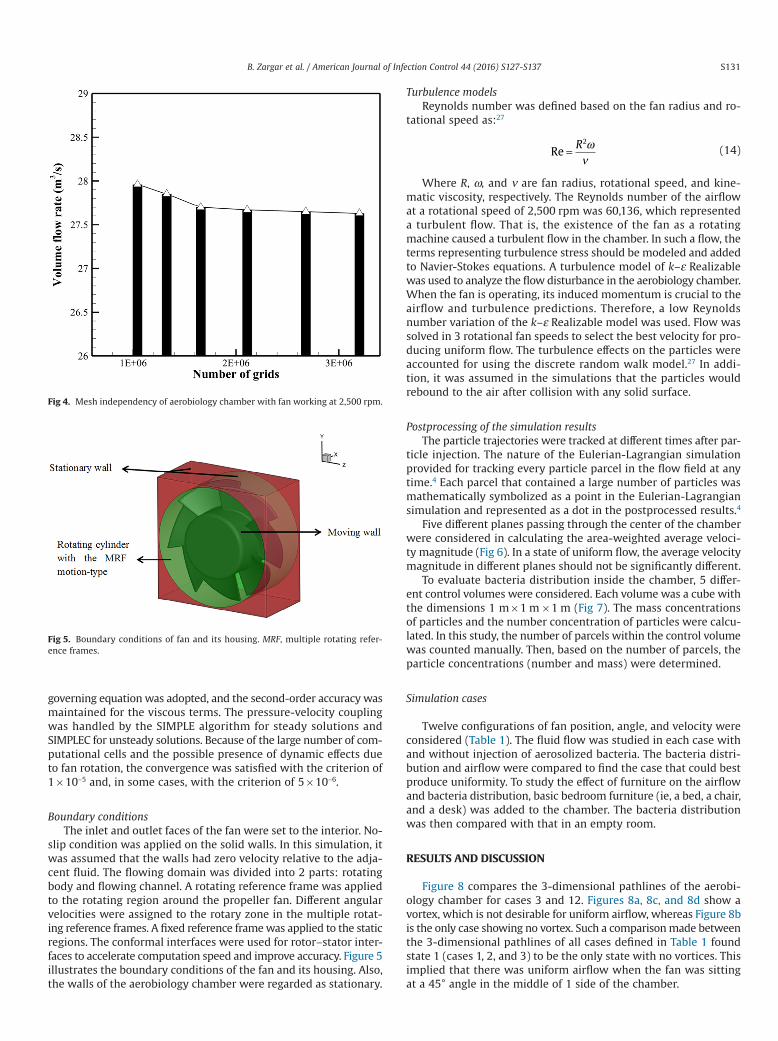
governing equationwas adopted, and the second-order accuracy wasmaintained for the viscous terms. The pressure-velocity couplingwas handled by the SIMPLE algorithm for steady solutions andSIMPLEC for unsteady solutions. Because of the large number of com-putational cells and the possible presence of dynamic effects dueto fan rotation, the convergence was satisfied with the criterion of1 × 10–5 and, in some cases, with the criterion of 5 × 10⁻⁶.
Boundary conditionsThe inlet and outlet faces of the fan were set to the interior. No-
slip condition was applied on the solid walls. In this simulation, itwas assumed that the walls had zero velocity relative to the adja-cent fluid. The flowing domain was divided into 2 parts: rotatingbody and flowing channel. A rotating reference frame was appliedto the rotating region around the propeller fan. Different angularvelocities were assigned to the rotary zone in the multiple rotat-ing reference frames. A fixed reference framewas applied to the staticregions. The conformal interfaces were used for rotor–stator inter-faces to accelerate computation speed and improve accuracy. Figure 5illustrates the boundary conditions of the fan and its housing. Also,the walls of the aerobiology chamber were regarded as stationary.
Turbulence modelsReynolds number was defined based on the fan radius and ro-
tational speed as:27
Re = R2ων
(14)
Where R, ω, and ν are fan radius, rotational speed, and kine-matic viscosity, respectively. The Reynolds number of the airflowat a rotational speed of 2,500 rpm was 60,136, which representeda turbulent flow. That is, the existence of the fan as a rotatingmachine caused a turbulent flow in the chamber. In such a flow, theterms representing turbulence stress should be modeled and addedto Navier-Stokes equations. A turbulence model of k–ε Realizablewas used to analyze the flow disturbance in the aerobiology chamber.When the fan is operating, its induced momentum is crucial to theairflow and turbulence predictions. Therefore, a low Reynoldsnumber variation of the k–ε Realizable model was used. Flow wassolved in 3 rotational fan speeds to select the best velocity for pro-ducing uniform flow. The turbulence effects on the particles wereaccounted for using the discrete random walk model.27 In addi-tion, it was assumed in the simulations that the particles wouldrebound to the air after collision with any solid surface.
Postprocessing of the simulation resultsThe particle trajectories were tracked at different times after par-
ticle injection. The nature of the Eulerian-Lagrangian simulationprovided for tracking every particle parcel in the flow field at anytime.4 Each parcel that contained a large number of particles wasmathematically symbolized as a point in the Eulerian-Lagrangiansimulation and represented as a dot in the postprocessed results.4
Five different planes passing through the center of the chamberwere considered in calculating the area-weighted average veloci-ty magnitude (Fig 6). In a state of uniform flow, the average velocitymagnitude in different planes should not be significantly different.
To evaluate bacteria distribution inside the chamber, 5 differ-ent control volumes were considered. Each volume was a cube withthe dimensions 1 m × 1 m × 1 m (Fig 7). The mass concentrationsof particles and the number concentration of particles were calcu-lated. In this study, the number of parcels within the control volumewas counted manually. Then, based on the number of parcels, theparticle concentrations (number and mass) were determined.
Simulation cases
Twelve configurations of fan position, angle, and velocity wereconsidered (Table 1). The fluid flow was studied in each case withand without injection of aerosolized bacteria. The bacteria distri-bution and airflow were compared to find the case that could bestproduce uniformity. To study the effect of furniture on the airflowand bacteria distribution, basic bedroom furniture (ie, a bed, a chair,and a desk) was added to the chamber. The bacteria distributionwas then compared with that in an empty room.
RESULTS AND DISCUSSION
Figure 8 compares the 3-dimensional pathlines of the aerobi-ology chamber for cases 3 and 12. Figures 8a, 8c, and 8d show avortex, which is not desirable for uniform airflow, whereas Figure 8bis the only case showing no vortex. Such a comparisonmade betweenthe 3-dimensional pathlines of all cases defined in Table 1 foundstate 1 (cases 1, 2, and 3) to be the only state with no vortices. Thisimplied that there was uniform airflow when the fan was sittingat a 45° angle in the middle of 1 side of the chamber.
Fig 4. Mesh independency of aerobiology chamber with fan working at 2,500 rpm.
Fig 5. Boundary conditions of fan and its housing. MRF, multiple rotating refer-ence frames.
S131B. Zargar et al. / American Journal of Infection Control 44 (2016) S127-S137
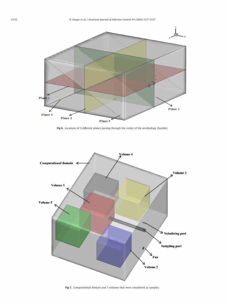
Fig 6. Locations of 5 different planes passing through the center of the aerobiology chamber.
Fig 7. Computational domain and 5 volumes that were considered as samples.
S132 B. Zargar et al. / American Journal of Infection Control 44 (2016) S127-S137
To have a better quantitative comparison between states, area-weighted average velocities were calculated on 5 different planes(Fig 9). The average and coefficient of variation (CV) of area-weighted velocities on 5 planes were calculated for each case andare reported in Table 2. Case 3 of state 1 had the smallest CV (6.5%),implying that the fan created the most uniform airflow when
positioned in the middle of 1 side of the chamber at an angle of45° and a speed of 2,800 rpm.
Bacteria were nebulized into the chamber through a port for 600seconds at a rate of 5,000 CFU/min. For each of the 12 cases, theaverage of particle concentration in 5 volumes and its CV were cal-culated 600 seconds after completing the nebulization process(Table 3). Figure 10 shows the average particle concentration in the5 volumes analyzed and the corresponding standard deviation. Case3 had the lowest CV, implying that the bacterial distribution in thiscase was themost uniform. This is in line with our finding from anal-ysis of the area-weighted average velocities. The small standarddeviation and CV between the 5 volumes implies that, after 900seconds, bacteria would be distributed uniformly inside the chamber,
(a) 90°, in the middle of one of the sides (b) 45°, in the middle of one of the sides
(d) 90°, in the center(c) 45°, in the center
Fig 8. The pathlines of the aerobiology chamber for the velocity of 2,800 rpm.
Table 1Different combinations (cases) of position, angle, and speed of the muffin fan
State Case Rotational speed (rpm) Angle Position
1 1 2,300 45° In the middle of1 of the sides2 2,500
3 2,800
2 4 2,300 45° In the center ofthe chamber5 2,500
6 2,800
3 7 2,300 90° In the center ofthe chamber8 2,500
9 2,800
4 10 2,300 90° In the middle of1 of the sides11 2,500
12 2,800
Fig 9. Average and standard variation of area-weighted average velocities on 5 dif-ferent planes for 12 cases.
S133B. Zargar et al. / American Journal of Infection Control 44 (2016) S127-S137
and there may not be any significant difference among the bacte-ria concentrations in the 5 different volumes analyzed.
The concentration of aerosolized bacteria in each volume wascalculated over time for the optimum case (case 3). Table 4 sum-marizes the results. Figure 11 shows log10 colony forming units permeters3 of samples over the 900 seconds of nebulization of the bac-terial suspension into the chamber. The bacteria concentrations inthe 5 volumes analyzed were different at the beginning of theprocess, but the curve of the 5 volumes converged after finishingthe nebulization at 600 seconds and reached steady state at 900seconds. This implies that 300 seconds (5 minutes) of stabiliza-tion time after completion of the nebulizing process will result ina uniform distribution of bacteria inside the chamber. That is, thebacteria are uniformly distributed, their concentration has reacheda plateau, and the air sampling process can start.
Analysis of variance was performed to determine whether thebacteria concentrations in the 5 volumes analyzed over the timewere significantly different. The results showed that the bacteria con-centrations were the same at a 99% confidence level (F4,40 = 0.29;P = .88), implying that each of these 5 volumes could be used as asampling site to calculate the airborne bacteria concentration insidethe chamber.
To study the influence of the furniture on bacteria distributionin the chamber, the fan was positioned at the optimum location ata 45° angle at 2,800 rpm (state 1, case 3). Figure 12 shows the sche-matics of the room with the furniture.
As with the roomwithout furniture, 600 seconds nebulizing timeand 300 seconds stabilizing time were considered, and the bacte-ria concentrations in the 5 volumes were analyzed. The results aresummarized in Table 5.
Figure 13 shows log10 colony forming units per meters3 for 900seconds after initiating bacterial nebulization into the chamber. Theconcentrations were different at the beginning of the nebulizationprocess but converged during the stabilization time and reached aplateau at the end of the stabilization time.
The bacteria concentrations in the 5 volumes during the nebu-lization and stabilization processes in the chamber with furniture
Table 2Area-weighted average velocity magnitude on 5 different planes for different cases
State 1 State 2 State 3 State 4
Case 1 2 3 4 5 6 7 8 9 10 11 12Speed (rpm) 2,300 2,500 2,800 2,300 2,500 2,800 2,300 2,500 2,800 2,300 2,500 2,800Plane 1 0.015 0.026 0.036 0.032 0.038 0.043 0.029 0.032 0.035 0.031 0.034 0.039Plane 2 0.017 0.029 0.036 0.033 0.039 0.043 0.035 0.039 0.039 0.041 0.045 0.052Plane 3 0.025 0.034 0.041 0.03 0.037 0.047 0.035 0.038 0.042 0.045 0.050 0.059Plane 4 0.012 0.030 0.036 0.03 0.034 0.038 0.035 0.039 0.040 0.029 0.032 0.037Plane 5 0.028 0.034 0.040 0.040 0.042 0.044 0.024 0.026 0.028 0.034 0.037 0.043Mean 0.020 0.031 0.038 0.033 0.038 0.043 0.031 0.035 0.037 0.036 0.040 0.046CV (%) 34.02 11.08 6.55 12.49 7.67 7.54 16.06 16.34 14.97 18.84 19.09 20.16
CV, coefficient of variation.
Table 3Log10 colony forming units per meters3 in 5 different volumes at 900 seconds for 12 cases
State Case Speed (rpm) Volume 1 Volume 2 Volume 3 Volume 4 Volume 5 Average CV (%)
1 1 2,300 4.685 4.565 4.636 4.562 4.679 4.626 0.642 2,500 4.674 4.568 4.620 4.601 4.688 4.630 0.543 2,800 4.662 4.597 4.630 4.631 4.700 4.644 0.42
2 4 2,300 4.678 4.592 4.661 4.541 4.685 4.631 0.675 2,500 4.663 4.570 4.687 4.555 4.692 4.633 0.716 2,800 4.639 4.566 4.725 4.590 4.709 4.646 0.76
3 7 2,300 4.722 4.551 4.664 4.522 4.676 4.627 0.938 2,500 4.713 4.542 4.706 4.556 4.683 4.640 0.909 2,800 4.692 4.497 4.718 4.575 4.697 4.636 1.03
4 10 2,300 4.747 4.564 4.668 4.544 4.692 4.643 0.9311 2,500 4.724 4.536 4.674 4.570 4.6988 4.640 0.8912 2,800 4.685 4.528 4.695 4.610 4.715 4.647 0.83
CV, coefficient of variation.
Fig 10. Average and standard variation of bacterial concentration in the 5 volumesanalyzed after 900 seconds for 12 cases.
Table 4Bacteria concentration (CFU/m3) in 5 volumes
Time (s) Volume 1 Volume 2 Volume 3 Volume 4 Volume 5
100 1,925 6,865 6,055 2,160 3,195200 4,315 11,795 10,310 4,965 7,275300 12,270 21,850 17,405 10,160 16,380400 21,575 26,960 19,855 17,725 26,120500 29,475 31,570 33,435 28,430 35,605600 36,710 40,685 39,695 35,110 41,815700 48,330 42,740 41,115 38,585 49,575800 41,860 40,105 43,320 41,010 48,660900 45,955 39,530 42,655 42,745 50,115
S134 B. Zargar et al. / American Journal of Infection Control 44 (2016) S127-S137
Fig 11. Log10 colony forming units per meters3 in 5 volumes during nebulization and stabilization process. CFU, colony-forming units.
Fig 12. Aerobiology chamber with furniture.
S135B. Zargar et al. / American Journal of Infection Control 44 (2016) S127-S137
were compared using analysis of variance. The bacteria concentra-tions in the 5 volumes were the same at the 99% confidence level(F4,40 = 0.23; P = .99). This implies that, in the presence of the fur-niture, a single sampling site is sufficient to represent the bacteriadistribution inside the chamber.
CONCLUSIONS
Environmental Protection Agency guidelines simply recom-mend the use of a sealed and empty 800-ft3 chamber for testingindoor air decontamination technologies, without further specifi-cations on design or operation. However, we considered additionaldetails, such as the time needed for producing a uniform distribu-tion of test bacteria in the chamber with and without basic furnitureand the position and number of sites for sampling air from withinthe chamber. This modeling study, based on CFD, was undertakento address those issues. Our main conclusions are as follows:
• A muffin fan placed at a 45° angle at the bottom of 1 side of achamber and operating at 2,800 rpm can provide sufficient airturbulence for uniform bacteria distribution throughout, evenin the presence of basic room furniture.
• A 5-minute postnebulization time is sufficient to distribute in-troduced bacteria aerosols uniformly throughout a chamber.
• Simulating the collection of airborne bacteria from 5 differentlocations in the chamber indicated that a single site at the centerof the chamber was sufficient to provide a representative profileof the concentration of the airborne bacteria.
This information should contribute to further standardization ofthe design and operation of aerobiology chambers for data gener-ation on the airborne survival of human pathogens, as well astechnologies for decontamination of indoor air.
References
1. Domgin JF, Huilier D, Burnage H, Gardin P. Coupling of a Lagrangian model witha CFD code: application to numerical modeling of the turbulent dispersion ofdroplets in a turbulent pipe flow. J Hydraul Res 1997;35:473-90.
2. Holmberg S, Li Y. Modeling of the indoor environment—particle dispersion anddeposition. Indoor Air 1998;8:113-22.
3. Lai ACK. Particle deposition indoors: a review. Indoor Air 2004;12:211-24.4. Zhang N. Motion and distribution of micro-sized solid particles in turbulent gas
flow [PhD dissertation]. Manhattan, KS: Kansas State University; 2005.5. Gao NP, Niu JL. Modeling particle dispersion and deposition in indoor
environments. Atmos Environ 2007;41:3862-76.6. Heederik D, Sigsgaard T, Thorne PS, Kline JN, Avery R, Bønløkke JH, et al. Health
effects of airborne exposures from concentrated animal feeding operations.Environ Health Perspect 2007;115:298-302.
7. Just N, Duchaine C, Singh B. An aerobiological perspective of dust in cagehousedand floor-housed poultry operations. J Occup Med Toxicol 2009;4:13.
8. Memarzadeh F. Effect of reducing ventilation rate on indoor air quality andenergy cost in laboratories. J Chem Health Saf 2009;16:20e6.
Fig 13. Bacterial concentration in the 5 different volumes analyzed during nebulization and stabilizing times in a chamber with furniture.
Table 5Bacterial concentration (CFU/m3) in the 5 different volumes analyzed with roomfurniture
Time (s) Volume 1 Volume 2 Volume 3 Volume 4 Volume 5
100 2,215 8,340 6,455 2,445 4,235200 4,850 13,355 12,530 6,070 8,390300 17,945 24,250 20,705 14,650 20,445400 28,550 29,960 20,645 25,685 34,080500 34,210 23,470 41,645 27,590 37,745600 38,480 41,280 52,695 35,280 43,415700 47,730 43,240 49,915 44,230 53,715800 51,460 38,300 44,310 42,280 49,810900 47,980 41,005 43,210 43,695 52,430
S136 B. Zargar et al. / American Journal of Infection Control 44 (2016) S127-S137
9. Ninomura P, Rousseau C, Bartley J. Updated Guidelines for Design andConstruction of Hospital and Health Care Facilities. ASHRAE J 2006;48:H33-H37.
10. Zhang Y. Indoor air quality engineering. New York (NY): CRC Press; 2004.11. American Society for Heating, Refrigeration & Air Conditioning Engineers. Position
Document on Airborne Infectious Diseases. Atlanta (GA): ASHRAE; 2014.12. Memarzadeh F, XuW. Role of air changes per hour (ACH) in possible transmission
of airborne infections. Build Simul 2012;5:15-28.13. Qian H, Li Y, Nielsen PV, Hyldgaard CE, Wong TW, Chwang ATY. Dispersion of
exhaled droplet nuclei in a two-bed hospital ward with three different ventilationsystems. Indoor Air 2006;16:111-28.
14. Lin Z, Wang J, Yao T, Chow TT, Fong KF. Numerical comparison of dispersion ofhuman exhaled droplets under different ventilation methods. World Rev SciTechnol Sustain Dev 2013;10:142-61.
15. Li Y, Leung GM, Tang JW, Yang X, Chao CY, Lin JZ, et al. Role of ventilation inairborne transmission of infectious agents in the built environment—amultidisciplinary systematic review. Indoor Air 2007;17:2-18.
16. Lai ACK, Cheng YC. Study of expiratory droplet dispersion and transport usinga new Eulerian modeling approach. Atmos Environ 2007;41:7473-84.
17. Qian H, Li Y, Nielsen PV, Hyldgaard CE. Dispersion of exhalation pollutants in atwo-bed hospital ward with a downward ventilation system. Build Environ2008;43:344-54.
18. McNeil J, Zhai Z. Critical review on hospital surgical room andmechanical systemsdesigns. World Rev Sci Technol Sustain Dev 2013;10:5-16.
19. Faulkner WB, Memarzadeh F, Riskowski G, Hamilton K, Chang CZ, Chang JR.Particulate concentrations within a reduced-scale room operated at various airexchange rates. Build Environ 2013;65:71-80.
20. Abduladheem A, Sahari KSM, Hasini H, Ahmed W, Mahdi RA. Ventilation airdistribution in hospital operating room-review. Int J Sci Res (IJSR) 2013;2.
21. Zhai Z, Osborne AL. Simulation-based feasibility study of improved airconditioning systems for hospital operating room. Front Archit Res 2013;2:468-75.
22. Zhao B, Zhang Z, Li XT. Numerical study of the transport of droplets orparticles generated by respiratory system indoors. Built Environ 2005;40:1032-9.
23. Liddament MW. A Review of Building Air Flow Simulation. Technical Note AIVC33.Air Infiltration and Ventilation Center. Coventry, Great Britain: University ofWarwick Science Park; 1991.
24. Haghighat F, Jiang Z, Wang JCY, Allard F. Air movement in buildings usingcomputational fluid dynamics. Trans ASME 1992;114:84-92.
25. US Environmental Protection Agency. Air sanitizers—efficacy datarecommendations. Test guideline no. #OCSPP 810.2500-Air Sanitizers-2013-03-12 [EPA 730-C-11- 003]. Environmental Protection Agency website. 2012.Available from: http://www.noticeandcomment.com/-ocspp-810-2500-air-sanitizers-2013-03-12-epa-730-c-11-003-fn-24288.aspx. Published March 13, 2013. Accessed December 4, 2015.
26. Sattar SA, Kibbee RJ, Zargar B, Wright KE, Rubino JR, Ijaz MK. Decontaminationof indoor air to reduce the risk of airborne infections: studies on survival andinactivation of airborne pathogens using an aerobiology chamber. Am J InfectControl 2016; [Epub ahead of print].
27. Ansys Fluent v.12.0 Usermanual, Available from: http://users.ugent.be/~mvbelleg/flug-12-0.pdf
28. Soltani M, Chen P. Shape design of internal flow with minimum pressure loss.Adv Sci Lett 2009;2:347-55.
S137B. Zargar et al. / American Journal of Infection Control 44 (2016) S127-S137
Major Article
Aerobiology of the built environment: Synergy between Legionellaand fungi
Absar Alum PhD a,b,*, Galahad Zachariah Isaacs BS b
a School of Sustainable Engineering and the Built Environment, Arizona State University, Tempe, AZb BioDetek Laboratory, Mesa, AZ
Key Words:Legionellaindoor airbuilt environmentfungibiofilms
Background: The modern built environment (BE) design creates unique ecological niches ideal for thesurvival and mutual interaction of microbial communities. This investigation focused on the synergisticrelations between Legionella and the fungal species commonly found in BEs and the impact of these syn-ergistic relationships on the survival and transmission of Legionella.Methods: A field study was conducted to identify the types and concentrations of fungi in BEs. The fungalisolates purified from BEs were cocultured with Legionella to study their synergistic association. CoculturedLegionella cells were aerosolized in an air-tight chamber to evaluate the efficacy of ultraviolet (UV) to in-activate these cells.Results: Aspergillus, Alternaria, and Cladosporiumwere the most common fungi detected in samples thattested positive for Legionella. After coculturing, Legionella cells were detected inside fungal hyphae. Themicroscopic observations of Legionella internalization in fungal hyphae were confirmed by molecular anal-yses. UV disinfection of the aerosolized Legionella cells that were cocultured with fungi indicated thatfungal spores and propagules act as a shield against UV radiation. The shield effect of fungal spores onLegionella cells was quantified at >2.5 log10.Conclusions: This study provides the first evidence, to our knowledge, of Legionella cell presence insidefungi detected in an indoor environment. This symbiotic relationship with fungi results in longer sur-vival of Legionella under ambient conditions and provides protection against UV rays.
© 2016 Published by Elsevier Inc. on behalf of Association for Professionals in Infection Control andEpidemiology, Inc.
BACKGROUND
The evolution in the architectural designs of living spaces and life-style changes has resulted in a significant shift of human activitiesfrom open-air environments to indoor. Modern living spaces are de-signed for energyefficiencyandcontrolledair conditions.1,2 Conditionsin the built environments (BEs) used for office or residential pur-poses are impacted by building design,maintenance operations, andoccupant activities. A compromise on these factors can potentially
result in sick building syndrome, adversely impacting the health ofoccupants. The severity of the adverse impact on health appears tobe linked to the time spent in such buildings.3,4
Indoor air pollution originates from nonbiologic and biologicsources. Bacteria, fungi, pollen, viruses, insect body parts, humansquames, animal dander, and bird droppings are the most commonsources of biologic contamination in BEs.5,6 In the indoor environ-ment, fungi are a serious public health threat.7,8 Fungal speciesbelonging toAscomycetes, Basidiomycetes, and the anamorphic fungi(Deuteromycota) are themost common causes of allergic reaction inoccupants.8,9
Legionella pneumophila is themost serious bacterial pathogen as-sociatedwith indoor environments. Since thediscoveryof Legionnairedisease in1976, bacteria of thegenus Legionellahavebecomea leadingpublic health concern.10 Legionella can be found in air as part ofaerosols generated from contaminated sources. It is known tocause Legionnaire disease and Pontiac fever, a milder and self-limiting respiratory infection.11 Aerosolization is an importantcomponent of Legionella transmission to the human respiratorysystem,12 and it can be accomplished by aerosol-generating systems,
* Address correspondence to Absar Alum, PhD, BioDetek Laboratory, 1815 W 1stAvenue, #125, Mesa, AZ 85202.
E-mail address: [email protected] (A. Alum).Funding/Support: Supported by EnviroPak and HKreation. Publication of this sup-
plement is primarily supported by RB, Montvale, New Jersey, with additional supportfrom MicroBioTest, a division of Microbac Laboratories, Inc., Sterling, Virginia. TheCity University of New York (CUNY) and the University of Ottawa, Ottawa, Canada,are academic sponsors. Editorial support was provided by Ashley O’Dunne, PhD;Shannon O’Sullivan, ELS; and Alanna Franchetti, ELS of Medergy (Yardley, PA), andfunded by RB.
Conflicts of Interest: None to report.
0196-6553/© 2016 Published by Elsevier Inc. on behalf of Association for Professionals in Infection Control and Epidemiology, Inc.http://dx.doi.org/10.1016/j.ajic.2016.06.004
American Journal of Infection Control 44 (2016) S138-S143
Contents lists available at ScienceDirect
American Journal of Infection Control
journal homepage: www.aj ic journal .org
American Journal of Infection Control
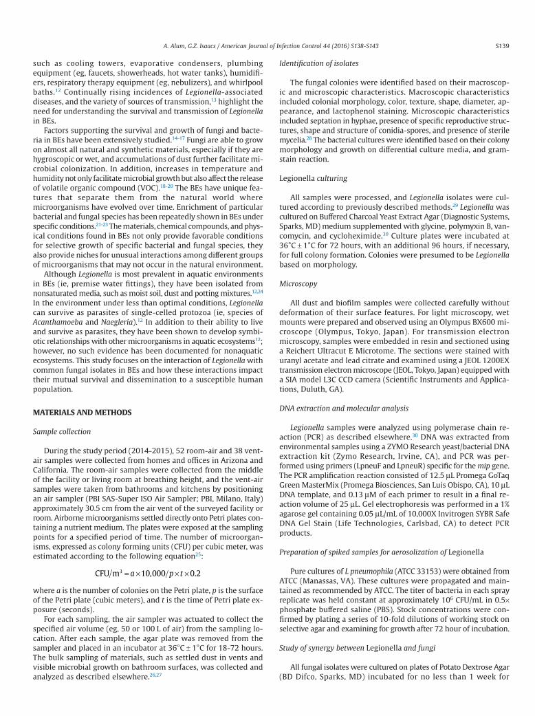
such as cooling towers, evaporative condensers, plumbingequipment (eg, faucets, showerheads, hot water tanks), humidifi-ers, respiratory therapy equipment (eg, nebulizers), and whirlpoolbaths.12 Continually rising incidences of Legionella-associateddiseases, and the variety of sources of transmission,13 highlight theneed for understanding the survival and transmission of Legionellain BEs.
Factors supporting the survival and growth of fungi and bacte-ria in BEs have been extensively studied.14-17 Fungi are able to growon almost all natural and synthetic materials, especially if they arehygroscopic orwet, and accumulations of dust further facilitatemi-crobial colonization. In addition, increases in temperature andhumiditynotonly facilitatemicrobial growthbut alsoaffect the releaseof volatile organic compound (VOC).18-20 The BEs have unique fea-tures that separate them from the natural world wheremicroorganisms have evolved over time. Enrichment of particularbacterial and fungal species has been repeatedly shown in BEs underspecific conditions.21-23 Thematerials, chemical compounds, andphys-ical conditions found in BEs not only provide favorable conditionsfor selective growth of specific bacterial and fungal species, theyalso provide niches for unusual interactions among different groupsof microorganisms that may not occur in the natural environment.
Although Legionella is most prevalent in aquatic environmentsin BEs (ie, premise water fittings), they have been isolated fromnonsaturatedmedia, such asmoist soil, dust and pottingmixtures.12,24
In the environment under less than optimal conditions, Legionellacan survive as parasites of single-celled protozoa (ie, species ofAcanthamoeba and Naegleria).12 In addition to their ability to liveand survive as parasites, they have been shown to develop symbi-otic relationshipswith othermicroorganisms in aquatic ecosystems12;however, no such evidence has been documented for nonaquaticecosystems. This study focuses on the interaction of Legionellawithcommon fungal isolates in BEs and how these interactions impacttheir mutual survival and dissemination to a susceptible humanpopulation.
MATERIALS AND METHODS
Sample collection
During the study period (2014-2015), 52 room-air and 38 vent-air samples were collected from homes and offices in Arizona andCalifornia. The room-air samples were collected from the middleof the facility or living room at breathing height, and the vent-airsamples were taken from bathrooms and kitchens by positioningan air sampler (PBI SAS-Super ISO Air Sampler; PBI, Milano, Italy)approximately 30.5 cm from the air vent of the surveyed facility orroom. Airbornemicroorganisms settled directly onto Petri plates con-taining a nutrient medium. The plates were exposed at the samplingpoints for a specified period of time. The number of microorgan-isms, expressed as colony forming units (CFU) per cubic meter, wasestimated according to the following equation25:
CFU m3 10 000 0 2= × × ×a p t, .
where a is the number of colonies on the Petri plate, p is the surfaceof the Petri plate (cubic meters), and t is the time of Petri plate ex-posure (seconds).
For each sampling, the air sampler was actuated to collect thespecified air volume (eg, 50 or 100 L of air) from the sampling lo-cation. After each sample, the agar plate was removed from thesampler and placed in an incubator at 36°C ± 1°C for 18-72 hours.The bulk sampling of materials, such as settled dust in vents andvisible microbial growth on bathroom surfaces, was collected andanalyzed as described elsewhere.26,27
Identification of isolates
The fungal colonies were identified based on their macroscop-ic and microscopic characteristics. Macroscopic characteristicsincluded colonial morphology, color, texture, shape, diameter, ap-pearance, and lactophenol staining. Microscopic characteristicsincluded septation in hyphae, presence of specific reproductive struc-tures, shape and structure of conidia-spores, and presence of sterilemycelia.28 The bacterial cultures were identified based on their colonymorphology and growth on differential culture media, and gram-stain reaction.
Legionella culturing
All samples were processed, and Legionella isolates were cul-tured according to previously described methods.29 Legionella wascultured on Buffered Charcoal Yeast Extract Agar (Diagnostic Systems,Sparks, MD) medium supplemented with glycine, polymyxin B, van-comycin, and cycloheximide.30 Culture plates were incubated at36°C ± 1°C for 72 hours, with an additional 96 hours, if necessary,for full colony formation. Colonies were presumed to be Legionellabased on morphology.
Microscopy
All dust and biofilm samples were collected carefully withoutdeformation of their surface features. For light microscopy, wetmounts were prepared and observed using an Olympus BX600 mi-croscope (Olympus, Tokyo, Japan). For transmission electronmicroscopy, samples were embedded in resin and sectioned usinga Reichert Ultracut E Microtome. The sections were stained withuranyl acetate and lead citrate and examined using a JEOL 1200EXtransmission electronmicroscope (JEOL, Tokyo, Japan) equippedwitha SIA model L3C CCD camera (Scientific Instruments and Applica-tions, Duluth, GA).
DNA extraction and molecular analysis
Legionella samples were analyzed using polymerase chain re-action (PCR) as described elsewhere.30 DNA was extracted fromenvironmental samples using a ZYMO Research yeast/bacterial DNAextraction kit (Zymo Research, Irvine, CA), and PCR was per-formed using primers (LpneuF and LpneuR) specific for themip gene.The PCR amplification reaction consisted of 12.5 μL Promega GoTaqGreen MasterMix (Promega Biosciences, San Luis Obispo, CA), 10 μLDNA template, and 0.13 μM of each primer to result in a final re-action volume of 25 μL. Gel electrophoresis was performed in a 1%agarose gel containing 0.05 μL/mL of 10,000X Invitrogen SYBR SafeDNA Gel Stain (Life Technologies, Carlsbad, CA) to detect PCRproducts.
Preparation of spiked samples for aerosolization of Legionella
Pure cultures of L pneumophila (ATCC 33153) were obtained fromATCC (Manassas, VA). These cultures were propagated and main-tained as recommended by ATCC. The titer of bacteria in each sprayreplicate was held constant at approximately 106 CFU/mL in 0.5×phosphate buffered saline (PBS). Stock concentrations were con-firmed by plating a series of 10-fold dilutions of working stock onselective agar and examining for growth after 72 hour of incubation.
Study of synergy between Legionella and fungi
All fungal isolates were cultured on plates of Potato Dextrose Agar(BD Difco, Sparks, MD) incubated for no less than 1 week for
S139A. Alum, G.Z. Isaacs / American Journal of Infection Control 44 (2016) S138-S143
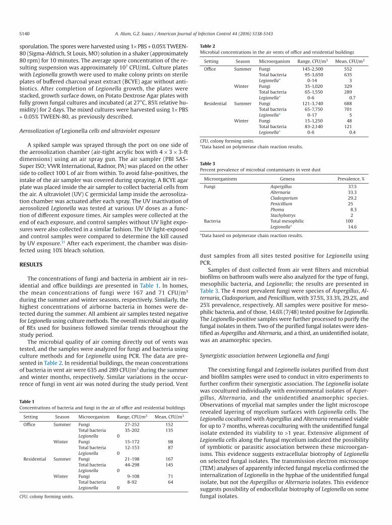
sporulation. The spores were harvested using 1× PBS + 0.05% TWEEN-80 (Sigma-Aldrich, St Louis, MO) solution in a shaker (approximately80 rpm) for 10 minutes. The average spore concentration of the re-sulting suspension was approximately 107 CFU/mL. Culture plateswith Legionella growth were used to make colony prints on sterileplates of buffered charcoal yeast extract (BCYE) agar without anti-biotics. After completion of Legionella growth, the plates werestacked, growth surface down, on Potato Dextrose Agar plates withfully grown fungal cultures and incubated (at 27°C, 85% relative hu-midity) for 2 days. The mixed cultures were harvested using 1× PBS+ 0.05% TWEEN-80, as previously described.
Aerosolization of Legionella cells and ultraviolet exposure
A spiked sample was sprayed through the port on one side ofthe aerosolization chamber (air-tight acrylic box with 4 × 3 × 3-ftdimensions) using an air spray gun. The air sampler (PBI SAS-Super ISO; VWR International, Radnor, PA) was placed on the otherside to collect 100 L of air from within. To avoid false-positives, theintake of the air sampler was covered during spraying. A BCYE agarplate was placed inside the air sampler to collect bacterial cells fromthe air. A ultraviolet (UV) C germicidal lamp inside the aerosoliza-tion chamber was actuated after each spray. The UV inactivation ofaerosolized Legionella was tested at various UV doses as a func-tion of different exposure times. Air samples were collected at theend of each exposure, and control samples without UV light expo-sures were also collected in a similar fashion. The UV light-exposedand control samples were compared to determine the kill causedby UV exposure.31 After each experiment, the chamber was disin-fected using 10% bleach solution.
RESULTS
The concentrations of fungi and bacteria in ambient air in res-idential and office buildings are presented in Table 1. In homes,the mean concentrations of fungi were 167 and 71 CFU/m3
during the summer and winter seasons, respectively. Similarly, thehighest concentrations of airborne bacteria in homes were de-tected during the summer. All ambient air samples tested negativefor Legionella using culturemethods. The overall microbial air qualityof BEs used for business followed similar trends throughout thestudy period.
The microbial quality of air coming directly out of vents wastested, and the samples were analyzed for fungi and bacteria usingculture methods and for Legionella using PCR. The data are pre-sented in Table 2. In residential buildings, the mean concentrationsof bacteria in vent air were 635 and 289 CFU/m3 during the summerand winter months, respectively. Similar variations in the occur-rence of fungi in vent air was noted during the study period. Vent
dust samples from all sites tested positive for Legionella usingPCR.
Samples of dust collected from air vent filters and microbialbiofilms on bathroomwalls were also analyzed for the type of fungi,mesophilic bacteria, and Legionella; the results are presented inTable 3. The 4 most prevalent fungi were species of Aspergillus, Al-ternaria, Cladosporium, and Penicillium, with 37.5%, 33.3%, 29.2%, and25% prevalence, respectively. All samples were positive for meso-philic bacteria, and of those, 14.6% (7/48) tested positive for Legionella.The Legionella-positive samples were further processed to purify thefungal isolates in them. Two of the purified fungal isolates were iden-tified as Aspergillus and Alternaria, and a third, an unidentified isolate,was an anamorphic species.
Synergistic association between Legionella and fungi
The coexisting fungal and Legionella isolates purified from dustand biofilm samples were used to conduct in vitro experiments tofurther confirm their synergistic association. The Legionella isolatewas cocultured individually with environmental isolates of Asper-gillus, Alternaria, and the unidentified anamorphic species.Observations of mycelial mat samples under the light microscoperevealed layering of mycelium surfaces with Legionella cells. TheLegionella cocultured with Aspergillus and Alternaria remained viablefor up to 7months, whereas coculturing with the unidentified fungalisolate extended its viability to >1 year. Extensive alignment ofLegionella cells along the fungal mycelium indicated the possibilityof symbiotic or parasitic association between these microorgan-isms. This evidence suggests extracellular biotrophy of Legionellaon selected fungal isolates. The transmission electron microscope(TEM) analyses of apparently infected fungal mycelia confirmed theinternalization of Legionella in the hyphae of the unidentified fungalisolate, but not the Aspergillus or Alternaria isolates. This evidencesuggests possibility of endocellular biotrophy of Legionella on somefungal isolates.
Table 1Concentrations of bacteria and fungi in the air of office and residential buildings
Setting Season Microorganism Range, CFU/m3 Mean, CFU/m3
Office Summer Fungi 27-252 152Total bacteria 35-202 135Legionella 0
Winter Fungi 15-172 98Total bacteria 12-153 87Legionella 0
Residential Summer Fungi 21-198 167Total bacteria 44-298 145Legionella 0
Winter Fungi 9-108 71Total bacteria 8-92 64Legionella 0
CFU, colony forming units.
Table 2Microbial concentrations in the air vents of office and residential buildings
Setting Season Microorganism Range, CFU/m3 Mean, CFU/m3
Office Summer Fungi 145-2,500 552Total bacteria 95-3,650 635Legionella* 0-14 3
Winter Fungi 35-1,020 329Total bacteria 65-1,550 289Legionella* 0-6 0.7
Residential Summer Fungi 121-3,740 688Total bacteria 65-7,750 701Legionella* 0-17 5
Winter Fungi 15-1,250 48Total bacteria 83-2,140 121Legionella* 0-6 0.4
CFU, colony forming units.*Data based on polymerase chain reaction results.
Table 3Percent prevalence of microbial contaminants in vent dust
Microorganisms Genera Prevalence, %
Fungi Aspergillus 37.5Alternaria 33.3Cladosporium 29.2Penicillium 25Phoma 8.3Stachybotrys 2
Bacteria Total mesophilic 100Legionella* 14.6
*Data based on polymerase chain reaction results.
S140 A. Alum, G.Z. Isaacs / American Journal of Infection Control 44 (2016) S138-S143
Aerosolization and UV disinfection of Legionella cocultured withfungi
Three independent experiments were performed using pure cul-tures of Legionella and Legionella cells cocultured with theenvironmental isolates of Aspergillus and Alternaria. Air samples werecollected before and after UV disinfection. The kill effect of UV dis-infection on Legionella is presented in Figure 1. The log10 reductionsof the pure culture of Legionella and Legionella cocultured with As-pergillus and Alternariawere 3.0, 0.2, and 0.3, respectively, indicatinga protective effect against UV radiation of fungal spores and propa-gules on Legionella cells.
DISCUSSION
This study included sampling sites located in Arizona and Cal-ifornia, and comparison of data sets from both states revealed noremarkable differences in terms of microbial concentrations and di-versity. BEs are known to contain a complexmixture of bioaerosolsconstituting fungi, bacteria, and allergens, along with nonbiologicparticles.32 Bioaerosols contribute between 5% and 34% of indoorair pollution.33 In general, sources of indoor bioaerosols are locatedoutdoors, and intrusion happens through openings in the buildingenvelope (ie, windows, doors). In this study, all sampling sites arelocated in similar ecogeographic zones (ie, Arizona, Southern Cal-ifornia) with similar microbial flora in outdoor and indoorenvironments.34 Therefore, a similar microbial complex detectedacross the study sites is not unexpected. However, variation in BEmanagement and maintenance practices are known to impact thehygienequality of indoor environments. Thehousekeeping andmain-tenance records of participating sites were not available; therefore,a correlation between the microbial quality of indoor environ-ments and stringency of housekeeping and maintenance practicescould not be established. In addition, within BEs, the existence ofmicroenvironments in different sections of a building is a commonphenomenon. Thesemicroenvironment islands are linkedwith use-based spaces (ie, bathrooms, kitchens), which are known to becharacteristically different from the main living spaces.35 In a pre-vious study, concentrations and types of fungi in bathrooms werefound to be different from the rest of a building’s sections.34-36 Thisstudy also found higher fungal concentrations in air samples col-lected from vents in bathrooms and kitchens. In addition, passivesampling (ie, dust and biofilm samples collected by swabbing) re-sulted in a higher percentage of Legionella-positive samples thanthe active sampling (ie, direct filtration of air). These results are
consistentwithpreviousfindings froman Italian study inwhichwaterwas flushed for 2 minutes from a known source of Legionella andthe surrounding areas were sampled for up to 8 hours using activeand passive sampling. Using passive sampling, airborne Legionellawas detected in twice as many samples as using active sampling.37
The environmental stage of Legionella is commonly found in soilsor in associationwith detritus and biofilms prevalent in freshwaterenvironments, which are known to have high concentrations of abroad range of heterotrophic bacteria. In oligotrophic environ-ments, the commonlyprevalentheterotrophicbacteria cangrowmorerapidly than Legionella. To avoid competition with other bacteria,Legionella has evolved into facultative intracellular parasites of free-living protozoa (ie, amoebae). The unusual nutrients required forthe growth of Legionella are rarely found in the environment38;however, as intracellularparasites, theyfindsequesteringniches insidetheir host,which provide for their special nutritional needs. In indoorenvironments, aerosolized bacteria eventually land on surfaces. Afterlanding on surfaces, their survival depends on the microenviron-ment of the sites they land on. For example, bathroom walls andventilation ducts are known for biofilms containing fungal masses.In this study, Legionellawas detected in only the samples (biofilmsand dust) collected frombathrooms and kitchens. All of the samplestested positive for Legionella were also tested positive for at leastone type of fungus. Further analyses of these samples confirmedthe presence of Legionella inside fungal hyphae, indicating the po-tential of Legionella to establish parasitic relationships withsaprophytic fungi. During evolution Legionella has developed elab-oratemechanisms for invading a broad range of host cells and resultsof this study is consistentwith the results of previous studies. Func-tionalDot/Icm type IV secretion apparatus of Legionella allowdeliveryof effector proteins into their host cells to evade host defenses.39 Dif-ferent strains of a single bacterial species may carry variable arrayof effector proteins, which determine their host range andspecificity.39,40 More than 300 effector proteins have been reportedfor Legionella,41,42 allowing them to parasitize a broad range of hosts,including human, animals, plant pathogenic fungi, nematodes, pro-tozoa, and amoebae.43 Legionella is known tohave enzymesbelongingto a novel phospholipase C (PLC) family. Among bacteria, PLC-likeproteins are present only in Legionella spp and some Pseudomonasspp.39,44 Legionella cells rely on Mip, a surface-exposed protein, forthe optimal infection of host cells.45 In addition, Mip is known toinfluence the secretion of novel PLC-like proteins. Molecular anal-yses of fungal isolates with Legionella internalized in their hyphaerevealed a homologous sequence in the conserved motifs of a PLC-like gene found in other Legionella hosts (data not presented).
Fig 1. Ultraviolet disinfection of single cells of Legionella and coculture with fungi.
S141A. Alum, G.Z. Isaacs / American Journal of Infection Control 44 (2016) S138-S143
In this study, the most prevalent fungi included Aspergillus, Al-ternaria,Cladosporium, andPenicillium. These fungal generahavebeenwidely documented as common in indoor environments at concen-trations similar to the ones recorded in this study.46-48 On average,healthy adults inhale approximately 0.5 L of air with every breath.Basedonaveragebreathinghabits, approximately10m3of air is trans-ported through our lungs every day.49 Considering the commonlydocumentedmicrobial concentrations in indoorair, sucha largevolumeof air intake can pose a substantial health risk for physiologically im-paired humans. Formost healthy individuals, however, the anatomyof the pulmonary system and the clumping-sequestering of micro-organisms (ie, increasing particle size) reduce the risk of infection.Nevertheless, reliable comprehensive environmentalmonitoringdataare needed to accurately assess the prevalence of Legionella in BEs.This is compounded by the lack of uniform guidance for preventa-tive environmental testing for Legionella.50
In the environment,microbial clumping not only reduces the riskof fungi intrusion in the lungs, it also imparts a shielding effect againstradiation (ie, sunlight, UV, etc). Microbial clumping can create biasin the results of disinfection studies, especially using UV or other ra-diation. It has been estimated that 61% of the incident radiation of254-nmwavelength is transmitted through a single microbial cell.51
A cluster surrounding 2 microbial cells permits only 37% of the in-cidentUV light to reach the third cell. Therefore, clumping ofmultiplemicroorganisms as clusters can provide protection against disinfec-tion. Much of the published literature on the microbicidal effect ofUV for airbornepathogens is basedonstudiesusingone typeofmostlymonodispersed cells,31,52 which does not represent a real-world sce-nario. In this study, Legionella coculturedwith fungi was aerosolizedto test the efficacy of UV. Aggregation of Legionella cells with othermicrobial cells appeared to provide protection against UV, and thisinformation should be considered when planning future studies.
CONCLUSIONS
The importance of evaluating the quality of the air in BEs cannotbe overemphasized. The microbial quality in BEs is indirectly in-fluenced by the architectural design and directly influenced byhuman occupancy, use patterns, and housekeeping practices. Thearchitectural features included in modern buildings for resource ef-ficacy and occupant comfort also serve as a means of creatingsuitable niches for the enrichment of many types of microbes. Fromthe operational aspect, the microbial quality of indoor air can bean indicator of effective housekeeping and cleanliness and can aidin determining sources of human discomfort. Bathrooms and kitch-ens were identified as hotspots of microbial communities, fromwhich microbial pathogens can potentially spread to other areas.These areas were found to be appropriate niches for the coexis-tence of diverse microorganisms, and this is the first study, to ourknowledge, to report the synergistic relationship between Legionellaand fungi in bathrooms. Fungal spores and propagules have highaerosolization capacities. Association with fungal propagules canpotentially facilitate Legionella cells to become airborne and spreadacross buildings. In addition, Legionella cells clustered with a fungusexhibited high levels of resistance to UV radiation. The results ofthis study highlight the importance of using appropriate levels oforganic material in microbial stocks for the UV disinfection of aero-solized pathogens. The synergistic association noted betweenLegionella and fungi can have significant implications for the suc-cessful management of air quality in BEs and emphasizes thesignificance of environmental testing andmonitoring. In the contextof the lack of uniform recommendations, various facilities followthe environmental testing program that suits their conditions. Thefact that bathroom and kitchens showed the highest level of mi-crobial contamination in residential or commercial use buildings
purport to their appropriateness for inclusion in an environmen-tal monitoring and testing program.
Acknowledgments
We thank Michael HimelarbMichael Himelfarb, Global Preven-tion Services, Scottsdale, AZ, for his technical support in the selectionof sampling sites and conduction of this study; Afnaan Moham-med for help in sample analyses; Suhayb Alsabbry and OmerAlsabbry for their help in field sampling; and Shamel Absar and IsraMishqat for their help in ultraviolet disinfection studies.
References
1. Chao HJ, Schwartz J, Milton DK, Burge HA. The work environment and workershealth in four large office buildings. Environ Health Perspect 2003;111:1242-8.
2. Molhave L. Sick building syndrome. In: Nriagu JO, editor. Encyclopedia ofenvironmental health. Philadelphia (PA): Elsevier Science; 2011. p. 61-7.
3. Ebbehøj NE, Hansen MO, Sigsgaard T, Larsen L. Building-related symptoms andmolds: a two-step intervention study. Indoor Air 2002;12:273-7.
4. Zeliger HI. Toxic effects of chemical mixtures. Arch Environ Health 2003;58:23-9.5. Nevalainen A, Seuri M. Of microbes andmen. Indoor Air 2005;15(Suppl 9):58-64.6. Khan AAH, Karuppayil SM. Potential natural disinfectants for indoor
environments. Int J Clin Aromather 2010;7:1-5.7. Samet JM, Spengler JD. Indoor environments and health: moving into the 21st
century. Am J Pub Health 2003;93:1489-93.8. Khan AAH, Karuppayil SM. Fungal pollution of indoor environments and its
management. Saudi J Biol Sci 2012;19:405-26.9. Portnoy JM. Evaluation of indoor mold exposure is what allergists do best. Ann
Allergy Asthma Immunol 2003;90:175.10. Brunkard JM, Ailes E, Roberts VA, Hill V, Hilborn ED, Craun GF, et al. Surveillance
for waterborne disease outbreaks associated with drinking water—United States,2007-2008. MMWRMorb Mortal Wkly Rep 2011;60:38-68.
11. Nakayama K, Morimoto K. Relationship between, lifestyle, mold and sick buildingsyndromes in newly built dwellings in Japan. Int J Immunopathol Pharmacol2007;20(Suppl 2):35-43.
12. US Environmental Protection Agency, Office of Water. Legionella human healthcriteria document. Washington (DC): US Environmental Protection Agency, Officeof Water; 1999.
13. Beer KD, Gargano JW, Roberts VA, Hill V, Garrison L, Kutty P, et al. Surveillancefor waterborne disease outbreaks associated with drinking water—United States,2011-2012. MMWRMorb Mortal Wkly Rep 2015;64:842-8.
14. Fischer PH, Kriz B, Martuzzi M, Wojtyniak B, Lebret E, Van Reeuwijk H, et al.Risk factors indoors and prevalences of childhood respiratory health in fourcountries in western and central Europe. Indoor Air 1988;8:244-54.
15. Garrett MH, Rayment PR, Hooper MA, Abramson MJ. Indoor airborne fungalspores, house dampness and associations with environmental factors andrespiratory health in children. Clin Exp Allergy 1988;28:459-67.
16. Kozak PP, Gallup J, Cummins LH, Gillman SA. Factors of importance indetermining the prevalence of indoor molds. Ann Allergy 1980;43:88-94.
17. Rylander R, Megevand Y. Environmental risk factors for respiratory infections.Arch Environ Health 2000;55:300-3.
18. Reijula K. Moisture-problem buildings with molds causing work-related diseases.Adv Appl Microbiol 2004;55:175-89.
19. Hatfield ML, Hartz KEH. Secondary organic aerosol from biogenic volatile organiccompounds mixtures. Atmos Environ 2011;45:2211-9.
20. Pieckova E, Jesenska Z. Microscopic fungi in dwellings and their healthimplications in humans. Ann Agric Environ Med 1999;6:1-11.
21. Anaissie EJ, Penzak SR, Dignani MC. The hospital water supply as a source ofnosocomial infections: a plea for action. Arch Intern Med 2002;162:1483-92.
22. Casini B, Valentini P, Baggiani A, Torracca F, Frateschi S, Nelli LC, et al. Molecularepidemiology of Legionella pneumophila serogroup 1 isolates following long-term chlorine dioxide treatment in a university hospital water system. J HospInfect 2008;69:141-7.
23. Szewzyk U, Szewzyk R, ManzW, Schleifer KH. Microbiological safety of drinkingwater. Annu Rev Microbiol 2000;54:81-127.
24. Steele TW, Lanser J, Sangster N. Isolation of Legionella longbeachae serogroup1 from potting mixes. Appl Environ Microbiol 1990;56:49-53.
25. Stryjakowska-Sekulska M, Piotraszewska-Pajak A, Szyszka A, Nowicki M, FilipiakM. Microbiological quality of indoor air in university rooms. Pol J Environ Stud2007;16:624-32.
26. O’Meara T, Tovey E. Monitoring personal allergen exposure. Clin Rev AllergyImmunol 2000;18:341-95.
27. Reponen T, Singh U, Schaffer C, Vesper S, Johansson E, Adhikari A, et al. Visuallyobserved mold and moldy odor versus quantitatively measured microbialexposure in homes. Sci Total Environ 2010;408:5565-74.
28. Zafar S, Aqil F, Ahmad I. Metal tolerance and biosorption potential of filamentousfungi isolated from metal contaminated agriculture soil. Bioresour Technol2007;98:2557-61.
S142 A. Alum, G.Z. Isaacs / American Journal of Infection Control 44 (2016) S138-S143
29. US Department of Health and Human Services, Centers for Disease Control andPrevention. Procedures for the recovery of Legionella from the environment. 2005Available from: http://www.cdc.gov/legionella/health-depts/inv-tools-cluster/lab-inv-tools/procedures-manual.pdf. Accessed April 17, 2016.
30. Wullings BA, Bakker G, van der Kooij D. Concentration and diversity ofuncultured Legionella spp. in two unchlorinated drinking water supplies withdifferent concentrations of natural organic matter. Appl Environ Microbiol2011;77:634-41.
31. King B, Kesavan J, Sagripanti J-L. Germicidal UV sensitivity of bacteria in aerosolsand on contaminated surfaces. Aerosol Sci Technol 2011;45:1-9.
32. Hargreaves M, Parappukkaran S, Morawska L, Hitchins J, Congrong H, GilbertD. A pilot investigation into associations between indoor airborne fungal andnonbiological particle concentrations in residential houses in Brisbane. Sci TotalEnviron 2003;312:89-101.
33. Srikanth P, Sudharsanam S, Steinberg R. Bioaerosols in indoor environment:composition, health effects and analysis. Indian J MedMicrobiol 2008;26:302-12.
34. Tsai FC, Macher JM. Concentrations of airborne culturable bacteria in 100 largeUS office buildings from the BASE study. Indoor Air 2005;15(Suppl 9):71-81.
35. Ojima M, Toshima Y, Koya E, Ara K, Tokuda H. Hygiene measures consideringactual distributions of microorganisms in Japanese households. J Appl Microbiol2002;93:800-9.
36. Hamada N, Abe N. Growth characteristics of fungal species from bathroom.Biocontrol Sci 2010;15:111-5.
37. Montagna MT, De Giglio O, Napoli C, Cannova L, Cristina ML, Deriu MG, et al.Legionella spp. contamination in indoor air: preliminary results of an Italianmulticenter study. Epidemiol Prev 2014;38(Suppl 2):62-5.
38. FonsecaMV, SwansonMS. Nutrient salvaging andmetabolism by the intracellularpathogen Legionella pneumophila. Front Cell Infect Microbiol 2014;4:12.
39. Aurass P, Schlegel M, Metwally O, Harding CR, Schroeder GN, Frankel G, et al.The Legionella pneumophila Dot/Icm-secreted effector PlcC/CegC1 togetherwith PlcA and PlcB promotes virulence and belongs to a novel zincmetallophospholipase C family present in bacteria and fungi. J Biol Chem2013;288:11080-92.
40. Alfano JR, Collmer A. Type III secretion system effector proteins: double agentsin bacterial disease and plant defense. Annu Rev Phytopathol 2004;42:385-414.
41. Burstein D, Zusman T, Degtyar E, Viner R, Segal G, Pupko T. Genome-scaleidentification of Legionella pneumophila effectors using a machine learningapproach. PLoS Pathog 2009;5:e1000508.
42. Huang L, Boyd D, Amyot WM, Hempstead AD, Luo ZQ, O’Connor TJ, et al. The Eblock motif is associated with Legionella pneumophila translocated substrates.Cell Microbiol 2010;13:227-45.
43. D’Auria G, Jiménez-Hernández N, Peris-Bondia F, Moya A, Latorre A. Legionellapneumophila pangenome reveals strain-specific virulence factors. BMC Genomics2010;11:181.
44. Engel J, Balachandran P. Role of Pseudomonas aeruginosa type III effectors indisease. Curr Opin Microbiol 2009;12:61-6.
45. Newton HJ, Ang DKY, van Driel IR, Hartland EL. Molecular pathogenesis ofinfections caused by Legionella pneumophila. ClinMicrobiol Rev 2010;23:274-98.
46. de Ana SG, Torres-Rodriguez JM, Ramirez EA, Garcia SM, Belmonte-Soler J.Seasonal distribution of Alternaria, Aspergillus, Cladosporium and Penicilliumspecies isolated in homes of fungal allergic patients. J Investig Allergol ClinImmunol 2006;16:357-63.
47. Haleem Khan AA, Mohan Karuppayil S. Fungal pollution of indoor environmentsand its management. Saudi J Biol Sci 2012;19:405-26.
48. Ren P, Jankun TM, Belanger K, Bracken MB, Leaderer BP. The relation betweenfungal propagules in indoor air and home characteristics. Allergy 2001;56:419-24.
49. Khandpur RS. Handbook of biomedical instrumentation. New Delhi, India: TataMcGraw-Hill Education; 2003.
50. Barker KA, Whitney EA, Blake S, Berkelman RL. A review of guidelines for theprimary prevention of legionellosis in long-term care facilities. J Am Med DirAssoc 2015;16:832-6.
51. Coohill TP. Virus cell interactions as probes for vacuum-ultraviolet radiationdamage and repair. Photochem Photobiol 1986;44:359-63.
52. Coohill TP, Sagripanti JL. Bacterial inactivation by solar ultraviolet radiationcompared with sensitivity to 254 nm radiation. Photochem Photobiol2009;85:1043-52.
S143A. Alum, G.Z. Isaacs / American Journal of Infection Control 44 (2016) S138-S143
Commentary
The Role of Indoor Air as a Vehicle for Human Pathogens: Summary ofPresentations, Knowledge Gaps, and Directions for the Future
Syed A. Sattar MSc, Dip Bact, MS, PhD a,*, M. Khalid Ijaz DVM, MSc(Honors), PhD b,c
a Professor Emeritus of Microbiology, Faculty of Medicine, University of Ottawa, Ottawa, ON, Canadab RB, Montvale, NJc Department of Biology, Medgar Evers College of the City University of New York (CUNY), Brooklyn, NY
BACKGROUND
This 1-day internationalworkshop on the role of indoor air in thespread of human pathogens comprised 6 invited presentations thatwere selected to meet a set of objectives. The event was organizedunder the auspices of and with support from ASTM International(http://www.astm.org/). TheCityUniversity of NewYork and theUni-versity of Ottawa (Canada) were the 2 academic sponsors, and thefinancial support for the workshop was provided by RB (Montvale,NJ;http://www.rb.com/)andMicrobiotest, adivisionofMicrobac (Ster-ling, VA; https://www.microbac.com/our-laboratories/sterling-va/).RB also funded publication of the conference proceedings.
Five of the speakers (A. Alum, C. Duchaine, Y. Li, S.A. Sattar, andB. Zargar) are university-associated experts in diverse aspects of re-search in the aerobiology of pathogens. The sixth speaker (M.K. Ijaz),who represents both industry and academia, also brought exten-sive experience in the study of airborne viruses and other types ofhuman pathogens (Fig 1).
The first presenter (Dr Sattar) set the stage with a brief historicalperspective on aerobiology, with particular focus on the significanceof indoor air as a vehicle for human pathogens, and then defined theobjectives of theworkshop andwhat to expect from it. Thefinal eventof the workshop was a panel discussion that engaged all speakersand the audience, addressing 3 preformulated questions.
Objective 1: Review the role of indoor air in pathogen spread
All presenters addressed this topic from the perspective of theirown research. Professor Li gave an overview of how pathogens can
spread via indoor air and the potential for airborne pathogens tocontaminate environmental surfaces for secondary spread. Dr Alumexpounded on the complexmicrobial ecology of biofilms in the builtenvironment and biofilms in premise plumbing as significant sourcesof opportunistic pathogens, such as legionellae. Dr Ijaz further elabo-rated on the complex interactions between airborne pathogensindoors and, in particular, the resuspension of microbial aerosolsfrom contaminated surfaces for further spread by air.
Objective 2: Methods to study pathogen survival in air
Dr Duchaine discussed this topic with specific reference to theuse of the rotating drum1 for the aging of experimentally aerosol-ized pathogens under different conditions of air temperature andrelative humidity and their subsequent inactivation by physical andchemical agents.
Dr Ijaz described a room-sized aerobiology chamber2 built in con-formance with the 2012 guideline from the U.S. EnvironmentalProtection Agency (EPA).3 He presented original data generated usingthis chamber that showed the airborne survival of several types ofvegetative bacteria that are used as surrogates for human patho-gens. He then illustrated and described the use of the chamber toassess commercially available devices claiming decontamination ofindoor air. These are the first data of their kind to be generated usingan EPA-specified aerobiology chamber.
Objective 3: Means of pathogen recovery from indoor air
This topic was covered only to a limited degree, except for themention of a biosampler by Dr Duchaine and a slit-to-agar samplerfor event-related profiling of viable bacteria and other microbes inair, which was discussed by Dr Sattar, with illustration of its use fordirect capture and enumeration of viable airborne bacteriophageMS-2 on a lawn of its Escherichia coli host borne on the surface ofa recovery medium.
Objective 4: Math modeling to validate and refine test facilities
Dr Zargar used mathematical modeling of the aerobiologychamber, described earlier by Dr Ijaz, to show that (1) the type, lo-cation, and speed of the fan was optimal for rapid and uniform
* Address correspondence to Syed A. Sattar, MSc, Dip Bact, MS, PhD, ProfessorEmeritus of Microbiology, Faculty of Medicine, University of Ottawa, Ottawa, ON,K1H 8M5, Canada.
E-mail address: [email protected] (S.A. Sattar).Publication of this supplement is primarily supported by RB,Montvale, New Jersey,
with additional support fromMicroBioTest, a division of Microbac Laboratories, Inc.,Sterling, Virginia. The City University of New York (CUNY) and the University of Ottawa,Ottawa, Canada, are academic sponsors. Editorial support was provided by AshleyO’Dunne, PhD; Shannon O’Sullivan, ELS; and Alanna Franchetti, ELS of Medergy(Yardley, PA), and funded by RB.
Conflicts of Interest: None to report.
0196-6553/© 2016 Association for Professionals in Infection Control and Epidemiology, Inc. Published by Elsevier Inc. All rights reserved.http://dx.doi.org/10.1016/j.ajic.2016.06.006
American Journal of Infection Control 44 (2016) S144-S146
Contents lists available at ScienceDirect
American Journal of Infection Control
journal homepage: www.aj ic journal .org
American Journal of Infection Control
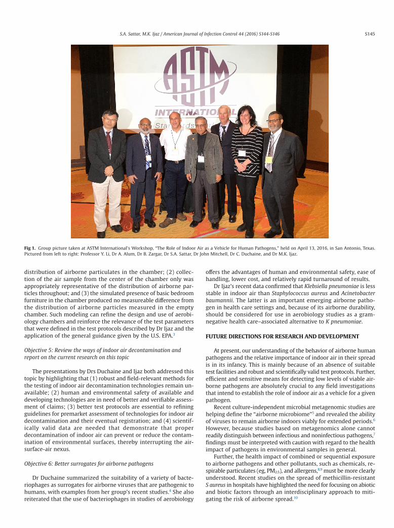
distribution of airborne particulates in the chamber; (2) collec-tion of the air sample from the center of the chamber only wasappropriately representative of the distribution of airborne par-ticles throughout; and (3) the simulated presence of basic bedroomfurniture in the chamber produced no measureable difference fromthe distribution of airborne particles measured in the emptychamber. Such modeling can refine the design and use of aerobi-ology chambers and reinforce the relevance of the test parametersthat were defined in the test protocols described by Dr Ijaz and theapplication of the general guidance given by the U.S. EPA.3
Objective 5: Review the ways of indoor air decontamination andreport on the current research on this topic
The presentations by Drs Duchaine and Ijaz both addressed thistopic by highlighting that (1) robust and field-relevant methods forthe testing of indoor air decontamination technologies remain un-available; (2) human and environmental safety of available anddeveloping technologies are in need of better and verifiable assess-ment of claims; (3) better test protocols are essential to refiningguidelines for premarket assessment of technologies for indoor airdecontamination and their eventual registration; and (4) scientif-ically valid data are needed that demonstrate that properdecontamination of indoor air can prevent or reduce the contam-ination of environmental surfaces, thereby interrupting the air-surface-air nexus.
Objective 6: Better surrogates for airborne pathogens
Dr Duchaine summarized the suitability of a variety of bacte-riophages as surrogates for airborne viruses that are pathogenic tohumans, with examples from her group’s recent studies.4 She alsoreiterated that the use of bacteriophages in studies of aerobiology
offers the advantages of human and environmental safety, ease ofhandling, lower cost, and relatively rapid turnaround of results.
Dr Ijaz’s recent data confirmed that Klebsiella pneumoniae is lessstable in indoor air than Staphylococcus aureus and Acinetobacterbaumannii. The latter is an important emerging airborne patho-gen in health care settings and, because of its airborne durability,should be considered for use in aerobiology studies as a gram-negative health care–associated alternative to K pneumoniae.
FUTURE DIRECTIONS FOR RESEARCH AND DEVELOPMENT
At present, our understanding of the behavior of airborne humanpathogens and the relative importance of indoor air in their spreadis in its infancy. This is mainly because of an absence of suitabletest facilities and robust and scientifically valid test protocols. Further,efficient and sensitive means for detecting low levels of viable air-borne pathogens are absolutely crucial to any field investigationsthat intend to establish the role of indoor air as a vehicle for a givenpathogen.
Recent culture-independent microbial metagenomic studies arehelping define the “airborne microbiome”5 and revealed the abilityof viruses to remain airborne indoors viably for extended periods.6
However, because studies based on metagenomics alone cannotreadily distinguish between infectious and noninfectious pathogens,7
findings must be interpreted with caution with regard to the healthimpact of pathogens in environmental samples in general.
Further, the health impact of combined or sequential exposureto airborne pathogens and other pollutants, such as chemicals, re-spirable particulates (eg, PM2.5), and allergens,8,9 must bemore clearlyunderstood. Recent studies on the spread of methicillin-resistantS aureus in hospitals have highlighted the need for focusing on abioticand biotic factors through an interdisciplinary approach to miti-gating the risk of airborne spread.10
Fig 1. Group picture taken at ASTM International’s Workshop, “The Role of Indoor Air as a Vehicle for Human Pathogens,” held on April 13, 2016, in San Antonio, Texas.Pictured from left to right: Professor Y. Li, Dr A. Alum, Dr B. Zargar, Dr S.A. Sattar, Dr John Mitchell, Dr C. Duchaine, and Dr M.K. Ijaz.
S145S.A. Sattar, M.K. Ijaz / American Journal of Infection Control 44 (2016) S144-S146
CONCLUDING REMARKS
Indoor air is increasingly understood to be a vehicle for a varietyof human pathogens via direct inhalation or contamination of sec-ondary vehicles, such as environmental surfaces. Further, pathogenson surfaces and objects initially contaminated by air can be resus-pended for further transport.
The study of human pathogens in air continues to present majorchallenges, including the following:
1. Experimental facilities for the study of survival and transportof airborne pathogens (viruses, in particular) remain rare becauseof a lack of specialized equipment, infrastructure, and techni-cal skills.
2. Practical and standardized processes for recovering viable patho-gens from field samples of air remain unavailable, therebypreventing linking of airborne exposure to a variety of infections.
3. Simultaneous or sequential exposures of hosts to airborne patho-gens and other harmful substances, possibly combining to createnegative health impacts, make risk assessment particularlychallenging.
4. Surrogate microbes often used for the study of the aerobiol-ogy of human pathogens may be unsuitable for the purposebecause of an inability to withstand aerosolization and remainviable in air. A major research need is the identification of ap-propriate bacteriophages because they are attractive surrogatesfor the study of airborne human pathogenic viruses. This defi-nitely is a topic for further investigation.
5. Despite the increasing number and variety of technologies claim-ing indoor air decontamination, robust and scientifically validprotocols remain unavailable for their validation.
Meaningful approaches to resolving the knowledge gaps that aredescribed here will require the joint efforts of microbiologists, ar-chitects, and specialists in indoor air handling systems.
Table 1 summarizes the research needed to refine the study ofaerobiology of human pathogens.
Other areas of evolving significance are microalgae11 and entericprotozoa12 as airborne pathogens. Although it is relatively easy todetect, by molecular means, the presence of a wide variety of mi-croorganisms in indoor air, relating such data to actual health riskremains a major challenge. As always, answers to these and otherquestions will require better and longer-term research funding.
References
1. Goldberg LJ, Watkins HM, Boerke EE, Chatigny MA. The use of a rotating drumfor the study of aerosols over extended periods of time. Am J Hyg 1958;68:85-93.
2. Sattar SA, Kibbee RJ, Zargar B, Wright KE, Rubino JR, Ijaz MK. Decontaminationof indoor air to reduce the risk of airborne infections: studies on survival andinactivation of airborne pathogens using an aerobiology chamber. Am J InfectControl 2016 Jun 30. [Epub ahead of print].
3. U.S. Environmental Protection Agency. Air sanitizers–efficacy datarecommendations. Test Guideline No. #OCSPP 810.2500-Air Sanitizers-2013-03-12 [EPA 730-C-11-003]. Available from: http://www.noticeandcomment.com/-ocspp-810-2500-air-sanitizers-2013-03-12-epa-730-c-11-003-fn-24288.aspx. Accessed December 4, 2015.
4. Turgeon N, Toulouse MJ, Martel B, Moineau S, Duchaine C. Comparison of fivebacteriophages as models for viral aerosol studies. Appl Environ Microbiol2014;80:4242-50.
5. Kembel SW, Jones E, Kline J, Northcutt D, Stenson J, Womack AM, et al.Architectural design influences the diversity and structure of the builtenvironment microbiome. ISME J 2012;6:1469-79.
6. Prussin AJ, Vikram A, Bibby KJ, Marr LC. Seasonal dynamics of the airbornebacterial community and selected viruses in a children’s daycare center. PLoSONE 2016;11:e0151004.
7. Prussin AJ, Garcia EB, Marr LC. Total virus and bacteria concentrations in indoorand outdoor air. Environ Sci Technol Lett 2015;2:84-8.
8. Chen R, Zhao A, Chen H, Zhao Z, Cai J, Wang C, et al. Cardiopulmonary benefitsof reducing indoor particles of outdoor origin: a randomized, double-blindcrossover trial of air purifiers. J Am Coll Cardiol 2015;65:2279-87.
9. Leung DYC. Outdoor-indoor air pollution in urban environment: challenges andopportunity. Front Environ Sci 2015;2:1-7.
10. Creamer E, Shore AC, Deasy EC, Galvin S, Dolan A, Walley N, et al. Air and surfacecontamination patterns of meticillin-resistant Staphylococcus aureus on eightacute hospital wards. J Hosp Infect 2014;86:201-8.
11. Tesson SV, Skjoth CA, Santl-Temkiv T, Londahl J. Airborne microalgae: insights,opportunities, and challenges. Appl Environ Microbiol 2016;82:1978-91.
12. Sponseller JK, Griffiths JK, Tzipori S. The evolution of respiratoryCryptosporidiosis: evidence for transmission by inhalation. Clin Microbiol Rev2014;27:575-86.
Table 1Continuing challenges in research on aerobiology of human pathogens
Factor(s) Refinements required
Experimental setup Space, biosafety, field relevance, ease of control, and monitoring of test parametersChallenge microbe selection Representative of airborne pathogens, ease of culture and recovery, stability during aerosolization and in air, and
preparation and concentration of cultureSuspension to be nebulized Safe and standardized soil load representing body fluids, antifoam, and physical tracer (if needed)Nebulization and particle size distribution Protection for microbe, generation of aerosols and droplet nuclei, granulometrics, and uniform distributionAging and exposure conditions Control of air temperature and RH, testing at an RH <20%, and harmonization for relevant microbial typesAerosol collection and sizing Protection of viability, optimal growth conditions, and neutralization of activesAssessing decontamination Proper controls, realistic efficacy criteria for method and device air decontamination technologies, and number of
replicationsInterpretation of data Statistical analyses, field relevance, and regulatory requirements
RH, relative humidity.
S146 S.A. Sattar, M.K. Ijaz / American Journal of Infection Control 44 (2016) S144-S146
Commentary
Workshop on “The Role of Indoor Air as a Vehicle forHuman Pathogens”: A Panel Discussion
Syed A. Sattar MSc, Dip Bact, MS, PhD *Professor Emeritus of Microbiology, Faculty of Medicine, University of Ottawa, Ottawa, ON, Canada
The primary objective of this panel discussion was to seek inputon the following 3 questions from the speakers at the workshop.Comments and questions were also entertained from the membersof the audience.
PANEL DISCUSSION
Question 1: Considering the potential advantages of microbialdecontamination of indoor air, what criteria should be used to selecta given technology?
Dr C. Duchaine: Technologies of interest will depend on the sit-uation and the point source of microbial contamination, for example,a person coughing in an emergency room, a patient vomiting in ahospital ward, or a student vomiting in a classroom. The interven-tion of choice must be relevant for the site, for instance, a hospitalversus a shoppingmall. Portable indoor air decontamination devicesmay be used in smaller areas, such as classrooms, but devices thatwould allow global decontamination (eg, at a hospital or a shop-ping mall) would be more challenging. When possible, routine andcontinuous air treatment may be better than an on-site intervention.
Dr Y. Li: Technologies that are effective for decontamination ofcirculating air in different situations would be more desirable. Oneexample is the use of UVGI in air circulation ducts if the potential-ly generated ozone can be effectively removed to avoid its entry intooccupied zones. The technology of choice must be safe when peopleare present, while also being energy efficient. Further, it must notonly be effective, but also scalable to suit the site of use. The resultsof testing in an aerobiology chamber can be much different thanthose in a large area, such as a hospital or a large shopping mall,where there are on-going changes in the air quality parameters. Thedebate continues as to whether indoor air for breathing should betreated in the same fashion as water for drinking.
Dr S.A. Sattar: Yes, it is certainly true that waters for drinkingand swimming are routinely decontaminated, but not indoor air inmost settings! Titanium dioxide filters in ventilation systems couldbe very valuable as they can work on an ongoing basis withoutadding any chemicals to the air. However, in situations of very closecontact between 2 people, it is virtually impossible to prevent ex-posure to airborne pathogens. There is an invention though thatclaims personal protection against airborne pathogens and othercontaminants based on 2 thimble-sized cups with filters to be placedinside the nostrils [http://www.breathecleanerair.org/]. I believe thatthis device requires independent testing and perhaps additionalrefinements.
Audience member 1: Much of what is being discussed has beendone before using algorithms and flow rates at the UNLV [Univer-sity of Nevada Las Vegas] with the U.S. EPA [Environmental ProtectionAgency] data. A peer review team consisted of NASA [National Aero-nautics and Space Administration] and the EPA radon/asbestosparticulate team. There is a rich body of data from the IEST [Insti-tute of Environmental Sciences and Technology, Arlington, IL] onclean room standards and aerospace standards that you should bereviewing. For the space industry, particle exposure testing was per-formed using microbeads as surrogates. Airflow could be studiedwithout the issue of contamination/decontamination of the chamber.The mass and shape of material contaminants can affect results andthe decisions made. Did you consider this? Solutions can be foundin other disciplines, and many materials do not require monitoring.
Dr Sattar: It is unfortunate that this original work is not morereadily accessible. The 2012 EPA guidelines do not reflect this back-ground information, which could have impacted chamber design.
Audience member 2: Decontamination is too strong of a termunless complete kill is demonstrated. Microbial reduction is a betterterm.
Dr Sattar: For indoor air quality, you can differentiate betweenphysical removals by filtration versus killing. Decontamination cap-tures both physical removal and killing of microorganisms. Reductionalso could be used to describe the situation.
Audience member 2: In real life, it is very difficult to decontam-inate a room completely, as microorganisms will be enteringcontinuously through doors, windows, and cracks. Unless the entirebuilding is sealed, there alwayswill be low levels of organisms present.
Dr Sattar: The purpose of air decontamination is not to steril-ize the room. The emphasis is on decontaminating the air and notthe surfaces. By decreasing the microbial content in the air, youwould hope to reduce the contamination present on surfaces as well.
* Address correspondence to Syed A. Sattar, MSc, Dip Bact, MS, PhD, ProfessorEmeritus of Microbiology, Faculty of Medicine, University of Ottawa, 451 Smyth Rd,Ottawa, ON, K1H8M5, Canada.
E-mail address: [email protected] (S.A. Sattar).Publication of this supplement is primarily supported by RB,Montvale, New Jersey,
with additional support fromMicroBioTest, a division of Microbac Laboratories, Inc.,Sterling, Virginia. The City University of New York (CUNY) and the University of Ottawa,Ottawa, Canada, are academic sponsors. Editorial support was provided by AshleyO’Dunne, PhD; Shannon O’Sullivan, ELS; and Alanna Franchetti, ELS of Medergy(Yardley, PA), and funded by RB.
Conflicts of Interest: None to report.
0196-6553/© 2016 Association for Professionals in Infection Control and Epidemiology, Inc. Published by Elsevier Inc. All rights reserved.http://dx.doi.org/10.1016/j.ajic.2016.06.007
American Journal of Infection Control 44 (2016) S147-S149
Contents lists available at ScienceDirect
American Journal of Infection Control
journal homepage: www.aj ic journal .org
American Journal of Infection Control
Although the EPA guideline mentions it, one should avoid the term“sanitization of air” and use “decontamination” instead. “Sanitiza-tion” is a nebulous term and is difficult to define. That term is notused in Europe.
Audience member 3: As with any technology, there can be un-foreseen consequences, for example, as observed with the high-efficiency plumbing. What are the potential consequences ofdecontamination of indoor air?
Dr Li: Some microbes are good and some are bad, and the goalshould be to remove those organisms that are contaminants in theair. People should not be encouraged to decontaminate ordinaryspaces unless known contaminants exist. Air decontamination is im-portant in health care settings, such as for hospital spaces. Overdoingmicrobial decontamination may have secondary environmentalimpacts. During the SARS [severe acute respiratory syndrome] out-break in Hong Kong, significant environmental decontamination andcommunity hygiene measures were associated with a reduction inthe reported incidence rates of other respiratory infections duringthat time.1 On the other hand, overuse of disinfectants can lead tomore pollution in the wastewater.
Dr M.K. Ijaz: No matter what the technology, it has to be safe.In case there are safety concerns to humans, the space being de-contaminated should not be occupied during treatment.With regardsto opening of doors and re-introduction of microbial contami-nants, the data I presented addressed this by showing that a properindoor air decontamination device can continually deal with on-going fluctuations in indoor air contamination. No one is suggestingto eliminate all microbes from the air, and, from a practical pointof view, it is impossible. The focus is on risk reduction, and a suit-able air decontamination device can lower the risk of airborne spreadof pathogens.
Dr B. Zargar: One needs to consider the location of the indoorair decontamination device in a given room and also make sure thatthe existing air-handling system is working properly. Occupants’ ex-posure to potentially harmful bacteria should thus be reduced asmuch as possible. CFD [computational fluid dynamics] is usedwidelyto simulate the behavior of airborne microorganisms in various set-tings. To the best of our knowledge, this is the first time that CFDhas been used to assess the design and performance of an aerobi-ology chamber to test the behavior of microorganisms in indoor air.Since the size of airborne particles is an important consideration,we have chosen for our simulation particles in the range of 0.5 to5 μm in diameter.
Dr Sattar: It is a valid concern not to introduce technologies thatwill have unforeseen consequences. Good communication is re-quired among engineers, designers, architects, and microbiologists.We are becoming wiser in introducing technologies that are safeand sustainable. Air decontamination goes beyond removing andkilling bacteria. Is it conceivable that air decontamination also mightremove allergens and pollen in addition to pathogens? Technolo-gies that have a broad base, beyond just microorganisms, might bea more attractive proposition to the consumer.
Audience member 2: If we continually operate the device, wedo not know the impact of reduced exposure to pathogens on ourimmune system. What is the effect of living like this?
Dr Ijaz:Microbial loadwill be reduced, but contaminants are con-tinually reintroduced. Contamination is minimized, but you do notremove it completely. This is a question of risk reduction.
Audience member 4:Whatwas the size of the chamber you usedfor testing and modeling of data?
Dr Sattar: Our chamber was built according to the EPA guide-line, but was slightly >800 ft3, or about 24 m3.
Dr Zargar: The minimum chamber dimensions suggested by theU.S. EPA guidelines are 10 feet × 10 feet × 8 feet (3.048 × 3.048 ×2.44 m).
Audience member 5: One needs to consider the size of the testchamber versus the size of the device. For example, UV [ultravio-let] decontamination is related to energy level, distance, and time.Is it fair to test a device designed to decontaminate a large area anda device meant for a small area in the same sized chamber? Doesthis give an advantage to the device designed for larger rooms? Thereare practical concerns with this.
Dr Ijaz: The test chamber built at the University of Ottawa is inaccordance with the EPA guideline and is designed as an average-sized room. The chamber is useful for screening technologies. Forlarger areas, field studies would be required to prove the effective-ness of the technology.
Dr Sattar: Look at HVAC [heating, ventilation, and air condi-tioning] systems for understanding. The equipment is scaled to thesize of the building. It is a matter of engineering rather than thedevice, and one has to cater to the needs of the home. It is possi-ble that more than one device will be required for decontamination.
Audience member 5: Scale the device to the chamber?Dr Sattar: Yes.Dr Ijaz: The research team at the University of Ottawa has already
tested a number of different devices. The key question is “how fastand how frequently can the chamber air be processed through thedevice?” One of the devices produced a 3-log10 reduction in exper-imentally aerosolized bacterial challenge in 45 minutes, whileanother one required >3 hours to achieve the same level of bacte-rial reduction. Therefore, the performance of the 2 devices wasdrastically different.
Audience member 2: The chamber was designed specifically formicrobial decontamination of a room, and the EPA provided the sizeas a standard. You need to know the size of the room you want todecontaminate. Future products would need to be labeled for thesize of the room and tested in this size.
Audience member 1: There are many companies active in thisarea. In the agricultural field, there are ISO [International Organi-zation for Standardization]- andWHO [World Health Organization]-approved protocols for decontaminating facilities such as chickenand egg houses. Construction and other materials in the roommayabsorb or adsorb the chemicals used to decontaminate the air, andthis must be considered. ASHRAE [American Society for Heating, Re-frigeration, and Air-Conditioning Engineers] has many standards inthis area. There are several guidance documents and standards avail-able in other areas upon which one can draw.
Dr Zargar: The range of airflow can be adjusted for different roomsizes, for example, a higher speed for larger rooms and a lower speedfor smaller rooms. Instead of building different-sized chambers fortesting different devices, mathematical modeling can be used tomapthe result of experiments in rooms of different sizes.
Audience member 6: Which came first—clean air or fresh air?Is there a standard definition?
Dr Sattar: The EPA standard is a 3 log10 reduction in the levelof viable bacteria. One needs to know the baseline values to deter-mine if a technology can result in a 3-log10 reduction in a specifiedperiod of time.
Question 2: What should be the essential elements for anexperimental aerobiology facility in terms of the biosafety level andthe size and configuration of the test chamber?
Dr J. Mitchell (moderator): This question has been partially an-swered in the discussion so far.
Dr Sattar: We sought input from the CDC [U.S. Centers for DiseaseControl and Prevention] as well as from the NIH [U.S. National In-stitutes of Health]. Based on the feedback, if we aerosolize a biosafetylevel-2 (BSL-2) organism, then the testing must be conducted in aBSL-3 laboratory. Although we have been using a BSL-3 facility thus
S148 S.A. Sattar / American Journal of Infection Control 44 (2016) S147-S149
far, our university has recently decided to downgrade that facilityto BSL-2 to reducemaintenance and operating costs and to cut downon the paper work for periodic certification as a BSL-3 lab. The de-cision was based on risk assessment considering that proper stafftraining and use of personal protective equipment can minimize ex-posure to the microbes being aerosolized. However, there is still nogeneral consensus on the biosafety containment level needed toworkwith aerosolized BSL-2 organisms.
Dr Duchaine: If we aerosolize a BSL-2 organism, we use a double-containment strategy so exposure is limited.
Dr Li: It is important to ensure that the chamber is fully mixedso that the organisms are uniformly dispersed and the environ-mental conditions remain constant. Organism survival is a functionof temperature and humidity. Within a chamber that is not uniform,the organism can be dispersed into different environmental con-ditions. Different-sized chambers are employed for different purposes(eg, VOC [volatile organic compound] release). This is probably re-flected in the fact that a typical room or building size does not exist,as different indoor environments are of different sizes.
Question 3: The U.S. EPA guideline (2012) for testing indoor airdecontamination technologies recommends the use of specific strainsof Staphylococcus aureus and Klebsiella pneumoniae as surrogatesfor airborne gram-positive and gram-negative vegetative bacterialpathogens, respectively. Are they the most suitable for the purpose?Also, should additional surrogates be considered for other classes ofairborne pathogens, such as viruses, fungi, mycobacteria, and spore-forming bacteria? If yes, what desirable attributes should weconsider in selecting such surrogates?
Dr Sattar: This is a crucial aspect where we want to consoli-date our views on selection of surrogates. This is not only importantfor aerobiology but for environmental microbiology as well.
Dr Duchaine: Several types of surrogates will be required, as theyall do not behave in the same manner. Eventually, one will need towork with the real contaminating organism to confirm the re-sponse. It is important to validate that the surrogate behaves likethe actual contaminating organism.
Audience member 3: Dr Alum, in your Legionella studies, whatwas the inoculum level used and does it represent the typical sit-uation that occurs during an outbreak?
Dr Alum: The level of Legionella used in the studies was 10 cfu[colony formingunits]/m3. Fornonoutbreak−associatedcooling towers,wehave found,with limited studies, that the Legionellanumberswerevery low, <10 cfu/mL. In cooling tower water associated with out-breaks, the Legionella concentration is relatively high.
Audience member 3: In selecting surrogates, do we also needto consider expected numbers present in the targeted situation?
Dr Sattar: Yes, but we always build into the study a certain re-dundancy and a certain higher level of performance. Hence, the levelobserved in the actual field situation may not be the level used intesting. The product label has to have assurances built in, so thisis the reason we use a higher level of test organisms. It is absolute-ly right that we need to have surrogates that closely represent thosepresent in air. Consider surrogates in 4 categories: vegetative bac-teria, spore-formers, fungi, and viruses. Microalgae are a new areathat I cannot comment on yet. If this is a health concern, it shouldbe considered as a fifth category.
Audience member 1: Algaewere studied in the 1970s and shownto be an allergen, producing an immune response. Since microalgaeexist in the normal environment, I would suggest we keep it on thelist as a fifth category.
Audience member 5: There are a lot of data for surface disin-fection that suggest a general hierarchy of resistance. Do you seethe same hierarchy for aerobiological studies? When norovirus isdetected in air from a contaminated area, what is the source? It isless likely from direct breathing, but rather from vomit or fecal aero-solization of norovirus. If norovirus is primarily transmitted by touch,then is it important to decontaminate the air if air transmission doesnot cause infection?
Dr Duchaine: The source is the patients themselves. The sam-pling device was present where the actively emitting patients were,and they may have vomited or had diarrhea. There may be up to1012 virus particles per gram of diarrheic feces, and this would bepart of the aerosol. Surfaces are cleaned, but if an airborne routeexists, this explains how other patients some distance away becomeinfected. One way to prevent spreading of infections is to decon-taminate the air. For the first part of your question, droplet nucleineed to come in contact with the product for an appropriate periodof time. Unlike surface testing, the air decontaminant contact timeis longer.
Audience member 5: How do you compare the resistance levelsof the different categories of organisms (bacteria, virus, and fungi)in air?
Dr Sattar: You need to develop comparative data, but one wouldexpect differences in comparing liquid disinfectants to air treat-ments. For liquid disinfectants, there is a larger volume to infectiousagent ratio. In air, the ratio is not the same, and the testing dynam-ics are different. Therefore, one cannot compare liquid disinfectiontesting to air decontamination. There is a need to consider surro-gate organisms for each of the 5 categories because there are practicaland ethical issues to deal with in working with actual pathogens.For example,M [Mycobacterium] tuberculosis can spread by air, butit is a relatively slow-growing pathogen and difficult to obtain inviability titers high enough for aerosolization. Also, how canwe growenough of the SARS virus to contaminate an aerobiology chamber?If regulators allow the use of surrogate organisms for testing en-vironmental surface disinfectants, why can their use not be extendedto air testing?
Audience member 7:We should not limit ourselves to key patho-gens, such as respiratory, as less known ones can become a problem.Could infection or airflow be modeled through adjoining rooms orhalls, rather than in a chamber? That might be beneficial to betteralign with the real world.
Dr Zargar: Modeling of a furnished chamber can be used as thestarting point for simulation of a hospital room. Modeling of theventilation system in any such setting would be crucial.
Audience member 2: Following up on the hierarchy question,AD [Antimicrobial Division of the U.S. EPA] for registration pur-poses allowed 60% glycol products to have an air sanitization claim.Data are required to support this, so registrants must provide thedata. This testing is in its infancy. As more data become available,a hierarchy for air decontamination can be developed. Selecting sur-rogates also is still in its infancy and currently is supported byhistorical selection.
Mr Rubino: It is time to close the session, and I would like tothank ASTM International for supporting this workshop. Specialthanks to Dr Sattar, Dr Mitchell, Dr Ijaz, and the rest of the team,as well as to the speakers.
Reference
1. Lo JY, Tsang TH, Leung YH, Yeung EY,Wu T, LimWW. Respiratory infections duringSARS outbreak, Hong Kong, 2003. Emerg Infect Dis 2005;11:1738-41.
S149S.A. Sattar / American Journal of Infection Control 44 (2016) S147-S149


![Vol 003s - [Supplement]](https://img.pdfslide.net/doc/110x75/568c3c261a28ab0235ace557/vol-003s-supplement.jpg)