Embed Size (px)
Citation preview
Sergio A. GehrkeMar�ıa P. Ram�ırez-FernandezJos�e Manuel Granero Mar�ınMarcos Barbosa SallesMassimo Del FabbroJos�e Luis Calvo Guirado
A comparative evaluation betweenaluminium and titanium dioxidemicroparticles for blasting the surfacetitanium dental implants: anexperimental study in rabbits
Authors’ affiliations:Sergio A. Gehrke, Biotecnos Research Center,Santa Maria, BrazilUniversity Catholica San Antonio de Murcia(UCAM), Murcia, SpainMar�ıa P. Ram�ırez-Fernandez, InternationalDentistry Research Cathedra, Faculty of Medicine,San Antonio Catholic University of Murcia(UCAM), Murcia, SpainJos�e Manuel Granero Mar�ın, International ResearchCathedra, UCAM, Universidad Cat�olica SanAntonio, Murcia, SpainMarcos Barbosa Salles, General Dentistry of the 9de Julho University, San Pablo, BrazilMassimo Del Fabbro, Research Center in OralHealth, Department of Biomedical, Surgical andDental Sciences, Universit�a degli Studi di Milano,IRCCS Istituto Ortopedico Galeazzi, Milano, ItalyJos�e Luis Calvo Guirado, International DentistryResearch Cathedra, Faculty of Medicine &Dentistry, San Antonio Catholic University ofMurcia (UCAM), Murcia, Spain
Corresponding author:Prof. Sergio A. Gehrke, DDS, PhDDepartment of Research, Biotecnos – Technologyand ScienceRua Dr. Bozano 571, CP 97015-001, Santa MariaRS, BrazilTel / Fax: +55 55 32227253e-mail: [email protected]
Key words: aluminium blasted, dental implants, SLA surface, surface treatment, titanium
blasted
Abstract
Objective: The aim of this study was to compare, through biomechanical and histological analysis,
the aluminium (AlO2) and titanium dioxide (TiO2) microparticles for blasting during the
sandblasting acid surface treatment in titanium dental implants using a rabbit tibia model.
Materials and methods: Forty-eight commercially available titanium dental implants were divided
into two test groups (n = 24 per group): implants with surface treated by AlO2 followed by acid
etching as control group (Con group) and implants with surface treated by TiO2 followed by acid
etching as test group (Test group). The implants were randomly installed in both tibias of eight
rabbits and block samples were removed 4 and 8 weeks after implantation. Resonance Frequency
Analyses were performed immediately after the implantation and at 8 weeks. Twelve implants of
each group were removed to measure the reverse torque. The remaining implants were used for
histological analysis. The data were compared using statistical tests (a = 0.05).
Results: In comparing the implant stability quotient at the two time points, no significant
statistical differences were found (P > 0.05), as well as in the removal torque test at 8 weeks after
implant placement, no found significant difference between the two groups was tested.
Histomorphometric analysis showed a high degree of bone organization in all samples with no
significant difference between groups in the bone-to-implant contact (P > 0.05).
Conclusion: Within the limitations of this study, the results indicate that the media of surface
blasting (AlO2 or TiO2 microparticles) did not show significant differences in the tested parameters
for assessing the osseointegration of the implants.
Success rates of titanium dental implants-
based therapy in dentistry have been docu-
mented to be over 98% (Buser et al. 1997;
Mangano et al. 2010). Implant success is
strictly related to the osseointegration pro-
cess that has been defined as the formation
of a direct bone–implant interface with no
intervening soft tissues (Br�anemark et al.
1969). Titanium surfaces can also be modi-
fied to increase their biological properties.
Such modifications are achieved by either
adding a coating consisting of different types
of bioactive substances or by removing por-
tions of the external layer with the use of
blasting materials of different particle sizes
or by the application of chemical treatments
and/or by physical means such as the laser
(Wennerberg & Albrektsson 2009). Among
these, blasting and acid etching have been
the most widely used by industry, and their
combination has shown improved biological
activity of the titanium surface in terms of
implant osseointegration as compared to
machined (turned) surfaces (Novaes et al.
2010).
The modification of the implant surface
can thus bring benefits to the response of the
surrounding bone tissue, accelerating the
healing process and/or improving the newly
formed bone quality (Novaes et al. 2010;
Wennerberg & Albrektsson 2010). Studies
have shown that osseointegration is related
to microgeometric features such as the degree
of surface roughness and can also depend on
factors such as physical and chemical surface
properties (Sul et al. 2005; Le Gu�ehennec
et al. 2007). The macrogeometric design such
as the implant body shape, height, density
Date:Accepted 14 August 2016
To cite this article:Gehrke SA, Ram�ırez-Fernandez MP, Granero Mar�ın JM,Barbosa Salles M, Del Fabbro M, Calvo Guirado JL. Acomparative evaluation between aluminium and titaniumdioxide microparticles for blasting the surface titanium dentalimplants: an experimental study in rabbits.Clin. Oral Impl. Res. 00, 2016, 1–6doi: 10.1111/clr.12973
© 2016 John Wiley & Sons A/S. Published by John Wiley & Sons Ltd 1
and cutting ability of the threads may affect
the biomechanics of the bone–implant inter-
locking, possibly improving implant stability
(Hallgren et al. 2003).
Several types of chemical and physical sur-
face treatments have been developed and
marketed by dental implant manufacturers
(Binon 2000). However, there is still no con-
sensus on what the optimal condition for
peri-implant bone growth should be. It is
known that the bone response can be influ-
enced by the implant surface topography at
the micrometer level, and some indications
exist that a nanometric surface can also have
an effect (Pan et al. 2012). However, the
mechanisms behind an optimal bone
response in relation to a given type of surface
still remain largely unknown. Some biologi-
cal processes involved in the activation of
the early stages of osseointegration, such as
protein adsorption, cell–surface interaction,
progenitor cell recruitment and differentia-
tion and tissue formation at the interface
between the body and the biomaterial, can be
affected by the implant surface microrough-
ness as well as by its physical–chemical
surface properties (Schliephake et al. 2006,
2009a,b; Lutz et al. 2010).
Surfaces known as sandblasting acid (SLA)
types, which are produced by sandblasting
with titanium particles followed by a strong
acid etching bath with a mixture of HCl/
H2SO4 at elevated temperature for several
minutes, are widely utilized and have been
well documented in the literature (Li et al.
2002; Esposito et al. 2005; Kim et al. 2015).
These are moderately rough surfaces that usu-
ally present fine 2–4-lm micropits superim-
posed on the rough-blasted surface. Although
well documented, the presence of residuals of
alumina embedding on its surface due to the
fabrication process has been regarded as a
potential risk for long-term osseointegration
(Piattelli et al. 2003; Gehrke et al. 2014,
2015). Alternatively, surfaces have been
blasted with other biocompatible media such
as calcium phosphate bioactive ceramics (Piat-
telli et al. 2002; Schliephake et al. 2006,
2009a) and titanium oxide (Zinger et al. 2004;
Gehrke et al. 2014, 2015). The first comprises
a resorbable medium that is actually bioactive,
while the second method consists of particles
that are made of the same biocompatible
material as the implant. Even though a wide
literature body exists for the alumina-blasted/
acid-etched surfaces relative to other surface
modification techniques (Li et al. 2002; Espos-
ito et al. 2005), a substantially smaller body of
evidence exists for the resorbable blasting
media and an even smaller one concerning the
characterization and in vivo evaluation of
TiO2-blasted surfaces.
The purpose of this study was to compare,
through biomechanical and histological analy-
ses, the effects of aluminium (AlO2) and tita-
nium dioxide (TiO2) microparticles for
blasting used to produce the SLA surface treat-
ment of two commercially available titanium
dental implants, using a rabbit tibia model.
Material and methods
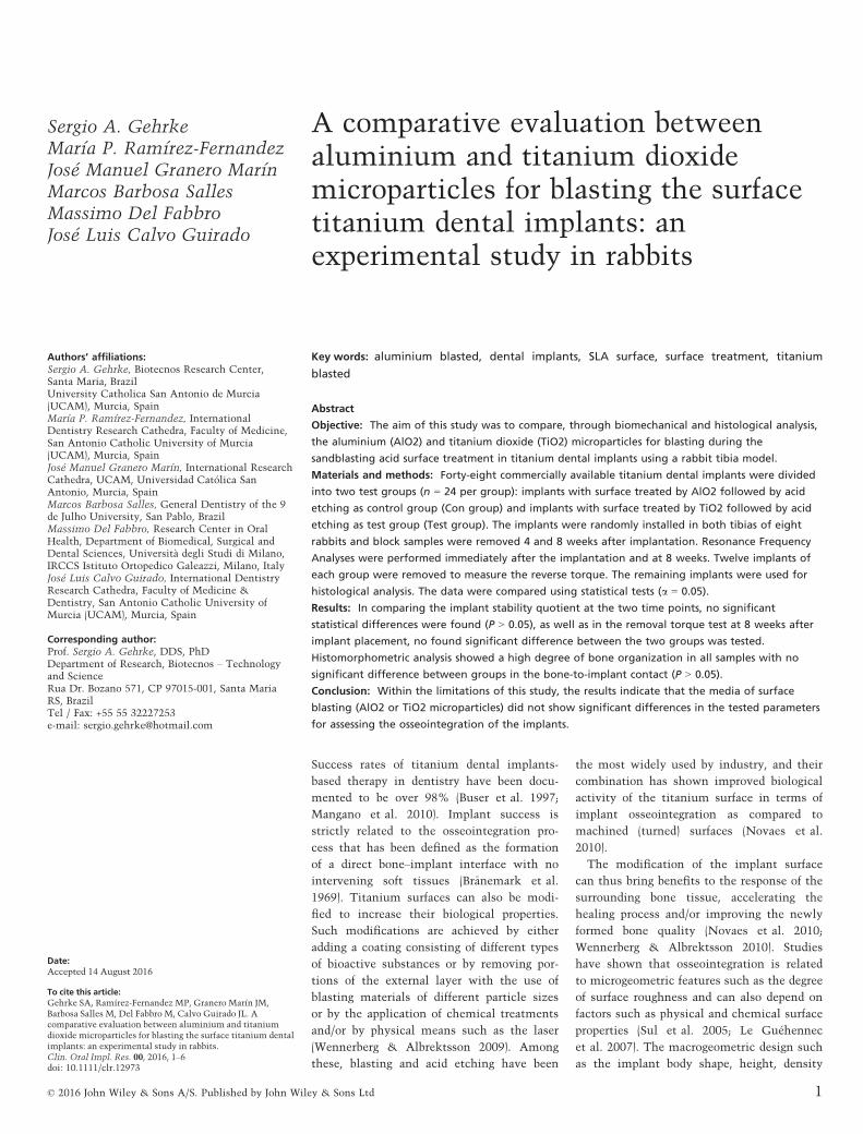
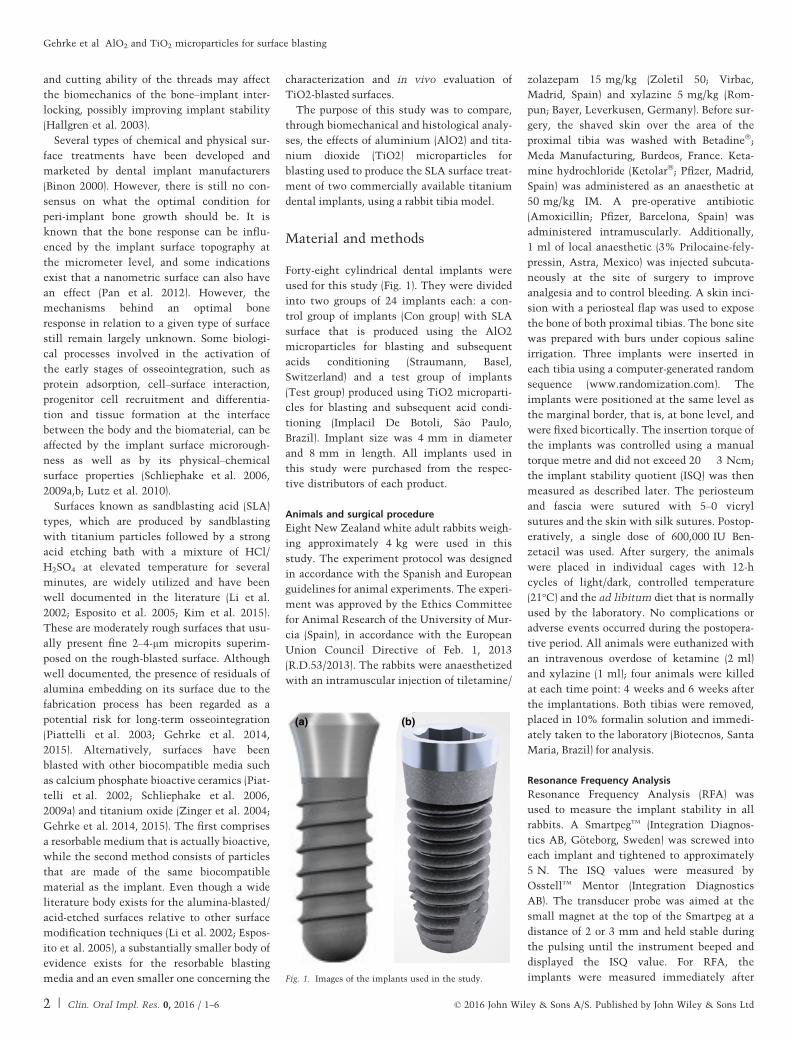
Forty-eight cylindrical dental implants were
used for this study (Fig. 1). They were divided
into two groups of 24 implants each: a con-
trol group of implants (Con group) with SLA
surface that is produced using the AlO2
microparticles for blasting and subsequent
acids conditioning (Straumann, Basel,
Switzerland) and a test group of implants
(Test group) produced using TiO2 microparti-
cles for blasting and subsequent acid condi-
tioning (Implacil De Botoli, S~ao Paulo,
Brazil). Implant size was 4 mm in diameter
and 8 mm in length. All implants used in
this study were purchased from the respec-
tive distributors of each product.
Animals and surgical procedure
Eight New Zealand white adult rabbits weigh-
ing approximately 4 kg were used in this
study. The experiment protocol was designed
in accordance with the Spanish and European
guidelines for animal experiments. The experi-
ment was approved by the Ethics Committee
for Animal Research of the University of Mur-
cia (Spain), in accordance with the European
Union Council Directive of Feb. 1, 2013
(R.D.53/2013). The rabbits were anaesthetized
with an intramuscular injection of tiletamine/
zolazepam 15 mg/kg (Zoletil 50; Virbac,
Madrid, Spain) and xylazine 5 mg/kg (Rom-
pun; Bayer, Leverkusen, Germany). Before sur-
gery, the shaved skin over the area of the
proximal tibia was washed with Betadine�;
Meda Manufacturing, Burdeos, France. Keta-
mine hydrochloride (Ketolar�; Pfizer, Madrid,
Spain) was administered as an anaesthetic at
50 mg/kg IM. A pre-operative antibiotic
(Amoxicillin; Pfizer, Barcelona, Spain) was
administered intramuscularly. Additionally,
1 ml of local anaesthetic (3% Prilocaine-fely-
pressin, Astra, Mexico) was injected subcuta-
neously at the site of surgery to improve
analgesia and to control bleeding. A skin inci-
sion with a periosteal flap was used to expose
the bone of both proximal tibias. The bone site
was prepared with burs under copious saline
irrigation. Three implants were inserted in
each tibia using a computer-generated random
sequence (www.randomization.com). The
implants were positioned at the same level as
the marginal border, that is, at bone level, and
were fixed bicortically. The insertion torque of
the implants was controlled using a manual
torque metre and did not exceed 20 � 3 Ncm;
the implant stability quotient (ISQ) was then
measured as described later. The periosteum
and fascia were sutured with 5–0 vicryl
sutures and the skin with silk sutures. Postop-
eratively, a single dose of 600,000 IU Ben-
zetacil was used. After surgery, the animals
were placed in individual cages with 12-h
cycles of light/dark, controlled temperature
(21°C) and the ad libitum diet that is normally
used by the laboratory. No complications or
adverse events occurred during the postopera-
tive period. All animals were euthanized with
an intravenous overdose of ketamine (2 ml)
and xylazine (1 ml); four animals were killed
at each time point: 4 weeks and 6 weeks after
the implantations. Both tibias were removed,
placed in 10% formalin solution and immedi-
ately taken to the laboratory (Biotecnos, Santa
Maria, Brazil) for analysis.
Resonance Frequency Analysis
Resonance Frequency Analysis (RFA) was
used to measure the implant stability in all
rabbits. A SmartpegTM (Integration Diagnos-
tics AB, G€oteborg, Sweden) was screwed into
each implant and tightened to approximately
5 N. The ISQ values were measured by
OsstellTM Mentor (Integration Diagnostics
AB). The transducer probe was aimed at the
small magnet at the top of the Smartpeg at a
distance of 2 or 3 mm and held stable during
the pulsing until the instrument beeped and
displayed the ISQ value. For RFA, the
implants were measured immediately after
(a) (b)
Fig. 1. Images of the implants used in the study.
2 | Clin. Oral Impl. Res. 0, 2016 / 1–6 © 2016 John Wiley & Sons A/S. Published by John Wiley & Sons Ltd
Gehrke et al �AlO2 and TiO2 microparticles for surface blasting

the installation and 8 weeks after the
implant installation. The ISQ values were
measured in two perpendicular directions
(proximal to distal and lateral to medial), and
an average value for each sample was deter-
mined.
Removal torque test
A total of 24 implants (12 per group) were
used in this test. The biological specimens
were processed immediately after the
removal of the tibiae. The samples were
maintained in liquid solution (10% buffered
formalin) and immediately evaluated (1 h
after removal) so as to avoid dehydration. A
torque testing machine was used – CME
(T�ecnica Industrial Oswaldo Filizola, Guarul-
hos, Brazil), which is fully controlled by soft-
ware DynaView Torque Standard/Pro M
(Fig. 2), performing calculations and generat-
ing reports automatically with test speed of
1 rpm and angular measuring system with a
resolution of 0.002°. Measurements of peak
torque to initiate reverse rotation were
recorded, and the mean torque values were
calculated for each group.
Samples treatment for histomorphometricanalysis
The others 24 samples (12 per group) were
dehydrated using an ascending series of alco-
hols and embedded in glycomethacrylate
resin (Technovit 9100 VLC; Kulzer, Frie-
drichsdorf, Germany) to produce undecalci-
fied sections. Undecalcified cut and ground
sections that contained the central part of
each implant and had a final thickness of
15 lm were produced using a macrocutting
and grinding system (Isomet 2000; Buehler,
Esslingen, Germany). The sections were
stained with toluidine blue and acid fuchsin,
and histomorphometric analysis was carried
out.
Specimens that had been prepared for the
histological analysis of the tissue surround-
ing the implant were examined using a light
microscope (EOS 200; Nikon, Tokyo, Japan).
After digitizing the phase of each specimen
under light microscope, the percentage of
bone-to-implant contact (BIC%) was mea-
sured using the program Image Tool version
5.02 for Microsoft WindowsTM. BIC% was cal-
culated as the percentage of bone that was in
direct contact with the implant surface, eval-
uated along the entire profile of the implant.
Data analysis
For comparison between groups at each time
in vivo, statistical analysis was performed by
multiple paired t-tests considering the animal
number per time in vivo as the statistical
unit. For comparing each experimental group
at different times in vivo, t-tests assuming
equal variances were utilized. All evaluations
were conducted at the 95% level of signifi-
cance.
Results
The surgical procedures were uneventful. All
animals presented appropriate healing during
the first week following the surgical proce-
dure. Post-surgical inspections for 2 weeks
postoperatively indicated the absence of
infection or inflammation. After the sched-
uled follow-up time, all implants were
osseointegrated.
Resonance Frequency Analysis (RFA)
The data and statistical analysis of resonance
frequency values for the times investigated of
the two groups are summarized in the
Table 1. Applying the test inside the groups
at the times period proposed (baseline and
8 weeks), the values showed statistically sig-
nificant differences (P < 0.05). Among the
groups, the variations in the RFA values
between the first and the second time point
were not significantly different (P > 0.05).
Removal torque test
In removal torque testing, all of the implants
were stable and anchored in bone after
8 weeks of healing. The mean resistance to
removal torque values and standard deviation
are summarized in the Table 2 and were not
significantly different (P > 0.05).
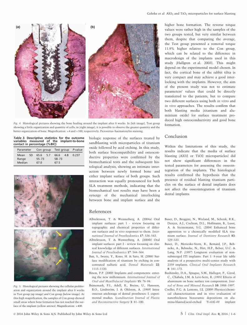
Histological analysis
Histological analysis showed a complete
bone organization and mineralization at
8 weeks in both groups (Figs 3 and 4). The
BIC% values are summarized in the Table 3
and did not show statistical differences
(P = 0.237). At high magnification, the sam-
ples of Con group showed small areas where
bone formation has not reached the surface
of the implant, probably because of some
physical–chemical components that pre-
vented the contact (Fig. 5).
Discussion
Over the past decades, a multitude of in vivo
studies examined the effect of the implant
surface on the bone healing and apposition
(Misch 1990; Hsu et al. 2007). Modifications
in implant surface morphology and roughness
have been initially attempted aiming not
only to hasten the host-to-implant response
but also to increase the level of mechanical
interlocking between bone and implant sur-
face, thus improving the initial stability, and
subsequent stress dissipation during func-
tional loading (Textor et al. 2001).
Histology-based investigations have shown
that surface texturing created by blasting led
to greater bone–implant contact as compared
with the machined surface (Ivanoff et al.
2001), which is a desirable response for
improving the overall system biomechanics.
Blasting the implant surface with gritting
agents made of materials other than the
implant core material may change the sur-
face composition and the implant biocompat-
ibility (Wennerberg et al. 1996). Abrasive
blasting increases the surface roughness, as
well as the metal surface reactivity (Wenner-
berg et al. 1996). With the use of a blasting
material such as Al2O3, a potential risk of
contamination by remnants of blasting parti-
cles with dissolution of aluminium ions into
the host tissue cannot be excluded (Wenner-
berg et al. 1996). It has been reported that Al
ions may inhibit normal differentiation of
bone marrow stromal cells and normal bone
deposition and mineralization (Stea et al.
1992), and aluminium has been shown to
induce net calcium efflux from cultured bone
(Bushinsky et al. 1995). Moreover, alu-
minium may compete with calcium during
the healing of implant bed. Aluminium hasFig. 2. Image of the computerized torque machine used in the removal torque test.
© 2016 John Wiley & Sons A/S. Published by John Wiley & Sons Ltd 3 | Clin. Oral Impl. Res. 0, 2016 / 1–6
Gehrke et al �AlO2 and TiO2 microparticles for surface blasting

also been shown to accumulate at the miner-
alization front and in the osteoid matrix
itself (Nimb et al. 1995). Therefore, other
alternative methods were developed to sand-
blasting to roughening the implant surface,
such as the use of resorbable particles based
on calcium (Albrektsson & Wennerberg
2004a,b) and particles of TiO2 (Albrektsson
& Wennerberg 2004a,b; Buser et al. 2004),
both of which are unproblematic when small
residues remain deposited after surface treat-
ment procedures.
The effects of sandblasting the implant
surface with titanium oxide as an alterna-
tive to aluminium oxide have been investi-
gated previously (Gotfredsen et al. 1992;
Toni et al. 1994; Wennerberg et al. 1995;
Ivanoff et al. 2001; Kohal et al. 2004; Smuk-
ler-Monkler et al. 2004; Sennerby et al.
2005; Gehrke et al. 2014, 2015). The
research protocols took into account biome-
chanical (removal torque), interfacial and
histological analyses as well as histomorpho-
metric and microhardness measurements.
Only one study observed and analysed the
specimens using both scanning electron
microscopy and histomorphometry, as well
as removal torque test in dogs (Gotfredsen
et al. 1992). The present study showed that
implants blasted with titanium dioxide par-
ticles or aluminium dioxide particles had a
good anchorage, with no difference in bone–
implant contact.
Different studies have reported that surface
acid etching reduces the concentrations of C,
Ti and N, but increases the amount of oxy-
gen, revealing a more oxidized surface com-
pared to baseline substrate alloy
characteristics (Hall & Lausmaa 2000). Thus,
either grit blasting alone or in combination
with a subsequent acid etching protocol
alters not only surface texture but also
surface chemistry and wettability, presenting
the potential to alter the early interaction
between the host biological fluids and
implant surface (Ban et al. 2006; Coelho &
Lemons 2009). The application of acid condi-
tioning after the sandblasting using both
microparticle media tested on the surface
promotes the roundness of the irregularities
created, making the surface topography more
uniform.
Studies reported that the feature known to
be of utmost importance during the initial
stages of osseointegration as textured sur-
faces’ ability to attract and retain the blood
clot responsible for the subsequent osteo-
genic cascade is enhanced by higher surface
wetting characteristics (Buser et al. 2004;
Yang et al. 2006). The blasting particle mate-
rial, either TiO2 or Al2O3, did not show any
difference in bone response with respect to
removal torque, bone-to-implant contact and
bone area after 12-week healing (Wennerberg
et al. 1996). Similar results were found in the
present study.
Animal models are essential in providing
phenomenological information on biological
reaction to implants inserted in bone (Piat-
telli et al. 1998). The rabbit represents a com-
mon model used in orthopedics (Wennerberg
et al. 2003). This animal model due to its
rather fast metabolism and the features of
the bone tissue, relatively similar to human
bone, provides ideal conditions for the inves-
tigation of bone regeneration and implant
osseointegration (Lopes & K€onig J�unior 2002;
Novaes et al. 2010). The tibia was chosen as
the implant site because of the simplicity of
the surgical access (Piattelli et al. 2003). In
the present study, the authors wanted to
evaluate the degree of the force of osseointe-
gration and the characteristics of the bone
around the surface after 8 weeks. In fact, pre-
vious researches had shown that the surface
characteristics were important in influencing
the bone–implant contact percentages, and
statistically significant differences were
observed in different implant surfaces (Piat-
telli et al. 1998). Histomorphometric and
removal torque measurements are two repre-
sentative tests in studying the nature of the
implant tissue interface (Meredith 1998).
Recently, Gehrke et al. 2015 evaluated
in vitro a surface SLA where the blasting pro-
cess of the surface was made using particles
of TiO2, and the conclusions were that repre-
sent an adequate option for the surface treat-
ment of dental implants, with minimal risk
of contamination by the residual debris from
the blasting procedure. Another study by
Gehrke et al. 2014 demonstrated an excellent
(a) (b)
Fig. 3. Histological pictures showing the bone healing around the implant after 8 weeks. In (left image), Con group
showing a little organization and quantity of cells; in (right image), it is possible to observe the greater quantity and
the better organization of bone. Magnification: 94 and 9100, respectively. Picrosirius–haematoxylin staining.
Table 1. Brunner–Langer test of ISQ measurements and analysis at baseline (initial) day and at8 weeks. Results as mean and medians were expressed in ISQ values
ISQ Value
Baseline 8 weeks
P valueMean � SD Median Mean � SD Median
Control group 71.3 � 1.4 71 74.1 � 1.7 74.0 0.031*Test group 72.1 � 1.9 71.5 75.2 � 1.3 75 0.027*P value (inter-group) 0.167 0.179
*Significant differences with P < 0.05.
Table 2. Descriptive statistics for the outcomevariables measured using removal torquemeasurements
ParameterControlgroup
Testgroup P-value
Mean � SD 104 � 6.9 118.9 � 7.5 0.0001Range 98–121 104–126Median 103.5 118.9
4 | Clin. Oral Impl. Res. 0, 2016 / 1–6 © 2016 John Wiley & Sons A/S. Published by John Wiley & Sons Ltd
Gehrke et al �AlO2 and TiO2 microparticles for surface blasting
biologic response of the surfaces treated by
sandblasting with microparticles of titanium
oxide followed by acid etching. In this study,
both surface biocompatibility and osteocon-
ductive properties were confirmed by the
biomechanical tests and the subsequent his-
tological analysis, showing an intimate inter-
action between newly formed bone and
either implant surface of both groups. Such
interaction was equally pronounced for both
SLA treatment methods, indicating that the
biomechanical test results may have been a
synergy of the mechanical interlocking
between bone and implant surface and the
higher bone formation. The reverse torque
values were rather high in the samples of the
two groups tested, but very similar between
them, despite that comparing the average,
the Test group presented a removal torque
11.9% higher relative to the Con group,
which can be related to the difference in
macrodesign of the implants used in this
study (Hallgren et al. 2003). This might
depend on the experimental model chosen. In
fact, the cortical bone of the rabbit tibia is
very compact and may achieve a good inter-
locking with the implants. However, the aim
of the present study was not to estimate
parameters’ values that could be directly
transferred to the patients, but to compare
two different surfaces using both in vitro and
in vivo approaches. The results confirm that
both blasting media (titanium and alu-
minium oxide) for surface treatment pro-
duced high osteoconductivity and good bone
formation.
Conclusion
Within the limitations of this study, the
results indicate that the media of surface
blasting (AlO2 or TiO2 microparticles) did
not show significant differences in the
tested parameters for assessing the osseoin-
tegration of the implants. The histological
results confirmed the hypothesis that the
presence of residual blasting titanium parti-
cles on the surface of dental implants does
not affect the osseointegration of titanium
dental implants.
References
Albrektsson, T. & Wennerberg, A. (2004a) Oral
implant surfaces: part 1 - review focusing on
topographic and chemical properties of differ-
ent surfaces and in vivo responses to them. Inter-
national Journal of Prosthodontics 17: 536–543.
Albrektsson, T. & Wennerberg, A. (2004b) Oral
implant surfaces: part 2 - review focusing on clin-
ical knowledge of different surfaces. International
Journal of Prosthodontics 17: 544–564.
Ban, S., Iwaya, Y., Kono, H. & Sato, H. (2006) Sur-
face modification of titanium by etching in con-
centrated sulfuric acid. Dental Materials 22:
1115–1120.
Binon, P.P. (2000) Implants and components: enter-
ing the new millennium. International Journal of
Oral and Maxillofacial Implants 15: 76–94.
Br�anemark, P.I., Adell, R., Breine, U., Hansson,
B.O., Lindstr€om, J. & Ohlsson, A. (1969) Intra-
osseous anchorage of dental prostheses: I. experi-
mental studies. Scandinavian Journal of Plastic
and Reconstructive Surgery 3: 81–100.
Buser, D., Broggini, N., Wieland, M., Schenk, R.K.,
Denzer, A.J., Cochran, D.L., Hoffmann, B., Lussi,
A. & Steinemann, S.G. (2004) Enhanced bone
apposition to a chemically modified SLA tita-
nium surface. Journal of Dentistry Research 83:
529–533.
Buser, D., Mericske-Stern, R., Bernard, J.P., Beh-
neke, A., Behneke, N., Hirt, H.P., Belser, U.C. &
Lang, N.P. (1997) Longterm evaluation of non-
submerged ITI implants. Part 1: 8-year life table
analysis of a prospective multi-center study with
2359 implants. Clinical Oral Implants Research
8: 161–172.
Bushinsky, D.A., Sprague, S.M., Hallegot, P., Girod,
C., Chabala, J.M. & Levi-Setti, R. (1995) Effects of
aluminum on bone surface ion composition. Jour-
nal of Bone and Mineral Research 10: 1988–1997.
Coelho, P.G. & Lemons, J.E. (2009) Physico/chemi-
cal characterization and in vivo evaluation of
nanothickness bioceramic depositions on alu-
mina-blasted/acid-etched Ti-6Al-4V implant
(a) (b)
Fig. 4. Histological pictures showing the bone healing around the implant after 8 weeks. In (left image), Test group
showing a little organization and quantity of cells; in (right image), it is possible to observe the greater quantity and the
better organization of bone. Magnification:94 and9100, respectively. Picrosirius–haematoxylin staining.
Table 3. Descriptive statistics for the outcomevariables measured of the implant-to-bonecontact in percentage (%BIC)
Parameter Con group Test group P-value
Mean � SD 65.6 � 5.7 66.6 � 4.8 0.237Range 55–73 58–73Median 67.0 67.5
(a)
(b)
Fig. 5. Histological pictures showing the cellular prolifer-
ation and organization around the implant after 8 weeks
in Test group (up image) and Con group (below image). At
this high magnification, the samples of Con group showed
small areas where bone formation has not reached the sur-
face of the implant (yellow arrows). Magnification: 9400.
© 2016 John Wiley & Sons A/S. Published by John Wiley & Sons Ltd 5 | Clin. Oral Impl. Res. 0, 2016 / 1–6
Gehrke et al �AlO2 and TiO2 microparticles for surface blasting

surfaces. Journal of Biomedical Materials
Research Part A 90: 351–361.
Esposito, M., Coulthard, P., Thomsen, P. & Wor-
thington, H.V. (2005) The role of implant surface
modifications, shape and material on the success
of osseointegrated dental implants. A Cochrane
systematic review. European Journal of
Prosthodontics Restorative Dentistry 13: 15–31.
Gehrke, S.A., Taschieri, S., Del Fabbro, M. &
Coelho, P.G. (2015) Positive biomechanical
effects of titanium oxide for sandblasting implant
surface as an alternative to aluminium oxide.
Journal of Oral Implantology 41: 515–522.
Gehrke, S.A., Zizzari, V.L., Iaculli, F., Mortellaro,
C., Tet�e, S. & Piattelli, A. (2014) Relationship
between the surface energy and the histologic
results of different titanium surfaces. Journal of
Craniofacial Surgery 25: 863–867.
Gotfredsen, K., Nimb, L., Hj€orting-Hansen, E., Jen-
sen, J.S. & Holm�en, A. (1992) Histomorphometric
and removal torque analysis for TiO2-blasted tita-
nium implants. An experimental study on dogs.
Clinical Oral Implants Research 3: 77–84.
Hall, J. & Lausmaa, J. (2000) Properties of a new
porous oxide surface on titanium implants.
Applied Osseointegration Research 1: 5–8.
Hallgren, C., Reimers, H., Chakarov, D., Gold, J. &
Wennerberg, A. (2003) An in vivo study of bone
response to implants topographically modified by
laser micromachining. Biomaterials 24: 701–710.
Hsu, S.H., Liu, B.S., Lin, W.H., Chiang, H.C.,
Huang, S.C. & Cheng, S.S. (2007) Characteriza-
tion and biocompatibility of a titanium dental
implant with a laser irradiated and dual-acid
etched surface. Biomedical Material Engineer 17:
53–68.
Ivanoff, C.J., Hallgren, C., Widmark, G., Sennerby,
L. & Wennerberg, A. (2001) Histologic evaluation
of the bone integration of TiO(2) blasted and
turned titanium microimplants in humans. Clini-
cal Oral Implants Research 12: 128–134.
Kim, H.K., Lee, E.Y. & Kim, J.J. (2015) Five-year ret-
rospective radiographic follow-up study of den-
tal implants with sandblasting with large grit,
and acid etching-treated surfaces. Journal of
Korean Association of Oral and Maxillofacial
Surgery 41: 317–321.
Kohal, R.J., Weng, D., B€achle, M. & Strub, J.R.
(2004) Loaded custom-made zirconia and tita-
nium implants show similar osseointegration: an
animal experiment. Journal of Periodontology 75:
1262–1268.
Le Gu�ehennec, L., Soueidan, A., Layrolle, P. &
Amouriq, Y. (2007) Surface treatments of tita-
nium dental implants for rapid osseointegration.
Dental Materials 23: 844–854.
Li, D., Ferguson, S.J., Beutler, T., Cochran, D.,
Sittig, C., Hirt, H.P. & Buser, D. (2002) Biome-
chanical comparison of the sandblasted
and acid-etched and the machined and acid-
etched titanium surface for dental implants.
Journal of Biomedical Materials Research 60:
325–332.
Lopes, C.C. & K€onig J�unior, B. (2002) Histological
findings of bone remodeling around smooth den-
tal titanium implants inserted in rabbit’s tibias.
Annals of Anatomy 184: 359–362.
Lutz, R., Srour, S., Nonhoff, J., Weisel, T., Damien,
C.J. & Schlegel, K.A. (2010) Biofunctionalization of
titanium implants with a biomimetic active pep-
tide (P-15) promotes early osseointegration. Clini-
cal Oral Implants Research 21: 726–734.
Mangano, C., Mangano, F., Piattelli, A., Iezzi, G.,
Mangano, A. & La Colla, L. (2010) Prospective
clinical evaluation of 307 single tooth taper-con-
nection implants: a multicenter study. Interna-
tional Journal of Oral Maxillofacial Implants 25:
394–400.
Meredith, N. (1998) Assessment of implant stability
as a prognostic determinant. International Jour-
nal of Prosthodontics 11: 491–501.
Misch, C.E. (1990) Density of bone: effect on treat-
ment plans, surgical approach, healing, and pro-
gressive bone loading. International Journal of
Oral Implantology 6: 23–31.
Nimb, L., Jensen, J.S. & Gotfredsen, K. (1995) Inter-
face mechanics and histomorphometric analysis
of hydroxyapatite-coated and porous glass-ceramic
implants in canine bone. Journal of Biomedical
Materials Research 29: 1477–1482.
Novaes, A.B., Jr, Souza, S.L.S., Barros, R.R.M., Per-
eira, K.K.Y., Iezzi, G. & Piattelli, A. (2010) Influ-
ence of Implant Surfaces on Osseointegration.
Brazilian Dental Journal 21: 471–481.
Pan, H.A., Hung, Y.C., Chiou, J.C., Tai, S.M.,
Chen, H.H. & Huang, G.S. (2012) Nanosurface
design of dental implants for improved cell
growth and function. Nanotechnology 23:
335703.
Piattelli, A., Degidi, M., Paolantonio, M., Mangano,
C. & Scarano, A. (2003) Residual aluminum oxide
on the surface of titanium implants has no effect on
osseointegration. Biomaterials 24: 4081–4089.
Piattelli, A., Manzon, L., Scarano, A., Paolantonio,
M. & Piattelli, M. (1998) Histologic and morpho-
logic analysis of the bone response to machined
and sandblasted titanium implants: an experi-
mental study in rabbit. International Journal of
Oral Maxillofacial Implants 13: 805–810.
Piattelli, M., Scarano, A., Paolantonio, M., Iezzi, G.,
Petrone, G. & Piattelli, A. (2002) Bone response
to machined and resorbable blast material tita-
nium implants: an experimental study in rabbits.
Journal of Oral Implantology 28: 2–8.
Schliephake, H., Aref, A., Scharnweber, D., Bier-
baum, S. & Sewing, A. (2009b) Effect of modifica-
tions of dual acid-etched implant surfaces on
peri-implant bone formation. Part I: organic
coatings. Clinical Oral Implants Research 20:
31–37.
Schliephake, H., Aref, A., Scharnweber, D., R€osler,
S. & Sewing, A. (2009a) Effect of modifications of
dual acid-etched implant surfaces on periimplant
bone formation. Part II: calcium phosphate
coatings. Clinical Oral Implants Research 20:
38–44.
Schliephake, H., Scharnweber, D., Roesseler, S.,
Dard, M., Sewing, A. & Aref, A. (2006)
Biomimetic calcium phosphate composite
coating of dental implants. International Journal
of Oral and Maxillofacial Implants 21:
738–746.
Sennerby, L., Dasmah, A., Larsson, B. & Iverhed, M.
(2005) Bone tissue responses to surface-modified
zirconia implants: A histomorphometric and
removal torque study in the rabbit.Clinical Implant
Dentistry and Related Research 7(Suppl 1): S13–
S20.
Smukler-Monkler, S., Testori, T. & Bernard, J.P.
(2004) Etched implants: a comparative surface
analysis of four implant systems. Journal of
Biomedical Materials Research B Applied Bioma-
terials 69: 46–57.
Stea, S., Savarino, L., Toni, A., Sudanese,
A., Giunti, A. & Pizzoferrato, A. (1992) Microra-
diographic and histochemical evaluation of min-
eralization inhibition at the bone–alumina
interface. Biomaterials 13: 664–667.
Sul, Y.T., Johansson, C., Wennerberg, A., Cho, L.R.,
Chang, B.S. & Albrektsson, T. (2005) Optimum
surface properties of oxidized implants for rein-
forcement of osseointegration: surface chemistry,
oxide thickness, porosity, roughness, and crystal
structure. International Journal of Oral and Max-
illofacial Implants 20: 349–359.
Textor, M., Sittig, C., Frauchiger, V., Tosatti, S. &
Brunette, D.M. (2001) Properties and biological
significance of natural oxide films on titanium
and its alloys. In: Brunette, D.M., Tengvall, P.,
Textor, M. & Thomsen, P., eds. Titanium in
Medicine, 171–230. Berlin: Springer.
Toni, A., Lewis, C.G., Sudanese, A., Stea, S., Cal-
ista, F., Savarino, L., Pizzoferrato, A. & Giunti, A.
(1994) Bone demineralization induced by cement-
less alumina-coated femoral stems. Journal of
Arthroplasty 9: 435–444.
Wennerberg, A. & Albrektsson, T. (2009) Effects of
titanium surface topography on bone integration:
a systematic review. Clinical Oral Implants
Research 20: 172–184.
Wennerberg, A. & Albrektsson, T. (2010) On
implant surfaces: a review of current knowledge
and opinions. International Journal of Oral Max-
illofacial Implants 25: 63–74.
Wennerberg, A., Albrektsson, T., Andersson, B. &
Krol, J.J. (1995) A histomorphometric and removal
torque study of screw-shaped titanium implants
with three different surface topographies. Clinical
Oral Implants Research 6: 24–30.
Wennerberg, A., Albrektsson, T., Johansson, C. &
Andersson, B. (1996) Experimental study of turned
and grit-blasted screw-shaped implants with spe-
cial emphasis on effects of blasting material and
surface topography. Biomaterials 17: 15–22.
Wennerberg, A., Ide-Ektessabi, A., Hatkamata, S.,
Sawase, T., Johansson, C., Albreksson, T., Marti-
nelli, A., Sodervall, U & Odelius, H. (2003) Tita-
nium release from implants prepared with
different surface roughness- an in vitro and
ex vivo study, Proceeding of 18th European Con-
ference on Biomaterials.
Yang, C., Tartaglino, U. & Persson, B.N.J. (2006)
Influence of surface roughness on superhydropho-
bicity. Physical Review Letters 97: 116103.
Zinger, O., Anselme, K., Denzer, A., Habersetzer,
P., Wieland, M., Jeanfils, J., Hardouin, P. & Lan-
dolt, D. (2004) Time-dependent morphology and
adhesion of osteoblastic cells on titanium model
surfaces featuring scale resolved topography. Bio-
materials 25: 2695–2711.
6 | Clin. Oral Impl. Res. 0, 2016 / 1–6 © 2016 John Wiley & Sons A/S. Published by John Wiley & Sons Ltd
Gehrke et al �AlO2 and TiO2 microparticles for surface blasting