Embed Size (px)
Citation preview
SERIES ‘‘SARCOIDOSIS FROM BENCH TO BEDSIDE’’Edited by V. Cottin and J. Muller-QuernheimNumber 3 in this Series
Imaging of sarcoidosis of the airways and
lung parenchyma and correlation with lung
functionHilario Nunes*, Yurdagul Uzunhan*, Thomas Gille#, Christine Lamberto#,Dominique Valeyre* and Pierre-Yves Brillet"
ABSTRACT: Imaging has a prominent role in the assessment of sarcoidosis diagnosis and outcome,
which are extremely variable. Chest radiography staging helps predict the probability of
spontaneous remission, and stage IV is associated with higher mortality. However, the
reproducibility of reading is poor and changes in radiography and lung function are inconsistently
correlated, which may be problematic for the monitoring of disease and treatment response. Chest
computed tomography (CT) makes a great diagnostic contribution in difficult cases. Bilateral hilar
lymphadenopathy with peri-lymphatic micronodular pattern is highly specific for sarcoidosis. CT is
important for the investigation of pulmonary complications, including aspergilloma and pulmonary
hypertension. CT improves the yield of bronchoscopy for obtaining a positive endobronchial or
transbronchial biopsy. CT findings may also discriminate between active inflammation and
irreversible fibrosis, with occasional influence on therapeutic decisions. Three CT patterns of
fibrotic sarcoidosis are identified, with different functional profiles: predominant bronchial distortion
is associated with obstruction; honeycombing is associated with restriction and lower diffusing
capacity of the lung for carbon monoxide; whereas functional impairment is relatively minor with
linear pattern. The clinical impact of correlations between CT severity scores and functional
impairment is uncertain, except for its utility elucidating the mechanisms of airflow limitation, which
include bronchial distortion, peribronchovascular thickening, air-trapping and bronchial
compression by lymphadenopathy.
KEYWORDS: Chest radiography, computed tomography, diagnosis, outcome, pulmonary function
testing, sarcoidosis
Sarcoidosis is a systemic granulomatousdisease of unknown cause that primarilyaffects the lungs and the lymphatic system
(in more than 90% of patients). Disease clinicalphenotypes, course and prognosis are highly he-terogeneous. The diagnosis of sarcoidosis is basedupon the association of compatible clinical andradiological findings, histological demonstrationof noncaseating granulomas, and exclusion of othergranulomatous disorders [1]. However, clinical andradiological findings are extraordinarily variableand histological confirmation is sometimes elusive.The value of chest radiography for the diagnosis
of sarcoidosis is unsatisfactory. Chest computedtomography (CT) is much more accurate, but it isnot required in all patients. Its true role has beendelineated more clearly in cases with difficultdiagnosis or suspected pulmonary complications.Imaging also makes a major contribution to theappraisal of prognosis. The radiography stagingdescribed by SCADDING [2] more than five decadesago continues to hold a prominent position in theassessment of sarcoidosis outcome, although draw-backs in the reproducibility of reading have recentlybeen underlined. Similarly, the role of radiogra-phy versus pulmonary function tests (PFTs) in the
AFFILIATIONS
*Dept of Pneumology, University of
Paris 13, UPRES EA 2363,
Assistance Publique-Hopitaux de
Paris, Avicenne Hospital, Bobigny,#Dept of Physiology, University of
Paris 13, UPRES EA 2363,
Assistance Publique-Hopitaux de
Paris, Avicenne Hospital, Bobigny,
and"Dept of Radiology, University of
Paris 13, UPRES EA 2363,
Assistance Publique-Hopitaux de
Paris, Avicenne Hospital, Bobigny,
France.
CORRESPONDENCE
Hilario Nunes
Service de Pneumologie
Hopital Universitaire Avicenne
125 rue de Stalingrad
93009 Bobigny
France
E-mail: [email protected]
Received:
Feb 10 2012
Accepted after revision:
June 11 2012
First published online:
July 12 2012
European Respiratory Journal
Print ISSN 0903-1936
Online ISSN 1399-3003
Previous articles in this series: No. 1: Schlobin OA, Nathan SD. Management of end-stage sarcoidosis: pulmonary hypertension and lung transplantation.
Eur Respir J 2012; 39: 1521–1534. No. 2: Drent M, Lower EE, De Vries J. Sarcoidosis-associated fatigue. Eur Respir J 2012; 40: 255–263.
750 VOLUME 40 NUMBER 3 EUROPEAN RESPIRATORY JOURNAL
Eur Respir J 2012; 40: 750–765
DOI: 10.1183/09031936.00025212
Copyright�ERS 2012
monitoring of disease against has been the subject of controversy.Numerous studies have correlated CT features with diseaseactivity and CT scores with functional abnormalities, essentially inan attempt to better understand the mechanisms of complicationssuch as airflow limitation. It is important to evaluate the clinicalimpact of these correlations between imaging and PFTs. In fact,there is a lack of consensus concerning the best end-point for themonitoring of disease and treatment response, which is critical inclinical trials.
In this article, we review current knowledge and emergingconcepts in the imaging of pulmonary sarcoidosis with parench-ymal and/or airway involvement, with a particular focus on thecorrelation with lung function. We will not deal with positronemission-computed tomography (PET-CT), a technique that mayhave a prominent place in the near future, because it is the topicof another article in this series.
CHEST RADIOGRAPHYConventional chest radiography should be performed in allsarcoidosis patients. It is abnormal in some way in more than90% of cases and is often the first investigation to suggest thediagnosis [1, 3, 4]. Between 30–60% of patients present withincidental radiographic abnormalities [4].
Radiographic featuresThe most salient feature of sarcoidosis is bilateral hilarlymphadenopathy (BHL), noted in 50–80% of cases, which istypically symmetrical and noncompressive [4, 5]. In patientswith thoracic lymphadenopathy, BHL is present in over 95% ofcases, often associated with enlargement of right paratrachealand aortic–pulmonic window lymph nodes (more than 70% ofcases with thoracic lymphadenopathy). Subcarinal (21%),anterior mediastinal (16%) and posterior mediastinal (2%)involvement are less frequent [6]. The presence of loneparatracheal, subcarinal or mediastinal enlarged lymph nodeswithout BHL is exceptional [6], as is unilateral hilar lympha-denopathy, and should raise the possibility of an alternativediagnosis (i.e. infection including tuberculosis or histoplasmo-sis, lymphoma, or bronchogenic or extra-thoracic carcinoma).Nodal size ranges from minimal to massive and tends to belargest at presentation, with gradual diminution leading, in amajority of cases, to complete regression within 2 years. Wheninitially unilateral, sarcoidosis lymph nodes usually becomebilateral within 3 months. In long-standing sarcoidosis, calci-fication is seen on chest radiography in more than 20% of casesafter 10 years of disease, appearing in most instances duringthe second or third decade after onset [7].
Pulmonary infiltrates are noted in 25–50% of sarcoidosis patients[4, 5]. Infiltrates are usually bilateral and symmetrical with afrank predilection for mid/upper lung zones. The pattern istypically micronodular or reticulomicronodular [4, 5]. Whenpresent, pulmonary fibrotic changes are variably marked onchest radiography with evidence of architectural distortion,upper lobe volume loss with hilar upward retraction, coarselinear bands, masses, and bullae in advanced disease [4, 5].
Radiographic findings are atypical in approximately 20% ofcases [8, 9] and are more frequent in patients over the age of50 yrs [10]. CT is of considerable aid when radiographic
presentation is atypical and not immediately diagnostic, asdiscussed later in this article.
Radiographic scoring systemsScadding staging
SCADDING [2] classified postero–anterior chest radiographyfindings in sarcoidosis into five stages: stage 0 (normal); stageI (BHL); stage II (BHL accompanied by pulmonary infiltrates);stage III (pulmonary infiltrates without BHL); and stage IV(overt pulmonary fibrosis). The distribution of patients accord-ing to radiographic stages largely depends on geographic orethnic origin and referral source. Overall, the frequency of eachstage at presentation is reported as: stage 0, 5–15%; stage I, 25–65%; stage II, 20–40%; stage III, 10–15%; and stage IV, appro-ximately 5% (table 1) [1, 3–5].
A major shortcoming with this staging classification is thepoor reproducibility of reading. In a recent study, the overallagreement between two expert radiologists was fair (weightedk50.43). The two major problems of interpretation were theassessment of lymphadenopathy and the presence of fibrosis[11]. Surprisingly, the interobserver agreement was quite goodin another study (weighted k.0.8), including for the adjudica-tion of stage IV disease [12].
Muers scoring systemThe International Labor Organization (ILO) radiographic scor-ing system, originally developed for pneumoconiosis, has beenmodified and applied to sarcoidosis. In this system, shadows arecategorised into four subtypes (R: reticulonodular; M: mass; C:confluent; or F: fibrosis), which are assigned a score based onextent and profusion separately [13]. Interobserver agreement isbetter than that of Scadding staging, with weighted k valuesranging from 0.327–0.578 in one study [11] and 0.67–0.87 inanother [13], depending on the type of score. R is the pre-dominant abnormality, with a strong correlation between extentand profusion components [13] and the best agreement betweenreaders [11, 13].
Role of chest radiography in the diagnosis of sarcoidosisIn the absence of pathological confirmation, clinical and/orradiographic features may be diagnostic in stage I (reliability of98%) or stage II (89%) sarcoidosis, but are less accurate forpatients with stage III (52%) or stage 0 (23%) disease [1]. Otherimportant causes of BHL, all much less frequent than sar-coidosis, are infection (mycobacterial or fungal) and malignancy(lymphoma, bronchogenic or extra-thoracic carcinoma). In a
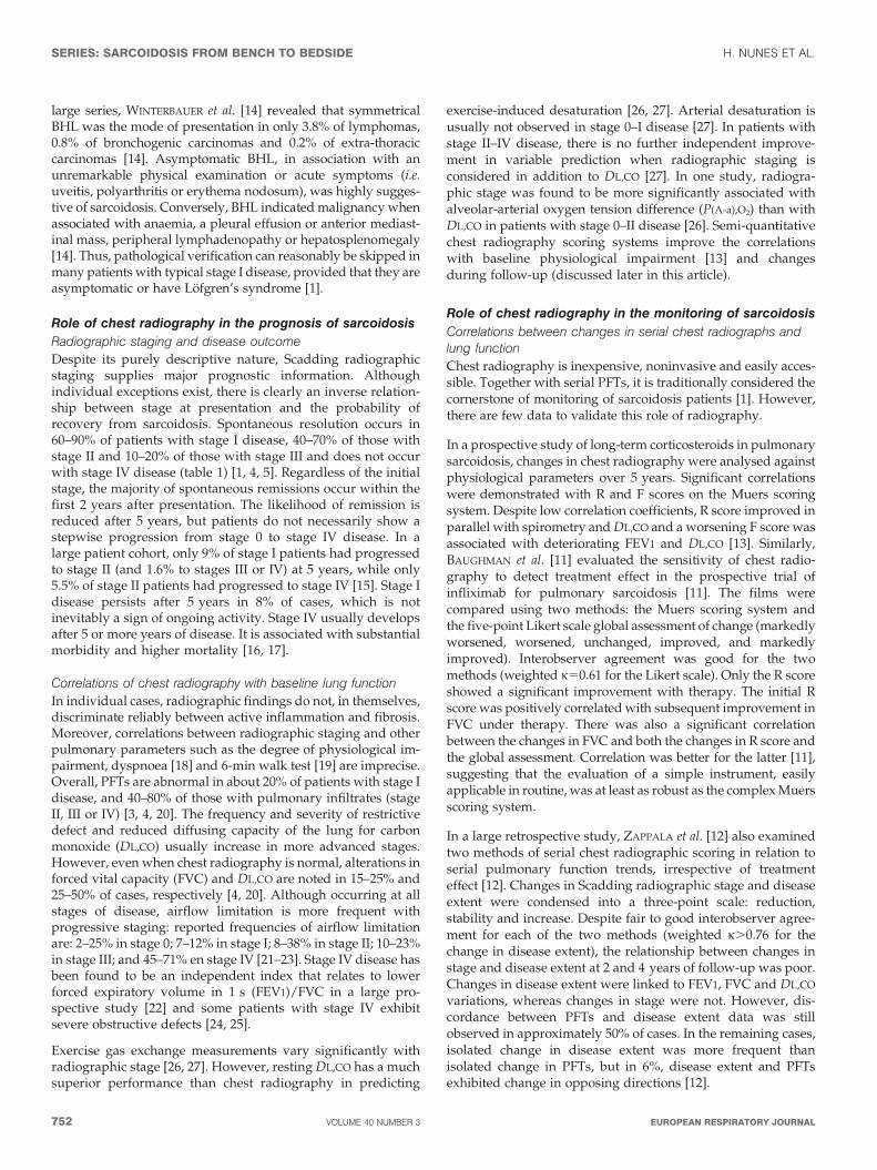
TABLE 1 Frequency of Scadding radiographic stages atpresentation and probability of spontaneousresolution of sarcoidosis
Radiographic stage Frequency % Resolution %
Stage 0 5–15
Stage I 25–65 60–90
Stage II 20–40 40–70
Stage III 10–15 10–20
Stage IV 5 0
H. NUNES ET AL. SERIES: SARCOIDOSIS FROM BENCH TO BEDSIDE
cEUROPEAN RESPIRATORY JOURNAL VOLUME 40 NUMBER 3 751
large series, WINTERBAUER et al. [14] revealed that symmetricalBHL was the mode of presentation in only 3.8% of lymphomas,0.8% of bronchogenic carcinomas and 0.2% of extra-thoraciccarcinomas [14]. Asymptomatic BHL, in association with anunremarkable physical examination or acute symptoms (i.e.uveitis, polyarthritis or erythema nodosum), was highly sugges-tive of sarcoidosis. Conversely, BHL indicated malignancy whenassociated with anaemia, a pleural effusion or anterior mediast-inal mass, peripheral lymphadenopathy or hepatosplenomegaly[14]. Thus, pathological verification can reasonably be skipped inmany patients with typical stage I disease, provided that they areasymptomatic or have Lofgren’s syndrome [1].
Role of chest radiography in the prognosis of sarcoidosis
Radiographic staging and disease outcome
Despite its purely descriptive nature, Scadding radiographicstaging supplies major prognostic information. Althoughindividual exceptions exist, there is clearly an inverse relation-ship between stage at presentation and the probability ofrecovery from sarcoidosis. Spontaneous resolution occurs in60–90% of patients with stage I disease, 40–70% of those withstage II and 10–20% of those with stage III and does not occurwith stage IV disease (table 1) [1, 4, 5]. Regardless of the initialstage, the majority of spontaneous remissions occur within thefirst 2 years after presentation. The likelihood of remission isreduced after 5 years, but patients do not necessarily show astepwise progression from stage 0 to stage IV disease. In alarge patient cohort, only 9% of stage I patients had progressedto stage II (and 1.6% to stages III or IV) at 5 years, while only5.5% of stage II patients had progressed to stage IV [15]. Stage Idisease persists after 5 years in 8% of cases, which is notinevitably a sign of ongoing activity. Stage IV usually developsafter 5 or more years of disease. It is associated with substantialmorbidity and higher mortality [16, 17].
Correlations of chest radiography with baseline lung function
In individual cases, radiographic findings do not, in themselves,discriminate reliably between active inflammation and fibrosis.Moreover, correlations between radiographic staging and otherpulmonary parameters such as the degree of physiological im-pairment, dyspnoea [18] and 6-min walk test [19] are imprecise.Overall, PFTs are abnormal in about 20% of patients with stage Idisease, and 40–80% of those with pulmonary infiltrates (stageII, III or IV) [3, 4, 20]. The frequency and severity of restrictivedefect and reduced diffusing capacity of the lung for carbonmonoxide (DL,CO) usually increase in more advanced stages.However, even when chest radiography is normal, alterations inforced vital capacity (FVC) and DL,CO are noted in 15–25% and25–50% of cases, respectively [4, 20]. Although occurring at allstages of disease, airflow limitation is more frequent withprogressive staging: reported frequencies of airflow limitationare: 2–25% in stage 0; 7–12% in stage I; 8–38% in stage II; 10–23%in stage III; and 45–71% en stage IV [21–23]. Stage IV disease hasbeen found to be an independent index that relates to lowerforced expiratory volume in 1 s (FEV1)/FVC in a large pro-spective study [22] and some patients with stage IV exhibitsevere obstructive defects [24, 25].
Exercise gas exchange measurements vary significantly withradiographic stage [26, 27]. However, resting DL,CO has a muchsuperior performance than chest radiography in predicting
exercise-induced desaturation [26, 27]. Arterial desaturation isusually not observed in stage 0–I disease [27]. In patients withstage II–IV disease, there is no further independent improve-ment in variable prediction when radiographic staging isconsidered in addition to DL,CO [27]. In one study, radiogra-phic stage was found to be more significantly associated withalveolar-arterial oxygen tension difference (P(A-a),O2) than withDL,CO in patients with stage 0–II disease [26]. Semi-quantitativechest radiography scoring systems improve the correlationswith baseline physiological impairment [13] and changesduring follow-up (discussed later in this article).
Role of chest radiography in the monitoring of sarcoidosis
Correlations between changes in serial chest radiographs andlung function
Chest radiography is inexpensive, noninvasive and easily acces-sible. Together with serial PFTs, it is traditionally considered thecornerstone of monitoring of sarcoidosis patients [1]. However,there are few data to validate this role of radiography.
In a prospective study of long-term corticosteroids in pulmonarysarcoidosis, changes in chest radiography were analysed againstphysiological parameters over 5 years. Significant correlationswere demonstrated with R and F scores on the Muers scoringsystem. Despite low correlation coefficients, R score improved inparallel with spirometry and DL,CO and a worsening F score wasassociated with deteriorating FEV1 and DL,CO [13]. Similarly,BAUGHMAN et al. [11] evaluated the sensitivity of chest radio-graphy to detect treatment effect in the prospective trial ofinfliximab for pulmonary sarcoidosis [11]. The films werecompared using two methods: the Muers scoring system andthe five-point Likert scale global assessment of change (markedlyworsened, worsened, unchanged, improved, and markedlyimproved). Interobserver agreement was good for the twomethods (weighted k50.61 for the Likert scale). Only the R scoreshowed a significant improvement with therapy. The initial Rscore was positively correlated with subsequent improvement inFVC under therapy. There was also a significant correlationbetween the changes in FVC and both the changes in R score andthe global assessment. Correlation was better for the latter [11],suggesting that the evaluation of a simple instrument, easilyapplicable in routine, was at least as robust as the complex Muersscoring system.
In a large retrospective study, ZAPPALA et al. [12] also examinedtwo methods of serial chest radiographic scoring in relation toserial pulmonary function trends, irrespective of treatmenteffect [12]. Changes in Scadding radiographic stage and diseaseextent were condensed into a three-point scale: reduction,stability and increase. Despite fair to good interobserver agree-ment for each of the two methods (weighted k.0.76 for thechange in disease extent), the relationship between changes instage and disease extent at 2 and 4 years of follow-up was poor.Changes in disease extent were linked to FEV1, FVC and DL,CO
variations, whereas changes in stage were not. However, dis-cordance between PFTs and disease extent data was stillobserved in approximately 50% of cases. In the remaining cases,isolated change in disease extent was more frequent thanisolated change in PFTs, but in 6%, disease extent and PFTsexhibited change in opposing directions [12].
SERIES: SARCOIDOSIS FROM BENCH TO BEDSIDE H. NUNES ET AL.
752 VOLUME 40 NUMBER 3 EUROPEAN RESPIRATORY JOURNAL
The utility of chest radiography in diagnosing exacerbations ofpulmonary sarcoidosis was addressed by JUDSON et al. [28],using the profusion score of the ILO reading system. Exa-cerbation was defined by worsening pulmonary symptomsthought to be related to pulmonary sarcoidosis (i.e. not infection)that responded to an increase of the corticosteroid dose. Asubset of these patients (66.7%) was identified with a spiro-metric decline of o10% in FVC or FEV1 from the previousbaseline visit. Interobserver agreement in the determination ofprofusion score appeared to be moderate (weighted k50.54).Although changes in profusion score tended to worsen duringexacerbations in both the whole population and the spirometricdecline subgroup, a large percentage of readings demonstratedno modification (32% and 27%, respectively) or an improvement(19% and 23%, respectively). As expected, a significant decreasein spirometric measurements was seen during exacerbations.There were no statistically significant correlations betweenchanges in the profusion score and changes in FVC or FEV1 foreither the whole population or the spirometric decline subgroup[28]. The authors concluded that radiography was inadequate toreliably detect exacerbations of pulmonary sarcoidosis.
Taken together, these results highlight the weaknesses of chestradiography for the monitoring of sarcoidosis. However, afurther advantage of serial chest radiographs over PFTs is thedetection of pulmonary complications, including aspergilloma.From this point of view, it is the combination of the two teststhat is, in essence, helpful in clinical practice.
Chest radiography as a guide for treatment decisions
Treatment for pulmonary sarcoidosis is usually dictated by theseverity of symptoms and/or functional impairment, but chestradiography per se may also guide therapeutic intervention[29–31]. A prospective randomised study conducted by theBritish Thoracic Society provides some support for long-termcorticosteroid therapy in asymptomatic patients with persistentpulmonary infiltrates for at least 6 months [29]. After adjust-ment for possible confounding factors, the average difference invital capacity between treated and untreated groups at finalassessment was 9% of the predicted value. In another placebo-controlled study, patients with newly detected stage I or II–IIIdisease and (sub)normal lung function were immediatelytreated with oral prednisolone for 3 months, followed byinhaled budesonide for 15 months [30]. Treated patients withinitial stage II–III, but not stage I, disease improved significantlymore in FVC and DL,CO compared with patients on placebo.Functional benefits were maintained after 5 years, but differ-ences were small [31]. It remains uncertain whether modestimprovement in asymptomatic stage II–III patients is clinicallypertinent and justifies the morbidity associated with treatment.
CHEST COMPUTED TOMOGRAPHY
Standard CT scanning protocolsMultidetector CT, combining helical volumetric acquisitionsand thin slice thickness (0.6–1.25 mm), is now accepted as theimaging reference for the initial work-up of patients with diffuselung diseases (DLDs) [32]. It allows multiplanar reconstructionsand post-processing techniques, which are likely to becomeincreasingly useful. Maximum intensity projections enhancethe detection and analysis of the distribution of micronodularstructures. Minimum intensity projections improve the detection
of areas of increased and reduced density and bronchial steno-sis [33]. However, multidetector CT acquisitions increase theradiation burden, which should stay ‘‘As Low As ReasonablyAchievable’’ [34]. Therefore, in our group, high-resolution CT(HRCT) protocols with acquisition of 1–1.5 mm thin slices at10-mm spaced intervals are still preferred in the follow-up ofyoung patients. Additional sections at end expiration can beuseful in the presence of airflow obstruction to identify airtrapping in patients with small-airway involvement. Admi-nistration of contrast agents can be useful to better discernlymphadenopathy and in patients with vascular complications,including pulmonary hypertension [35, 36].
Indications for chest CTAccording to the American Thoracic Society/European Res-piratory Society/ World Association of Sarcoidosis and OtherGranulomatous Disorders expert consensus statement onsarcoidosis [1], CT is warranted in the following circumstances:1) atypical clinical and/or radiographic findings; 2) normalchest radiography but a clinical suspicion of sarcoidosis; and 3)detection of pulmonary complications. In addition, CT mayimprove the diagnostic yield of bronchoscopy for obtaining apositive endobronchial [37] or transbronchial biopsy [38] and itis required before endobronchial ultrasound-guided transbron-chial needle aspiration (EBUS-TBNA) [39]. CT findings maydiscriminate between active inflammation and irreversible fibro-sis [40–44], with occasional influence on therapeutic decisions.Furthermore, correlations between disease severity on CT andfunctional impairment have been examined in numerous studies[23, 35, 40, 44–54].
We will first describe pulmonary sarcoidosis as seen on CT andthen try to delineate the real contribution of CT in thesedifferent situations.
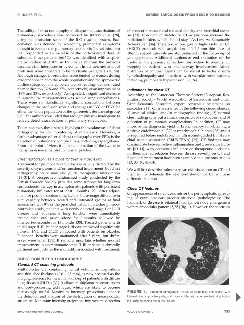
Chest CT featuresCT appearances of sarcoidosis mirror the perilymphatic spread-ing of granulomatous process observed pathologically. Thehallmark of disease is bilateral hilar lymph node enlargementwith micronodular pattern [5, 55] (fig. 1). However, the spectrum
FIGURE 1. Computed tomographic image of pulmonary sarcoidosis with
bilateral hilar lymphadenopathy and micronodules with a perilymphatic distribution
including spreading along the fissures.
H. NUNES ET AL. SERIES: SARCOIDOSIS FROM BENCH TO BEDSIDE
cEUROPEAN RESPIRATORY JOURNAL VOLUME 40 NUMBER 3 753
of disease on CT is extraordinarily protean [5, 55] and multiplefeatures or patterns more or less characteristic of the disease maybe variously associated in individual patients. Readers can referto the Fleischner Society glossary for the definition of terms usedin this section [56].
Thoracic lymphadenopathyAlthough not necessary in typical stage I disease, CT is moresensitive to detect enlarged lymph nodes than a chest radio-graph [57]. Overall, hilar or mediastinal lymphadenopathy areencountered on CT in 47–94% of patients with sarcoidosis,irrespective of radiographic staging [35, 42, 57–60]. Lymphnodes are usually bilateral but with right-sided predominance[57–60]. In two studies based on the American Thoracic Societylymph node map, the most commonly involved nodal stations,in decreasing order of frequency, were: 4R (right lowerparatracheal); 10R (right hilar); 7 (sub-carinal); 5 (sub-aortic,i.e. aorto-pulmonic window); 11R (right interlobar); and 11 L(left interlobar) [59, 60]. A median of three lymph nodes wereenlarged, as defined by a maximum short-axis diametero10 mm, and diameter was o20 mm in 29.1% of cases [59].Lymphadenopathy is also common in collagen vascular disease(present in 70% of cases), idiopathic pulmonary fibrosis (IPF)(67%), extrinsic allergic alveolitis (53%) and organising pneu-monia (36%) [59]. However, the number of enlarged lymphnodes is higher in sarcoidosis and a size greater than 20 mm inshort-axis diameter increases the chance of sarcoidosis [59].
Sarcoidosis lymph nodes are usually non-necrotic and non-compressive, with nodal calcification frequent in longstandingdisease. Calcification is present at presentation in 20% of cases,increasing to 44% 4 years later [42], with egg-shell aspectpresent in 9% [58]. The mean diameter of calcified nodes issignificantly larger in sarcoidosis, calcium deposition is morecommonly focal in sarcoidosis and diffuse in tuberculosis and,when present, hilar nodal calcification is much more likely to bebilateral in sarcoidosis than in tuberculosis (65% versus 8%) [58].
Lymphadenopathy may be unilateral or localised in an unusualsite. Although possible in sarcoidosis, the enlargement of inter-nal mammary and pericardial lymph nodes requires exclusionof lymphoma. Enlarged lymph nodes, essentially when calci-fied, and/or mediastinal fibrosis can rarely provoke extrinsiccompression on adjacent organs, including bronchi [61], largepulmonary arteries [36] or veins [62, 63], the superior vena cava[64], the oesophagus [65], the left recurrent nerve [66] or thethoracic duct [67].
Pulmonary patternsMicronodular opacities with a perilymphatic distributionNodules represent aggregates of granulomas [68]. They are seenin 80–100% of all patients [40–42, 44, 45, 53, 54, 69, 70] but are lessfrequent in stage IV disease [35]. Most are small, measuringbetween 1–10 mm in diameter, and have irregular poorlycircumscribed margins. Maximum intensity projection may behelpful to detect micronodules and specify their topographicdistribution [33]. They usually show a predilection for the mid/upper and posterior parts of the lungs with a perilymphaticdistribution (fig. 1). They tend to be more abundant aroundbronchovascular structures and sub-pleurally, along the con-cavity of chest wall, the mediastinum and/or the fissures, andalong the bronchovascular sheath and the interlobular septa
[5, 55]. Nodularity can result in a fissural or bronchovascular‘‘beaded’’ aspect, which is widely accepted as virtually patho-gnomonic of sarcoidosis (although this has never been formallyvalidated). Irregularity or thickening of the bronchovascularbundles is as a second cardinal sign [4, 55]. Peribronchovascularthickening often emanates from the hilar regions in an axialfashion and occasionally gives rise to luminal stenosis [61].Other diseases distributed along the lymphatics, such aslymphangitic carcinomatosis and lymphoma, are usually read-ily distinguished from sarcoidosis on CT [71].
Micronodules may be sparse in distribution rather than wide-spread or asymmetric. They may be dispersed throughout thelungs without any topographic predilection or may exceptionallyadopt a haematogenous or a centrilobular configuration ratherthan perilymphatic, simulating miliary tuberculosis or metastasesand hypersensitivity pneumonitis, respectively [72].
Nodular and alveolar opacities
Small nodules can coalesce into larger ones or masses, which canvery rarely cavitate [73–75]. Alveolar or pseudoalveolar con-solidations are seen in 12–38% of patients [35, 41, 44, 45, 54, 76].However, a predominance of multiple large nodules/masses
a)
b)
R
P
FIGURE 2. a) Chest radiograph and b) computed tomographic image of
‘‘alveolar’’ sarcoidosis associated with profuse micronodular involvement and
cavitary lesions on the left.
SERIES: SARCOIDOSIS FROM BENCH TO BEDSIDE H. NUNES ET AL.
754 VOLUME 40 NUMBER 3 EUROPEAN RESPIRATORY JOURNAL
and multifocal consolidations is uncommon in sarcoidosis andcan mimic organising pneumonia or malignancy. According to arecent series by MALAISAMY et al. [74], the presentation of such aform of disease is usually acute and symptomatic with anexcellent prognosis and it may particularly affect smokers [74].Nodules/masses and consolidations are homogeneous or inho-mogeneous, measure 10–80 mm in diameter, and are usuallylocated in the mid/upper lobes, along the bronchovascularbundles or sub-pleurally, sometimes with the presence of acentral air bronchogram [74, 77]. They are characterised by ill-defined contours as they fade to a micronodular pattern towardthe surrounding lung (fig. 2). In addition to these lesions, othermore representative abnormalities such as lymph nodes areusually associated [74, 77]. Solitary mass-like nodule and alveolarconsolidation are exceedingly rare in sarcoidosis [74, 78].
Necrotising sarcoid granulomatosis, which is defined patholo-gically by a sarcoid-like granulomatous reaction with vasculitis(involving both arteries and veins) and noncaseating necrosis,shares many clinical and radiological features with alveolarsarcoidosis but is traditionally viewed as a separate entity [79].
Cavitary lesions in sarcoidosis are believed to result from eitherischaemic necrosis (with extrusion of hyaline material fromconglomerate granulomas) or angiitis [73]. Reported rates ofcavitary lesions were 3.4% and 6.8% in two CT studies,respectively [40, 75]. According to a recent series by HOURS
et al. [73], cavitary lesions manifest as thin-walled cysts in mostcases or as cavities with thick wall or developing inside nodulesor condensations [73] (fig. 2). Cavitary lesions are variable in
size and, although occasionally found in isolation, they are morelikely to be multiple and bilateral. They usually arise in patientswith severe and active sarcoidosis. The evolution of cavitarylesions is variable. A wall thinning is usually observed undertreatment whereas a wall thickening is always associated withan infectious complication [73]. Pneumothorax can occur [73, 80].Because primary cavitary sarcoidosis is rare, granulomatosiswith polyangiitis (Wegener’s) and superimposed infectionshould always be excluded.
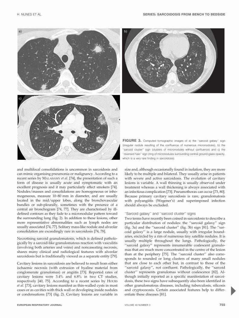
‘‘Sarcoid galaxy’’ and ‘‘sarcoid cluster’’ signs
Two terms have recently been coined in sarcoidosis to describe aparticular distribution of nodules: the ‘‘sarcoid galaxy’’ sign(fig. 3a) and the ‘‘sarcoid cluster’’ (fig. 3b) sign [81]. The ‘‘sar-coid galaxy’’ is a large nodule, usually with irregular bound-aries, encircled by a rim of numerous tiny satellite nodules. It isusually multiple throughout the lungs. Pathologically, the‘‘sarcoid galaxy’’ represents innumerable coalescent granulo-mas that are much more concentrated in the centre of the lesionthan at the periphery [75]. The ‘‘sarcoid cluster’’ also corre-sponds to rounded or long clusters of many small nodulesthat are close to each other but, in contrast to those of the‘‘sarcoid galaxy’’, not confluent. Pathologically, the ‘‘sarcoidcluster’’ represents granulomas without coalescence [82]. Al-though initially reported as a specific manifestation of sarcoi-dosis, these two signs have subsequently also been identified inother granulomatous diseases, including tuberculosis, silicosisand cryptococosis. Certain associated features help to differ-entiate these diseases [81].
a)
c)
b)
FIGURE 3. Computed tomographic images of a) the ‘‘sarcoid galaxy’’ sign
(irregular nodule resulting of the confluence of numerous micronodules), b) the
‘‘sarcoid cluster’’ sign (clusters of micronodules without confluence) and c) the
‘‘reversed halo’’ sign (ring of micronodules surrounding central ground-glass opacity,
which is a very rare finding in sarcoidosis).
H. NUNES ET AL. SERIES: SARCOIDOSIS FROM BENCH TO BEDSIDE
cEUROPEAN RESPIRATORY JOURNAL VOLUME 40 NUMBER 3 755
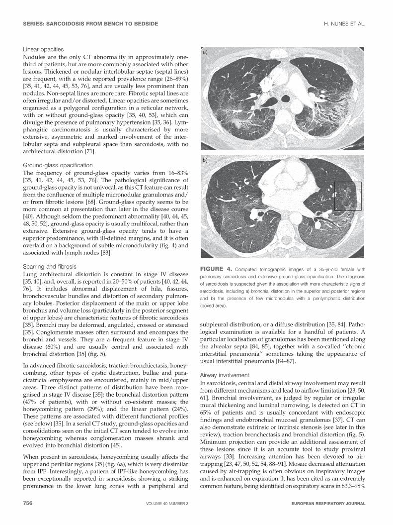
Linear opacitiesNodules are the only CT abnormality in approximately one-third of patients, but are more commonly associated with otherlesions. Thickened or nodular interlobular septae (septal lines)are frequent, with a wide reported prevalence range (26–89%)[35, 41, 42, 44, 45, 53, 76], and are usually less prominent thannodules. Non-septal lines are more rare. Fibrotic septal lines areoften irregular and/or distorted. Linear opacities are sometimesorganised as a polygonal configuration in a reticular network,with or without ground-glass opacity [35, 40, 53], which candivulge the presence of pulmonary hypertension [35, 36]. Lym-phangitic carcinomatosis is usually characterised by moreextensive, asymmetric and marked involvement of the inter-lobular septa and subpleural space than sarcoidosis, with noarchitectural distortion [71].
Ground-glass opacificationThe frequency of ground-glass opacity varies from 16–83%[35, 41, 42, 44, 45, 53, 76]. The pathological significance ofground-glass opacity is not univocal, as this CT feature can resultfrom the confluence of multiple micronodular granulomas and/or from fibrotic lesions [68]. Ground-glass opacity seems to bemore common at presentation than later in the disease course[40]. Although seldom the predominant abnormality [40, 44, 45,48, 50, 52], ground-glass opacity is usually multifocal, rather thanextensive. Extensive ground-glass opacity tends to have asuperior predominance, with ill-defined margins, and it is oftenoverlaid on a background of subtle micronodularity (fig. 4) andassociated with lymph nodes [83].
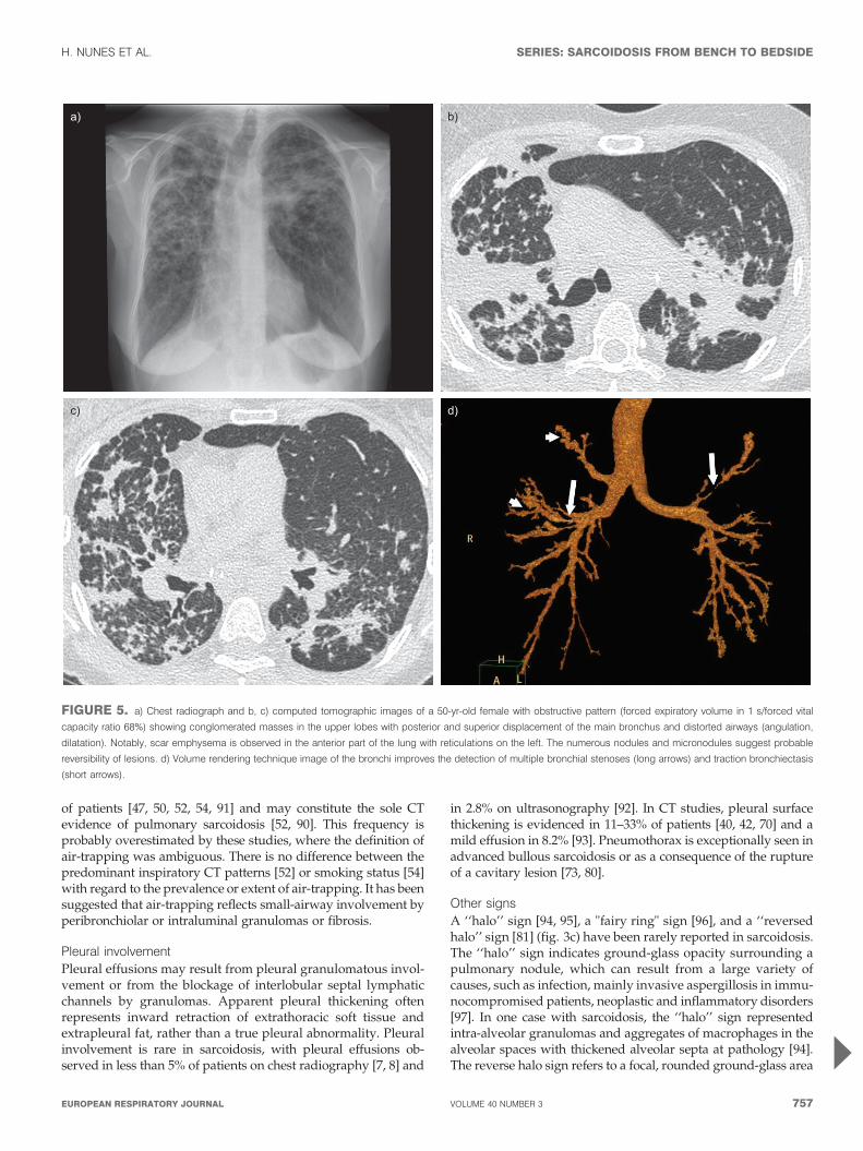
Scarring and fibrosisLung architectural distortion is constant in stage IV disease[35, 40], and, overall, is reported in 20–50% of patients [40, 42, 44,76]. It includes abnormal displacement of hila, fissures,bronchovascular bundles and distortion of secondary pulmon-ary lobules. Posterior displacement of the main or upper lobebronchus and volume loss (particularly in the posterior segmentof upper lobes) are characteristic features of fibrotic sarcoidosis[35]. Bronchi may be deformed, angulated, crossed or stenosed[35]. Conglomerate masses often surround and encompass thebronchi and vessels. They are a frequent feature in stage IVdisease (60%) and are usually central and associated withbronchial distortion [35] (fig. 5).
In advanced fibrotic sarcoidosis, traction bronchiectasis, honey-combing, other types of cystic destruction, bullae and para-cicatricial emphysema are encountered, mainly in mid/upperareas. Three distinct patterns of distribution have been reco-gnised in stage IV disease [35]: the bronchial distortion pattern(47% of patients), with or without co-existent masses; thehoneycombing pattern (29%); and the linear pattern (24%).These patterns are associated with different functional profiles(see below) [35]. In a serial CT study, ground-glass opacities andconsolidations seen on the initial CT scan tended to evolve intohoneycombing whereas conglomeration masses shrank andevolved into bronchial distortion [45].
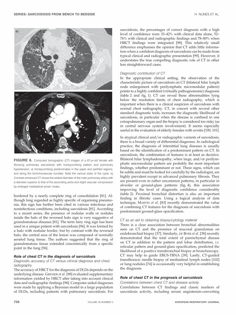
When present in sarcoidosis, honeycombing usually affects theupper and perihilar regions [35] (fig. 6a), which is very dissimilarfrom IPF. Interestingly, a pattern of IPF-like honeycombing hasbeen exceptionally reported in sarcoidosis, showing a strikingprominence in the lower lung zones with a peripheral and
subpleural distribution, or a diffuse distribution [35, 84]. Patho-logical examination is available for a handful of patients. Aparticular localisation of granulomas has been mentioned alongthe alveolar septa [84, 85], together with a so-called ‘‘chronicinterstitial pneumonia’’ sometimes taking the appearance ofusual interstitial pneumonia [84–87].
Airway involvement
In sarcoidosis, central and distal airway involvement may resultfrom different mechanisms and lead to airflow limitation [23, 50,61]. Bronchial involvement, as judged by regular or irregularmural thickening and luminal narrowing, is detected on CT in65% of patients and is usually concordant with endoscopicfindings and endobronchial mucosal granulomas [37]. CT canalso demonstrate extrinsic or intrinsic stenosis (see later in thisreview), traction bronchectasis and bronchial distortion (fig. 5).Minimum projection can provide an additional assessment ofthese lesions since it is an accurate tool to study proximalairways [33]. Increasing attention has been devoted to air-trapping [23, 47, 50, 52, 54, 88–91]. Mosaic decreased attenuationcaused by air-trapping is often obvious on inspiratory imagesand is enhanced on expiration. It has been cited as an extremelycommon feature, being identified on expiratory scans in 83.3–98%
a)
b)
FIGURE 4. Computed tomographic images of a 35-yr-old female with
pulmonary sarcoidosis and extensive ground-glass opacification. The diagnosis
of sarcoidosis is suspected given the association with more characteristic signs of
sarcoidosis, including a) bronchial distortion in the superior and posterior regions
and b) the presence of few micronodules with a perilymphatic distribution
(boxed area).
SERIES: SARCOIDOSIS FROM BENCH TO BEDSIDE H. NUNES ET AL.
756 VOLUME 40 NUMBER 3 EUROPEAN RESPIRATORY JOURNAL
of patients [47, 50, 52, 54, 91] and may constitute the sole CTevidence of pulmonary sarcoidosis [52, 90]. This frequency isprobably overestimated by these studies, where the definition ofair-trapping was ambiguous. There is no difference between thepredominant inspiratory CT patterns [52] or smoking status [54]with regard to the prevalence or extent of air-trapping. It has beensuggested that air-trapping reflects small-airway involvement byperibronchiolar or intraluminal granulomas or fibrosis.
Pleural involvement
Pleural effusions may result from pleural granulomatous invol-vement or from the blockage of interlobular septal lymphaticchannels by granulomas. Apparent pleural thickening oftenrepresents inward retraction of extrathoracic soft tissue andextrapleural fat, rather than a true pleural abnormality. Pleuralinvolvement is rare in sarcoidosis, with pleural effusions ob-served in less than 5% of patients on chest radiography [7, 8] and
in 2.8% on ultrasonography [92]. In CT studies, pleural surfacethickening is evidenced in 11–33% of patients [40, 42, 70] and amild effusion in 8.2% [93]. Pneumothorax is exceptionally seen inadvanced bullous sarcoidosis or as a consequence of the ruptureof a cavitary lesion [73, 80].
Other signs
A ‘‘halo’’ sign [94, 95], a "fairy ring" sign [96], and a ‘‘reversedhalo’’ sign [81] (fig. 3c) have been rarely reported in sarcoidosis.The ‘‘halo’’ sign indicates ground-glass opacity surrounding apulmonary nodule, which can result from a large variety ofcauses, such as infection, mainly invasive aspergillosis in immu-nocompromised patients, neoplastic and inflammatory disorders[97]. In one case with sarcoidosis, the ‘‘halo’’ sign representedintra-alveolar granulomas and aggregates of macrophages in thealveolar spaces with thickened alveolar septa at pathology [94].The reverse halo sign refers to a focal, rounded ground-glass area
a) b)
c) d)
FIGURE 5. a) Chest radiograph and b, c) computed tomographic images of a 50-yr-old female with obstructive pattern (forced expiratory volume in 1 s/forced vital
capacity ratio 68%) showing conglomerated masses in the upper lobes with posterior and superior displacement of the main bronchus and distorted airways (angulation,
dilatation). Notably, scar emphysema is observed in the anterior part of the lung with reticulations on the left. The numerous nodules and micronodules suggest probable
reversibility of lesions. d) Volume rendering technique image of the bronchi improves the detection of multiple bronchial stenoses (long arrows) and traction bronchiectasis
(short arrows).
H. NUNES ET AL. SERIES: SARCOIDOSIS FROM BENCH TO BEDSIDE
cEUROPEAN RESPIRATORY JOURNAL VOLUME 40 NUMBER 3 757
bordered by a nearly complete ring of consolidation [81]. Al-though long regarded as highly specific of organising pneumo-nia, this sign has further been cited in various infectious andnoninfectious conditions, including sarcoidosis [81]. Accordingto a recent series, the presence of nodular walls or nodulesinside the halo of the reversed halo sign is very suggestive ofgranulomatous diseases [81]. The term fairy ring sign has beenused in a unique patient with sarcoidosis [96]. It was formed bya halo with nodular border, but by contrast with the reversedhalo, the central area of the lesion was composed of normallyaerated lung tissue. The authors suggested that the ring ofgranulomatous tissue extended concentrically from a specificpoint in the lung [96].
Role of chest CT in the diagnosis of sarcoidosisDiagnostic accuracy of CT versus clinical diagnosis and chestradiographyThe accuracy of HRCT for the diagnosis of DLDs depends on theunderlying disease. GRENIER et al. [98] evaluated supplementaryinformation yielded by HRCT after taking into account clinicaldata and radiographic findings [98]. Computer-aided diagnoseswere made by applying a Bayesian model in a large populationof DLDs, including patients with pulmonary sarcoidosis. For
sarcoidosis, the percentages of correct diagnosis with a highlevel of confidence were 33–42% with clinical data alone, 52–76% with clinical and radiographic findings and 78–80% whenHRCT findings were integrated [98]. This relatively smalldifference emphasises the opinion that CT adds little informa-tion when a confident diagnosis of sarcoidosis can be made fromtypical clinical and radiographic presentation [99]. However, itunderstates the true compelling diagnostic role of CT in otherless straightforward cases.
Diagnostic contribution of CT
In the appropriate clinical setting, the observation of thecharacteristic picture of sarcoidosis on CT (bilateral hilar lymphnode enlargement with perilymphatic micronodular pattern)points to a highly confident (virtually pathognomonic) diagnosis(table 2 and fig. 1). CT can reveal these abnormalities lyingbelow the resolution limits of chest radiography, which isimportant when there is a clinical suspicion of sarcoidosis withnormal chest radiography. CT, in concert with several otherstandard diagnostic tools, increases the diagnostic likelihood ofsarcoidosis, in particular when the disease is confined to oneextrapulmonary organ and the biopsy is considered too risky (asin central nervous system involvement). It seems especiallyuseful in the evaluation of elderly females with uveitis [100, 101].
In atypical clinical and/or radiographic variants of sarcoidosis,there is a broad variety of differential diagnoses. In radiologicalpractice, the diagnosis of interstitial lung diseases is usuallybased on the identification of a predominant pattern on CT. Insarcoidosis, the combination of features is at least as decisive.Bilateral hilar lymphadenopathy, when large, and/or perilym-phatic micronodular pattern are probably the most importantfindings, whether predominant or not. These signs, which canbe subtle and must be looked for carefully by the radiologist, arehighly prevalent except in advanced pulmonary fibrosis. Theyare present even in rather uncommon patterns, in particular inalveolar or ground-glass patterns (fig. 4), this associationimproving the level of diagnostic confidence considerably(table 2). Proximal bronchial distortion is also an importantfinding in fibrotic cases. Using a logical analysis of datatechnique, MARTIN et al. [83] recently demonstrated the valueof combining CT features for the diagnosis of sarcoidosis withpredominant ground-glass opacification.
CT as an aid to obtaining biopsy/cytology material
There is a clear association between bronchial abnormalitiesseen on CT and the presence of mucosal granulomas onendobronchial biopsy [37]. Similarly, DE BOER et al. [38] recentlydemonstrated that the total extent of parenchymal diseaseon CT in addition to the pattern and lobar distribution, i.e.reticular pattern and ground-glass opacification, predicted thelikelihood of a positive transbronchial biopsy at bronchoscopy.CT may help to guide EBUS-TBNA [39]. Lastly, CT-guidedtransthoracic needle biopsy of mediastinal lymph nodes [102]or lung nodules [74] is occasionally very helpful in establishingthe diagnosis.
Role of chest CT in the prognosis of sarcoidosis
Correlations between chest CT and disease activity
Correlations between CT findings and classic markers ofsarcoidosis activity, including serum angiotensin-converting
a)
b)
FIGURE 6. Computed tomographic (CT) images of a 67-yr-old female with
fibrosing pulmonary sarcoidosis with honeycombing pattern and pulmonary
hypertension. a) Honeycombing predominates in the upper and perihilar regions,
and along the bronchovascular bundles. Note the various sizes of the cysts. b)
Contrast-enhanced CT shows the widest diameter of the main pulmonary artery with
a diameter superior to that of the ascending aorta and slight vascular compression
by enlarged mediastinal lymph nodes.
SERIES: SARCOIDOSIS FROM BENCH TO BEDSIDE H. NUNES ET AL.
758 VOLUME 40 NUMBER 3 EUROPEAN RESPIRATORY JOURNAL
enzyme (SACE) levels, bronchoalveolar lavage (BAL) lympho-cytosis, and gallium scan signal, are inconclusive or discrepant[40, 41, 43, 44, 46]. Much more has been learned from studies ofthe reversibility of CT features (spontaneously or under treat-ment) on serial examinations (table 3) [42, 44, 45, 76]. Architec-tural distortion, traction bronchiectasis, honeycombing andbullae are consistently irreversible. Micronodules, nodules,peribronchovascular thickening and consolidation are whollyor partially reversible in most, but not all, cases. The evolution ofground-glass and linear opacities is more variable. Ground-glassopacity may steady, worsen or improve over time, reflectingthe fact that it may represent either granulomas or fine fibrosis
[68, 103]. A coarse texture or concomitant traction bronchiectasisincreases the likelihood of underlying fibrosis [103]. Similarly,septal thickening from intense granulomatous infiltration tendsto reverse, whereas irregular distorted lines are more likely to befibrotic.
Thus, discrimination between active inflammation and irrever-
sible fibrosis with CT may occasionally be helpful when the deci-
sion to initiate or continue potentially toxic treatment is marginal.
For instance, in stage IV disease a trial of therapy might be war-
ranted if a potentially reversible component is still visible on CT
[35]. This role of CT may be supplanted by PET-CT in the future.
TABLE 2 Diagnostic confidence for sarcoidosis according to the predominant pattern and relevant associated features oncomputed tomography
Predominant pattern Associated features Diagnostic confidence
Large bilateral hilar lymphadenopathy Isolated High"
Perilymphatic micronodules Very high
Unilateral lymphadenopathy Isolated Very low
Perilymphatic micronodules Intermediate+
Mediastinal, internal mammary or pericardial lymphadenopathy Isolated Very low
Perilymphatic micronodules Low
Perilymphatic micronodules Cluster sign High
Large bilateral hilar lymphadenopathy Very high
Nodules/masses or condensations Unique Very low
Perilymphatic micronodules High
Sarcoid galaxy High
Large bilateral hilar lymphadenopathy High
Cavitations# Isolated Very low
Perilymphatic micronodules Intermediate+
Sarcoid galaxy Intermediate+
Large bilateral hilar lymphadenopathy Intermediate+
Ground-glass opacification Isolated Low
Perilymphatic micronodules High
Large bilateral hilar lymphadenopathy High
Pulmonary fibrosis Basal and peripheral honeycombing Very low
Upper/mid and central honeycombing Intermediate
Bronchial distortion High1
#: cavitations usually develop inside nodules/masses or condensations; ": in asymptomatic patients with an unremarkable physical examination or acute symptoms (i.e.
uveitis, polyarthritis or erythema nodosum); +: need to exclude superimposed comorbidities, including infections, before attributing the pattern to sarcoidosis; 1: when
bronchial distortion has an upper predominance, with deformed, angulated, crossed or stenosed proximal bronchi and posterior displacement of the main or upper lobe
bronchus.
TABLE 3 Reversibility of sarcoidosis features observed on computed tomography (spontaneously or under therapy)
Reversible features Irreversible features Variable reversibility
Micronodules Architectural distortion Consolidation#
Nodules Bronchial distortion Ground-glass opacification"
Peribronchovascular thickening Honeycombing Linear opacities+
Bullae
#: consolidations are wholly or partially reversible in most cases, in particular those with surrounding micronodules, representing coalescent granulomas. ": a coarse
texture or concomitant traction bronchiectasis increases the likelihood of underlying fibrosis. +: irregular distorted lines are more likely to be fibrotic.
H. NUNES ET AL. SERIES: SARCOIDOSIS FROM BENCH TO BEDSIDE
cEUROPEAN RESPIRATORY JOURNAL VOLUME 40 NUMBER 3 759
Correlations between chest CT and baseline lung functionMany studies have explored the relationships between CTfindings and PFTs [22, 23, 35, 40, 44–48, 50–54] with variableresults, probably reflecting the diversity of imaging analysisand scoring. In the study of REMY-JARDIN et al. [44], lungs weredivided into three zones and a percentage involvement scorewas assigned for abnormal parenchymal patterns (nodules,consolidation, lung distortion, septal and nonseptal linear,ground-glass opacity and honeycombing). The overall extentof disease was the summation of the scores for each type ofabnormality. Profusion of septal lines was the only CT findingthat correlated with initial disease activity (as assessed bySACE levels and BAL lymphocytosis). Significant but lowcorrelation was observed between the scores of CT abnormal-ities and either initial FEV1, FVC or DL,CO, except for nodules.CT findings could not help predict the further evolution ofdisease activity and functional changes over time [44]. A moresimple semi-quantitative CT scoring system, first described byOBERSTEIN et al. [43], served in the correlation study of DRENT
et al. [48]. This consisted of coarse quantification of the extentof abnormal parenchymal patterns (thickening or irregularity ofthe bronchovascular bundle, nodules, septal and nonseptal lines,and consolidation, including ground-glass opacity) using a four-point scale (0 5 no lesions found; 1 5 up to 33%; 2 5 up to 66%;and 3 5 more than 66%), and the extent of focal pleural thickeningand lymph node enlargement (0 5 no pathological findings; 1 5
minor; 2 5 moderate; and 3 5 pronounced changes). The totalscore was obtained by adding the individual subscores. Interob-server agreement was moderate for the subscores (weighted k
0.34–0.65, with worse results for bronchovascular bundle andlymph nodes), but excellent results for the total score (intraclasscorrelation coefficient 0.99). All CT subscores, except lymph nodesenlargement, were correlated with FEV1, FVC, DL,CO, maximalarterial oxygen tension and maximal P(A-a),O2, whereas the chestradiographic stage was not.
In other studies, CT was classified according to subjective apprai-sal of the predominant pattern of involvement. LOPES et al. [51]investigated the relationship between outcome measures ofcardiopulmonary exercise testing (CPET) and predominant CTpattern: nodules, ground-glass opacity, and traction bronchiec-tasis plus honeycombing. PFTs and CPET results were close tonormal only for patients with predominant nodules. Patientswith predominant ground-glass opacity showed intermediatevalues of FEV1, FVC and DL,CO compared with patients of theother two groups. Interestingly, only the CPET results were ableto differentiate patients with predominant ground-glass opacityand those with predominant traction bronchiectasis and honey-combing, with a significantly decreased peak oxygen uptakeand breathing reserve, and increased P(A-a),O2 for the latter [51].As discussed previously, ABEHSERA et al. [35] separated three CTpatterns of fibrotic pulmonary sarcoidosis, which were asso-ciated with different functional profiles. The interobserveragreement for recognising the main CT pattern was very goodas observers agreed in 80% of cases (k50.87). Bronchial dis-tortion pattern was associated with lower expiratory airflowrates, while honeycombing pattern was associated with restric-tion and lower DL,CO, and functional impairment was relativelyminor when linear pattern predominated [35].
In summary, the degree of functional alteration is usually linkedto an overall CT score but most correlations are weak [40, 46, 48,
53]. Total CT score seems superior to chest radiographic staging[48] but its real clinical impact is questionable. Correlationsbetween PFTs and the score of specific patterns are hardlyinterpretable because of considerable overlap among CT cate-gories, but they sometimes give clues to the understanding of
TABLE 4 Underlying mechanisms of airflow limitationobserved on computed tomography andprobability of response to therapy
Mechanism# Response to therapy
Bronchial distortion No
Peribronchovascular thickening Yes
Intrinsic bronchial stenosis Variable
Extrinsic bronchial compression
by lymphadenopathy
Yes"
Air-trapping Variable
#: these mechanisms are usually admixed; ": when lymph nodes are not calcified.
b)
a)
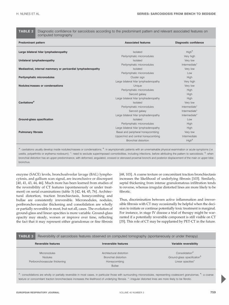
FIGURE 7. a) Chest radiography and b) computed tomographic image
(performed in procubitus) in a 43-yr-old female with cystic lung lesions complicated
with aspergilloma (fungus ball with a crescent sign in the right lung).
SERIES: SARCOIDOSIS FROM BENCH TO BEDSIDE H. NUNES ET AL.
760 VOLUME 40 NUMBER 3 EUROPEAN RESPIRATORY JOURNAL
disease complications, in particular airflow limitation [22, 23,50]. The reported frequency of airflow limitation is wide insarcoidosis, with an estimate of 8.8% of patients in a recentprospective study [22]. It is known to portend a higher risk ofmortality [25]. CT is a reliable method to identify the underlyingmechanisms of airflow limitation and it enables prediction ofthe therapeutic response (table 4). In a case–control study,NACCACHE et al. [23] demonstrated that CT patterns of airwayinvolvement (i.e. bronchial distortion, peribronchovascularthickening, air-trapping, and bronchial compression by enlargedlymph nodes) were found more frequently, scored higher, andwere more often multiple in patients with airflow obstructionthan in those without. Furthermore, functional improvementunder treatment was observed more frequently in patients withpredominant peribronchovascular thickening in comparison tothose with predominant bronchial distortion. Notably, inter-observer agreement was good for identifying the CT patterns ofairway involvement (agreement for 89% of cases, k50.85) [23].In the study by HANDA et al. [22], the only CT morphologicaldeterminant of lower FEV1/FVC was peribronchovascular
thickening. HANSELL et al. [50] demonstrated that the extent ofreticular pattern was independently associated with severalindices of airflow obstruction, including FEV1, FEV1/FVC,maximal expiratory flow (MEF) at 25% above residual volume(RV), MEF at 50% above RV and RV/total lung capacity (TLC).Air-trapping is associated with evidence of small-airway ob-struction including maximal expiratory flow between of FVC,RV and/or RV/TLC in some studies [47, 52, 90], whereas inothers it only contributes little to airflow obstruction [50, 54].
Role of chest CT in the monitoring of sarcoidosisOnce the diagnosis of sarcoidosis is secure, CT makes only asmall contribution to monitoring in most subjects. The cost andthe radiation hazard of repeated and superfluous exams inyoung patients have to be kept in mind. Moreover, although nostudy has clearly outlined the additional information providedby serial CTs compared with PFTs and chest radiography, it isprobable that CT is too sensitive, revealing changes that are notnecessarily relevant in the practical care of patients. Instead,clinical experience amply supports its role in the detection
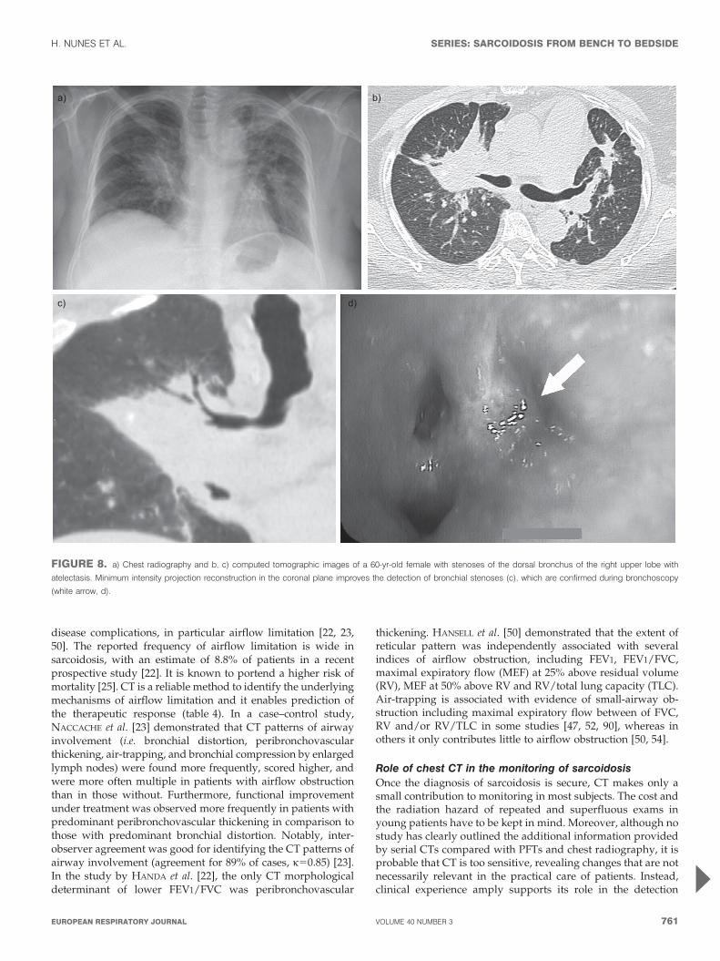
a) b)
c) d)
FIGURE 8. a) Chest radiography and b, c) computed tomographic images of a 60-yr-old female with stenoses of the dorsal bronchus of the right upper lobe with
atelectasis. Minimum intensity projection reconstruction in the coronal plane improves the detection of bronchial stenoses (c), which are confirmed during bronchoscopy
(white arrow, d).
H. NUNES ET AL. SERIES: SARCOIDOSIS FROM BENCH TO BEDSIDE
cEUROPEAN RESPIRATORY JOURNAL VOLUME 40 NUMBER 3 761
of pulmonary complications, in particular when a patient pre-sents with unexplained worsening of respiratory symptoms,haemoptysis, disproportionately impaired lung function orairflow obstruction, uncertain chest radiographic abnormal-ities, aspergilloma or pulmonary hypertension.
Aspergilloma
Mycetoma formation has been reported in about 2% ofsarcoidosis patients, essentially those with advanced fibrocys-tic or cavitary disease [104]. Aspergilloma is more readilyvisualised using chest CT than radiography, which generallyreveals a cavity containing a solid mass, with or without acharacteristic air crescent. The fungus ball can be mobilewithin the cavity when the patient is turned from the supine tothe prone position (fig. 7). In the presence of a cavitary lesion,the thickening of the wall and adjacent pleura points toaspergilloma and is sometimes the earliest sign before anychanges are visible inside the cavity [73]. Aspergilloma may bemultiple and bilateral [104], and this is best appreciated by CTand imperative to recognise before surgery.
Pulmonary hypertension
The prevalence of pre-capillary pulmonary hypertension doesnot traditionally exceed 5% of all sarcoidosis patients but itlargely depends on the stage of disease. Although usuallyattributed to the destruction of distal capillary bed by fibroticprocess and/or to the resultant chronic hypoxaemia, variousalternative mechanisms come into play, including extrinsiccompression of large pulmonary arteries by enlarged lymphnodes or mediastinal fibrosis [4], specific granulomatous orfibrotic vascular involvement that sometimes simulates pulmon-ary veno-occlusive disease [36, 105], and vasoconstriction byvasoactive factors. Contrast-enhanced CT may raise the possibi-lity of pulmonary hypertension when the widest diameter of themain pulmonary artery is larger than 30 mm or superior to thatof the ascending aorta (fig. 6b) but this sign is poorly reliable[106]. Contrast-enhanced CT can also help to recognise under-lying mechanisms by showing vascular compression or bothextensive septal reticulations and ground glass opacity in caseof pulmonary veno-occlusive disease [36]. Most importantly,contrast-enhanced CT rules out pulmonary embolism, which hasbeen recently associated with sarcoidosis [107].
Bronchial stenosis
Significant bronchial stenosis is very rare and may occur at anystage of the disease [108]. Obstruction may be the consequenceof endobronchial granulomatous involvement and extrinsiccompression by enlarged lymph nodes [61]. The stenoses canbe solitary or multiple, lobar or segmental and may cause orcontribute to pulmonary symptoms [61, 108]. The left upperlobe (44.5%), the right upper and middle lobes (15.5% each),and the left lower lobe (11%) are most often affected. Apart fromdepicting atelectasis and nodal external reduction of bronchiallumen, CT is useful in determining the extent and nature ofbronchial stenosis (fig. 8) [37, 108]. However, it cannot replacebronchoscopy, as it leads to false-positive results, incorrectlypredicting the presence of focal bronchial abnormalities in upto 14% of patients [37, 109]. New techniques of reconstructionof the bronchial tree may improve the assessment of airwayinvolvement.
OTHER IMAGING TECHNIQUESDespite inherent pitfalls, due mainly to the composition oflung tissue and physiological motion (cardiac pulsation andrespiration), magnetic resonance imaging (MRI) is gainingattention in pulmonary imaging [110]. However, data arelacking and relatively old in sarcoidosis [111–115] usingunenhanced T1 and T2-weighted MRI. GAETA et al. [116] firstused gadolinium enhanced thoracic MRI in a cohort of DLDs,including 10 patients with sarcoidosis, and showed enhance-ment of pulmonary lesions in five out of seven patients withactive disease [116]. More recent development in diffusion orperfusion imaging could be of major interest to evaluateregional disease activity [117].
CONCLUSIONImaging makes a major contribution to the diagnosis and mana-gement of sarcoidosis patients. Despite substantial disagree-ment about the recognition of radiographic stages, Scaddingclassification still provides a rough but invaluable evaluation ofdisease outcome. The comparison of serial chest radiographywith pulmonary function for the detection of change in diseaseseverity and for the assessment of treatment response requiresfurther studies. Although not necessary in all patients, chest CT isof outstanding utility for the diagnosis of sarcoidosis and itscomplications in selected cases. CT scores are better correlatedwith disease activity and functional impairment than radio-graphy, but the real clinical relevance of such correlations is un-certain. A better definition of end-points is crucial in sarcoidosis,in particular for the quality of clinical trials. Apart from PET-CT,chest MRI may also become a technique of choice in theforeseeable future, given its advantage of being radiation-free.
STATEMENT OF INTERESTA statement of interest for D. Valeyre can be found at www.erj.ersjournals.com/site/misc/statements.xhtml
REFERENCES1 American Thoracic Society, European Respiratory Society, World
Association of Sarcoidosis and Other Granulomatous Disorders.Statement on sarcoidosis. Am J Respir Crit Care Med 1999; 160:736–755.
2 Scadding JG. Prognosis of intrathoracic sarcoidosis in England.A review of 136 cases after five years’ observation. Br Med J 1961;2: 1165–1172.
3 Baughman RP, Teirstein AS, Judson MA, et al. Clinicalcharacteristics of patients in a case control study of sarcoidosis.Am J Respir Crit Care Med 2001; 164: 1885–1889.
4 Lynch JP 3rd, Ma YL, Koss MN, et al. Pulmonary sarcoidosis.Semin Respir Crit Care Med 2007; 28: 53–74.
5 Nunes H, Brillet PY, Valeyre D, et al. Imaging in sarcoidosis.Semin Respir Crit Care Med 2007; 28: 102–120.
6 Bein ME, Putman CE, McLoud TC, et al. A reevaluation ofintrathoracic lymphadenopathy in sarcoidosis. AJR Am J Roentgenol
1978; 131: 409–415.
7 Israel HL, Lenchner G, Steiner RM. Late development of mediast-inal calcification in sarcoidosis. Am Rev Respir Dis 1981; 124: 302–305.
8 Littner MR, Schachter EN, Putman CE, et al. The clinicalassessment of roentgenographically atypical pulmonary sarcoi-dosis. Am J Med 1977; 62: 361–368.
9 Rockoff SD, Rohatgi PK. Unusual manifestations of thoracicsarcoidosis. AJR Am J Roentgenol 1985; 144: 513–528.
SERIES: SARCOIDOSIS FROM BENCH TO BEDSIDE H. NUNES ET AL.
762 VOLUME 40 NUMBER 3 EUROPEAN RESPIRATORY JOURNAL
10 Conant EF, Glickstein MF, Mahar P, et al. Pulmonary sarcoidosisin the older patient: conventional radiographic features. Radiology1988; 169: 315–319.
11 Baughman RP, Shipley R, Desai S, et al. Changes in chestroentgenogram of sarcoidosis patients during a clinical trial ofinfliximab therapy: comparison of different methods of evalua-tion. Chest 2009; 136: 526–535.
12 Zappala CJ, Desai SR, Copley SJ, et al. Optimal scoring of serialchange on chest radiography in sarcoidosis. Sarcoidosis Vasc
Diffuse Lung Dis 2011; 28: 130–138.
13 Muers MF, Middleton WG, Gibson GJ, et al. A simple radio-graphic scoring method for monitoring pulmonary sarcoidosis:relations between radiographic scores, dyspnoea grade andrespiratory function in the British Thoracic Society Study ofLong-Term Corticosteroid Treatment. Sarcoidosis Vasc Diffuse
Lung Dis 1997; 14: 46–56.
14 Winterbauer RH, Belic N, Moores KD. Clinical interpretation ofbilateral hilar adenopathy. Ann Intern Med 1973; 78: 65–71.
15 Hillerdal G, Nou E, Osterman K, et al. Sarcoidosis: epidemiologyand prognosis. A 15-year European study. Am Rev Respir Dis1984; 130: 29–32.
16 Reich JM. Mortality of intrathoracic sarcoidosis in referral vspopulation-based settings: influence of stage, ethnicity, andcorticosteroid therapy. Chest 2002; 121: 32–39.
17 Nardi A, Brillet PY, Letoumelin P, et al. Stage IV sarcoidosis:comparison of survival with the general population and causesof death. Eur Respir J 2011; 38: 1368–1373.
18 Yeager H, Rossman MD, Baughman RP, et al. Pulmonary andpsychosocial findings at enrollment in the ACCESS study.Sarcoidosis Vasc Diffuse Lung Dis 2005; 22: 147–153.
19 Baughman RP, Sparkman BK, Lower EE. Six-minute walk testand health status assessment in sarcoidosis. Chest 2007; 132:207–213.
20 Alhamad EH, Lynch JP 3rd, Martinez FJ. Pulmonary functiontests in interstitial lung disease: what role do they have? ClinChest Med 2001; 22: 715–750.
21 Harrison BD, Shaylor JM, Stokes TC, et al. Airflow limitation insarcoidosis – a study of pulmonary function in 107 patients withnewly diagnosed disease. Respir Med 1991; 85: 59–64.
22 Handa T, Nagai S, Fushimi Y, et al. Clinical and radiographicindices associated with airflow limitation in patients withsarcoidosis. Chest 2006; 130: 1851–1856.
23 Naccache JM, Lavole A, Nunes H, et al. High-resolutioncomputed tomographic imaging of airways in sarcoidosispatients with airflow obstruction. J Comput Assist Tomogr 2008;32: 905–912.
24 Sharma OP, Johnson R. Airway obstruction in sarcoidosis. Astudy of 123 nonsmoking black American patients withsarcoidosis. Chest 1988; 94: 343–346.
25 Viskum K, Vestbo J. Vital prognosis in intrathoracic sarcoidosiswith special reference to pulmonary function and radiologicalstage. Eur Respir J 1993; 6: 349–353.
26 Medinger AE, Khouri S, Rohatgi PK. Sarcoidosis: the value ofexercise testing. Chest 2001; 120: 93–101.
27 Barros WG, Neder JA, Pereira CA, et al. Clinical, radiographic andfunctional predictors of pulmonary gas exchange impairment atmoderate exercise in patients with sarcoidosis. Respiration 2004;71: 367–373.
28 Judson MA, Gilbert GE, Rodgers JK, et al. The utility of the chestradiograph in diagnosing exacerbations of pulmonary sarcoido-sis. Respirology 2008; 13: 97–102.
29 Gibson GJ, Prescott RJ, Muers MF, et al. British Thoracic SocietySarcoidosis study: effects of long term corticosteroid treatment.Thorax 1996; 51: 238–247.
30 Pietinalho A, Tukiainen P, Haahtela T, et al. Oral prednisolonefollowed by inhaled budesonide in newly diagnosed pulmonarysarcoidosis: a double-blind, placebo-controlled multicenter study.
Finnish Pulmonary Sarcoidosis Study Group. Chest 1999; 116:424–431.
31 Pietinalho A, Tukiainen P, Haahtela T, et al. Early treatment ofstage II sarcoidosis improves 5-year pulmonary function. Chest2002; 121: 24–31.
32 Bradley B, Branley HM, Egan JJ, et al. Interstitial lung diseaseguideline: the British Thoracic Society in collaboration with theThoracic Society of Australia and New Zealand and the IrishThoracic Society. Thorax 2008; 63: Suppl. 5, v1–v58.
33 Beigelman-Aubry C, Hill C, Guibal A, et al. Multi-detector rowCT and postprocessing techniques in the assessment of diffuselung disease. Radiographics 2005; 25: 1639–1652.
34 Amis ES Jr. CT radiation dose: trending in the right direction.Radiology 2011; 261: 5–8.
35 Abehsera M, Valeyre D, Grenier P, et al. Sarcoidosis withpulmonary fibrosis: CT patterns and correlation with pulmonaryfunction. AJR Am J Roentgenol 2000; 174: 1751–1757.
36 Nunes H, Humbert M, Capron F, et al. Pulmonary hypertensionassociated with sarcoidosis: mechanisms, haemodynamics andprognosis. Thorax 2006; 61: 68–74.
37 Lenique F, Brauner MW, Grenier P, et al. CT assessment ofbronchi in sarcoidosis: endoscopic and pathologic correlations.Radiology 1995; 194: 419–423.
38 de Boer S, Milne DG, Zeng I, et al. Does CT scanning predict thelikelihood of a positive transbronchial biopsy in sarcoidosis?Thorax 2009; 64: 436–439.
39 Eckardt J, Olsen KE, Jorgensen OD, et al. Minimally invasivediagnosis of sarcoidosis by EBUS when conventional diagnosticsfail. Sarcoidosis Vasc Diffuse Lung Dis 2010; 27: 43–48.
40 Brauner MW, Grenier P, Mompoint D, et al. Pulmonarysarcoidosis: evaluation with high-resolution CT. Radiology 1989;172: 467–471.
41 Leung AN, Brauner MW, Caillat-Vigneron N, et al. Sarcoidosisactivity: correlation of HRCT findings with those of 67Gascanning, bronchoalveolar lavage, and serum angiotensin-con-verting enzyme assay. J Comput Assist Tomogr 1998; 22: 229–234.
42 Murdoch J, Muller NL. Pulmonary sarcoidosis: changes on follow-up CT examination. AJR Am J Roentgenol 1992; 159: 473–477.
43 Oberstein A, von Zitzewitz H, Schweden F, et al. Non invasiveevaluation of the inflammatory activity in sarcoidosis with high-resolution computed tomography. Sarcoidosis Vasc Diffuse Lung
Dis 1997; 14: 65–72.
44 Remy-Jardin M, Giraud F, Remy J, et al. Pulmonary sarcoidosis:role of CT in the evaluation of disease activity and functionalimpairment and in prognosis assessment. Radiology 1994; 191:675–680.
45 Akira M, Kozuka T, Inoue Y, et al. Long-term follow-up CT scanevaluation in patients with pulmonary sarcoidosis. Chest 2005;127: 185–191.
46 Bergin CJ, Bell DY, Coblentz CL, et al. Sarcoidosis: correlation ofpulmonary parenchymal pattern at CT with results of pulmon-ary function tests. Radiology 1989; 171: 619–624.
47 Davies CW, Tasker AD, Padley SP, et al. Air trapping insarcoidosis on computed tomography: correlation with lungfunction. Clin Radiol 2000; 55: 217–221.
48 Drent M, De Vries J, Lenters M, et al. Sarcoidosis: assessment ofdisease severity using HRCT. Eur Radiol 2003; 13: 2462–2471.
49 Handa T, Nagai S, Miki S, et al. Incidence of pulmonaryhypertension and its clinical relevance in patients with sarcoi-dosis. Chest 2006; 129: 1246–1252.
50 Hansell DM, Milne DG, Wilsher ML, et al. Pulmonary sarcoi-dosis: morphologic associations of airflow obstruction at thin-section CT. Radiology 1998; 209: 697–704.
51 Lopes AJ, de Menezes SL, Dias CM, et al. Comparison betweencardiopulmonary exercise testing parameters and computedtomography findings in patients with thoracic sarcoidosis. Lung
2011; 189: 425–431.
H. NUNES ET AL. SERIES: SARCOIDOSIS FROM BENCH TO BEDSIDE
cEUROPEAN RESPIRATORY JOURNAL VOLUME 40 NUMBER 3 763

52 Magkanas E, Voloudaki A, Bouros D, et al. Pulmonary sar-
coidosis. Correlation of expiratory high-resolution CT findingswith inspiratory patterns and pulmonary function tests. Acta
Radiol 2001; 42: 494–501.
53 Muller NL, Mawson JB, Mathieson JR, et al. Sarcoidosis:
correlation of extent of disease at CT with clinical, functional,and radiographic findings. Radiology 1989; 171: 613–618.
54 Terasaki H, Fujimoto K, Muller NL, et al. Pulmonary sarcoidosis:
comparison of findings of inspiratory and expiratory high-resolution CT and pulmonary function tests between smokers
and nonsmokers. AJR Am J Roentgenol 2005; 185: 333–338.
55 Criado E, Sanchez M, Ramirez J, et al. Pulmonary sarcoidosis:
typical and atypical manifestations at high-resolution CT withpathologic correlation. Radiographics, 30: 1567–1586.
56 Hansell DM, Bankier AA, MacMahon H, et al. Fleischner Society:
glossary of terms for thoracic imaging. Radiology 2008; 246: 697–722.
57 Sider L, Horton ES Jr. Hilar and mediastinal adenopathy in
sarcoidosis as detected by computed tomography. J Thorac
Imaging 1990; 5: 77–80.
58 Gawne-Cain ML, Hansell DM. The pattern and distribution of
calcified mediastinal lymph nodes in sarcoidosis and tubercu-losis: a CT study. Clin Radiol 1996; 51: 263–267.
59 Niimi H, Kang EY, Kwong JS, et al. CT of chronic infiltrative lungdisease: prevalence of mediastinal lymphadenopathy. J Comput
Assist Tomogr 1996; 20: 305–308.
60 Patil SN, Levin DL. Distribution of thoracic lymphadenopathy insarcoidosis using computed tomography. J Thorac Imaging 1999;
14: 114–117.
61 Polychronopoulos VS, Prakash UB. Airway involvement in
sarcoidosis. Chest 2009; 136: 1371–1380.
62 Morawiec E, Hachulla-Lemaire AL, Chabrol J, et al. Venoatrialcompression by lymphadenopathy in sarcoidosis. Eur Respir J
2010; 35: 1188–1191.
63 Yangui F, Battesti JP, Valeyre D, et al. Fibrosing mediastinitis as a
rare mechanism of pulmonary oedema in sarcoidosis. Eur Respir
J 2010; 35: 455–456.
64 Narayan D, Brown L, Thayer JO. Surgical management of superiorvena caval syndrome in sarcoidosis. Ann Thorac Surg 1998; 66:
946–948.
65 Cappell MS. Endoscopic, radiographic, and manometric findingsin dysphagia associated with sarcoid due to extrinsic esophageal
compression from subcarinal lymphadenopathy. Am J Gastroenterol
1995; 90: 489–492.
66 Tobias JK, Santiago SM, Williams AJ. Sarcoidosis as a cause ofleft recurrent laryngeal nerve palsy. Arch Otolaryngol Head Neck
Surg 1990; 116: 971–972.
67 Jarman PR, Whyte MK, Sabroe I, et al. Sarcoidosis presentingwith chylothorax. Thorax 1995; 50: 1324–1325.
68 Nishimura K, Itoh H, Kitaichi M, et al. Pulmonary sarcoidosis:correlation of CT and histopathologic findings. Radiology 1993;
189: 105–109.
69 Brauner M, Dumas JL, Beigelman C. Imagerie de la sarcoıdosepulmonaire. [Imaging of pulmonary sarcoidosis.] Ann Radiol
(Paris) 1994; 37: 216–221.
70 Hamper UM, Fishman EK, Khouri NF, et al. Typical and atypical
CT manifestations of pulmonary sarcoidosis. J Comput Assist
Tomogr 1986; 10: 928–936.
71 Honda O, Johkoh T, Ichikado K, et al. Comparison of high
resolution CT findings of sarcoidosis, lymphoma, and lymphan-gitic carcinoma: is there any difference of involved interstitium?
J Comput Assist Tomogr 1999; 23: 374–379.
72 Gruden JF, Webb WR, Naidich DP, et al. Multinodular disease:
anatomic localization at thin-section CT – multireader evaluationof a simple algorithm. Radiology 1999; 210: 711–720.
73 Hours S, Nunes H, Kambouchner M, et al. Pulmonary cavi-
tary sarcoidosis: clinico-radiologic characteristics and natural
history of a rare form of sarcoidosis. Medicine (Baltimore) 2008; 87:142–151.
74 Malaisamy S, Dalal B, Bimenyuy C, et al. The clinical andradiologic features of nodular pulmonary sarcoidosis. Lung 2009;187: 9–15.
75 Nakatsu M, Hatabu H, Morikawa K, et al. Large coalescentparenchymal nodules in pulmonary sarcoidosis: "sarcoid galaxy"sign. AJR Am J Roentgenol 2002; 178: 1389–1393.
76 Brauner MW, Lenoir S, Grenier P, et al. Pulmonary sarcoido-sis: CT assessment of lesion reversibility. Radiology 1992; 182:349–354.
77 Johkoh T, Ikezoe J, Takeuchi N, et al. CT findings in "pseu-doalveolar" sarcoidosis. J Comput Assist Tomogr 1992; 16: 904–907.
78 Judson MA, Uflacker R. Treatment of a solitary pulmonarysarcoidosis mass by CT-guided direct intralesional injection ofcorticosteroid. Chest 2001; 120: 316–317.
79 Quaden C, Tillie-Leblond I, Delobbe A, et al. Necrotising sarcoidgranulomatosis: clinical, functional, endoscopical and radio-graphical evaluations. Eur Respir J 2005; 26: 778–785.
80 Froudarakis ME, Bouros D, Voloudaki A, et al. Pneumothorax asa first manifestation of sarcoidosis. Chest 1997; 112: 278–280.
81 Marchiori E, Zanetti G, Barreto MM, et al. Atypical distributionof small nodules on high resolution CT studies: patterns anddifferentials. Respir Med, 105: 1263–1267.
82 Herraez Ortega I, Alonso Orcajo N, Lopez Gonzalez L. El‘‘cumulo sarcoideo’’. Un nuevo signo en tomografıa computar-izada de torax de alta resolucion. [The "sarcoid cluster sign".A new sign in high resolution chest CT.] Radiologia 2009; 51:495–499.
83 Martin SG, Kronek LP, Valeyre D, et al. High-resolutioncomputed tomography to differentiate chronic diffuse interstitiallung diseases with predominant ground-glass pattern usinglogical analysis of data. Eur Radiol 2010; 20: 1297–1310.
84 Padley SP, Padhani AR, Nicholson A, et al. Pulmonarysarcoidosis mimicking cryptogenic fibrosing alveolitis on CT.Clin Radiol 1996; 51: 807–810.
85 Aisner SC, Albin RJ. Diffuse interstitial pneumonitis and fibrosisin sarcoidosis. Chest 1988; 94: 193–195.
86 Nobata K, Kasai T, Fujimura M, et al. Pulmonary sarcoidosiswith usual interstitial pneumonia distributed predominantly inthe lower lung fields. Intern Med 2006; 45: 359–362.
87 Shigemitsu H, Oblad JM, Sharma OP, et al. Chronic interstitialpneumonitis in end-stage sarcoidosis. Eur Respir J 2010; 35: 695–697.
88 Bartz RR, Stern EJ. Airways obstruction in patients withsarcoidosis: expiratory CT scan findings. J Thorac Imaging 2000;15: 285–289.
89 Fazzi P, Sbragia P, Solfanelli S, et al. Functional significance ofthe decreased attenuation sign on expiratory CT in pulmonarysarcoidosis: report of four cases. Chest 2001; 119: 1270–1274.
90 Gleeson FV, Traill ZC, Hansell DM. Evidence of expiratory CTscans of small-airway obstruction in sarcoidosis. AJR Am J
Roentgenol 1996; 166: 1052–1054.
91 Nishino M, Kuroki M, Roberts DH, et al. Bronchomalacia insarcoidosis: evaluation on volumetric expiratory high-resolutionCT of the lung. Acad Radiol 2005; 12: 596–601.
92 Huggins JT, Doelken P, Sahn SA, et al. Pleural effusions ina series of 181 outpatients with sarcoidosis. Chest 2006; 129:1599–1604.
93 Szwarcberg JB, Glajchen N, Teirstein AS. Pleural involvement inchronic sarcoidosis detected by thoracic CT scanning. Sarcoidosis
Vasc Diffuse Lung Dis 2005; 22: 58–62.
94 Harada T, Nabeshima K, Matsumoto T, et al. Histologicalfindings of the computed tomography halo in pulmonarysarcoidosis. Eur Respir J 2009; 34: 281–283.
95 Marten K, Rummeny EJ, Engelke C. The CT halo: a new sign inactive pulmonary sarcoidosis. British J Radiol 2004; 77: 1042–1045.
SERIES: SARCOIDOSIS FROM BENCH TO BEDSIDE H. NUNES ET AL.
764 VOLUME 40 NUMBER 3 EUROPEAN RESPIRATORY JOURNAL
96 Marlow TJ, Krapiva PI, Schabel SI, et al. The "fairy ring": a newradiographic finding in sarcoidosis. Chest 1999; 115: 275–276.
97 Lee YR, Choi YW, Lee KJ, et al. CT halo sign: the spectrum ofpulmonary diseases. Br J Radiol 2005; 78: 862–865.
98 Grenier P, Chevret S, Beigelman C, et al. Chronic diffuseinfiltrative lung disease: determination of the diagnostic valueof clinical data, chest radiography, and CT and Bayesian analysis.Radiology 1994; 191: 383–390.
99 Mana J, Teirstein AS, Mendelson DS, et al. Excessive thoraciccomputed tomographic scanning in sarcoidosis. Thorax 1995; 50:1264–1266.
100 Kaiser PK, Lowder CY, Sullivan P, et al. Chest computerizedtomography in the evaluation of uveitis in elderly women. Am J
Ophthalmol 2002; 133: 499–505.101 Kosmorsky GS, Meisler DM, Rice TW, et al. Chest computed
tomography and mediastinoscopy in the diagnosis of sarcoidosis-associated uveitis. Am J Ophthalmol 1998; 126: 132–134.
102 Klein JS, Johnson A, Watson E, et al. CT-guided transthoracicneedle biopsy in the diagnosis of sarcoidosis. J Thorac Imaging
2009; 24: 23–30.103 Remy-Jardin M, Giraud F, Remy J, et al. Importance of ground-
glass attenuation in chronic diffuse infiltrative lung disease:pathologic-CT correlation. Radiology 1993; 189: 693–638.
104 Pena TA, Soubani AO, Samavati L. Aspergillus lung disease inpatients with sarcoidosis: a case series and review of theliterature. Lung 2011; 189: 167–172.
105 Hoffstein V, Ranganathan N, Mullen JB. Sarcoidosis simulatingpulmonary veno-occlusive disease. Am Rev Respir Dis 1986; 134:809–811.
106 Devaraj A, Wells AU, Meister MG, et al. The effect of diffusepulmonary fibrosis on the reliability of CT signs of pulmonaryhypertension. Radiology 2008; 249: 1042–1049.
107 Crawshaw AP, Wotton CJ, Yeates DG, et al. Evidence forassociation between sarcoidosis and pulmonary embolism from35-year record linkage study. Thorax 2011; 66: 447–448.
108 Chambellan A, Turbie P, Nunes H, et al. Endoluminal stenosis ofproximal bronchi in sarcoidosis: bronchoscopy, function, andevolution. Chest 2005; 127: 472–481.
109 Naidich DP, Lee JJ, Garay SM, et al. Comparison of CT andfiberoptic bronchoscopy in the evaluation of bronchial disease.AJR Am J Roentgenol 1987; 148: 1–7.
110 Kauczor HU, Ley-Zaporozhan J, Ley S. Imaging of pulmonarypathologies: focus on magnetic resonance imaging. Proc Am
Thorac Soc 2009; 6: 458–463.111 Craig DA, Colletti PM, Ratto D, et al. MRI findings in pulmonary
sarcoidosis. Magn Reson Imaging 1988; 6: 567–573.112 McFadden RG, Carr TJ, Wood TE. Proton magnetic resonance
imaging to stage activity of interstitial lung disease. Chest 1987;92: 31–39.
113 Mendelson DS, Gray CE, Teirstein AS. Magnetic resonancefindings in sarcoidosis of the thorax. Magn Reson Imaging 1992;10: 523–529.
114 Muller NL, Mayo JR, Zwirewich CV. Value of MR imaging in theevaluation of chronic infiltrative lung diseases: comparison withCT. AJR Am J Roentgenol 1992; 158: 1205–1209.
115 Primack SL, Mayo JR, Hartman TE, et al. MRI of infiltrative lungdisease: comparison with pathologic findings. J Comput Assist
Tomogr 1994; 18: 233–238.116 Gaeta M, Blandino A, Scribano E, et al. Chronic infiltrative lung
diseases: value of gadolinium-enhanced MRI in the evaluation ofdisease activity – early report. Chest 2000; 117: 1173–1178.
117 Henzler T, Schmid-Bindert G, Schoenberg SO, et al. Diffusionand perfusion MRI of the lung and mediastinum. Eur J Radiol2010; 76: 329–336.
H. NUNES ET AL. SERIES: SARCOIDOSIS FROM BENCH TO BEDSIDE
EUROPEAN RESPIRATORY JOURNAL VOLUME 40 NUMBER 3 765