Embed Size (px)
Citation preview
Serum amyloid A opposes lipoxin A4 to mediateglucocorticoid refractory lung inflammation in chronicobstructive pulmonary diseaseSteven Bozinovskia,1, Mohib Uddinb, Ross Vlahosa, Michelle Thompsonc, Jonathan L. McQualtera, Anne-Sophie Merritta,Peter A. B. Warkd,e, Anastasia Hutchinsonc, Louis B. Irvingc, Bruce D. Levyb,2, and Gary P. Andersona,f,2
aDepartment of Pharmacology, and fDepartment of Medicine, University of Melbourne, Parkville, Victoria, Australia 3010; bPulmonary and Critical CareMedicine, Brigham and Women’s Hospital and Harvard Medical School, Boston, MA 02115; cDepartment of Respiratory Medicine, Royal Melbourne Hospital,Parkville, Victoria, Australia 3010; dCentre for Asthma and Respiratory Disease, Hunter Medical Research Institute, University of Newcastle, Australia 2305;and eDepartment of Respiratory and Sleep Medicine, John Hunter Hospital, Australia 2305
Edited by Bruce D. Hammock, University of California, Davis, CA, and approved December 7, 2011 (received for review July 5, 2011)
Chronic obstructive pulmonary disease (COPD) will soon be thethird most common cause of death globally. Despite smokingcessation, neutrophilic mucosal inflammation persistently dam-ages the airways and fails to protect from recurrent infections.This maladaptive and excess inflammation is also refractory toglucocorticosteroids (GC). Here, we identify serum amyloid A(SAA) as a candidate mediator of GC refractory inflammation inCOPD. Extrahepatic SAA was detected locally in COPD bronchoal-veolar lavage fluid, which correlated with IL-8 and neutrophilelastase, consistent with neutrophil recruitment and activation.Immunohistochemistry detected SAA was in close proximity toairway epithelium, and in vitro SAA triggered release of IL-8 andother proinflammatory mediators by airway epithelial cells in anALX/FPR2 (formyl peptide receptor 2) receptor-dependent manner.Lipoxin A4 (LXA4) can also interact with ALX/FPR2 receptors andlead to allosteric inhibition of SAA-initiated epithelial responses(pA2 13 nM). During acute exacerbation, peripheral blood SAA lev-els increased dramatically and were disproportionately increasedrelative to LXA4. Human lung macrophages (CD68+) colocalizedwith SAA and GCs markedly increased SAA in vitro (THP-1, pEC50
43 nM). To determine its direct actions, SAA was administered intomurine lung, leading to induction of CXC chemokine ligand 1/2 anda neutrophilic response that was inhibited by 15-epi-LXA4 but notdexamethasone. Taken together, these findings identify SAA asa therapeutic target for inhibition and implicate SAA as a mediatorof GC-resistant lung inflammation that can overwhelm organprotective signaling by lipoxins at ALX/FPR2 receptors.
resolution | leukocyte activation | G protein-coupled receptor |innate immunity
Airway mucosal inflammation is an essential effector arm ofinnate host defense, but the principal determinants of its
intensity and persistence remain poorly understood. In particu-lar, molecular aberrations in diseases where inflammation isintractable and resolution is impaired have not been well de-lineated. Understanding these control mechanisms may help toredress disease states, such as chronic obstructive pulmonarydisease (COPD), an incurable fatal lung disease where gluco-corticoid (GC)-insensitive inflammation (reviewed in ref. 1)persists, despite smoking cessation (2). COPD is already the fifthleading cause of death worldwide and, given its long disease la-tency, its impact will continue to rise in coming decades (3). Theprimary cause of COPD is chronic exposure to cigarette smoke,which is known to permanently alter the lung transcriptome (4).Because the underlying cause of persistent innate immune acti-vation remains poorly understood in COPD, there is an urgentneed to identify new insights into the pathobiology of COPD thatwill lead to novel therapeutic strategies.The lead member of a new class of endogenous proresolving
mediators is lipoxin A4 (LXA4) (5), which is generated in the lung
during cell–cell interactions including in COPD (6), when in-filtrating leukocytes interact with airway epithelial cells (reviewedin ref. 7). LXA4 interacts withALX/FPR2 (formyl peptide receptor2) receptors to transduce anti-inflammatory signals that blockneutrophil activation and recruitment as part of a resolution pro-gram for airway inflammation. For example, LXA4 opposes theactions of the 5-lipoxygenase–derived eicosanoid leukotriene B4(LTB4), which is a potent agonist for neutrophil transmigration andactivation (reviewed in ref. 5). Of interest, serum amyloid A (SAA)can also interact with ALX/FPR2 receptors (8) and, in sharpcontrast to LXA4, stimulates neutrophil chemotaxis and functionalresponses (9, 10). SAA is a hepatic acute-phase protein, the serumlevels of which strongly predict the intensity of acute exacerbationsof COPD (AECOPD) (11) that are triggered by airway infectionand associated with further increases in inflammation (12). Thus,as SAA is increased in COPD,we reasoned that if it were present inthe lung it might account for persistent lung inflammation.Here, we have shown that SAA is present in inflamed lungs,
elevated in AECOPD, and is closely related to neutrophilic ac-tivation in COPD airways. During AECOPD, levels of circulatingSAAwere substantially greater than either of the anti-inflammatorymediators LXA4 or annexin A1 (ANXA1), a steroid-inducibleanti-inflammatory protein. In vitro, SAA potently activatedproinflammatory mediator release from airway epithelial cells inan ALX/FPR2-dependent manner. In vivo, SAA elicited a robustneutrophilic response that was counteracted by administration ofthe ALX/FPR2 anti-inflammatory ligand 15-epi-LXA4. Impor-tantly, SAA’s proinflammatory actions appeared to underlie theGC refractory nature of neutrophil-mediated inflammation inCOPD (and potentially other diseases of chronic neutrophilicinflammation), as GCs, the most common anti-inflammatory drugin current disease management, induced extrahepatic SAA ex-pression and failed to suppress extrahepatic SAA production.These data identify SAA as a candidate pathogenic mediator oflung inflammation and mechanism for defective resolution, rais-ing the possibility of SAA and ALX/FPR2 receptors as previouslyunexplored therapeutic targets in COPD.
Author contributions: S.B., M.U., R.V., L.B.I., B.D.L., and G.P.A. designed research; S.B.,M.U., R.V., M.T., J.L.M., A.-S.M., P.A.B.W., A.H., and L.B.I. performed research; S.B.,M.U., R.V., M.T., J.L.M., A.-S.M., P.A.B.W., A.H., L.B.I., B.D.L., and G.P.A. analyzed data;J.L.M. contributed new reagents/analytic tools; and S.B., J.L.M., B.D.L., and G.P.A. wrotethe paper.
Conflict of interest statement: B.D.L. is a co-inventor on patents on lipoxins in airwaydisease that are assigned to Brigham and Women’s Hospital and licensed forclinical development.
This article is a PNAS Direct Submission.1To whom correspondence should be addressed. E-mail: [email protected]. and G.P.A. contributed equally to this work.
This article contains supporting information online at www.pnas.org/lookup/suppl/doi:10.1073/pnas.1109382109/-/DCSupplemental.
www.pnas.org/cgi/doi/10.1073/pnas.1109382109 PNAS Early Edition | 1 of 6
MED
ICALSC
IENCE
S
ResultsSAA in COPD Bronchoalveolar Lavage Fluid Is Associated with IL-8 andNeutrophil Activation. The relationship between airway SAA andneutrophilic inflammation in COPD was explored by measuringSAA and neutrophil elastase (NE) activity, a marker of neutro-phil activation, in bronchoalveolar lavage fluid (BALF) archivedfrom subjects with stable (nonexacerbating) COPD. BALF SAAlevels positively correlated with NE (P = 0.001, r = 0.72,) (Fig.1A and Table S1). In the same cohort, NE also associated withIL-8, an independent and major neutrophil chemotactic factor(P = 0.002, r = 0.47) (Fig. 1B) and a significant association wasobserved between SAA and IL-8 levels (Fig. 1C) (P = 0.03, r =0.35). SAA levels in BALF collected during stable disease werenot significantly different across the Global Initiative for Ob-structive Lung Disease disease severity stages.
SAA Promotes Mucosal Epithelial Inflammatory Responses via ALX/FPR2. In control lung, modest ALX/ FPR2 staining was observedin airway epithelium (Fig. 2A, Upper). In COPD lung, ALX/FPR2 expression displayed more prominent apical and baso-lateral decoration of the airway mucosal lining (Fig. 2A, Lower),implicating the epithelium as a potentially major target for SAA.Staining for SAA in the same specimens demonstrated wide-spread and intense SAA staining that was prominent within thesubmucosa in close proximity to the basolateral epithelium.Lung epithelial A549 (null-A549; deficient for ALX/FPR2)
and hALX-A549 cells genetically engineered to express the
receptor were exposed to recombinant SAA. In the absence ofALX/FPR2, SAA did not induce significant amounts of mono-cyte chemoattractant protein-1 (MCP-1) or GM-CSF (Fig. 2 Band C). In the presence of ALX/FPR2, SAA markedly increasedboth MCP-1 (Fig. 2B) and GM-CSF levels (Fig. 2C). In hALX-A549 cells, SAA also induced a concentration dependent in-crease in IL-8 (Fig. 2D). As little as 10−9 M SAA, led to 3.79 ±1.97 ng IL-8/mL, and 50 ng SAA/mL (an SAA concentrationfound in COPD BALF) (Fig. 1A) resulted in a marked increaseto 37.47 ± 7.92 ng IL-8/mL (Fig. 2D). The EC50 of SAA forhALX-A549 generated IL-8 was 8.5 nM. In addition to A549 cells,SAA induced production of the inflammatory mediators MCP-1,GM-CSF, and IL-8 in the bronchial epithelial BEAS-2B cell lineand primary human COPD epithelial cells (Figs. S1 and S2).Because ALX/FPR2 engagement by LXs blocks proin-
flammatory mediator release by injured airway epithelial cells(13), the impact of LXA4 and 15-epi LXA4 on SAA-initiatedIL-8 release was determined. LXA4-exposed cells demonstrateda rightward shift in the SAA concentration response with de-pressed maxima (Fig. 2D). Both LXA4 and 15-epi-LXA4 wereequipotent at 10−7 M, with similar IC50 values (25.74 nM forLXA4 and 24.83 nM for 15-epi-LXA4) for SAA-mediated IL-8release (Fig. 2 D and E). However, the rightward shift and de-pressed SAA maxima caused by both LXs (Fig. 2 D and E),precluded simple competitive antagonism at a single binding siteas a mechanism for inhibition of IL-8 release. We thereforeapplied classic Schild regression analysis, which yielded a re-gression slope less than unity (0.34) with an estimated pA2 of 13nM; indicating negative cooperative binding secondary to allo-steric modulation as the basis for antagonism.
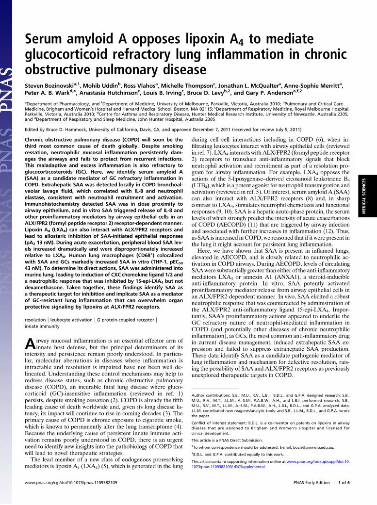
SAA Is Disproportionately Expressed Relative to LXs During anAECOPD in Humans. Because both SAA and LXA4 can serve asendogenous ALX/FPR2 ligands, the relative time course of theirsystemic formation was determined prospectively from the sameindividual during AECOPD and Resolution. There was a trendfor increased plasma LXA4 concentrations during AECOPD(2.0 ± 0.79 nM) vs. Resolution (0.80 ± 0.09 nM) (Fig. 3A), with7 of 10 matching samples displaying increased LXA4 levels atAECOPD. SAA concentrations rose sharply and were signifi-cantly elevated during AECOPD relative to Resolution (Fig. 3B)[18,595 ± 7,112 nM (AECOPD) vs. 510 ± 800 nM (Resolution),P < 0.05 for n = 10]. As a consequence, the amount of SAArelative to LXA4 was significantly increased during the acutephase of an exacerbation (Figs. 3C) [8,791 ± 6,092 SAA/LXA4(AECOPD) vs. 735 ± 1,305 SAA/LXA4 (Resolution), P < 0.05for n = 10]. In addition, SAA/LXA4 ratios were determinedfrom a control cohort with normal lung function (Fig. 3D). TheSAA/LXA4 ratio for Control (432 ± 166 SAA/LXA4) was evenlower than AECOPD resolution (735 ± 1,305 SAA/LXA4), al-though this difference did not reach statistical significance at thissample size (P = 0.25). No association between LXA4 and SAAlevels (Fig. 3E) (r = 0.152, P = 0.68) was observed duringAECOPD, whereas LXA4 and the proinflammatory 5-lip-oxygenase-derived eicosanoid LTB4 were significantly correlated(Fig. 3F) (r= 0.87, P= 0.002). Because ANXA1 can also opposeneutrophilic inflammation via its interaction with ALX/FPR2receptors (14), plasma ANXA1 levels were determined in matchedsamples. Levels for ANXA1 increased during an AECOPD (Fig.S3A). As observed for LXA4, the levels of SAA relative to ANXA1were also significantly elevated during an AECOPD (Fig. S3B). Inaddition, an inverse relationship between SAA and ANXA1 wasobserved during AECOPD (Fig. S3C).
SAA Elicits Airway Neutrophilic Inflammation That Is Inhibitable by15-epi-LXA4. To determine if the actions of SAA were opposed byLXs in vivo, a murine airway challenge model was established.Intranasal SAA (2 μg) challenge elicited an acute inflammatory
C
B
A
Fig. 1. Relationship between lung SAA, IL-8 and neutrophil activity. Cor-relation between secreted (A) SAA and NE activity; (B) IL-8 and NE, and SAAand IL-8 were determined in BALF from COPD subject (n = 41). (δP < 0.05,Pearson correlation).
2 of 6 | www.pnas.org/cgi/doi/10.1073/pnas.1109382109 Bozinovski et al.

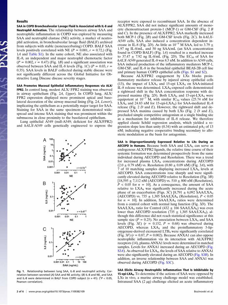
response peaking at 24 h that was neutrophilic in nature (8.4 ±1.2 × 10⁵ BALF neutrophils at 24 h) (Fig. 4 A and B). A sig-nificant increase in secreted CXC chemokine ligand (CXCL) 1 inBALF was seen during this early phase (6 h) (Fig. 4C). An in-crease in transcript expression of the neutrophil chemokines,CXCL1 and CXCL2 (the murine homologs of KC and MIP-2),as assessed by TaqMan quantitative PCR (qPCR) (Fig. 4 D andE), was also observed. Of note, secreted CXCL2 in BALF wasnot increased above control levels, which may indicate rapidconsumption of this neutrophil chemokine at the site of SAA-mediated inflammation. The murine ALX/FPR2 receptor path-way was targeted in vivo by locally delivering 15-epi-LXA4 con-currently with SAA, which reduced CXCL1 and CXCL2expression by 61% and 54%, respectively (Fig. 4F). Recruitmentof BALF neutrophils was similarly reduced by 15-epi-LXA4 atearly (6 h) and peak (24 h) time points, respectively (Fig. 4G andH). In contrast to 15-epi-LXA4, when dexamethasone (DEX)(0.5 mg/kg, i.p.) was given 2 h before SAA challenge, there wasno significant change in airway neutrophils (Fig. 4I). In addition,DEX did not inhibit SAA induction of CXCL1/2 expression (Fig.4J). As a control for acute inflammation, the same dose of DEXdisplayed a 49% reduction in LPS (1 μg, i.n.) triggered BALFneutrophilia (LPS; 7.0 ± 0.7 × 10⁵ vs. DEX-LPS 3.5 ± 0.5 × 10⁵,P < 0.05). To evaluate whether local SAA effects were alsomediated by LTB4, levels of LTB4 in BALF were measured. SAAdid not induce measureable local LTB4 production (EIA, Cay-man Chemicals; detection limit = 13 pg/mL), and treatment with15-epi-LXA4 did not significantly change LTB4 levels.
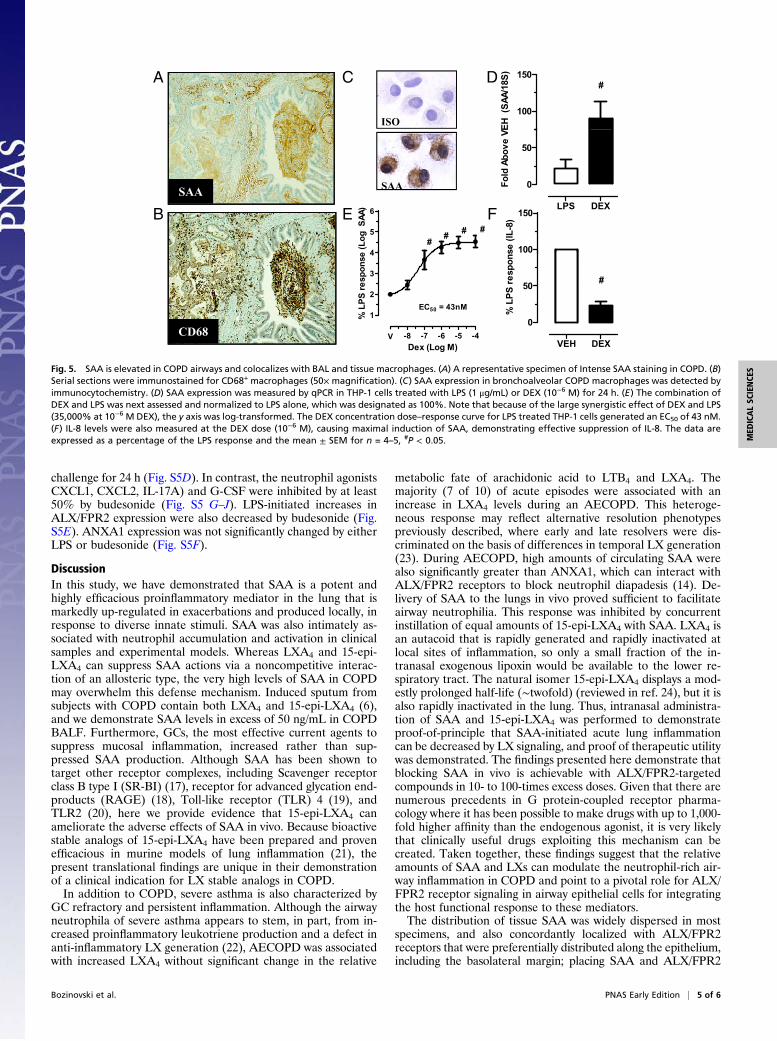
SAA Localized with Macrophages in COPD Lungs. The presence andcellular distribution of SAA was assessed immuno-histochemi-cally in resected human lung tissue (Table S2). Control subjectswere negative for SAA in seven of eight lung specimens, with onespecimen displaying moderate staining. In contrast, a moderatestaining pattern within the parenchyma was observed in 7 of 22COPD lung specimens. No reactivity was observed in 9 of 22 lung
specimens with clinically stable comorbid COPD. Intense SAAimmunoreactivity was observed in 6 of 22 COPD specimens thatwere diffuse in nature and localized to the submucosa, vascular,and bronchoalveolar compartments (Fig. 5A). CD68+ macro-phages were present in BALF and lung parenchyma (Fig. 5B)and colocalized with SAA-positive regions. Because SAA canserve as a precursor for amyloid protein AA serial sections werestained with Congo red. No sample showed observable bi-refringence, indicating the absence of insoluble AA amyloid fibrildeposition. Smoking history was not significantly different acrossthe three SAA staining profiles [No SAA: mean 47 pack years(10–140), moderate SAA: 59 pack years (30–120), and intenseSAA: 36 pack years (25–48)]. Additionally FEV1% predicted wasnot significantly different across the staining profiles [No SAA:mean 79% (54–89), moderate SAA 77% (52–91), and intenseSAA 78% (66–85)].A positive correlation between SAA mRNA and protein was
found in BALF (P = 0.01, r = 0.83) (Table S1), implicating al-veolar macrophages as a major candidate for production ofBALF SAA. BALF cytospot immunostaining confirmed thatalveolar macrophages from COPD subjects were positive forSAA, with prominent staining observed in the cytoplasmic region(Fig. 5C). The regulation of SAA expression was further in-vestigated in human macrophages (THP-1 cell line) exposed tobacterial LPS (1 μg/mL), which is abundant in the airways duringAECOPD (15). LPS induced THP-1 SAA mRNA levels by 25 ±12-fold (Fig. 5D). DEX also induced SAA mRNA by 90 ± 23-fold, and synergistically increased LPS-induced SAA expressionin a dose-dependent manner with 10−6 M DEX plus LPS, re-sulting in a 2,564 ± 754-fold increase. The calculated half max-imal concentration (EC50) was 43 nM (Fig. 5E). As a control,DEX (10−6 M) effectively decreased LPS-induced IL-8 releaseby 76% in the same incubations (Fig. 5F). To further investigatewhether innate stimuli relevant to COPD and infectiousAECOPD can directly promote de novo local synthesis of SAAin airway tissue, SAA mRNA in whole lung from mice treated
40
80
120
M-C
SF (p
g/m
L)
#CA Btrol
50
100
150 null- A549
rhALX- A549
CP-
1 (n
g/m
L)
#
#
ALXVEH SAA
0
GM
Con
ISO
PD
VEH SAA
0
MC
D E
80
120 Vehicle100 nM 15-epi-LXA4
#
of m
axim
al)
80
120 Vehicle10 nM LXA4
100 nM LXA4#
% o
f max
imal
)
ALX SAA
COP
BL
-10 -9 -8 -7
0
40
SAA [log M]
IL-8
rele
ase
(%
-10 -9 -8 -7
0
40
SAA [log M]
IL-8
rele
ase
(%
Fig. 2. ALX/FPR2 is expressed in COPD lungs and is a functional receptor for SAA in human airway epithelial cells. (A) Representative Isotype (ISO), ALX/FPR2(ALX) and SAA staining on serial Control (Upper) and COPD sections (Insets: 100× magnification). ALX/FPR2 inset demonstrates prominent apical andbasolateral (BL) epithelial ALX/FPR2 expression in COPD. MCP-1 (B) and GM-CSF (C) levels in cell-free supernatants were measured in null- A549 cells (openbar) and rhALX- A549 cells (black bar) treated with Vehicle (VEH) or recombinant SAA (1 mg/mL) for 24 h. rhALX- A549 cells were preincubated with LXA4
(10−8 M or 10−7 M) (D) or 15-epi-LXA4 (10−7 M) (E) for 30 min followed by recombinant SAA (10−10 M to 10−7 M, 24 h). Release of IL-8 was measured in cell-free
supernatants by ELISA (see SI Methods). The data are expressed as a percentage of the maximal SAA-stimulated IL-8 release and the mean ± SEM for n = 5–7measured in duplicate, #P < 0.05.
Bozinovski et al. PNAS Early Edition | 3 of 6
MED
ICALSC
IENCE
S
under three separate conditions were determined (Fig. S4).BALB/c mice subchronically exposed to cigarette smoke for 4 ddisplayed a 34-fold increase in SAA expression (Fig. S4A). In-tranasal administration of LPS (10 μg per mouse) induced a 350-fold peak increase in SAA 24 h following challenge (Fig. S4B). Inaddition, intranasal administration of influenza (HKx31) tran-siently induced SAA expression, peaking at day 5 postinfection(17-fold) (Fig. S4C).The acute phase SAA isoforms (SAA1 and SAA2) are 95%
homologous and indistinguishable by qPCR, whereas mouseSAA3 (71% sequence identify with SAA1) is a pseudogene inhumans (16). The in vivo expression of SAA isoforms wasassessed in murine lung tissue following LPS challenge in thepresence of a clinically used inhaled GC, budesonide (Fig. S5).Budesonide (intranasally) induced SAA1/2 expression ∼fivefoldand LPS induced SAA1/2 by greater than 20-fold (Fig. S5B).Airway administration of budesonide (10 μg) 6 h after LPS (3 μg)did not inhibit peak neutrophilic airway inflammation (Fig. S5A)or SAA1/2 lung transcript expression (Fig. S5B), whereas SAA3was reduced by 50% (Fig. S5C). Secreted SAA in BALF wasincreased by budesonide, in particular after concomitant LPS
F
D
BA
C
E
Fig. 3. Relationship between SAA and LXA4 during AECOPD. (A) LXA4 and(B) SAA concentration (nM) in matching blood samples from COPD subjectsduring (i) the acute phase of an exacerbation (AECOPD) and at (ii) clinicalrecovery (Resolution) (n = 10, *P < 0.05 by Wilcoxon-matched paired t test).(C) The relative ratio of SAA/LXA4 was determined for the matching AECOPDand recovery samples. (D) The SAA/LXA4 ratio was also determined fora control cohort with normal lung function (#P < 0.05 by ANOVA vs. Control).The association of LXA4 with (E) SAA and (F) LTB4 was compared during theAECOPD phase, demonstrating a significant correlation with LTB4 only (δP =0.002, r = 0.87 Pearson correlation).
8
12#
#
AL
ce
lls (
x10
5
)
20
30
40
d in
cre
ase
CL
1 / 1
8S
)
A D
0 24 48 72
0
4
SAA treatment (Hours)
To
tal B
A
12
#
10
5
)
0 24 48 72
0
10
SAA treatment (Hours)
Fo
ld
(C
XC
B E120
0 24 48 72
0
4
8
#
SAA treatment (Hours)
To
tal B
AL
Ne
uts (
x
0 24 48 72
0
40
80
SAA treatment (Hours)
Fo
ld in
cre
ase
(C
XC
L2 / 1
8S
)
C F
10
20
30VEH
15-epi-LXA4
Fo
ld
in
cre
as
e
(T
arg
et / 1
8S
)
0
2
4
6
8
#
CX
CL
1 (
ng
/mL
)
CXCL1 CXCL2
0
0 24 48 72
0
SAA treatment (Hours)
6
9
12
15- epi-LXA4
VEH
utro
ph
ils (
x10
5
)
0.4
0.6
0.8 VEH
15-epi-LXA4
utro
ph
ils (
x10
5
)
G H
SAA (24Hrs)
0
3
BA
L n
eu
SAA (6Hrs)
0.0
0.2
BA
L n
eu
I J
6
9
12
DEX (0.5mg/kg)
ph
ils (
x10
5
)
80
120DEX (0.5mg/kg)
cle
tre
ate
d
SAA (24Hrs)
0
3
6
BA
L n
eu
tro
CXCL1 CXCL2
0
40
% V
eh
ic
Fig. 4. Local SAA delivery promotes lung inflammation and CXCL1/2 that issuppressed by 15-epi-LXA4. Mice were challenged with SAA (2 μg) via in-tranasal administration and at the indicated timepoints (h) were killed. (A)Total BAL cells and differential analysis was used to determine (B) neutrophilBAL numbers. (C) Secreted CXCL1 concentration in BALF was determined byELISA. (D and E) CXCL1 and CXCL2 mRNA in response to SAA and the effectof concurrent 15-epi-LXA4 (4 μg) on CXCL1/2 expression (F) and neutrophilrecruitment at 6 h (G) and 24 h (H) were determined by TaqMan QPCR. (I) Ina separate experiment, mice were pretreated with DEX (0.5 mg/kg i.p.) for2 h before intranasal SAA challenge (1 μg, 24 h). (J) The effect of DEX onSAA-mediated CXCL1/2 expression in lung tissue was determined by qPCR.Data are presented at a percentage of the SAA+VEH-treated group. Resultsare expressed as mean ± SEM (n = 4–8) where #P < 0.05 by ANOVA andδP < 0.05 by unpaired t test.
4 of 6 | www.pnas.org/cgi/doi/10.1073/pnas.1109382109 Bozinovski et al.
challenge for 24 h (Fig. S5D). In contrast, the neutrophil agonistsCXCL1, CXCL2, IL-17A) and G-CSF were inhibited by at least50% by budesonide (Fig. S5 G–J). LPS-initiated increases inALX/FPR2 expression were also decreased by budesonide (Fig.S5E). ANXA1 expression was not significantly changed by eitherLPS or budesonide (Fig. S5F).
DiscussionIn this study, we have demonstrated that SAA is a potent andhighly efficacious proinflammatory mediator in the lung that ismarkedly up-regulated in exacerbations and produced locally, inresponse to diverse innate stimuli. SAA was also intimately as-sociated with neutrophil accumulation and activation in clinicalsamples and experimental models. Whereas LXA4 and 15-epi-LXA4 can suppress SAA actions via a noncompetitive interac-tion of an allosteric type, the very high levels of SAA in COPDmay overwhelm this defense mechanism. Induced sputum fromsubjects with COPD contain both LXA4 and 15-epi-LXA4 (6),and we demonstrate SAA levels in excess of 50 ng/mL in COPDBALF. Furthermore, GCs, the most effective current agents tosuppress mucosal inflammation, increased rather than sup-pressed SAA production. Although SAA has been shown totarget other receptor complexes, including Scavenger receptorclass B type I (SR-BI) (17), receptor for advanced glycation end-products (RAGE) (18), Toll-like receptor (TLR) 4 (19), andTLR2 (20), here we provide evidence that 15-epi-LXA4 canameliorate the adverse effects of SAA in vivo. Because bioactivestable analogs of 15-epi-LXA4 have been prepared and provenefficacious in murine models of lung inflammation (21), thepresent translational findings are unique in their demonstrationof a clinical indication for LX stable analogs in COPD.In addition to COPD, severe asthma is also characterized by
GC refractory and persistent inflammation. Although the airwayneutrophila of severe asthma appears to stem, in part, from in-creased proinflammatory leukotriene production and a defect inanti-inflammatory LX generation (22), AECOPD was associatedwith increased LXA4 without significant change in the relative
metabolic fate of arachidonic acid to LTB4 and LXA4. Themajority (7 of 10) of acute episodes were associated with anincrease in LXA4 levels during an AECOPD. This heteroge-neous response may reflect alternative resolution phenotypespreviously described, where early and late resolvers were dis-criminated on the basis of differences in temporal LX generation(23). During AECOPD, high amounts of circulating SAA werealso significantly greater than ANXA1, which can interact withALX/FPR2 receptors to block neutrophil diapadesis (14). De-livery of SAA to the lungs in vivo proved sufficient to facilitateairway neutrophilia. This response was inhibited by concurrentinstillation of equal amounts of 15-epi-LXA4 with SAA. LXA4 isan autacoid that is rapidly generated and rapidly inactivated atlocal sites of inflammation, so only a small fraction of the in-tranasal exogenous lipoxin would be available to the lower re-spiratory tract. The natural isomer 15-epi-LXA4 displays a mod-estly prolonged half-life (∼twofold) (reviewed in ref. 24), but it isalso rapidly inactivated in the lung. Thus, intranasal administra-tion of SAA and 15-epi-LXA4 was performed to demonstrateproof-of-principle that SAA-initiated acute lung inflammationcan be decreased by LX signaling, and proof of therapeutic utilitywas demonstrated. The findings presented here demonstrate thatblocking SAA in vivo is achievable with ALX/FPR2-targetedcompounds in 10- to 100-times excess doses. Given that there arenumerous precedents in G protein-coupled receptor pharma-cology where it has been possible to make drugs with up to 1,000-fold higher affinity than the endogenous agonist, it is very likelythat clinically useful drugs exploiting this mechanism can becreated. Taken together, these findings suggest that the relativeamounts of SAA and LXs can modulate the neutrophil-rich air-way inflammation in COPD and point to a pivotal role for ALX/FPR2 receptor signaling in airway epithelial cells for integratingthe host functional response to these mediators.The distribution of tissue SAA was widely dispersed in most
specimens, and also concordantly localized with ALX/FPR2receptors that were preferentially distributed along the epithelium,including the basolateral margin; placing SAA and ALX/FPR2
A
ISO
C
100
150#
H (
SAA/
18S)D
SAA
4
5
6
## # #
(Log
SAA
)
SAA
B ELPS DEX
0
50
Fold
Abo
ve V
E
F 150
e (IL
-8)
CD68 -9 -8 -7 -6 -5 -4
1
2
3
V
EC50 = 43nM
Dex (Log M)%
LPS
resp
onse
VEH DEX
0
50
100
#
% L
PS re
spon
s
Fig. 5. SAA is elevated in COPD airways and colocalizes with BAL and tissue macrophages. (A) A representative specimen of Intense SAA staining in COPD. (B)Serial sections were immunostained for CD68+ macrophages (50× magnification). (C) SAA expression in bronchoalveolar COPD macrophages was detected byimmunocytochemistry. (D) SAA expression was measured by qPCR in THP-1 cells treated with LPS (1 μg/mL) or DEX (10−6 M) for 24 h. (E) The combination ofDEX and LPS was next assessed and normalized to LPS alone, which was designated as 100%. Note that because of the large synergistic effect of DEX and LPS(35,000% at 10−6 M DEX), the y axis was log-transformed. The DEX concentration dose–response curve for LPS treated THP-1 cells generated an EC50 of 43 nM.(F) IL-8 levels were also measured at the DEX dose (10−6 M), causing maximal induction of SAA, demonstrating effective suppression of IL-8. The data areexpressed as a percentage of the LPS response and the mean ± SEM for n = 4–5, #P < 0.05.
Bozinovski et al. PNAS Early Edition | 5 of 6
MED
ICALSC
IENCE
S

in close proximity. Here, ALX/FPR2 expression markedly en-hanced SAA-mediated mucosal inflammatory responses by hu-man airway epithelial cells. Of note, genetic deletion of ALX/FPR2 in mice identified a critical role for this receptor in SAA-mediated leukocyte chemotaxis (25). ALX/FPR2 expression wasprominent at the apical and basolateral epithelial surfaces ofCOPD airways. Bronchial cell injury induces ALX/FPR2 epi-thelial expression, in part, via prostaglandin E2 (PGE2) (13) andthe staining pattern in COPD airways is consistent with increasedPGE2 levels observed in COPD airways (26). Of interest, GCs canup-regulate ALX/FPR2 expression in leukocytes (27), suggestingpotential amplification of proinflammatory signaling in conjunc-tion with GC-induced increases in SAA.Although extrahepatic SAA production by tissue macrophages
has recently been recognized in other chronic inflammatoryconditions, notably rheumatoid arthritis (28, 29) and atheroscle-rosis (30), the findings herein point to SAA as a major diseaselocus in lung disease, with CD68+ macrophages as a potentiallyimportant source for airway SAA (but does not exclude epithe-lium, fibroblasts or other cells as extrahepatic contributors). Be-cause SAA expression was markedly induced by DEX, SAAmay prove to be a major candidate for GC-induced neutrophilsurvival. Annexin A1 (Lipocortin 1) is another prominent GC-inducible protein, but Annexin A1 and related degradative pep-tides serve as ALX/FPR2 ligands that, like LXs, transduce anti-inflammatory signals for neutrophils (14). The marked inductionof SAA by GCs and in AECOPD would likely also block ALX/FPR2 signaling by Annexin A1 and related anti-inflammatorypeptides. Our results are unique in their characterization of denovo SAA isoform expression in inflamed lungs and demonstratethat budesonide, a clinically used GC, increased SAA1/2 lung
expression. These findings are consistent with SAA1 containinga functional glucocortoid response element (GRE) in its pro-moter region that is induced by GCs through a GC receptor(GR)-dependent manner (31, 32). Taken together, these findingsmay help to explain the clinical observations that inflammation inCOPD is refractory to inhaled GCs (33).In summary, our results demonstrate that SAA and LXs can
regulate airway inflammatory responses in vitro and in vivo viainteractions with ALX/FPR2 receptors, and that proinflam-matory SAA is disproportionately expressed relative to pro-resolving LXs in AECOPD. Because SAA is associated withAECOPD severity (11), the imbalance during severe events mayrepresent a fundamental mechanism for prolonging and in-tensifying inflammation that is associated with AECOPD-relatedhospitalization, respiratory failure, and death. Findings presentedhere suggest that harnessing natural LX counter regulatorypathways with stable LXA4 analogs holds promise as a uniqueGC-independent therapeutic approach to reducing SAA-medi-ated inflammation in COPD and dampening the severity of epi-sodes of AECOPD.
MethodsFull details are provided in SI Methods. Briefly, there were three separatepatient cohorts involving a total of 91 patients, as summarized in Table S3.See Table S4 for AECOPD characteristics.
ACKNOWLEDGMENTS. We thank Yilin Zhang, Maria Bishara, and Huei JiunnSoew (University of Melbourne) for their technical assistance, and PhilipAntippa (Royal Melbourne Hospital, Melbourne, Australia) for providinglung resection specimens. The work was funded in part by National Healthand Medical Research Council of Australia (S.B.) and by Grants HL68669 andHL090927 (to B.D.L.).
1. Barnes PJ, Adcock IM (2009) Glucocorticoid resistance in inflammatory diseases.Lancet 373:1905–1917.
2. Willemse BW, et al. (2005) Effect of 1-year smoking cessation on airway inflammationin COPD and asymptomatic smokers. Eur Respir J 26:835–845.
3. Pauwels RA, Rabe KF (2004) Burden and clinical features of chronic obstructive pul-monary disease (COPD). Lancet 364:613–620.
4. Spira A, et al. (2004) Effects of cigarette smoke on the human airway epithelial celltranscriptome. Proc Natl Acad Sci USA 101:10143–10148.
5. Serhan CN, Savill J (2005) Resolution of inflammation: The beginning programs theend. Nat Immunol 6:1191–1197.
6. Vachier I, et al. (2005) Severe asthma is associated with a loss of LX4, an endogenousanti-inflammatory compound. J Allergy Clin Immunol 115:55–60.
7. Haworth O, Levy BD (2007) Endogenous lipid mediators in the resolution of airwayinflammation. Eur Respir J 30:980–992.
8. Su SB, et al. (1999) A seven-transmembrane, G protein-coupled receptor, FPRL1,mediates the chemotactic activity of serum amyloid A for human phagocytic cells.J Exp Med 189:395–402.
9. El Kebir D, et al. (2007) Aspirin-triggered lipoxins override the apoptosis-delayingaction of serum amyloid A in human neutrophils: a novel mechanism for resolution ofinflammation. J Immunol 179:616–622.
10. He R, Sang H, Ye RD (2003) Serum amyloid A induces IL-8 secretion through a Gprotein-coupled receptor, FPRL1/LXA4R. Blood 101:1572–1581.
11. Bozinovski S, et al. (2008) Serum amyloid a is a biomarker of acute exacerbations ofchronic obstructive pulmonary disease. Am J Respir Crit Care Med 177:269–278.
12. Hurst JR, Perera WR, Wilkinson TM, Donaldson GC, Wedzicha JA (2006) Systemic andupper and lower airway inflammation at exacerbation of chronic obstructive pul-monary disease. Am J Respir Crit Care Med 173:71–78.
13. Bonnans C, Fukunaga K, Levy MA, Levy BD (2006) Lipoxin A(4) regulates bronchialepithelial cell responses to acid injury. Am J Pathol 168:1064–1072.
14. Perretti M, et al. (2002) Endogenous lipid- and peptide-derived anti-inflammatorypathways generated with glucocorticoid and aspirin treatment activate the lipoxinA4 receptor. Nat Med 8:1296–1302.
15. Sethi S (2000) Infectious etiology of acute exacerbations of chronic bronchitis. Chest117(5, Suppl 2):380S–385S.
16. Kluve-Beckerman B, Drumm ML, Benson MD (1991) Nonexpression of the humanserum amyloid A three (SAA3) gene. DNA Cell Biol 10:651–661.
17. Marsche G, et al. (2007) The lipidation status of acute-phase protein serum amyloidA determines cholesterol mobilization via scavenger receptor class B, type I. BiochemJ 402:117–124.
18. Okamoto H, Katagiri Y, Kiire A, Momohara S, Kamatani N (2008) Serum amyloid Aactivates nuclear factor-kappaB in rheumatoid synovial fibroblasts through bindingto receptor of advanced glycation end-products. J Rheumatol 35:752–756.
19. Sandri S, et al. (2008) Is serum amyloid A an endogenous TLR4 agonist? J Leukoc Biol83:1174–1180
20. Cheng N, He R, Tian J, Ye PP, Ye RD (2008) Cutting edge: TLR2 is a functional receptorfor acute-phase serum amyloid A. J Immunol 181:22–26.
21. Levy BD, et al. (2007) Lipoxin A4 stable analogs reduce allergic airway responses viamechanisms distinct from CysLT1 receptor antagonism. FASEB J 21:3877–3884.
22. Levy BD, et al.; Severe Asthma Research Program, National Heart, Lung, and BloodInstitute (2005) Diminished lipoxin biosynthesis in severe asthma. Am J Respir CritCare Med 172:824–830.
23. Morris T, et al. (2010) Dichotomy in duration and severity of acute inflammatoryresponses in humans arising from differentially expressed proresolution pathways.Proc Natl Acad Sci USA 107:8842–8847.
24. Chiang N, et al. (2006) The lipoxin receptor ALX: Potent ligand-specific and stereo-selective actions in vivo. Pharmacol Rev 58:463–487.
25. Dufton N, et al. (2010) Anti-inflammatory role of the murine formyl-peptide receptor2: Ligand-specific effects on leukocyte responses and experimental inflammation.J Immunol 184:2611–2619.
26. Chen Y, Chen P, Hanaoka M, Droma Y, Kubo K (2008) Enhanced levels of prosta-glandin E2 and matrix metalloproteinase-2 correlate with the severity of airflowlimitation in stable COPD. Respirology 13:1014–1021.
27. Sawmynaden P, Perretti M (2006) Glucocorticoid upregulation of the annexin-A1receptor in leukocytes. Biochem Biophys Res Commun 349:1351–1355.
28. Mullan RH, et al. (2006) Acute-phase serum amyloid A stimulation of angiogenesis,leukocyte recruitment, and matrix degradation in rheumatoid arthritis through anNF-kappaB-dependent signal transduction pathway. Arthritis Rheum 54:105–114.
29. O’Hara R, Murphy EP, Whitehead AS, FitzGerald O, Bresnihan B (2004) Local expres-sion of the serum amyloid A and formyl peptide receptor-like 1 genes in synovialtissue is associated with matrix metalloproteinase production in patients with in-flammatory arthritis. Arthritis Rheum 50:1788–1799.
30. Fyfe AI, et al. (1997) Association between serum amyloid A proteins and coronaryartery disease: Evidence from two distinct arteriosclerotic processes. Circulation 96:2914–2919.
31. Thorn CF, Lu ZY, Whitehead AS (2003) Tissue-specific regulation of the human acute-phase serum amyloid A genes, SAA1 and SAA2, by glucocorticoids in hepatic andepithelial cells. Eur J Immunol 33:2630–2639.
32. Thorn CF, Whitehead AS (2002) Differential glucocorticoid enhancement of the cy-tokine-driven transcriptional activation of the human acute phase serum amyloid Agenes, SAA1 and SAA2. J Immunol 169:399–406.
33. Culpitt SV, et al. (2003) Impaired inhibition by dexamethasone of cytokine release byalveolar macrophages from patients with chronic obstructive pulmonary disease. AmJ Respir Crit Care Med 167:24–31.
6 of 6 | www.pnas.org/cgi/doi/10.1073/pnas.1109382109 Bozinovski et al.


![Glucocorticoid-induced Cell Death Requires …...[CANCER RESEARCH 59, 1378–1385, March 15, 1999] Glucocorticoid-induced Cell Death Requires Autoinduction of Glucocorticoid Receptor](https://img.pdfslide.net/doc/110x75/5e5646d0314f24389e233453/glucocorticoid-induced-cell-death-requires-cancer-research-59-1378a1385.jpg)