Embed Size (px)
Citation preview

Shigella Pathogenesis
Prof WWMaw

Pathogenesis & Pathology of Dysentery
Causal organisms• S. dysenteriae (13 serotypes)• S. flexneri (6 serotypes) • S. boydii (18 serotypes)• S. sonnei
8/12/2016 Prof WWM 2

Pathogenesis & Pathology of Dysentery
Causal organisms• S. dysenteriae, S. flexneri and S. boydii cause
severe infectionReservoir• Humans are only reservoir for these bacteria
8/12/2016 Prof WWM 3

Pathogenesis & Pathology of Dysentery
MOT • Disease spread person to person by fecal-oral
route (by “food, fingers, faeces, and flies”• less commonly in water
8/12/2016 Prof WWM 4

Pathogenesis & Pathology of Dysentery
Infective dose• As few as 100 to 200 bacteria can establish
disease • highly infectious whereas it usually is 105-108 for
salmonellae and vibrios
8/12/2016 Prof WWM 5

Pathogenesis & Pathology of Dysentery
• Infections are almost always limited to the gastrointestinal tract
• invading and replicating in cells lining the colon
• Bloodstream invasion is quite rare
8/12/2016 Prof WWM 6

Pathogenesis• CO ; fecal-oral Route1 .Lipopolysaccharide - the irritation of the bowel wall. 2 .Enterotoxins - ShET1 and ShET2 (80% of shigellae ) - an early non-bloody,
voluminous diarrhea and the 3 .invasion of the large intestine -later dysentery with blood and pus in stools.
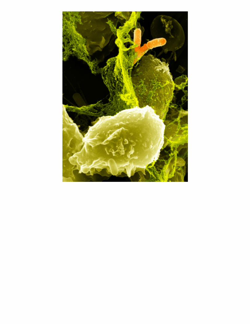
• Invasion (M, Map)A. induced phagocytosis type III secretion system IpaA, IpaB, IpaC, IpaD
membrane ruffling escape from the phagocytic vacuole, multiply spread within the epithelial cell cytoplasm, and passage to adjacent cells.
B. inducing programmed cell death (apoptosis) C. release of IL-1β, resulting in the attraction of polymorphonuclear leukocytes
into the infected tissues.• destabilizes the integrity of the intestinal wall and allows the bacteria to
reach the deeper epithelial cells.
8/12/2016 Prof WWM 7

Pathogenesis & Pathology of Dysentery
• First• Upon autolysis, • all shigellae release their toxic
lipopolysaccharide
• the irritation of the bowel wall
8/12/2016 Prof WWM 8

Pathogenesis & pathology of dysentery
• Second• Enterotoxins – • ShET1 and ShET2 (80% of shigellae ) • produce an early non-bloody, voluminous
diarrhea and
8/12/2016 Prof WWM 9

Pathogenesis & pathology of dysentery
• third• the invasion of the large intestine result in
• later dysentery with blood and pus in stools
8/12/2016 Prof WWM 1 0
Pathogenesis & pathology of dysentery
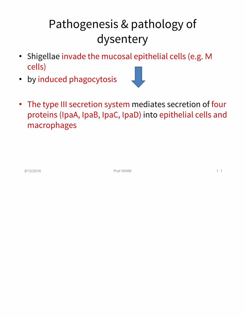
• Shigellae invade the mucosal epithelial cells (e.g. M cells)
• by induced phagocytosis
• The type III secretion system mediates secretion of four proteins (IpaA, IpaB, IpaC, IpaD) into epithelial cells andmacrophages
8/12/2016 Prof WWM 1 1
Pathogenesis & pathology of dysentery
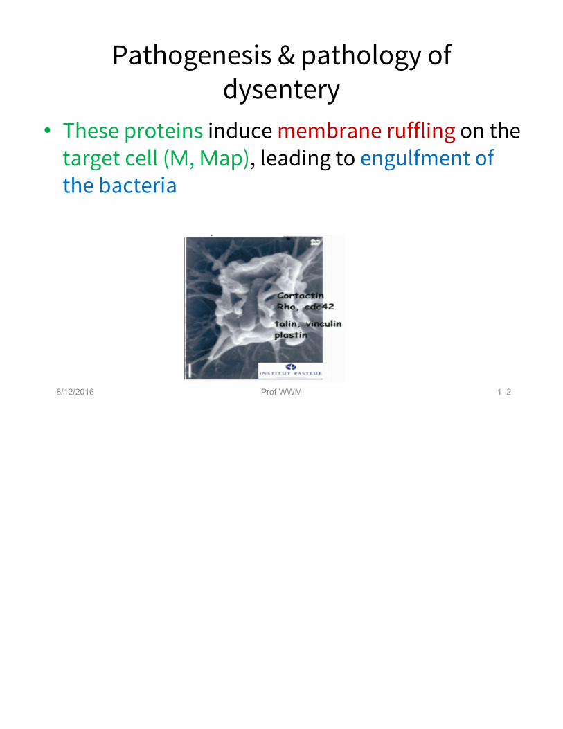
• These proteins induce membrane ruffling on thetarget cell (M, Map), leading to engulfment of the bacteria
8/12/2016 Prof WWM 1 2


Pathogenesis & pathology of dysentery
• Shigellae escape from the phagocytic vacuole,
• multiplication and
• spread within the epithelial cell cytoplasm and
• passage to adjacent cells
8/12/2016 Prof WWM 1 5

Pathogenesis & pathology of dysentery
• Shigellae survive phagocytosis by • inducing programmed cell death (apoptosis)
• This process also leads to the release of IL-1β,
• resulting in the attraction of polymorphonuclearleukocytes into the infected tissues
8/12/2016 Prof WWM 1 7

Pathogenesis & pathology of dysentery
• This in turn destabilizes the integrity of the intestinal wall and
• allows the bacteria to reach the deeper epithelial cells
8/12/2016 Prof WWM 1 8

Pathogenesis & pathology of dysentery
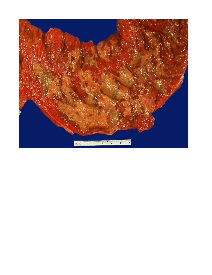
• Microabscesses in the wall of • the large intestine and terminal ileum
– lead to necrosis of the mucous membrane, – superficial ulceration, – bleeding and – formation of a "pseudomembrane" on the ulcerated
area in the wall of the large intestine and terminal ileum
8/12/2016 Prof WWM 1 9

Pathogenesis & pathology of haemorrhagic colitis
• S. dysenteriae strains produce an exotoxin, Shiga toxin
• has one A subunit and five B subunits
• E coli - VT
8/12/2016 Prof WWM 2 0

Pathogenesis & pathology of haemorrhagic colitis
• The B subunits
• bind to a host cell glycolipid (GB3) and
• facilitate transfer of the A subunit into the cell
8/12/2016 Prof WWM 2 1

Pathogenesis & pathology of haemorrhagic colitis
• The A subunit • cleaves the 28S rRNA in the 60S ribosomal subunit,
• thereby preventing the binding of aminoacyl-transfer RNA and
• disrupting protein synthesis
8/12/2016 Prof WWM 2 2

Pathogenesis & pathology of haemorrhagic colitis
• The primary manifestation of toxin activity is damage to the intestinal epithelium
• (Haemorrhagic colitis)
8/12/2016 Prof WWM 2 3

Pathogenesis & pathology of haemolytic uraemic syndrome (HUS)
• However, in a small subset of patients, the Shiga
• toxin can mediate damage to the glomerular endothelial cells,
• resulting in renal failure (HUS)
8/12/2016 Prof WWM 2 4

Pathogenesis & pathology of haemolytic uraemic syndrome (HUS)
• During pathogenesis, the release of the inflammatory mediators tumour necrosis factor (TNF) and interleukin-I (IL-l)
• increase the number of Gb3 receptors on the surface of eukaryotic cells,
• increasing the binding of toxin to these cells
8/12/2016 Prof WWM 2 5
Pathogenesis & pathology of haemolytic uraemic syndrome (HUS)
• Shiga toxin has also been shown to have neurotoxic properties
8/12/2016 Prof WWM 2 6
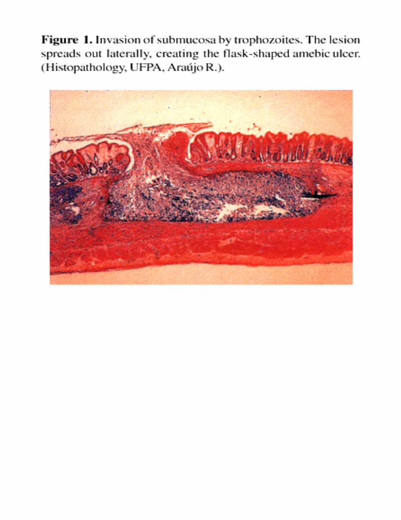

Pathology of Amebiasis

Clinical findings of dysentery
• Short incubation period (1-2 days)• A sudden onset of • abdominal pain, • fever, and • watery diarrhea
• A day or so later, as the infection involves the ileum and colon
8/12/2016 Prof WWM 3 0

Clinical findings of dysentery
• The number of stools increases (>10 times) • They are less liquid but often contain mucus and
blood
8/12/2016 Prof WWM 3 1

Clinical findings of dysentery
• Each bowel movement is accompanied by
• straining and tenesmus (rectal spasms),
• with resulting lower abdominal pain• In more than half of adult cases, • fever and diarrhea subside spontaneously in 2-5
days8/12/2016 Prof WWM 3 2

Clinical findings of dysentery
• However, in children and the elderly,
• loss of water and electrolytes may lead to
• dehydration, acidosis and even death
8/12/2016 Prof WWM 3 3

Clinical findings of dysentery
• The illness due to S. dysenteriae may be particularly severe
• On recovery,• most persons shed dysentery bacilli for only a
short period, • but a few remain chronic intestinal carriers and
may have recurrent bouts of the disease
8/12/2016 Prof WWM 3 4
Clinical findings of dysentery
• Upon recovery from the infection, most persons
• develop circulating antibodies to shigellae,
• but these do not protect against re-infection
8/12/2016 Prof WWM 3 5

Clinical findings of haemorrhagic colitis
• Bloody diarrhoea
8/12/2016 Prof WWM 3 6

Clinical findings HUS
• The condition, with its triad of : - haemolytic anaemia- thrombocytopenia - acute renal failure
8/12/2016 Prof WWM 3 7

Pathogenesis• CO ; fecal-oral Route1 .Lipopolysaccharide - the irritation of the bowel wall. 2 .Enterotoxins - ShET1 and ShET2 (80% of shigellae ) - an early non-bloody,
voluminous diarrhea and the 3 .invasion of the large intestine -later dysentery with blood and pus in stools.
• Invasion (M, Map)A. induced phagocytosis type III secretion system IpaA, IpaB, IpaC, IpaD
membrane ruffling escape from the phagocytic vacuole, multiply spread within the epithelial cell cytoplasm, and passage to adjacent cells.
B. inducing programmed cell death (apoptosis) C. release of IL-1β, resulting in the attraction of polymorphonuclear leukocytes
into the infected tissues.• destabilizes the integrity of the intestinal wall and allows the bacteria to
reach the deeper epithelial cells.
8/12/2016 Prof WWM 3 8

M cellShigella
Macrophage
Actin tail

M cellShigella
Macrophage

Host cellShigella
actin tail

Host cell
Shigella
actin tail

• Microabscesses in the wall of the large intestine and terminal ileum
• lead to necrosis of the mucous membrane,
• superficial ulceration,
• bleeding, and
• formation of a "pseudomembrane" on the ulcerated area in the wall of the large intestine and terminal ileum.
8/12/2016 Prof WWM 4 4