Embed Size (px)
Citation preview

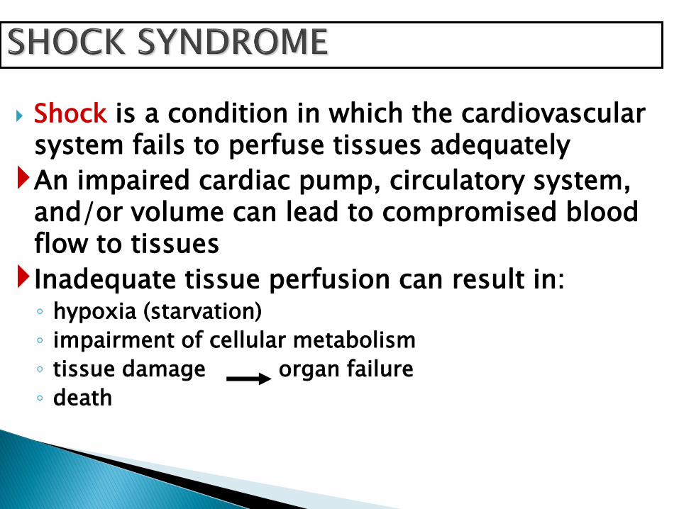
Shock is a condition in which the cardiovascular system fails to perfuse tissues adequately
An impaired cardiac pump, circulatory system, and/or volume can lead to compromised blood flow to tissues
Inadequate tissue perfusion can result in:◦ hypoxia (starvation)
◦ impairment of cellular metabolism
◦ tissue damage organ failure
◦ death

Impaired tissue perfusion occurs when an imbalance develops between cellular oxygen supply and cellular oxygen demand.
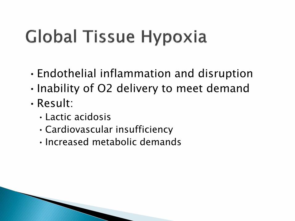
•Endothelial inflammation and disruption
• Inability of O2 delivery to meet demand
•Result: •Lactic acidosis
•Cardiovascular insufficiency
• Increased metabolic demands

Cells switch from aerobic to anaerobic metabolism
lactic acid production
Cell function ceases & swells
membrane becomes more permeable
electrolytes & fluids seep in & out of cell
Na+/K+ pump impaired
mitochondria damage
cell death

SNS - Neurohormonal response
Stimulated by baroreceptors
Increased heart rate
Increased contractility
Vasoconstriction (SVR-Afterload)
Increased Preload

SNS - Hormonal: Renin-angiotension system
Decrease renal perfusion
Releases renin angiotension I
angiotension II potent vasoconstriction &
releases aldosterone adrenal cortex
sodium & water retention
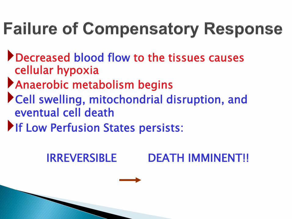
Decreased blood flow to the tissues causes cellular hypoxia
Anaerobic metabolism begins
Cell swelling, mitochondrial disruption, and eventual cell death
If Low Perfusion States persists:
IRREVERSIBLE DEATH IMMINENT!!

Initial stage - tissues are under perfused, decreased CO, increased anaerobic metabolism, lactic acid is building
Compensatory stage - Reversible. SNS activated by low CO, attempting to compensate for the decrease tissue perfusion.
Progressive stage - Failing compensatory mechanisms: profound vasoconstriction from the SNS ISCHEMIA Lactic acid production is high
metabolic acidosis Irreversible or refractory stage - Cellular necrosis and
Multiple Organ Dysfunction Syndrome may occur DEATH IS IMMINENT!!!!

Net results of cellular shock:
systemic lactic acidosis
decreased myocardial contractility
decreased vascular tone
decrease blood pressure, preload, and cardiac output
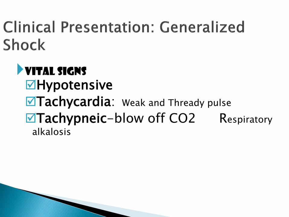
Vital signs
Hypotensive
Tachycardia: Weak and Thready pulse
Tachypneic-blow off CO2 Respiratory
alkalosis

Mental status: (LOC)
restless, irritable, apprehensive
unresponsive, painful stimuli onlyDecreased Urine output

•ABCDE•Airway
•control work of Breathing
•optimize Circulation
•assure adequate oxygen Delivery
•achieve End points of resuscitation

•Determine need for intubation but remember: intubation can worsen hypotension•Sedatives can lower blood pressure
•Positive pressure ventilation decreases preload
•May need volume resuscitation prior to intubation to avoid hemodynamic collapse
•Respiratory muscles consume a significant amount of oxygen
•Tachypnea can contribute to lactic acidosis
•Mechanical ventilation and sedation improves survival
• Isotonic crystalloids
•Titrated to:•CVP 8-12 mm Hg
•Urine output 0.5 ml/kg/hr (30 ml/hr)
• Improving heart rate
•May require 4-6 L of fluids
•No outcome benefit from colloids

•Decrease oxygen demands•Provide analgesia and anxiolytics to relax
muscles and avoid shivering
•Maintain arterial oxygen saturation/content•Give supplemental oxygen•Maintain Hemoglobin > 10 g/dL
•Serial lactate levels or central venous oxygen saturations to assess tissue oxygen extraction

• Inadequate volume resuscitation
•Pneumothorax
•Cardiac tamponade
•Hidden bleeding
•Adrenal insufficiency
•Medication allergy

•Progression of physiologic effects as shock ensues•Cardiac depression
•Respiratory distress
•Renal failure
•DIC
•Result is end organ failure
Hypovolemic Shock◦blood VOLUME problem
Cardiogenic Shock◦blood PUMP problem
Distributive Shock [septic;anaphylactic;neurogenic]
◦blood VESSEL problem

Loss of circulating volume “Empty tank ”
decrease tissue perfusion general shock response
ETIOLOGY: ◦ Internal or External fluid loss
◦ Intracellular and extracellular compartments
Most common causes:Hemmorhage
Dehydration
Fluid loss: Dehydration◦ Nausea & vomiting, diarrhea, massive diuresis, extensive burns,
Bowel obstruction, pancreatitis
Blood loss: ◦ trauma: blunt and penetrating◦ BLOOD YOU SEE◦ BLOOD YOU DON’T SEE
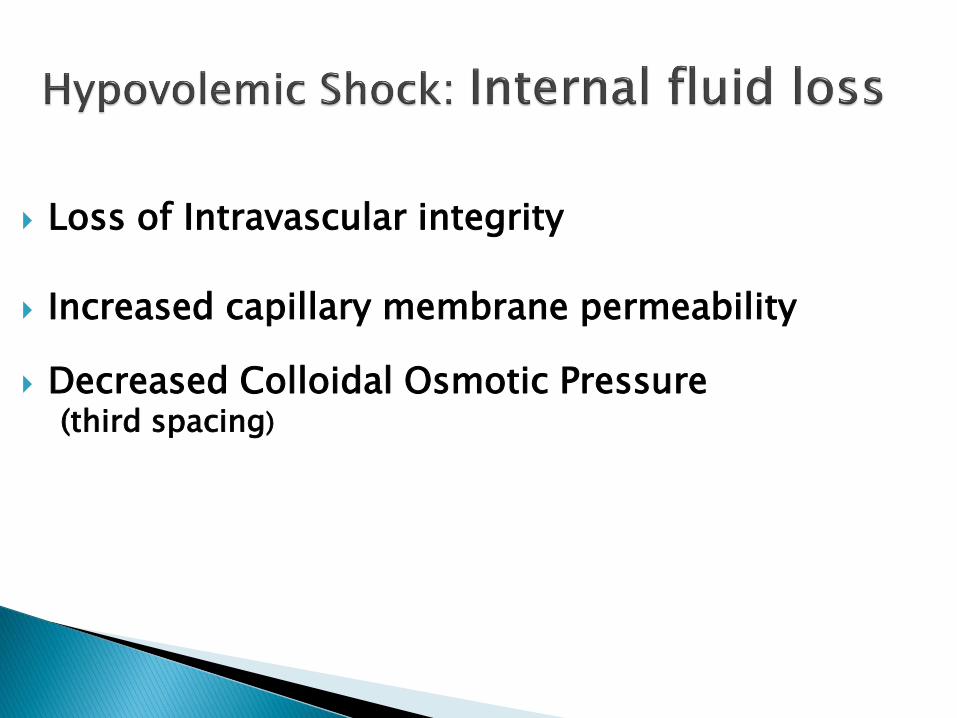
Loss of Intravascular integrity
Increased capillary membrane permeability
Decreased Colloidal Osmotic Pressure(third spacing)
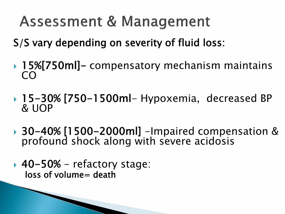
S/S vary depending on severity of fluid loss:
15%[750ml]- compensatory mechanism maintains CO
15-30% [750-1500ml- Hypoxemia, decreased BP & UOP
30-40% [1500-2000ml] -Impaired compensation & profound shock along with severe acidosis
40-50% - refactory stage:loss of volume= death
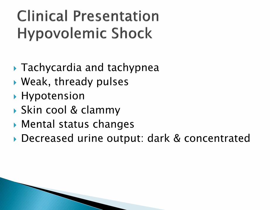
Tachycardia and tachypnea
Weak, thready pulses
Hypotension
Skin cool & clammy
Mental status changes
Decreased urine output: dark & concentrated
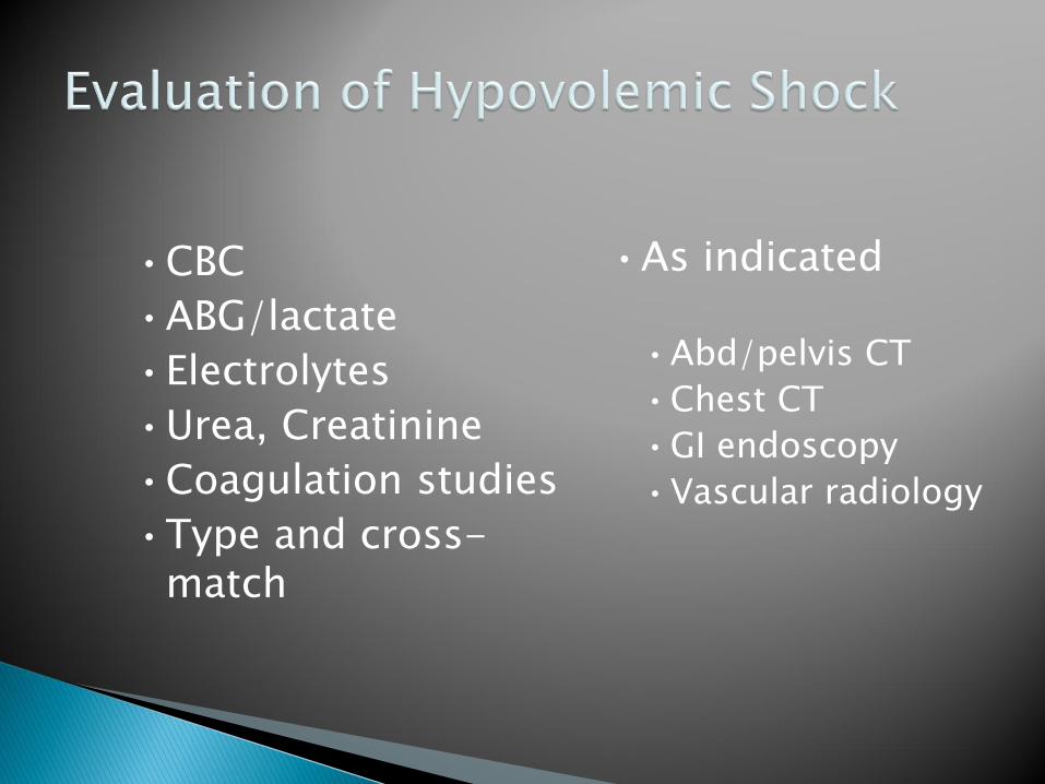
•CBC
•ABG/lactate
•Electrolytes
•Urea, Creatinine
•Coagulation studies
•Type and cross-match
•As indicated
•Abd/pelvis CT
•Chest CT
•GI endoscopy
•Vascular radiology

Management goal: ABCs Establish 2 large bore IVs or a central line Crystalloids
•Normal Saline or Lactate Ringers•Up to 3 liters
PRBCs•O negative or cross matched
Control any bleeding Arrange definitive treatment
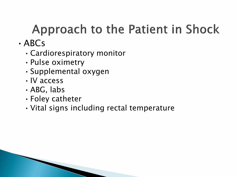
•ABCs•Cardiorespiratory monitor•Pulse oximetry•Supplemental oxygen • IV access•ABG, labs•Foley catheter•Vital signs including rectal temperature

The impaired ability of the heart to pump blood
Pump failure of the right or left ventricle
Most common cause is LV MI (Anterior)
Occurs when > 40% of ventricular mass damage
Mortality rate of 80 % or >

•Signs:•Cool, mottled skin
•Tachypnea
•Hypotension
•Altered mental status
•Narrowed pulse pressure
•Rales, murmur
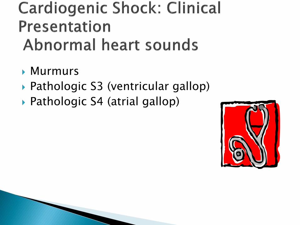
Murmurs
Pathologic S3 (ventricular gallop)
Pathologic S4 (atrial gallop)

Pericardial tamponade◦ muffled heart tones, elevated neck veins
Tension pneumothorax◦ tracheal deviation, decreased or absent
unilateral breath sounds, and chest hyperresonance on affected side
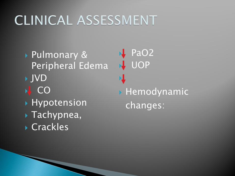
Pulmonary & Peripheral Edema
JVD
CO
Hypotension
Tachypnea,
Crackles
PaO2
UOP
Hemodynamic
changes:
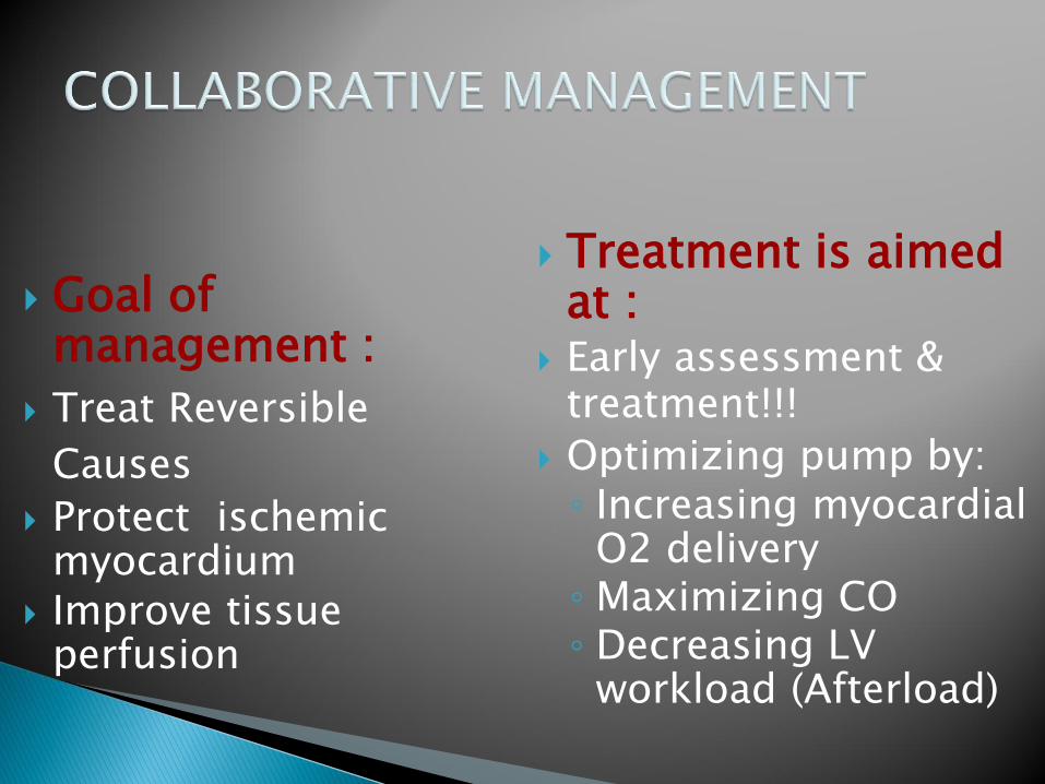
Goal of management :
Treat Reversible
Causes
Protect ischemic myocardium
Improve tissue perfusion
Treatment is aimed at :
Early assessment & treatment!!!
Optimizing pump by:◦ Increasing myocardial
O2 delivery◦ Maximizing CO◦ Decreasing LV
workload (Afterload)
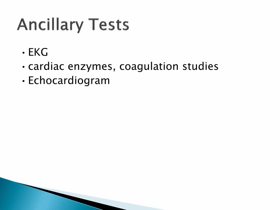
•EKG
•cardiac enzymes, coagulation studies
•Echocardiogram

•Goals- Airway stability and improving myocardial pump function
•Cardiac monitor, pulse oximetry
•Supplemental oxygen, IV access
•Intubation will decrease preload and result in hypotension •Be prepared to give fluid bolus

• AMI• Aspirin, beta blocker, morphine, heparin• If no pulmonary edema, IV fluid challenge• If pulmonary edema
• Dopamine – will ↑ HR and thus cardiac work• Dobutamine – May drop blood pressure• Combination therapy may be more effective
• thrombolytics, , angioplasty in specific cases
• Acute mitral regurgitation or VSD • Pressors (Dobutamine and Nitroprusside)
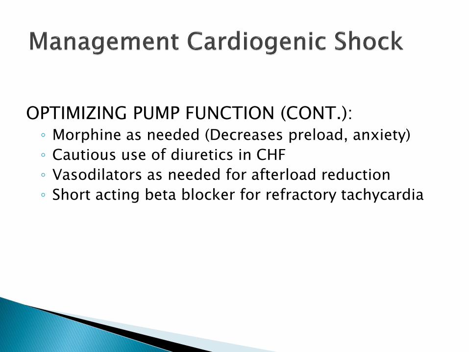
OPTIMIZING PUMP FUNCTION (CONT.):◦ Morphine as needed (Decreases preload, anxiety)
◦ Cautious use of diuretics in CHF
◦ Vasodilators as needed for afterload reduction
◦ Short acting beta blocker for refractory tachycardia
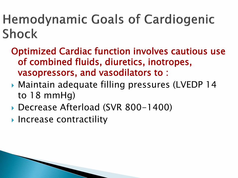
Optimized Cardiac function involves cautious use of combined fluids, diuretics, inotropes, vasopressors, and vasodilators to :
Maintain adequate filling pressures (LVEDP 14 to 18 mmHg)
Decrease Afterload (SVR 800-1400)
Increase contractility

Inadequate perfusion of tissues through maldistribution of blood flow
Intravascular volume is maldistributed because of alterations in blood vessels
Cardiac pump & blood volume are normal but blood is not reaching the tissues
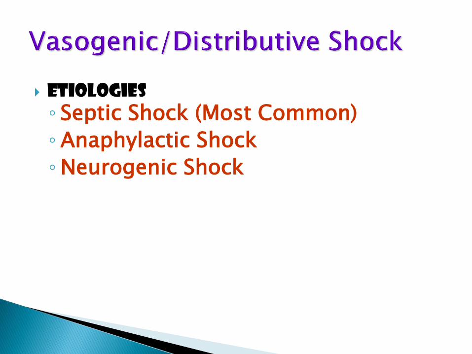
Etiologies
◦ Septic Shock (Most Common)
◦ Anaphylactic Shock
◦Neurogenic Shock
A type of distributive shock that results from widespread systemic allergic reaction to an antigen
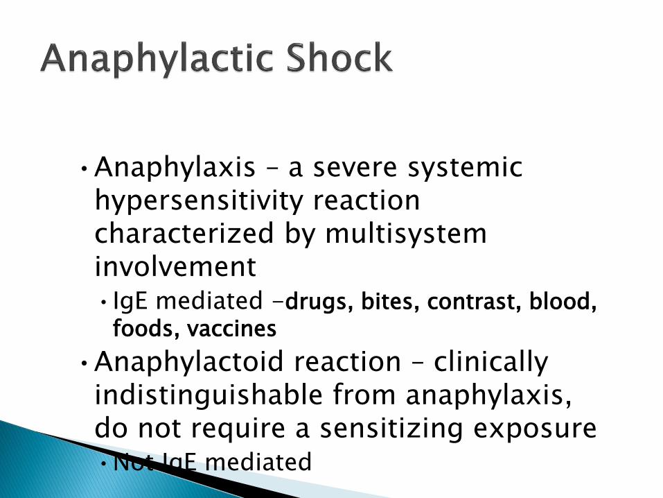
•Anaphylaxis – a severe systemic hypersensitivity reaction characterized by multisystem involvement •IgE mediated -drugs, bites, contrast, blood,
foods, vaccines
•Anaphylactoid reaction – clinically indistinguishable from anaphylaxis, do not require a sensitizing exposure•Not IgE mediated

Antigen exposure
body stimulated to produce IgE antibodies specific to antigen◦ drugs, bites, contrast, blood, foods, vaccines
Reexposure to antigen◦ IgE binds to mast cells and basophils
Anaphylactic response

Vasodilatation
Increased vascular permeability
Bronchoconstriction
Increased mucus production
Increased inflammatory mediators recruitment to sites of antigen interaction

• Mild, localized urticaria can progress to full anaphylaxis
• Symptoms usually begin within 60 minutes of exposure
• Faster the onset of symptoms = more severe reaction
• Biphasic phenomenon occurs in up to 20% of patients• Symptoms return 3-4 hours after initial reaction has
cleared

Almost immediate response to inciting antigen
Cutaneous manifestations◦ urticaria, erythema, pruritis, angioedema
Respiratory compromise◦ stridor, wheezing, bronchorrhea, resp. distress
Circulatory collapse◦ tachycardia, vasodilation, hypotension

•ABC’s•Angioedema and respiratory compromise require
immediate intubation
• IV, cardiac monitor, pulse oximetry• IVFs, oxygen•Epinephrine•Second line•Drugs

• Corticosteroids• Methylprednisolone 125 mg IV • Prednisone 60 mg PO
• Antihistamines• H1 blocker- Diphenhydramine 25-50 mg IV• H2 blocker- Ranitidine 50 mg IV
• Bronchodilators• Albuterol nebulizer• Atrovent nebulizer• Magnesium sulfate 2 g IV over 20 minutes
• Glucagon• For patients taking beta blockers and with refractory hypotension• 1 mg IV q5 minutes until hypotension resolves
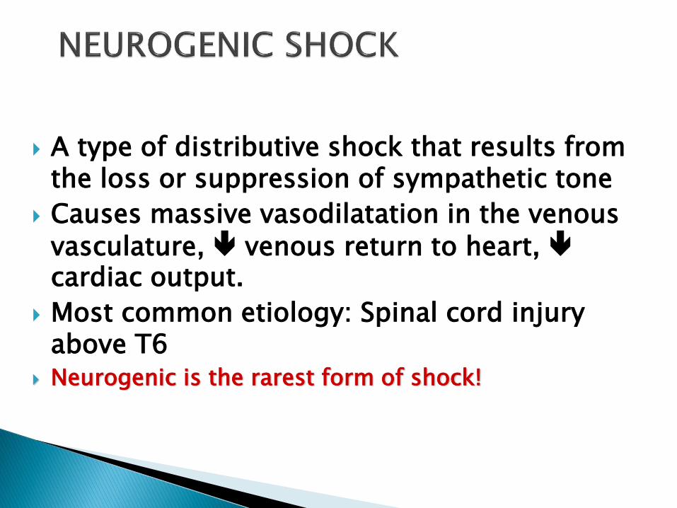
A type of distributive shock that results from the loss or suppression of sympathetic tone
Causes massive vasodilatation in the venous
vasculature, venous return to heart, cardiac output.
Most common etiology: Spinal cord injury above T6
Neurogenic is the rarest form of shock!

Distruption of sympathetic nervous system
Loss of sympathetic tone
Venous and arterial vasodilation
Decreased venous return
Decreased stroke volume
Decreased cardiac output
Decreased cellular oxygen supply
Impaired tissue perfusion
Impaired cellular metabolism

PATIENT ASSESSMENT Hypotension Bradycardia Hypothermia Warm, dry skin
CO Flaccid paralysis
below level of the spinal lesion
MEDICAL MANAGEMENT
Goals of Therapy are to treat or remove the cause & prevent cardiovascular instability, & promote optimal tissue perfusion
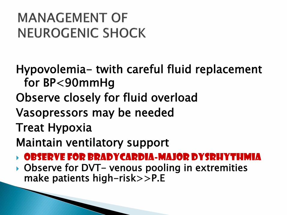
Hypovolemia- twith careful fluid replacement for BP<90mmHg
Observe closely for fluid overload
Vasopressors may be needed
Treat Hypoxia
Maintain ventilatory support
Observe for Bradycardia-major dysrhythmia Observe for DVT- venous pooling in extremities
make patients high-risk>>P.E

◦ Alpha agonist to augment tone if perfusion still inadequate
dopamine at alpha doses (> 10 mcg/kg per min)
ephedrine (12.5-25 mg IV every 3-4 hour)
◦ Treat bradycardia with atropine 0.5-1 mg doses to maximum 3 mg
may need transcutaneous or transvenous pacing temporarily
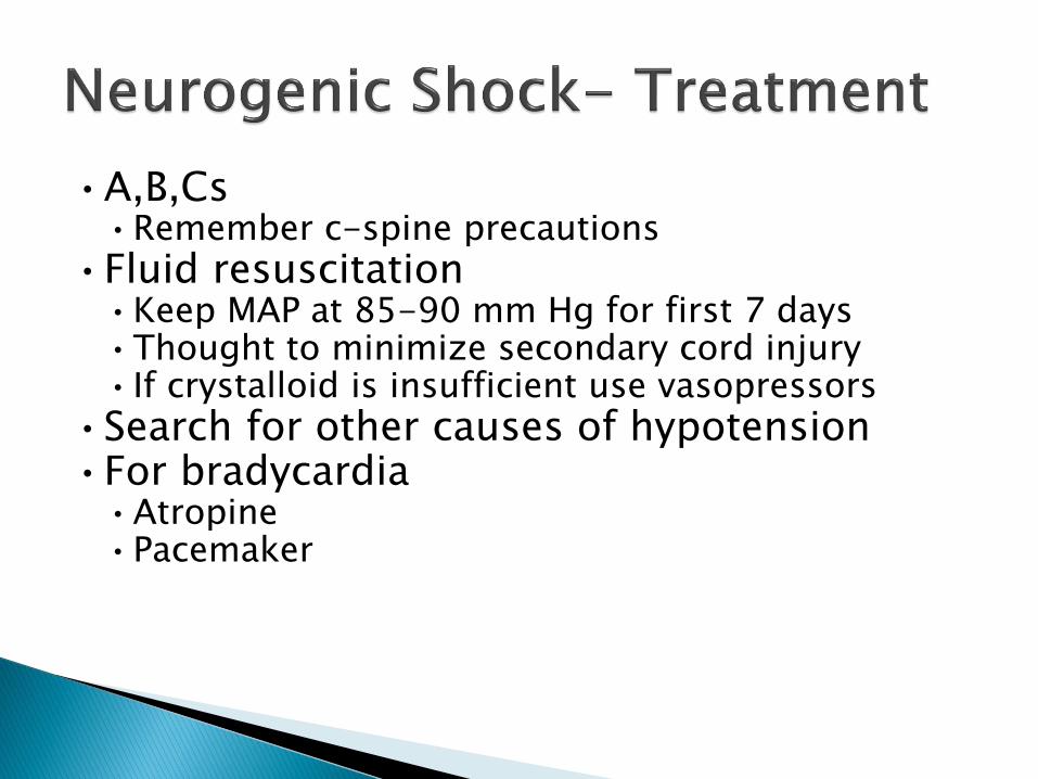
•A,B,Cs•Remember c-spine precautions
•Fluid resuscitation•Keep MAP at 85-90 mm Hg for first 7 days•Thought to minimize secondary cord injury• If crystalloid is insufficient use vasopressors
•Search for other causes of hypotension•For bradycardia
•Atropine•Pacemaker

•Methylprednisolone•Used only for blunt spinal cord injury
•High dose therapy for 23 hours
•Must be started within 8 hours
•Controversial- Risk for infection, GI bleed

Systemic Inflammatory Response (SIRS) to INFECTION manifested by two or > of following:◦ Temp > 38 or < 36 centigrade
◦ HR > 90
◦ RR > 20 or PaCO2 < 32
◦ WBC > 12,000

Age
Malnutrition
General debilitation
Use of invasive catheters
Traumatic wounds
Drug Therapy

Initiated by gram-negative (most common) or gram positive bacteria, fungi, or viruses
Cell walls of organisms contain Endotoxins
Endotoxins release inflammatory mediators (systemic inflammatory response) causes…...
Vasodilation & increase capillary permeability leads to
Shock due to alteration in peripheral circulation & massive dilation
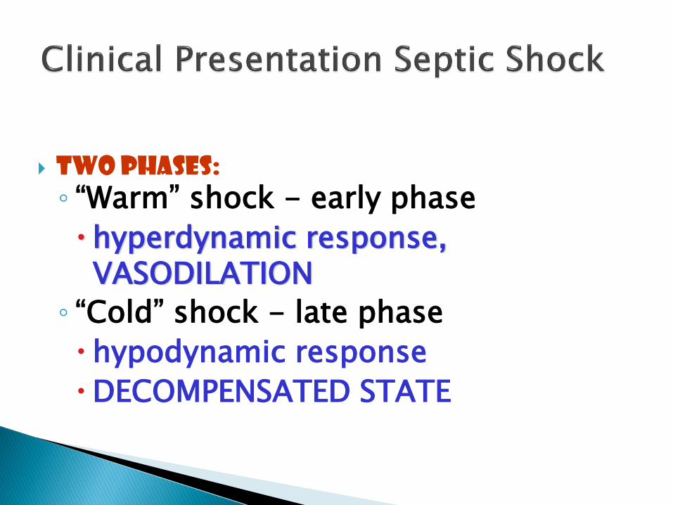
Two phases:
◦ “Warm” shock - early phase
hyperdynamic response, VASODILATION
◦ “Cold” shock - late phase
hypodynamic response
DECOMPENSATED STATE

EARLY---HYPERDYNAMIC STATE---COMPENSATION
◦ Massive vasodilation
◦ Pink, warm, flushed skin
◦ Increased Heart RateFull bounding
pulse
◦ Tachypnea

L ATE--HYPODYNAMIC STATE--DECOMPENSATION
◦ Vasoconstriction
◦ Skin is pale & cool
◦ Significant tachycardia
◦ Decreased BP
◦ Increase Systematic vascular resistance
◦ Decreased CO
◦ Decreased UOP
◦ Metabolic & respiratory acidosis with hypoxemia

Prevention !!! Find and kill the
source of the infection
Fluid Resuscitation Vasoconstrictors Inotropic drugs
Maximize O2 delivery Support
Nutritional Support
Comfort & Emotional support

The effects of the bacteria’s endotoxins can continue even after the bacteria is dead!!!

•2 large bore IVs•NS IVF bolus- 1-2 L wide open (if no
contraindications)
•Supplemental oxygen
•Empiric antibiotics, based on suspected source, as soon as possible

•Antibiotics- Survival correlates with how quickly the correct drug was given
•Cover gram positive and gram negative bacteria• ceftriaxone 1 gram IV or• Imipenem 1 gram IV
•Add additional coverage as indicated• Pseudomonas- Gentamicin or Cefepime• MRSA- Vancomycin • Intra-abdominal or head/neck anaerobic infections-
Clindamycin or Metronidazole • Asplenic- Ceftriaxone for N. meningitidis, H. infuenzae• Neutropenic – Cefepime or Imipenem


![MG [Režim kompatibility]) - upjs.sk](https://img.pdfslide.net/doc/110x75/6169ea8e11a7b741a34cc9cb/mg-reim-kompatibility-upjssk.jpg)


