Embed Size (px)
Citation preview

Shrink-Induced Superhydrophobic and AntibacterialSurfaces in Consumer PlasticsLauren R. Freschauf1, Jolie McLane1, Himanshu Sharma2, Michelle Khine1,2*
1 Department of Biomedical Engineering, University of California Irvine, Irvine, California, United States of America, 2 Department of Chemical Engineering and Materials
Science, University of California Irvine, Irvine, California, United States of America
Abstract
Structurally modified superhydrophobic surfaces have become particularly desirable as stable antibacterial surfaces.Because their self-cleaning and water resistant properties prohibit bacteria growth, structurally modified superhydrophobicsurfaces obviate bacterial resistance common with chemical agents, and therefore a robust and stable means to preventbacteria growth is possible. In this study, we present a rapid fabrication method for creating such superhydrophobicsurfaces in consumer hard plastic materials with resulting antibacterial effects. To replace complex fabrication materials andtechniques, the initial mold is made with commodity shrink-wrap film and is compatible with large plastic roll-to-rollmanufacturing and scale-up techniques. This method involves a purely structural modification free of chemical additivesleading to its inherent consistency over time and successive recasting from the same molds. Finally, antibacterial propertiesare demonstrated in polystyrene (PS), polycarbonate (PC), and polyethylene (PE) by demonstrating the prevention of gram-negative Escherichia coli (E. coli) bacteria growth on our structured plastic surfaces.
Citation: Freschauf LR, McLane J, Sharma H, Khine M (2012) Shrink-Induced Superhydrophobic and Antibacterial Surfaces in Consumer Plastics. PLoS ONE 7(8):e40987. doi:10.1371/journal.pone.0040987
Editor: Vipul Bansal, RMIT University, Australia
Received February 26, 2012; Accepted June 14, 2012; Published August 20, 2012
Copyright: � 2012 Freschauf et al. This is an open-access article distributed under the terms of the Creative Commons Attribution License, which permitsunrestricted use, distribution, and reproduction in any medium, provided the original author and source are credited.
Funding: This research was supported by the Undergraduate Research Opportunities Program (UROP) sponsored by the Division of Undergraduate Education atthe University of California, Irvine (442870-19900) and by the Integrative Graduate Education and Research Traineeship Program (IGERT) sponsored by theNational Science Foundation (NSF) (NSFIGERT DGE 0549479). Additional support is due to the Defense Advanced Research Projects Agency (DARPA) N/MEMS S&TFundamentals Program under grant no. N66001-1-4003 issued by the Space and Naval Warfare Systems Center Pacific (SPAWAR) to the Micro/nano FluidicsFundamentals Focus (MF3)Center. The funders had no role in study design, data collection and analysis, decision to publish, or preparation of the manuscript.
Competing Interests: The authors have declared that no competing interests exist.
* E-mail: [email protected]
Introduction
The spread of bacteria is a common problem and is the main
source of health associated infections. In 2009, such health
associated infections cost the healthcare industry $28–45 billion
and ranged from food poisoning to septicemia, often leading to
extensive hospital care and even death [1,2]. Bacterial exposure
can occur during surgical procedures or can be transferred patient-
to-patient from infected hospital surfaces [3]. Hospitals are a
major source of bacterial spread, but everyday facilities also act as
distributors of bacterial disease. Flores et al. has shown that public
restrooms house at least nineteen strains of bacteria, ranging from
skin, gut, and soil sources that can be transferred by touch [4].
Furthermore, multiple bacterial strands are capable of growing on
plastics and fabric surfaces for days and even months [4–7].
Therefore, there is a growing demand for reliable antibacterial
surfaces to combat this common occurrence of contamination.
Currently, there are fabrication methods for antibacterial
reagents and structurally modified antibacterial surfaces. Silver
nanoparticles have been used as a bacterial growth inhibitor as the
heavy metals disrupt and inactivate the proteins in bacteria,
preventing growth [8,9]. Functional groups on self-assembled gold
monolayers have also been used to decrease bacterial motility and
attachment, preventing cell adherence, growth of bacteria on
surfaces, and the formation of biofilms [10]. Zheng et al. has
shown that high molecular weights of chitosan inhibit gram-
positive bacteria such as Staphylococus aureus due to lack of nutrient
adsorption whereas low molecular weights of chitosan inhibit
gram-negative bacteria such as E. coli due to a disturbed
metabolism [11]. Chemically modified superhydrophobic surfaces
have also been shown to inhibit bacterial growth because of the
low surface energy and minimal contact with the surface for
bacterial adhesion [12]. While many antibacterial reagents and
chemicals effectively inhibit the growth of bacteria, they can lead
to bacterial resistance and become ineffective over time [13].
Purely structural antibacterial surfaces, however, do not induce
bacterial resistance and are therefore ideal for preventing the
spread of infectious bacteria. Superhydrophobic surfaces have
become particularly desirable as stable antibacterial surfaces
because of their self-cleaning and water resistant properties.
Such superhydrophobic surfaces in nature include the lotus leaf
[14], springtails (Collembola, Entognatha) [15], and termite wings
(Nasutitermes sp.) [16] which demonstrate properties such as self-
cleaning, bacterial resistance, and flight efficiency. Superhydro-
phobicity can be achieved artificially through structural [17,18] or
chemical [19,20] alterations to allow for free movement of water
across a surface due to water’s high contact angle (CA) and low
sliding angle (SA). Current fabrication techniques employ complex
production methods such as photolithography [21,22], chemical
vapor deposition [23], and self assembled monolayers [24] to
create highly organized structures. It is also possible for
heterogeneous micro and nanoscale structures to yield super-
hydrophobicity using gels, colloids, and oxides [12,17,18,25].
However, all of these methods pose a time consuming and costly
PLOS ONE | www.plosone.org 1 August 2012 | Volume 7 | Issue 8 | e40987
barrier to production. By simplifying the fabrication process and
enabling its scale up and its structural integration into existing
surfaces, the benefits of superhydrophobic surfaces can be readily
available to a range of materials for various biomedical (e.g.
implants, coatings) as well as consumer applications.
In this study, we create multi-scale structures ranging from the
nano to the micro-range by leveraging the buckling of metal
coated shrink film; these structures can be readily transferred into
any plastic using a rapid cast and mold method, resulting in
superhydrophobic surfaces in hard plastics for antibacterial
applications. Hard plastics such as PS, PC, and PE are commonly
used in commercial applications because they are nonreactive, are
biocompatible [26,27], and can be manufactured using inexpen-
sive techniques such as roll-to-roll manufacturing. Polydimethylsi-
loxane (PDMS), a widely used polymer for sealing, coating, and
molding [28], is used as a mold for casting because of its thermal
stability and the ability to imprint high aspect ratio and high
resolution features with good fidelity. The superhydrophobic
properties are achieved without chemical alteration. With the
initial substrate, we are able to produce multiple superhydropho-
bic PDMS casts for molding. Each of these superhydrophobic
PDMS substrates is capable of imprinting roughened features into
the aforementioned hard plastics, creating a substantial number of
superhydrophobic hard plastics from an initial mold. The final
superhydrophobic hard plastics utilize non-wetting properties to
induce antibacterial effects, which could be highly beneficial for
commercial application.
TheoryThe phenomenon of superhydrophobicity is explained in part
by a triad of equations centered upon the contact of water with the
surface. The surface tension created between water and a surface
can be calculated using Young’s equation [29] where the three
interfaces, solid-vapor (lSV), solid-liquid (lSL), and liquid-vapor
(lLV), describe the material’s resulting water CA (hY) during
thermodynamic equilibrium (1).
lSV {lSL{lLV cos hY ~0 ð1Þ
In particular, as the solid-liquid surface tension increases, the CA
increases due to less physical contact [30]. Further analysis of
wetting can be performed with Wenzel’s theory [31] where the
roughness factor (r), determined by a ratio of the geometric surface
to the apparent surface, is directly associated with the change in
CA (hW) of the roughened surface (2).
cos hW ~r cos hY ð2Þ
In more general terms, this equation explains the ability to
increase hydrophobicity on hydrophobic surfaces and increase
hydrophilicity on hydrophilic surfaces merely through roughening
the surface. However, another model was developed by Cassie and
Baxter [32] in which water can only contact the peaks of the
roughened surface versus wetting the entire surface in the Wenzel
model. This occurs due to the formation of air pockets between the
water and surface, decreasing the contact between the solid and
liquid phases. For multi-scale (nano to micro) roughness substrates
such as the lotus leaf, the Cassie-Baxter model better predicts the
equilibrium state [14]. Here, the CA on the roughened surface (hC)
is additionally described by the fraction of the droplet directly in
contact with the solid surface (W) (3).
cos hC~W cos hY zW{1 ð3Þ
The increase in solid-liquid surface tension is the primary key to
creating superhydrophobicity or the lotus effect.
Superhydrophobicity is achieved when the CA exceeds 150uand the SA is reduced to less than 10u. The high surface tension,
minimal surface contact, and ease of movement exhibited by water
on superhydrophobic surfaces can be attributed to the presence of
multiscale structures [33]. Cheng et al. demonstrated the
importance of these features on the lotus leaf by removing the
nanostructures which resulted in a decrease in water contact angle
[14]. Furthermore, surfaces must be inherently hydrophobic [31]
and have a low surface energy [34] to become superhydrophobic
when structurally modified.
Thus, leveraging these superhydrophobic surfaces for antibac-
terial applications is feasible. Due to the minimal solid-liquid
contact, the inherently low surface energy of the material, and low
SA of the substrate, bacteria prefer to remain in solution rather
than adhere to the surface [35,36]. When a droplet containing
bacteria contacts a superhydrophobic surface, there is minimal
contact where the bacteria can adhere to the surface. Additionally,
in this low contact area, there is low surface energy which allows
Figure 1. Process flow of the superhydrophobic substratesformed from shrink film paired with their respective CA. (i) POfilm is plasma treated with oxygen for 30 seconds (ii) Treated PO film issputter coated with 60 nm of silver and 60 nm of gold (iii) PO film isshrunk at 160uC to induce buckling and folding (iv) PDMS is pouredover fully shrunk PO film for casting (paired photo features flat PDMS)(v) Superhydrophobic PDMS cast is removed from shrunk PO (vi) Hardplastics are casted into superhydrophobic PDMS mold by applyingpressure and heat (paired photo features flat PC) (vii) Superhydropho-bic PC casted from superhydrophobic PDMS.doi:10.1371/journal.pone.0040987.g001
Superhydrophobic Antibacterial Plastics
PLOS ONE | www.plosone.org 2 August 2012 | Volume 7 | Issue 8 | e40987

Figure 2. Top down SEM images and AFM of the structurally modified surfaces’ multiscale structures were taken. Features are shownin (A) shrunk, bimetallic PO, (B) transferred in PDMS, and (C) imprinted in PS from PDMS. Scale bar is 10 mm for the large SEM images and 2 mm forthe insets. (D) AFM 3D image of the morphology and height profile.doi:10.1371/journal.pone.0040987.g002
Figure 3. Graphs depicting CA and SA for the structurally modified surfaces compared to flat. (A) Contact angle measurements ofstructurally modified and flat PDMS, PS, PC, and PE. (B) Sliding angle measurements of structurally modified and flat PDMS, PS, PC, and PE.+represents measurements .90u.doi:10.1371/journal.pone.0040987.g003
Superhydrophobic Antibacterial Plastics
PLOS ONE | www.plosone.org 3 August 2012 | Volume 7 | Issue 8 | e40987

only weak interactions between the surface and bacteria,
preventing bacterial adhesion [37]. Since the superhydrophobic
surface also has a low SA, bacteria in solution easily slide off the
surface when tilted and do not adhere to the surface. Privett et al.
even show that structural modification dominates over chemically
modified hydrophobic surfaces such as fluorination for antibacte-
rial properties [12]. With solely a structural modification, a
superhydrophobic surface will repel bacteria in solution rather
than kill them, negating the potential for resistance as would occur
due to chemical reagents.
Materials and Methods
Structurally Modified Superhydrophobic SurfacesBy utilizing a novel shrink method, superhydrophobic hard
plastics were created from shrink film, pre-stressed polyolefin (PO).
PO (Sealed Air) was first pretreated with oxygen plasma (SPI
Supplies) for 30 seconds to temporarily increase the surface energy
for better adhesion and was then sputter coated (Quorom) with
60 nm of silver and 60 nm of gold (Figure S1). After the bimetallic
coating, the PO film was heated to 160uC, causing the PO to fully
shrink. While the PO shrinks due to heating, the metallic films at
the surface buckle and fold, creating extremely rough, high-aspect,
and multiscale structures [38]. PDMS (Dow Corning Co.) is used
to cast these features into a thermally and mechanically stable
medium. These features are further transferred into the hard
plastics PS (Grafix Plastics), PC (McMaster-Carr), and PE
(McMaster-Carr). To produce structurally modified PS, pre-
stressed PS was heated to 135uC to fully shrink the polymer and
then casted to the superhydrophobic PDMS mold by applying
uniform pressure and heat at 150uC [39]. The PC and PE were
produced using the same casting technique at 150uC. Figure 1
depicts a brief process flow of this fabrication method paired with
CA images for each step.
The superhydrophobic properties of the structurally modified
substrates and the original flat substrates were characterized with
CA and SA measurements. A contact angle meter (Drop Shape
Analysis System DSA100, KRUSS) was used to measure the CA
of initial PDMS molds. Further CA measurements were taken with
a drop analysis program [40] on PS, PC, and PE. The SA
measurements were performed using a tool clamp with a 90urotational arm.
Antibacterial SurfacesAntibacterial testing was performed on equally sized PS, PC,
and PE samples for both flat and superhydrophobic substrates
using DH5-a gram-negative E. coli. E. coli was inoculated in 10 mL
of Luria Broth (LB) (Difco) overnight in an air bath shaker
(Environ Shaker) at 37uC and 300 rpm to reach the exponential
growth phase. The bacteria was then diluted 1,0006 or 10,0006in LB. Using the spread plate method, plating concentrations were
determined as 105 colony forming units (CFU)/mL for PS and PC
and 2.66104 CFU/mL for PE. For testing antibacterial proper-
ties, 10 mL of bacterial solution was placed on the surface of each
substrate. Substrates were tilted at 90u to allow bacterial solution
to roll off, if possible. Subsequently, samples were either rinsed
with 50 mL of sterile phosphate buffered saline (PBS) or not rinsed.
The surfaces of the substrates were then stamped face-down in
agar (Fisher Scientific) plates to transfer residual bacteria. 50 mL of
PBS was added to the agar dish to aid in spreading, and bacteria
was spread using a sterile glass loop and a turntable per the spread
plate method. 10 mL of bacterial solution was added directly to the
control agar plates along with 50 mL of sterile PBS for performing
the spread plate method. The agar plates were incubated for
24 hours at 37uC in a humidified incubator (VWR Scientific
Products). Images were taken after 24 hours, and CFU counts
were performed to compare bacterial growth. Agar was prepared
prior to experiments according to the manufacture’s protocol.
Results
Structurally Modified Superhydrophobic SurfacesThe heterogeneous nano and microstructures of the metal,
PDMS, and PS were analyzed using a scanning electron
microscope (SEM) (Hitachi S-4700-2 FES) shown in Figure 2(A–
C). The roughness from the shrunk, bimetallic PO mold is
translated directly into the PDMS and subsequently into the PS,
PC, and PE. Nanostructures can be seen on the surface of the
microstructures, leading to the enhanced hydrophobicity ex-
plained by the Cassie-Baxter theory [32]. Further visualization of
morphology and height was achieved using Atomic Force
Microscopy (AFM) (Asylum MPF3D), shown in Figure 2(D),
displaying a three dimensional view of the shrunk, bimetallic PO
Figure 4. The low SA of superhydrophobic PDMS allows thewater droplet to easily roll off the surface. (A–C) A droplet beingplaced on the surface of superhydrophobic PDMS retracts onto thedropper. (D–F) A droplet rolling off the same surface immediately afterplacement at a 5u angle.doi:10.1371/journal.pone.0040987.g004
Table 1. Calculated values of the solid fraction (W) werefound using the average flat CA (hY) and the averagestructurally modified CA (hC).
Material hC (6) hY (6) W
PDMS 152 108 .17
PS 145 70 .14
PC 151 95 .14
PE 155 87 .09
A low value of W represents minimal water contact with the surface.doi:10.1371/journal.pone.0040987.t001
Superhydrophobic Antibacterial Plastics
PLOS ONE | www.plosone.org 4 August 2012 | Volume 7 | Issue 8 | e40987
mold with a heterogeneous microstructure height range of 2.8 mm
and a root mean square (RMS) value of 700 nm.
CAs averaged above 150u with a maximum of 167u measured
with the KRUSS system, and the average SA was below 5u with a
minimum of less than 2u in PDMS, as shown in Figure 3. PC and
PE yielded similarly high CAs and low SAs indicative of
superhydrophobicity. PS produced slightly lower CAs and higher
SAs but showed hydrophobic enhancement from its flat compar-
ison. The low SA of superhydrophobic PDMS is depicted in
Figure 4 and Video S1.
Over the course of three casts from the shrunk, bimetallic PO to
PDMS, the CA remained consistently above 150u (data not
shown). In addition, casting PS, PC, or PE from a single PDMS
mold has yielded superhydrophobic substrates for more than 30
casts. The thermal stability of the superhydrophobicity in PDMS
molds was also investigated and remained stable across a range of
heat exposure from 25–100uC. PDMS samples were placed on a
hotplate at 10uC intervals and allowed to acclimatize to the
indicated temperature over the course of 5 minutes with a 5 mL
water droplet until CA was taken (data not shown).
Calculation of the solid fraction (W) from the Cassie-Baxter
equation (3) can be calculated using the average flat CA (hY) and
the average structurally modified CA (hC) for each surface (4).
W~cos hCz1
cos hY z1ð4Þ
The solid fraction W is a ratio of the properties of the structured
surface to the flat surface. Since all structures are imprinted from
the same initial metal PO mold to the polymers, each polymer
would theoretically have the same solid fraction W. However, the
initial hY is different for each polymer due to intrinsic chemical
differences, causing variation in W between materials. Table 1
shows calculated values of W for our roughened substrates. The
low values are similar to the findings of Zhu et al. whose calculated
W was typically less than 0.1, indicating a highly structured surface
[41]. As apparent from equations 3 and 4, as W approaches 0, hC
approaches 180u.
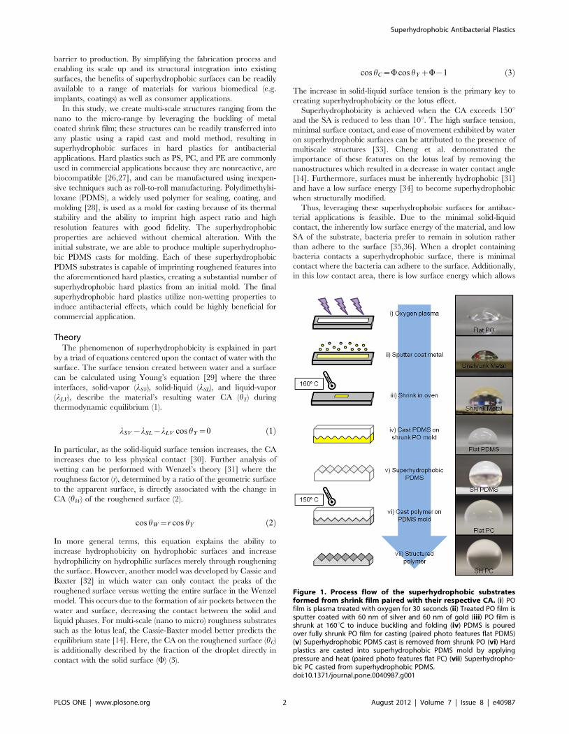
Antibacterial SurfacesSuperhydrophobic surfaces exhibit a significantly reduced
amount of bacterial growth over flat surfaces, as shown in
Figure 5. Control agar plates of PS and PC had 100,100 CFUs,
and PE had 25,800 CFUs for all conditions. Rinsed super-
hydrophobic surfaces yielded ,100 CFUs for PS and PE, and no
bacteria was observed on rinsed superhydrophobic PC (Table 2).
A small fraction (,.1%) of bacteria was retained from the initial
droplet on all rinsed superhydrophobic samples. The flat rinsed
Figure 5. PS, PC, and PE structured and flat substrates were contaminated with a bacteria solution and either rinsed or not rinsed.The resulting bacterial growth can be observed in each plate in the form of colonies following 24 hour incubation. (A) Substrates were rinsed with50 mL of PBS after bacteria solution was deposited on the surface. (B) Substrates were not rinsed.doi:10.1371/journal.pone.0040987.g005
Superhydrophobic Antibacterial Plastics
PLOS ONE | www.plosone.org 5 August 2012 | Volume 7 | Issue 8 | e40987
surfaces had much higher CFU counts where 10% of the initial
number of cells placed on the flat surfaces was transferred to the
agar plates even after rinsing. The no rinse superhydrophobic
surfaces were also effective at preventing bacterial adhesion with
only ,2% of the original number of cells plated in the final CFU
count. Not rinsed flat surfaces had ,34% of the original number
of bacteria plated. Note that all samples experienced a loss of
bacteria due to gravity during the tilting step of the experiment.
Discussion
With our cast and mold method, we induced superhydrophobic
properties on PDMS, PS, PC, and PE. The bimetallic layer
deposited on the preshrunk PO mold provided the initial necessary
mismatch in stiffness during the shrinking process to create highly
structured features after complete shrinking. When casted with
PDMS, the bimetallic PO mold transfers its physical shape,
producing multiscale roughening on the PDMS surface and
enhancing its natural hydrophobic properties. PDMS was used to
imprint these features in PS, PC, and PE to yield similar
heterogeneous rough structures and superhydrophobic properties.
The consistency of superhydrophobic properties is due in part to
the natural properties of PDMS, PS, PC, and PE as well as the
features transferred from the metal coated, shrunk PO into hard
plastics. With our cast and mold method, the surface of the
polymer becomes superhydrophobic due to the highly intercut
high aspect ratio structures passed on from mold to cast. PDMS
serves as the ideal medium to transfer these structures into the
hard plastics because of its pliability yet high thermal stability.
However, we found that higher levels of hydrophobicity were
achieved through structural modification of initially more hydro-
phobic polymers (PC and PE) versus initially less hydrophobic
polymers (PS). While roughening of the PS surface did increase
hydrophobicity, it did not achieve characteristic values to be truly
superhydrophobic because of its naturally less hydrophobic state
when flat. Nevertheless, antibacterial testing for the structurally
modified PS was favorable over the flat PS in both the rinse and no
rinse conditions but to a lesser degree than PC and PE. Thus, for
optimal hydrophobic and antibacterial surfaces, beginning with a
more hydrophobic polymer seems favorable.
Superhydrophobic surfaces are antibacterial because of their
minimal solid-liquid contact at the surface, weak surface interac-
tions with bacteria, and low SA. As a result of these properties, it is
energetically favorable for the bacteria to remain in solution and to
roll off the surface when tilted rather than adhere to the
superhydrophobic surface. This self-cleaning principle is the key
to antibacterial properties of superhydrophobic surfaces. Dirt and
bacteria adhere to water better than the surface and are, therefore,
cleansed easily by simple rinsing, mitigating the need for
antibacterial reagents. Since this antibacterial design is purely
structural, a product with permanent features can be manufac-
tured for everyday use with minimal maintenance for the
customer.
This fabrication method has the potential for further develop-
ment at a larger manufacturing scale and into additional materials.
The PO polymer used to create the initial mold, in addition to the
resulting molded hard plastics, are compatible with roll-to-roll
manufacturing methods. While we demonstrate the ability to
create superhydrophobic characteristics by transferring these
features into only three hard plastics, this method is applicable
to virtually any inherently hydrophobic plastic.
Conclusion
Here we have presented a novel method of producing a
superhydrophobic surface from PO by simply molding our unique
multi-scale features into PDMS and again into the hard plastics
PS, PC, and PE. This process is rapid, reproducible, and yields
antibacterial surfaces on these hard plastics. By eliminating the
need for chemical alterations to the surface, these superhydro-
phobic surfaces become much more robust due to the reliance
solely on physical geometry at the surface. In addition, using
PDMS as a means to transfer the superhydrophobic nano and
microscale structures presents the opportunity to produce a
substantial number of superhydrophobic hard plastics from a
single mold. Finally, this technique is compatible with roll-to-roll
manufacturing and scale-up production methods due to the use of
the polymers PO, PS, PC, and PE, making this process potentially
accessible for many different applications.
Supporting Information
Figure S1 Various thicknesses of metal depositionproduce different contact angles. To yield consistent super-
hydrophobicity, 60 nm of silver and 60 nm of gold was chosen as
the optimal metal thickness on the PO. CAs were taken on casted
PDMS.
(TIF)
Video S1 Water does not wet the superhydrophobicsurface. Water favors the dropper rather than the super-
hydrophobic surface. Next, the low sliding angle of the super-
hydrophobic surface allows the water droplet to roll off the surface
with ease. This video can also be viewed at http://shrink.eng.uci.
edu/research.html.
(WMV)
Table 2. CFU counts for structured versus flat surfaces.
Condition Substrate PS PC PEAdherence (Average of Experimental/Control)
Rinse Structured 70 0 30 ,0.1%
Rinse Flat 15,700 10,700 900 10%
No Rinse Structured 2,100 1,500 300 2%
No Rinse Flat .36,900* 30,700 8,900 .34%
Control Control 100,100 100,100 25,800 100%
*One agar plate yielded a condensed area of cell growth, hindering the ability to count individual colonies. Thus, this value is an underestimate.doi:10.1371/journal.pone.0040987.t002
Superhydrophobic Antibacterial Plastics
PLOS ONE | www.plosone.org 6 August 2012 | Volume 7 | Issue 8 | e40987
Acknowledgments
Special thanks to Anna Hoang of the Complex Fluids and Interfacial
Physics laboratory at the University of California, Los Angeles for her aid
in obtaining initial CA measurements and to Kamran Ali of University of
California, Irvine for providing the DH5a E. coli strain. Also thanks to Nick
Sharac and Dharmakeerthi Nawarathna at University of California, Irvine
for their specialized AFM technique.
Author Contributions
Conceived and designed the experiments: MK. Performed the experi-
ments: LRF JM. Analyzed the data: LRF JM MK. Contributed reagents/
materials/analysis tools: HS. Wrote the paper: LRF JM HS MK. Helped
with characterization: HS.
References
1. Scott RD (2008) The direct medical costs of healthcare-associated infections in
US hospitals and the benefits of prevention. Center for Disease Control.
Available at http://www.cdc.gov/ncidod/dhqp/pdf/Scott_CostPaper.pdf. Ac-
cessed 7/1/2009.
2. Gaidelyte A, Vaara M, Bamford DH (2006) Bacteria, Phages and Septicemia.
PloS ONE 2: 11.
3. Klevens RM, Edwards JR, Richards CL, Horan T, Gaynes R, et al. (2007)
Estimating healthcare-associated infections in U.S. hospitals, 2002. Public
Health Report 122: 160–166.
4. Flores GE, Bates ST, Knights D, Lauber CL, Stomaugh J, et al. (2011) Microbial
Biogeography of Public Restroom Surfaces. PLoS ONE 6: 11.
5. Getchell-White SI, Donowitz LG, Groschel DHM (1989) The Inanimate
Environment of an Intensive Care Unit as a Potential Source of Nosocomial
Bacteria: Evidence for Long Survival of Acinetabacter calcoaceticus. Infectious
Control and Hopsital Epidemiology 10: 402–407.
6. Brookes JS, Annand JW, Hammer A, Dembkowski K, Shulman S (2009)
Investigation of Bacterial Pathogens on 70 Frequently Used Environmental
Surfaces in a Large Urban U.S. University. Journal of Environmental Health 71:
17–21.
7. Srinkanth P, Rajaram E, Sudharsanam S, Lakshumanam A, Mariappan USS,
et al. (2010) Mobile phones: emerging threat for infection control. Journal of
Infection Prevention 11: 87–90.
8. Sondi I, Sondi BS (2004) Silver nanoparticles as antimicrobial agent: a case study
on E. coli as a model for Gram-negative bacteria. J of Colloid and Interface
Science 275: 177–182.
9. Cho KH, Park JE, Osaka T, Park SG (2005) The study of antimicrobial activity
and preservative effects of nanosilver ingredient. Electrochemica Acta 51: 956–
960.
10. Hou S, Burton EA, Simon KA, Blodgett D, Luk Y, et al. (2007) Inhibition ofEscherichia coli Biofilm Formation by Self-Assembled Monolayers of Functional
Alkanethiols on Gold. App And Environ Microbio 73: 4300–4307.
11. Zheng L, Zhu J (2003) Study on antimicrobial activity of chitosan with different
molecular weights. Elsevier 54: 527–530.
12. Privett BJ, Youn J, Hong SA, Lee J, Han J, et al. (2011) Antibacterial
Fluorinated Silica Colliod Superhydrophobic Surfaces. Langmuir 27: 9597–
9601.
13. Andersson DI, Hughes D (2010) Antibiotic resistance and its cost: is it possible to
reverse resistance? Nat Rev Microbio 8: 260–271.
14. Cheng YT, Rodak DE, Wong CA, Hayden CA (2006) Effects of micro-and
nano-structures of the self-cleaning behavior of lotus leaves. Nanotechnology 17:
1359–1362.
15. Helbig R, Nickerl J, Neinhuis C, Werner C (2011) Smart Skin Patterns Protect
Springtails. PLoS ONE 6: 9.
16. Watson GS, Cribb BW, Watson JA (2011) Contrasting Micro/Nano
Architecture on Termite Wings: Two Divergent Strategies for Optimising
Success of Colonisation Flights. PLoS ONE 6: 9.
17. Xu QF, Wang JN, Sanderson KD (2010) Organic-Inorganic CompositeNanocoatings with Superhydrophobicity, Good Transparency, and Thermal
Stability. ACS Nano 4: 2201–2209.
18. Ebril HY, Demirel AL, Avci Y, Mert O (2003) Transformation of a Simple
Plastic into a Superhydrophobic Surface. Science 299: 1377–1380.
19. Gomez GB, Flendrig LM, Cooper JM (2010) Hysteresis and Reversibility of a
Superhydrophobic Photopatternable Silicon Elsatomer. Langmuir 26: 7248–
7253.
20. Oner D, McCarthy TJ (2000) Ultrahydrophobic Surfaces. Effects of Topogra-phy Length Scales on Wettability. Langmuir 16: 3453–3456.
21. Jokinen V, Sainiemi L, Franssila S (2008) Complex Droplets on ChemicallyModified Silicon Nanograss. Adv Mater 20: 3453–3456.
22. Guo SS, Sun MH, Shi J, Liu YJ, Huang WH, et al. (2007) Patterning of
Hydrophilic Micro Arrays with Superhydrophobic Surrounding Zones. Elsevier84: 1673–1676.
23. Cortese B, D’Amone S, Manca M, Viola I, Cingolani R, et al. (2007)Superhydrophobicity Due to the Hierarchical Scale Roughness of PDMS
Surface. Langmuir 24: 2712–2718.
24. Genzer J, Efimenko K (2000) Creating Long-Lived Superhydrophobic PolymerSurfaces Through Mechanically Assembled Monolayers. Science 290: 2130–
2132.25. Wu J, Xia J, Lei W, Wang B (2010) Superhydrophobic Surface Based on a
Coral-Like Hierarchical Structure of ZnO. PLoS ONE 5: 12.26. Vaquette C, Fawzi-Grancher S, Lavalle P, Frochot C, Viriot ML (2006) In vitro
biocompatibility of different polyester membranes. Bio-Med Mat And Eng 16:
S131–S136.27. Zeus Industrial Products Inc. (2005) Biocompatibility of Plastics. Technical
WhitePaper.28. Xia Y, Whitesides GM (1998) Soft Lithography. Annu Rev Mater Sci 28: 153–
184.
29. Young T (1805) An Essay on the Cohesion of Fluids. Philos Trans R SocLondon 95: 65–87.
30. Temenoff JS, Mikos AG (2008) Biomaterials: The Intersection of Biology andMaterials Science. Pearson Prentice Hall 249–251 p.
31. Wenzel RN (1936) Resistance of Solid Surfaces to Wetting by Water. Ind EngChem 28: 988–994.
32. Cassie ABD, Baxter S (1944) Wettability of Porous Surfaces. Faraday Soc 40:
546–551.33. Whitney HM, Poetes R, Steiner U, Chittka L, Glover BJ (2011) Determining the
Contribution of Epidermal Cell Shape to Petal Wettability Using IsogenicAntirrhinum Lines. PLoS ONE 6: 3.
34. Ma M, Hill RM (2006) Superhydrophobic Surfaces. Elsevier 11: 193–202.
35. Hallab NJ, Bundy KJ, O’Connor K, Moses RL, Jacobs JJ (2001) Evaluation ofMetallic and Polymeric Biomaterial Surface Energy and Surface Roughness
Characteristics for Directed Cell Adhesion. Tissue Engineering 7: 55–71.36. Yang H, Deng Y (2008) Preparation and physical properties of super-
hydrophobic papers. J Colloid Inter Sci 325: 588–593.
37. Pringle JH, Fletcher M (1986) Influence of Substratum Hydration and AdsorbedMacromolecules on Bacterial Attachment to Surfaces. App And Environ
MicroBio 51: 1321–1325.38. Fu CC, Grimes A, Long M, Ferri CGL, Rich BD, et al. (2009) Tunable
Nanowrinkles on Shape Memory Polymer Sheets. Advanced Materials, 21: 1–5.39. Goral VN, Hsieh YC, Petzold ON, Faris RA, Yuen PK (2011) Hot embossing of
plastic microfluidic devices using poly(dimethylsiloxane) molds. J Micromech
Microeng 21: 017002.40. Stalder AF, Melchior T, Muller M, Sage D, Blu T, et al. (2010) Low-Bond
Axisymmetric Drop Shape Analysis for Surface Tension and Contact AngleMeasurements of Sessile Drops. Colloids and Surfaces A: Physicochemical and
Engineering Aspects 364: 72–81.
41. Zhu L, Xiu Y, Xu J, Tamirisa PA, Hess DW, et al. (2005) Superhydrophobicityon Two-Tier Rough Surfaces Fabricated by Controlled Growth of Aligned
Carbon Nanotube Arrays Coated with Fluorocarbon. Langmuir 21: 11208–11212.
Superhydrophobic Antibacterial Plastics
PLOS ONE | www.plosone.org 7 August 2012 | Volume 7 | Issue 8 | e40987