Embed Size (px)
Citation preview

The Plant Cell, S265–S276, Supplement 2002, www.plantcell.org © 2002 American Society of Plant Biologists
Signaling In and Out: Control of Cell Division and Differentiation in the Shoot and Root
Keiji Nakajima
a
and Philip N. Benfey
b,1
a
Graduate School of Biological Sciences, Nara Institute of Science and Technology, 8916-5 Takayama, Ikoma, Nara630-0101, Japan
b
Department of Biology, New York University, 1009 Main Building, 100 Washington Square East, New York, New York 10003
INTRODUCTION
To build a plant requires strict control of stem cell popula-tions as well as specification of appropriate cell fates oncecells enter a differentiation program. It is now accepted thatboth intracellular and intercellular signaling play importantroles in controlling cell division patterns and cell specifica-tion (Westhoff et al., 1998; Scheres, 2001). The molecularnature of this signaling, however, has long been elusive. Re-cent molecular and genetic studies have begun to reveal thesignaling mechanisms that regulate cell differentiation inboth shoot and root. In this review, we discuss recent ad-vances in the areas of stem cell control in both vegetativeand floral meristems as well as in pattern formation in roots.Our knowledge of the molecular mechanisms underlyingthese processes has been obtained primarily from Arabi-dopsis. Whether common mechanisms operate in otherspecies awaits experimental proof by the isolation and char-acterization of orthologous genes.
SIGNALING AT THE TOP END: MAINTENANCE OF THE SHOOT APICAL MERISTEM
The shoot apical meristem (SAM) is the source of cells for allaerial organs produced after germination. The SAM is firstformed at the globular stage of embryogenesis and devel-ops into a dome shape in the mature embryo. Upon germi-nation, the SAM starts a highly coordinated cell divisionprogram that continues throughout vegetative growth.Based on histological studies, the SAM of mature plants hasbeen subdivided into three domains: the central zone, theperipheral zone, and the rib zone (Figure 1A, left) (for re-views, see Steeves and Sussex, 1989; Howell, 1998). As its
name indicates, the central zone is a small, centrally locatedregion toward the top of the SAM. The central zone containsa population of stem cells that divide relatively slowly. Asthese cells divide, their peripheral daughters are displacedgradually from the central zone and enter the peripheral andrib zones. Cells in the peripheral zone and the rib zone arerich in cytoplasm and divide rapidly. Later, these cells arerecruited into the cell division programs of lateral organsand the stem, respectively.
Cells in the SAM also can be grouped according to theirclonal relationships: the epidermal, subepidermal, and un-derlying layers (Figure 1A, right) (Satina et al., 1940; re-viewed by Steeves and Sussex, 1989). Cells in theepidermal and subepidermal layers divide in a plane per-pendicular to the layers (anticlinal division). These cells areultimately incorporated into the epidermal and subepidermallayers of lateral organs (i.e., leaves and floral organs). Cellsin the underlying layer divide in a more complex manner,and their daughter cells differentiate into the inner tissues oflateral organs as well as into the pith in the stem. Despiteclear clonal distinctions between the three layers, their celldivisions are highly coordinated with each other, indicatingan intimate intercellular communication that allows pro-grammed development of organs with fixed shape and size.
Key Signaling Components
Maintaining a strict balance between the number of stemcells and the programmed differentiation of their progeny iscritical to SAM function. In Arabidopsis, a stem cell popula-tion persists even after the transition from vegetative to re-productive growth, allowing the inflorescence SAM toproduce an indeterminate number of flowers on its flanks.The maintenance of stem cells requires the WUSCHEL(WUS) homeodomain transcription factor. Loss-of-function
wus
mutants have defects in the SAM at all developmentalstages (Laux et al., 1996; Mayer et al., 1998). The
wus
vege-tative shoot apex is flat and lacks intensely stained cells
1
To whom correspondence should be addressed. E-mail [email protected]; fax 212-995-4204.Article, publication date, and citation information can be found atwww.plantcell.org/cgi/doi/10.1105/tpc.010471.

S266 The Plant Cell
typically found in the wild-type SAM (Figure 1B, left). Leafprimordia are formed slowly and fail to develop into matureleaves. At later stages, inflorescence stems emerge from theflank of the defective shoot apex as well as from leaf axils,giving a “wuschel” (tousled) appearance to the mutants.From these observations, it has been proposed that the pri-mary role of WUS is to confer stem cell fate (Laux et al.,
1996; Mayer et al., 1998). Expression analyses revealed that
WUS
mRNA is localized to a few cells underlying the stemcells (Figure 1B, right). Therefore, WUS controls stem cellfate in a non-cell-autonomous manner (Mayer et al., 1998).
shoot-meristemless
(
stm
) mutants exhibit defects in SAMformation/maintenance similar to those of
wus
(Barton andPoethig, 1993; Clark et al., 1996; Endrizzi et al., 1996).Plants with strong
stm
alleles fail to establish a SAM duringembryogenesis (Figure 1C, left). Leaf primordia are formedoccasionally between the fused cotyledon petioles, but theydo not develop into typical rosette leaves. The
STM
geneencodes a putative transcription factor with a homeoboxDNA binding domain (Long et al., 1996).
STM
is transcribedthroughout the SAM except in the regions corresponding toincipient leaf primordia (Figure 1C, right). All of these fea-tures indicate that the primary function of STM is either toinhibit cells from entering differentiation programs or to pro-mote cell proliferation in the center of the SAM (Endrizzi etal., 1996; Long et al., 1996).
Mutations in three
CLAVATA
loci (
CLV1
,
CLV2
, and
CLV3
)result in a phenotype opposite to those of
wus
and
stm
(Clark et al., 1993, 1995; Kayes and Clark, 1998). The SAMin the mutant embryo is slightly larger than that of wild-typeembryos. During postembryonic growth, organ initiation isretarded, whereas the SAM gradually increases in size byaccumulating undifferentiated cells (Figure 1D, left). Thesemutant phenotypes indicate a role for the
CLV
genes in re-stricting the size of the stem cell population. All three
CLV
genes have been identified, and their protein products likelyconstitute a single receptor–ligand complex (Figure 2), con-sistent with the three mutants having an almost identicalphenotype in the SAM (Clark et al., 1997; Fletcher et al.,1999; Jeong et al., 1999). The CLV1 protein is a receptor-like kinase composed of a Leu-rich repeat–containing extra-cellular domain with putative receptor function and a cyto-plasmic Ser kinase domain linked through a transmembranedomain (Clark et al., 1997). CLV2 is structurally similar toCLV1 but lacks a cytoplasmic kinase domain (Jeong et al.,1999).
CLV3
encodes a small polypeptide that contains aputative signal sequence for secretion. Otherwise, CLV3shows no apparent homology with other proteins withknown biochemical functions (Fletcher et al., 1999).
In both cauliflower and Arabidopsis, the CLV1 protein ispresent in two complexes of 185 and 450 kD (Trotochaud etal., 1999) (Figure 2). Convincing but indirect evidence indi-cates that the 185-kD complex is a disulfide-linked het-erodimer of CLV1 and CLV2 (Trotochaud et al., 1999). Thelarger 450-kD complex includes the 185-kD complex, theCLV3 peptide, and at least two other noncovalently boundsubunits: a kinase-associated protein phosphatase (KAPP)and a Rho GTPase–related protein (Rop) (Trotochaud et al.,1999, 2000). KAPP is likely to be a modulator of CLV1 ki-nase activity, whereas Rho may participate in the down-stream signal transduction pathway analogous to the animalmitogen-activated protein kinase cascade (Hirt, 1997;Williams et al., 1997; Stone et al., 1998; Trotochaud et al.,
Figure 1. Schemes Depicting Wild-Type and Mutant ArabidopsisSAM Structures and Expression Patterns of Key Regulatory Genes.
(A) Subdivision of SAM domains based on histological observations(left) and clonal relationships of cell layers (right).(B) SAM structure of loss-of-function wus mutants (left) and wild-typeWUS gene expression (right) (Laux et al., 1996; Mayer et al., 1998).(C) SAM structure of strong loss-of-function stm mutants (left) andwild-type STM gene expression (right) (Barton and Poethig, 1993;Endrizzi et al., 1996; Long et al., 1996). In strong stm mutants, theregion corresponding to the wild-type SAM is reduced to a smallnumber of cells between the cotyledon petioles. Leaf primordia arisein the center of this region.(D) SAM structure caused by a loss-of-function mutation in CLV1,CLV2, or CLV3 (left) and gene expression patterns of CLV1 andCLV3 (right) (Clark et al., 1993, 1995, 1997; Kayes and Clark, 1998;Fletcher et al., 1999).CP, cotyledon petiole; CZ, central zone; LA, leaf anlagen; LP, leafprimordium; L1, L2, and L3, epidermal, subepidermal, and underlyinglayers, respectively; PZ, peripheral zone; RZ, rib zone; WT, wild type.

Cell Division and Differentiation S267
1999). Assembly of the 450-kD complex requires the CLV3peptide, because CLV1 was found exclusively in the 185-kDcomplex in strong
clv3
mutants (Trotochaud et al., 1999).Furthermore, a CLV1/CLV2 receptor expressed on the yeastcell surface has been shown to bind CLV3 from a cauliflower
extract (Trotochaud et al., 2000). All of these observationsare consistent with a model in which the CLV3 peptidebinds to and activates the 185-kD CLV1/CLV2 heterodimerthrough autophosphorylation, which then becomes a 450-kDcomplex that includes KAPP and Rop (Figure 2) (Trotochaudet al., 1999). In cauliflower, 76% of CLV3 was found in the450-kD complex, whereas the remaining 24% was found ina 25-kD multimer (Trotochaud et al., 2000). It is not known,however, what CLV3 partner is in the 25-kD multimer orwhether CLV3 binds to the CLV1/CLV2 receptor as a multi-mer or as a monomer.
CLV1
and
CLV3
are expressed in distinct regions of theSAM (Figure 1D, right). Although
CLV3
mRNA accumulatesspecifically in the stem cells in the central zone,
CLV1
is ex-pressed in the center of the rib zone. The
CLV1
expressiondomain overlaps that of
CLV3
only slightly (Clark et al.,1997; Fletcher et al., 1999). This is consistent with the possi-ble extracellular secretion of the CLV3 peptide (Fletcher etal., 1999): CLV3 may be secreted from the stem cells andperceived by the
CLV1
-expressing cells just below the stemcells.
CLV2
is expressed in most organs and may have addi-tional roles in other signaling pathways (Kayes and Clark,1998; Jeong et al., 1999).
Stem Cell Maintenance
How do the key regulatory genes act in maintaining theSAM? Recent molecular genetic studies have revealed in-terdependence between the
WUS
and
CLV
pathways(Brand et al., 2000; Schoof et al., 2000) (Figure 3A). First, theCLV signal downregulates
WUS
expression. This is basedon the observations that (1) the size of the
WUS
expressiondomain is enlarged in
clv
mutant backgrounds (Schoof etal., 2000); and (2) ectopic expression of
CLV3
eliminates
WUS
-expressing cells, thereby causing a
wus
-like pheno-type (Brand et al., 2000). Second, because
CLV3
expressionis specific to the stem cells, whose maintenance requires
WUS
, the
CLV
pathway may be regulated indirectly by
WUS
(Laux et al., 1996; Mayer et al., 1998). This has been con-firmed by the ectopic expression of
WUS
. Transgenic plantsexpressing
WUS
under either the
CLV1
or
AINTEGUMENTA
(
ANT
) promoters accumulate undifferentiated cells that ex-press
CLV3
(Schoof et al., 2000).Based on this relationship, a mechanism involving a self-
regulatory loop has been proposed for stem cell mainte-nance (Figure 3A). If the number of stem cells is increased inthe central zone, more CLV3 peptide is produced, whichthen signals the
CLV1
-expressing cells in the rib zone todownregulate
WUS
expression. Fewer
WUS
-expressingcells would reduce the number of stem cells. Conversely, iftoo few stem cells are left in the SAM, signaling by CLV3would be attenuated, which would lead to more cells ex-pressing
WUS
and hence more stem cells (Schoof et al.,2000). In
ANT promoter
::
WUS
transgenic plants,
WUS
ex-pression was uncoupled from the self-regulatory loop,
Figure 2. Predicted Signal Transduction Mechanism by the CLVSignaling Complex.
CLV1 and CLV2 form a 185-kD heterodimer via thioester bonds onthe plasma membrane. Their N-terminal Leu-rich repeat (LRR) regionsare considered to be facing outside the cell to form a receptor domain,whereas the C-terminal kinase domain is located in the cytoplasm.Here, the LRR region of each CLV monomer is assumed to take acylindrical form, based on three-dimensional modeling of plant-spe-cific LRRs to a known LRR crystal structure (Kajava, 1998). Free CLV3ligand likely forms a multimer, although its molecular nature is notknown (a homodimer-like structure is assumed in this scheme). Uponbinding of the CLV3 ligand to the CLV1/CLV2 receptor, the CLV1kinase domain is phosphorylated (P), probably by other CLV1/CLV2complexes. The phosphorylated kinase domain then is recognized byseveral protein molecules, including a Rho GTPase-related protein(Rop) and a kinase associated protein phosphatase (KAPP), forming a450-kD complex. Rop is presumed to act via a mechanism analogousto the mitogen-activated protein kinase cascade. KAPP is anchoredto the inner surface of the plasma membrane through its uncleavedsignal peptide. KAPP negatively regulates the Rop-mediated signaltransduction pathway, probably through its phosphatase activity. Thescheme is drawn based on the publications by Stone et al. (1994,1998), Williams et al. (1997), and Trotochaud et al. (1999, 2000).

S268 The Plant Cell
resulting in a phenotype similar to that of loss-of-function
clv
mutants, despite ectopic
CLV3
expression.Both
WUS
and
CLV3
can signal across cell layers. For
CLV3
signaling, this probably is accomplished by apoplasticmovement of the CLV3 peptide (Fletcher et al., 1999). Themolecular mechanism for the non-cell-autonomous actionof
WUS
is not yet known. Likewise, it is not clear how
STM
acts in relation to the
WUS/CLV
regulatory pathway. Basedon its expression pattern in the mature SAM,
STM
appearsto reserve the region composed of undifferentiated cellsupon which
WUS
and
CLV
expression patterns are speci-fied. During embryogenesis, however,
WUS
expression canbe detected as early as the 16-cell-embryo stage, when nei-ther
STM
expression nor a visible SAM structure has beenestablished (Mayer et al., 1998). Furthermore, neither
WUS
nor
STM
expression in the embryo requires the function of
the other (Mayer et al., 1998). Double mutant analyses havedemonstrated that although either the
stm
or the
clv
pheno-type can be rescued partially by mutation of the other, the
wus
mutation enhances the defects caused by weak
stm
al-leles (Clark et al., 1996; Endrizzi et al., 1996). These obser-vations indicate that the
WUS/CLV
signaling pathway andSTM act at different levels but are not correlated in a simpleepistatic order. Moreover, their interdependence may changedepending on the developmental stage.
When Stem Cells Come to an End
The young flower primordium retains stem cells at its apex;therefore, it is called a floral meristem (FM). Both mutantphenotypes and gene expression patterns indicate that the
WUS/CLV
signaling pathway acts to maintain stem cells inthe FM.
wus
mutant flowers form normal numbers of sepalsand petals, but the central two whorls are replaced with asingle stamen (Laux et al., 1996). This defect results fromthe inability of
wus
to maintain a sufficient quantity of stemcells to form the correct numbers of stamens and carpels. Incontrast, the FM of
clv
mutants accumulates stem cells andgives rise to a flower with increased numbers of floral or-gans, especially carpels (Clark et al., 1993, 1995; Kayes andClark, 1998).
WUS
is expressed in a few cells in the centerof the FM, whereas
CLV1
and
CLV3
are expressed in thecenter and apex of the FM, respectively, similar to their ex-pression patterns in the SAM (Clark et al., 1997; Mayer etal., 1998; Fletcher et al., 1999).
In the wild-type FM, the ability of the stem cells to maintaina constant population size must end late in flower develop-ment, because the central part of the FM is programmed tobecome a determinate number of carpels. The
AGAMOUS
(
AG
) gene has been implicated in this process because, in ad-dition to having defects in floral organ specification,
ag
mutantflowers produce indeterminate numbers of floral organs(Bowman et al., 1989). This phenotype requires functionalWUS, because flowers of
ag wus
double mutants show de-fects similar to those of
wus
single mutants (Laux et al., 1996).Recent genetic analyses by two groups have demon-
strated a role for
AG
in the regulation of
WUS
(Lenhard etal., 2001; Lohmann et al., 2001). In the wild-type FM, ex-pression of both
WUS
and
CLV3
diminishes as flower devel-opment proceeds and then disappears completely by thetime carpel primordia initiate. In the FM of
ag
mutants, both
WUS
and
CLV
expression remain long after floral organ de-velopment is completed (Lenhard et al., 2001; Lohmann etal., 2001). The repression of
WUS
by
AG
appears to be in-dependent of
CLV signaling, because in the FM of ag clv1double mutants, WUS expression is not only prolonged butexpanded spatially (Lohmann et al., 2001). Conversely, theectopic expression of WUS in various regions of the FM re-sults in indeterminate organ formation in the correspondingfloral region (Lenhard et al., 2001; Lohmann et al., 2001). Allof these observations indicate a requirement for AG in the
Figure 3. Models of Stem Cell Regulation.
(A) Stem cell maintenance in the vegetative and inflorescence mer-istems (Brand et al., 2000; Schoof et al., 2000). The size of the stemcell population is controlled by a regulatory loop between WUS andCLV. When the number of stem cells is increased, more CLV3 ligandis released from the stem cells, which is perceived by the CLV1/CLV2 receptor kinase in underlying layers. This results in fewer cellsexpressing WUS, thereby attenuating stem cell–promoting activity(left). In contrast, when the number of stem cells is decreased, lessCLV3 ligand is released. Consequently, more cells start to expressWUS, thereby promoting stem cell identity (right).(B) Termination of the stem cell population in a FM (Lenhard et al.,2001; Lohmann et al., 2001). In early flower primordia (stage 2, left),the meristem identity gene LFY is expressed throughout the primordia(Weigel et al., 1992), whereas WUS is expressed in the center to re-serve a stem cell population for later developmental stages (Mayer etal., 1998). Later, in stage 3 (middle), LFY and WUS together activateAG expression in the center of the primordia. As flower developmentproceeds (stage 7, right), WUS expression becomes downregulatedby AG. AG expression persists in the center of the primordia and de-termines floral organ identities in whorls 3 and 4 (Drews et al., 1991).

Cell Division and Differentiation S269
downregulation of WUS and hence in the termination ofstem cell maintenance.
The involvement of AG in stem cell termination raises thequestion of how AG expression is induced in the FM at thecorrect time and place. There is strong evidence that AG ex-pression depends on WUS and on the floral meristem iden-tity gene LEAFY (LFY) (Lenhard et al., 2001; Lohmann et al.,2001). The ectopic expression of WUS results in the tran-scription of a �-glucuronidase (GUS) reporter controlled bythe AG cis regulatory region. This is consistent with the abil-ity of ectopic WUS to form an enlarged stem cell populationand an enhanced “C” function mediated by AG, as shownby an indeterminate number of stamens and carpels. BothGUS expression and the indeterminate organ formation arelargely absent when the same experiments are performed ina lfy mutant background. The interaction of WUS/LFY andAG was investigated further at the molecular level. Both invitro and in vivo studies demonstrated direct binding of LFYand WUS to the cis elements in the AG second intron(Busch et al., 1999; Lohmann et al., 2001). A quantitativeanalysis using a yeast expression system indicated thatWUS and LFY act synergistically to activate AG expression,even though cooperative binding of the two proteins wasnot observed in vitro (Lohmann et al., 2001).
Based on these observations, a signaling pathway forstem cell termination has been proposed (Lenhard et al.,2001; Lohmann et al., 2001) (Figure 3B). In young flower pri-mordia, the WUS/CLV signaling pathway maintains a stemcell population in the apex. WUS then activates AG tran-scription by binding directly to the AG promoter. The tran-scription of AG by WUS is enhanced synergistically by LFY,which is expressed in flower primordia but not in the SAM.Because WUS expression is limited to a small number ofcells in the FM, strong AG expression occurs only at thecenter of the flower primordium, where it specifies stamenand carpel identities. Later in flower development, AG down-regulates WUS expression, thereby terminating the stemcell population. Thus, WUS and AG constitute a negativefeedback loop in which WUS activates the transcription ofAG, which in turn represses WUS expression. This signalingloop is similar to the WUS/CLV regulatory loop in the SAM.An important difference is that the WUS/AG pathway doesnot involve cell-to-cell communication. One should note,however, that the WUS/AG pathway requires an existingprepattern, as does the WUS/CLV pathway. Furthermore, incontrast to the WUS/CLV pathway, which continues in theSAM throughout plant development, the WUS/AG pathwayappears to function only once in each FM.
SIGNALING UNDER THE GROUND: CELL DIVISION AND DIFFERENTIATION IN THE ROOT
Compared with the SAM, the root meristem (RM) has fewercells and a simpler structure. In Arabidopsis, the cell division
pattern in the RM has been well characterized, and a nearlycomplete fate map can be drawn for every cell type (Dolanet al., 1993). The mature part of the Arabidopsis root is com-posed of concentrically organized cell layers, which from in-side to outside form vasculature, pericycle, endodermis,cortex, and epidermis (Figure 4A). The vasculature and peri-cycle together constitute the stele. Cells in each layer havetheir origin in the “initial cells” located at the RM, which re-peat a highly stereotyped sequence of divisions. One of thetwo daughter cells remains as an initial cell, whereas theother enters an appropriate differentiation pathway. Some ofthe root cell files share the same clonal origin; for example,epidermis and lateral root cap originate from the same initialcells, and the two ground tissue layers, endodermis andcortex, also share the same initial cells (Figure 4A).
Intercellular Signaling in Root Development
The highly coordinated division of the initial cells suggests thepresence of extensive cell-to-cell communication. A laser-ablation study first gave solid evidence for the importance ofpositional signaling in root pattern formation (van den Berg etal., 1995). When a cortex/endodermis initial cell is ablated, anadjacent pericycle cell invades the ablated position and per-forms a periclinal division. The outer daughter cell then be-haves as a cortex/endodermis initial cell: it undergoes atransverse cell division. The upper daughter cell then dividespericlinally, giving rise to the first cells in the cortex and endo-dermis lineages. These pericycle-derived endodermal cellshave differentiated attributes of endodermis, as revealed bythe presence of a casparian strip. A similar invasion by neigh-boring cells, followed by a corresponding cell fate change,was observed when an epidermis/lateral root cap initial cellwas ablated (van den Berg et al., 1995).
Respecification of cell fate is not limited to mechanical abla-tion but seems to occur in nature. Kidner et al. (2000) investi-gated root cell lineage using genetic mosaics of GUS-positivecells that had been generated by heat shock–induced transpo-son excision from a CaMV35S::GUS transgene. Analysis of theroots after heat shock treatment revealed that the cells in thecortex/endodermis lineage invaded both the outer epidermisand inner stele layers. Surprisingly, those roots retained a nor-mal root radial pattern with no apparent increase or decrease inthe number of cell layers or in the number of cells in each layer.Although heat shock treatment may have enhanced the fre-quency of cell death, this observation demonstrated that theroot radial pattern is not perturbed even when cells in the mer-istem region are lost accidentally. Clearly, intercellular signalingis key to the self-maintenance capacity of the RM.
Stem Cell Maintenance in the RM
The root initial cells can be thought of as functionally equiv-alent to the CLV3-expressing stem cells of the SAM. The

S270 The Plant Cell
initial cells are arranged around a few mitotically inactivecells in the center of the RM that constitute a “quiescentcenter” (QC). The QC appears to play an important role inmaintaining RM activity. When a QC cell is laser ablated, theabutting initial cells lose their ability to perform as stem cellsand, instead, start to differentiate or divide in a mannercharacteristic of their daughter cells (van den Berg et al.,1997). Differentiation of initial cells also was observed whenlaser ablation was performed on a mutant that lackspostembryonic root cell divisions, showing that this functionof the QC is independent of cell divisions (van den Berg etal., 1997). These observations indicate that the QC main-tains the identity of the surrounding initial cells by inhibitingtheir differentiation (Figure 4B). A similar conclusion wasdrawn from a study in which root cell differentiation waspromoted genetically through modulation of the expressionlevel of CAK, a cyclin-dependent kinase–activating kinase(Umeda et al., 2000). The ability of the QC to inhibit initialcell divisions is similar to the function of the WUS-expressingcells in the SAM. An important difference, however, is that
WUS-expressing cells appear to be replaced continuously,whereas QC cells are maintained for a long period.
Although a few loci have been reported to affect the for-mation and activity of the RM (Willemsen et al., 1998;Vernoux et al., 2000; Frugier et al., 2001), no putative signal-ing components have been identified, nor are the root counter-parts for WUS and CLV documented. This implies that celldivision and the differentiation of shoot and root are con-trolled by different mechanisms, although both include inter-cellular signaling to maintain the stem cell population.Alternatively, root stem cells may be maintained by redun-dant pathways that to date have avoided identificationthrough genetic analyses.
Auxin Signaling and Root Distal Patterning
The plant hormone auxin influences cell division and differ-entiation as well as cell elongation. Because of its highlypleiotropic effects, however, it has been difficult to deter-
Figure 4. Schemes of Wild-Type and Mutant Arabidopsis Root Structures.
(A) Wild-type root (Dolan et al., 1993). Cell types are given in the key at bottom. Abbreviations shown in parentheses are used in all subsequent figures.(B) QC cells function as an organizing center of the RM by inhibiting the differentiation of surrounding initial cells (stem cells) (van den Berg et al.,1997; Umeda et al., 2000).(C) Defective root radial pattern of three Arabidopsis mutants, scr, shr, and wol (Benfey et al., 1993; Di Laurenzio et al., 1996; Helariutta et al.,2000; Mähönen et al., 2000).

Cell Division and Differentiation S271
mine the role of auxin in each developmental program bygenetic approaches. Recently, a physiological study linkedthe distribution of auxin to patterning of the root apical re-gion (Sabatini et al., 1999). Using a GUS reporter gene fusedto a synthetic auxin-responsive promoter (DR5::GUS), amaximum of auxin concentration was localized to the col-umella initial cells. This auxin distribution was either lost ordisturbed in mutants of known auxin signal transducers andtransporters. In mutants of the putative auxin efflux carrierPIN-FORMED1, abnormal cell division and elongation werefound in the tissue in which the auxin concentration was in-creased ectopically.
A more dramatic change in root pattern was inducedwhen wild-type plants were grown in the presence of auxintransport inhibitors. The auxin concentration maximum wasexpanded to include the cortex and apical epidermis to-gether with the original maximum at the columella initials.The change in auxin distribution was accompanied by re-specification of cell fates and modification of cell divisionprograms: the cells in the positions of endodermis, cortex,and epidermis were respecified as QC, columella initial, andlateral root cap, respectively. The new columella initials di-vided, and their daughter cells contained amyloplasts, afunctional marker of the columella. Cell fate specificationwas not dependent on the absolute concentration of auxinbut was determined by the position of the auxin concentra-tion maximum relative to the vascular tissue (Sabatini et al.,1999). Similarly, misspecification of epidermis cells as lateralroot cap cells has been reported for tornado1 (trn1) and trn2mutants (Cnops et al., 2000). Although the mutated gene hasyet to be identified, TRN1 is likely to be involved in the auxin-dependent cell fate determination pathway, because polarauxin transport was impaired in a trn1 allele (lop1) (Carlandand McHale, 1996). These observations strongly suggest arole for auxin distribution in root distal patterning.
Radial Signaling in Ground Tissue Patterning
The radial pattern of cell layers seen in the mature root isdetermined initially during early embryogenesis. Upon ger-mination, the initial cells in the RM are activated and start astereotyped cell division sequence that maintains a radialpattern identical to that formed in the embryo. Two lines ofexperimental data have suggested that the correct radialpattern formation requires a “top-down” flow of positionalinformation from mature cells to the initial cells. First, mu-tants with defective radial patterns have analogous defectsin the embryo and the mature root (Scheres et al., 1995).Second, when intercellular communication between a cor-tex/endodermis initial cell and the more mature cells aboveit is blocked by laser ablation, the initial cell ceases to dividecorrectly (van den Berg et al., 1995). Postembryonic induc-tion experiments with a key regulatory gene, however, indi-cate that top-down signaling is not essential for groundtissue patterning (our unpublished results).
In contrast to the top-down model, analysis of two Arabi-dopsis mutants, scarecrow (scr) and short-root (shr), hasprovided compelling evidence for a radial flow of informa-tion during root patterning. Both scr and shr loss-of-functionmutants lack asymmetric cell division of the cortex/endo-dermis initial daughter cell, resulting in a single ground tis-sue layer in place of the normal two layers of cortex andendodermis (Figure 4C) (Benfey et al., 1993; Di Laurenzio etal., 1996; Helariutta et al., 2000). The two mutants differ inthat the single layer of scr has differentiated attributes ofboth cortex and endodermis, whereas that of shr has onlycortex characteristics (Figure 4C). Therefore, SCR is neces-sary for correct cell division of the cortex/endodermis initialdaughter cell, whereas SHR is required for both cell divisionand endodermal cell fate specification. SCR and SHR en-code putative transcription factors that belong to the sameplant-specific GRAS family (Di Laurenzio et al., 1996; Pyshet al., 1999; Helariutta et al., 2000). Importantly, the SCRand SHR genes are transcribed in mutually exclusive butadjacent layers in the root (Figure 5A).
SCR is transcribed in the endodermis as well as in theQC, cortex/endodermis initial, and its daughter (Di Laurenzioet al., 1996; Helariutta et al., 2000). In contrast, SHR is tran-scribed in the entire stele, including stele initials and peri-cycle (Helariutta et al., 2000). This expression pattern in-dicates non-cell-autonomous action of SHR in two processes.First, SHR controls the differentiation of the endodermis,where SHR is not transcribed. Second, SHR is necessary forthe correct division of the cortex/endodermis initial daughtercells, which again do not transcribe SHR. Although thedownstream targets of SHR for endodermal specificationare unknown, there is strong evidence that SHR actsthrough SCR to effect the division of the cortex/endodermisinitial daughter cell (Helariutta et al., 2000; Nakajima et al.,2001).
What is the mechanism of SHR’s non-cell-autonomousactivity? Most likely, it is dependent on intercellular proteinmovement (Nakajima et al., 2001). Expression of an SHR::greenfluorescent protein (GFP) fusion protein in the shr mutantunder the control of the SHR promoter results in completerescue of the root radial pattern, indicating that the fusionprotein is fully functional. In these transgenic roots, GFPfluorescence clearly is localized to the nuclei of the endo-dermis, the QC, and the cortex/endodermis initial and itsdaughter, in addition to the nuclei and cytoplasm of thestele cells (Figure 5A). These cells adjacent to the stele arelocated precisely where mutations in SHR have their ef-fects. The localization of the SHR protein was confirmedby the use of antibodies specific to SHR. The difference inthe mRNA and protein localization have led to a model inwhich SHR transmits positional information from the steleto a single outer layer by its own movement (Figure 5B, left)(Nakajima et al., 2001). Because SCR seems to function inthe same cell types in which the gene is transcribed (ourunpublished results), intercellular movement does not ap-pear to be a general feature of GRAS proteins. Because
S272 The Plant Cell
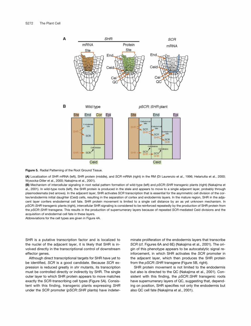
SHR is a putative transcription factor and is localized tothe nuclei of the adjacent layer, it is likely that SHR is in-volved directly in the transcriptional control of downstreameffector genes.
Although direct transcriptional targets for SHR have yet tobe identified, SCR is a good candidate. Because SCR ex-pression is reduced greatly in shr mutants, its transcriptionmust be controlled directly or indirectly by SHR. The singleouter layer to which SHR protein appears to move matchesexactly the SCR-transcribing cell types (Figure 5A). Consis-tent with this finding, transgenic plants expressing SHRunder the SCR promoter (pSCR::SHR plants) have indeter-
minate proliferation of the endodermis layers that transcribeSCR (cf. Figures 6A and 6E) (Nakajima et al., 2001). The ori-gin of this phenotype appears to be autocatalytic signal re-inforcement, in which SHR activates the SCR promoter inthe adjacent layer, which then produces the SHR proteinfrom the pSCR::SHR transgene (Figure 5B, right).
SHR protein movement is not limited to the endodermisbut also is directed to the QC (Nakajima et al., 2001). Con-sistent with this finding, the pSCR::SHR transgenic rootshave supernumerary layers of QC, suggesting that, depend-ing on position, SHR specifies not only the endodermis butalso QC cell fate (Nakajima et al., 2001).
Figure 5. Radial Patterning of the Root Ground Tissue.
(A) Localization of SHR mRNA (left), SHR protein (middle), and SCR mRNA (right) in the RM (Di Laurenzio et al., 1996; Helariutta et al., 2000;Wysocka-Diller et al., 2000; Nakajima et al., 2001).(B) Mechanism of intercellular signaling in root radial pattern formation of wild-type (left) and pSCR::SHR transgenic plants (right) (Nakajima etal., 2001). In wild-type roots (left), the SHR protein is produced in the stele and appears to move to a single adjacent layer, probably throughplasmodesmata (red arrows). In the adjacent layer, SHR activates SCR transcription that is essential for the asymmetric cell division of the cor-tex/endodermis initial daughter (Ceid) cells, resulting in the separation of cortex and endodermis layers. In the mature region, SHR in the adja-cent layer confers endodermal cell fate. SHR protein movement is limited to a single cell distance by an as yet unknown mechanism. InpSCR::SHR transgenic plants (right), intercellular SHR signaling is considered to be reinforced repeatedly by the production of SHR protein fromthe pSCR::SHR transgene. This results in the production of supernumerary layers because of repeated SCR-mediated Ceid divisions and theacquisition of endodermal cell fate in these layers.Abbreviations for the cell types are given in Figure 4A.

Cell Division and Differentiation S273
A Common Mechanism in Root and ShootRadial Patterning
Radial symmetry is not limited to the root but is found in mostplant organs. In Arabidopsis, hypocotyls have a similar radialpattern to that of the root, except that hypocotyls have twolayers of cortex instead of the single layer found in the root.Inflorescence stems also have a similar radial pattern, exceptthat the vascular bundles are positioned circumferentiallyaround the central pith tissue. How common are the mecha-nisms that control root and shoot radial patterning? SCR istranscribed in the cognate layers in roots, stems, and hypo-cotyls (Wysocka-Diller et al., 2000). Roots and stems of scrmutants show analogous radial pattern defects. Furthermore,scr mutant embryos lack asymmetric cell division in the earlyheart-stage embryo that first separates the cortex and endo-dermis cell lineages (Scheres et al., 1995). This results in amutant hypocotyl with two ground tissue layers instead of thenormal three (Scheres et al., 1995; Fukaki et al., 1998).
In the shoot, lack of an endodermis layer results in an in-
ability to respond to gravity, because the shoot endodermispossesses amyloplasts that sediment according to the grav-ity vector, thereby acting as a gravity-sensing “statolith”(Fukaki et al., 1998). pSCR::SHR plants show very similartransgenic phenotypes in hypocotyls, embryos, and roots.The hypocotyls of the pSCR::SHR plants have supernumer-ary ground tissue layers similar to those seen in roots. Inthese supernumerary layers, amyloplasts sediment towardthe gravity vector (Figures 6F and 6G). SCR expression alsois increased in the transgenic hypocotyl and embryo, in apattern similar to that seen in the root (Figures 6F and 6H)(Nakajima et al., 2001). All of these observations indicatethat a common mechanism operates in ground tissue pat-terning in both root and shoot.
Root Vascular Patterning
In contrast to the epidermis and ground tissue, the for-mation of the vascular cylinder requires more complex
Figure 6. Comparison of Wild-Type and pSCR::SHR Transgenic Plants.
(A) to (D) Wild type.(E) to (H) pSCR::SHR transgenic plants.(A) and (E) Confocal images of roots. Red indicates propidium iodide staining of cell walls. Green indicates GFP fluorescence showing the site ofSCR transcription.(B) and (F) Confocal images of dark-grown hypocotyls. Red indicates autofluorescence of plastids. Green indicates GFP fluorescence showingSCR transcription.(C) and (G) Longitudinal sections of light-grown hypocotyls. Arrows indicate amyloplasts sedimenting toward the gravity vector. Sedimentingamyloplasts occur specifically in the endodermis of wild-type plants ([C], arrow), whereas they are found in all layers between the stele and theepidermis of transgenic plants ([G], arrows).(D) and (H) Epifluorescence images of mature embryos. Green represents the site of SCR transcription. In the wild-type embryo, GFP fluorescence isbarely detected above the strong red autofluorescence ([D], arrowheads), whereas strong GFP fluorescence is found in the transgenic embryo (H).Amy, amylose. Abbreviations for the cell types are given in Figure 4A. Bars � 50 �m.

S274 The Plant Cell
patterning, including specification of multiple cell types withspecialized functions, such as xylem and phloem (Figure4A). In Arabidopsis, the average number of stele initials is11, whereas �31 cells are seen in cross-sections from ma-ture regions, indicating that more than two formative divi-sions must take place among the progeny of each initial(Mähönen et al., 2000).
Mutation of the WOODEN-LEG (WOL) gene results in a re-duced number of cells in the vascular cylinder of roots andhypocotyls, and all root vascular cells differentiate into pro-toxylem (Figure 4C, right). Genetic analyses have indicatedthat WOL controls cell divisions but not cell differentiation.When cell divisions in wol are promoted by the epistatic mu-tation fass, the root vasculature of the wol fass double mu-tant produces the full range of cell types (Scheres et al.,1995; Mähönen et al., 2000). WOL encodes a novel two-component His kinase, which recently has been shown tobe allelic to the cytokinin receptor CRE1 (Inoue et al., 2001).WOL/CRE1 is expressed in all cells in the root vascular cyl-inder as well as in the procambium of the embryo (Mähönenet al., 2000). Therefore, WOL/CRE1 has been hypothesizedto sense extracellular cytokinin on the vascular cell surfaceand to transmit a signal to the nucleus (Hwang and Sheen,2001; Inoue et al., 2001). The signal is thought to act ulti-mately to promote vascular cell divisions, allowing the differ-entiation of various cell types.
In root vascular development, the specification of xylemcells precedes that of other vascular cell types, althoughphloem differentiation becomes visible first (Esau, 1977;Bowman, 1994; Mähönen et al., 2000). Therefore, the wolphenotype has been attributed to a failure to produce asufficient number of cells that can accommodate cell typesother than xylem. To date, little is known about how xylemcell fate is specified in the root vascular cylinder. Numer-ous studies on leaf vein patterning have suggested thatauxin plays a major role in vein pattern formation (for re-view, see Dengler and Kang, 2001). The specification ofroot xylem cells just below the existing xylem poles ap-pears to support signaling from the mature xylem tissue. Incontrast, classic dissection studies suggested that the rootxylem pattern is determined autonomously by the RM (forreview, see Raghaven, 2000). These opposing models canbe tested by manipulating the number of stele cells alongthe root axis, possibly through the induction or repressionof WOL function.
CONCLUDING REMARKS
Past studies based on physiological and genetic analyseshave highlighted the importance of positional information inplant development. The molecular nature of cell-to-cellcommunication, however, has long been elusive. Compo-nents of the predicted signaling networks are beginning toemerge in many developmental studies, and some of these
are now understood at the molecular level. In shoot stemcell maintenance, a ligand–receptor interaction appears toconstitute an important part of the intercellular signalingpathway, whereas transcription factor movement appears tobe responsible for ground tissue patterning. The presenceof a large number of CLV3 homologs expressed in a varietyof organs suggests that similar ligand–receptor interactionsoperate in other organs outside the SAM (Cock andMcCormick, 2001). On the other hand, a number of tran-scription factors have been reported to move across celllayers in the SAM and flower organs, although the develop-mental significance of this movement is obscure (Jackson etal., 1994; Perbal et al., 1996; Sessions et al., 2000). The rel-ative importance of intercellular signaling by ligand–receptorinteractions versus protein movement in plant developmentwill be revealed in future studies.
The characterization of various patterning processes hasemphasized the importance of plant hormones, especiallyauxin. In Arabidopsis, cellular auxin levels are thought to becontrolled primarily by polar auxin transport, which in turndepends on the distribution of auxin influx and efflux carriers(Estelle, 1998). Not only are efflux carriers expressed in dif-ferent cell types, but the proteins are targeted to differentcell surfaces (Galweiler et al., 1998; Muller et al., 1998).Therefore, cell fate changes caused by modified auxin distri-bution could lead to redistribution of the efflux carriers,thereby affecting local auxin transport. This may act tomaintain auxin homeostasis: a given auxin distribution couldstabilize a transporter profile, thus causing the distributionpattern to be perpetuated. Because the Arabidopsis ge-nome contains at least 18 potential auxin carrier genes(Swarup et al., 2000), it is conceivable that auxin distributionin the plant is determined by a complex expression patternof many transporter genes in combination with their intracel-lular protein localization. Reverse genetic analysis for eachof these genes will contribute to a comprehensive under-standing of the role of auxin in plant developmental pattern-ing. It also may lead to an understanding of the genes thatcontrol carrier protein expression, which must be responsi-ble for setting up a prepattern of primary importance.
In the past decade, key regulatory genes have been iden-tified based on visible phenotypic alterations. These genesnow are undergoing detailed functional studies. Once a ma-jor foundation of a signaling pathway is clarified, more elab-orate screening procedures can be designed to searchspecifically for other molecules in the same signaling path-way. Reverse genetic approaches are facilitated greatly bythe availability of the entire Arabidopsis genome sequenceand recent technical advances in the production of desiredknockouts. Microarray analyses also can be used to identifydownstream components of signaling pathways. In the nextfew years, we will obtain a much clearer view of the signal-ing networks that underlie plant development.
Received October 29, 2001; accepted January 16, 2002.

Cell Division and Differentiation S275
REFERENCES
Barton, M.K., and Poethig, R.S. (1993). Formation of the shoot api-cal meristem in Arabidopsis thaliana: An analysis of developmentin the wild type and in the shoot meristemless mutant. Develop-ment 119, 823–831.
Benfey, P.N., Linstead, P.J., Roberts, K., Schiefelbein, J.W.,Hauser, M.-T., and Aeschbacher, R.A. (1993). Root develop-ment in Arabidopsis: Four mutants with dramatically altered rootmorphogenesis. Development 119, 57–70.
Bowman, J. (1994). ARABIDOPSIS: An Atlas of Morphology andDevelopment. (New York: Springer-Verlag).
Bowman, J.L., Smyth, D.R., and Meyerowitz, E.M. (1989). Genesdirecting flower development in Arabidopsis. Plant Cell 1, 37–52.
Brand, U., Fletcher, J.C., Hobe, M., Meyerowitz, E.M., andSimon, R. (2000). Dependence of stem cell fate in Arabidopsis on afeedback loop regulated by CLV3 activity. Science 289, 617–619.
Busch, M.A., Bomblies, K., and Weigel, D. (1999). Activation of afloral homeotic gene in Arabidopsis. Science 285, 585–587.
Carland, F.M., and McHale, N.A. (1996). LOP1: A gene involved inauxin transport and vascular patterning in Arabidopsis. Develop-ment 122, 1811–1819.
Clark, S.E., Running, M.P., and Meyerowitz, E.M. (1993).CLAVATA1, a regulator of meristem and flower development inArabidopsis. Development 119, 397–418.
Clark, S.E., Running, M.P., and Meyerowitz, E.M. (1995).CLAVATA3 is a specific regulator of shoot and floral meristemdevelopment affecting the same processes as CLAVATA1. Devel-opment 121, 2057–2067.
Clark, S.E., Jacobsen, S.E., Levin, J.Z., and Meyerowitz, E.M.(1996). The CLAVATA and SHOOT MERISTEMLESS loci competi-tively regulate meristem activity in Arabidopsis. Development 122,1567–1575.
Clark, S.E., Williams, R.W., and Meyerowitz, E.M. (1997). TheCLAVATA1 gene encodes a putative receptor kinase that controlsshoot and floral meristem size in Arabidopsis. Cell 89, 575–585.
Cnops, G., Wang, X., Linstead, P., Van Montagu, M., VanLijsebettens, M., and Dolan, L. (2000). Tornado1 and tornado2are required for the specification of radial and circumferential pat-tern in the Arabidopsis root. Development 127, 3385–3394.
Cock, J.M., and McCormick, S. (2001). A large family of genes thatshare homology with CLAVATA3. Plant Physiol. 126, 939–942.
Dengler, N., and Kang, J. (2001). Vascular patterning and leafshape. Curr. Opin. Plant Biol. 4, 50–56.
Di Laurenzio, L., Wysocka-Diller, J., Malamy, J., Pysh, L., Helariutta,Y., Freshour, G., Hahn, M., Feldmann, K., and Benfey, P.(1996). The SCARECROW gene regulates an asymmetric cell divi-sion that is essential for generating the radial organization of theArabidopsis root. Cell 86, 423–433.
Dolan, L., Janmaat, K., Willemsen, V., Linstead, P., Poethig, S.,Roberts, K., and Scheres, B. (1993). Cellular organisation of theArabidopsis thaliana root. Development 119, 71–84.
Drews, G.N., Bowman, J.L., and Meyerowitz, E.M. (1991). Nega-tive regulation of the Arabidopsis homeotic gene AGAMOUS bythe APETALA2 product. Cell 65, 991–1002.
Endrizzi, K., Moussian, B., Haecker, A., Levin, J.Z., and Laux, T.(1996). The SHOOT MERISTEMLESS gene is required for mainte-nance of undifferentiated cells in Arabidopsis shoot and floralmeristems and acts at a different regulatory level than the mer-istem genes WUSCHEL and ZWILLE. Plant J. 10, 967–979.
Esau, K. (1977). The root: Primary state of growth. In Anatomy ofSeed Plants. (New York: John Wiley & Sons), pp. 215–242.
Estelle, M. (1998). Polar auxin transport: New support for an oldmodel. Plant Cell 10, 1775–1778.
Fletcher, J., Brand, U., Running, M., Simon, R., and Meyerowitz,E. (1999). Signaling of cell fate decisions by CLAVATA3 in Arabi-dopsis shoot meristems. Science 283, 1911–1914.
Frugier, F., Folmer, S., Blilou, I., Willemsen, V., Wolkenfelt, H.,Ferreira, P., and Scheres, B. (2001). HOBBIT, a component ofthe APC involved in control of cell division and cell fate (abstr.179). In 12th International Conference on Arabidopsis Research.
Fukaki, H., Wysocka-Diller, J., Kato, T., Fujisawa, H., Benfey,P.N., and Tasaka, M. (1998). Genetic evidence that the endoder-mis is essential for shoot gravitropism in Arabidopsis thaliana.Plant J. 14, 425–430.
Galweiler, L., Guan, C., Müller, A., Wisman, E., Mendgen, K.,Yephremov, A., and Palme, K. (1998). Regulation of polar auxintransport by AtPIN1 in Arabidopsis vascular tissue. Science 282,2226–2230.
Helariutta, Y., Fukaki, H., Wysocka-Diller, J., Nakajima, K., Jung,J., Sena, G., Hauser, M., and Benfey, P. (2000). The SHORT-ROOT gene controls radial patterning of the Arabidopsis rootthrough radial signaling. Cell 101, 555–567.
Hirt, H. (1997). Multiple roles of MAP kinases in signal transductionin plants. Trends Plant Sci. 2, 11–15.
Howell, S.H. (1998). Molecular Genetics of Plant Development.(Cambridge, UK: Cambridge University Press).
Hwang, I., and Sheen, J. (2001). Two-component circuitry in Arabi-dopsis cytokinin signal transduction. Nature 413, 383–389.
Inoue, T., Higuchi, M., Hashimoto, Y., Seki, M., Kobayashi, M.,Kato, T., Tabata, S., Shinozaki, K., and Kakimoto, T. (2001).Identification of CRE1 as a cytokinin receptor from Arabidopsis.Nature 409, 1060–1063.
Jackson, D., Veit, B., and Hake, S. (1994). Expression of maizeKNOTTED1 related homeobox genes in the shoot apical meristempredicts patterns of morphogenesis in the vegetative shoot.Development 120, 405–413.
Jeong, S., Trotochaud, A.E., and Clark, S.E. (1999). The Arabidop-sis CLAVATA2 gene encodes a receptor-like protein required forthe stability of the CLAVATA1 receptor-like kinase. Plant Cell 11,1925–1934.
Kajava, A.V. (1998). Structural diversity of leucine-rich repeat pro-teins. J. Mol. Biol. 277, 519–527.
Kayes, J.M., and Clark, S.E. (1998). CLAVATA2, a regulator of mer-istem and organ development in Arabidopsis. Development 125,3843–3851.
Kidner, C., Sundaresan, V., Roberts, K., and Dolan, L. (2000).Clonal analysis of the Arabidopsis root confirms that position, notlineage, determines cell fate. Planta 211, 191–199.
Laux, T., Mayer, K.F., Berger, J., and Jurgens, G. (1996). The

S276 The Plant Cell
WUSCHEL gene is required for shoot and floral meristem integrityin Arabidopsis. Development 122, 87–96.
Lenhard, M., Bohnert, A., Jurgens, G., and Laux, T. (2001). Termi-nation of stem cell maintenance in Arabidopsis floral meristems byinteractions between WUSCHEL and AGAMOUS. Cell 105, 805–814.
Lohmann, J.U., Hong, R.L., Hobe, M., Busch, M.A., Parcy, F.,Simon, R., and Weigel, D. (2001). A molecular link between stemcell regulation and floral patterning in Arabidopsis. Cell 105, 793–803.
Long, J.A., Moan, E.I., Medford, J.I., and Barton, M.K. (1996). Amember of the KNOTTED class of homeodomain proteinsencoded by the STM gene of Arabidopsis. Nature 379, 66–69.
Mähönen, A.P., Bonke, M., Kauppinen, L., Riikonen, M., Benfey,P.N., and Helariutta, Y. (2000). A novel two-component hybridmolecule regulates vascular morphogenesis of the Arabidopsisroot. Genes Dev. 14, 2938–2943.
Mayer, K.F., Schoof, H., Haecker, A., Lenhard, M., Jurgens, G.,and Laux, T. (1998). Role of WUSCHEL in regulating stem cellfate in the Arabidopsis shoot meristem. Cell 95, 805–815.
Muller, A., Guan, C., Galweiler, L., Tanzler, P., Huijser, P., Marchant,A., Parry, G., Bennett, M., Wisman, E., and Palme, K. (1998).AtPIN2 defines a locus of Arabidopsis for root gravitropism con-trol. EMBO J. 17, 6903–6911.
Nakajima, K., Sena, G., Nawy, T., and Benfey, P.N. (2001). Inter-cellular movement of the putative transcription factor SHR in rootpatterning. Nature 413, 307–311.
Perbal, M., Haughn, G., Saedler, H., and Schwarz-Sommer, Z.(1996). Non-cell-autonomous function of the Antirrhinum floralhomeotic proteins DEFICIENS and GLOBOSA is exerted by theirpolar cell-to-cell trafficking. Development 122, 3433–3441.
Pysh, L., Wysocka-Diller, J., Camilleri, C., Bouchez, D., and Benfey,P. (1999). The GRAS gene family in Arabidopsis: Sequence char-acterization and basic expression analysis of the SCARECROW-LIKE genes. Plant J. 18, 111–119.
Raghaven, V. (2000). Developmental Biology of Flowering Plants.(New York: Springer-Verlag).
Sabatini, S., Beis, D., Wolkenfelt, H., Murfett, J., Guilfoyle, T.,Malamy, J., Benfey, P., Leyser, O., Bechtold, N., Weisbeek, P.,and Scheres, B. (1999). An auxin-dependent distal organizer ofpattern and polarity in the Arabidopsis root. Cell 99, 463–472.
Satina, S., Blakeslee, A.F., and Avery, A. (1940). Demonstration ofthe three germ layers in the shoot apex of Datura by means ofinduced polyploidy in periclinal chimeras. Am. J. Bot. 27, 895–905.
Scheres, B. (2001). Plant cell identity: The role of position and lin-eage. Plant Physiol. 125, 112–114.
Scheres, B., Laurenzio, L.D., Willemsen, V., Hauser, M.-T.,Janmaat, K., Weisbeek, P., and Benfey, P.N. (1995). Mutationsaffecting the radial organisation of the Arabidopsis root displayspecific defects throughout embryonic axis. Development 121,53–62.
Schoof, H., Lenhard, M., Haecker, A., Mayer, K.F., Jurgens, G.,and Laux, T. (2000). The stem cell population of Arabidopsisshoot meristems is maintained by a regulatory loop between theCLAVATA and WUSCHEL genes. Cell 100, 635–644.
Sessions, A., Yanofsky, M.F., and Weigel, D. (2000). Cell-cell sig-
naling and movement by the floral transcription factors LEAFYand APETALA1. Science 287, 419–421.
Steeves, T.A., and Sussex, I.M. (1989). Patterns in Plant Develop-ment. (Cambridge, UK: Cambridge University Press).
Stone, J.M., Collinge, M.A., Smith, R.D., Horn, M.A., and Walker,J.C. (1994). Interaction of a protein phosphatase with an Arabi-dopsis serine-threonine receptor kinase. Science 266, 793–795.
Stone, J.M., Trotochaud, A.E., Walker, J.C., and Clark, S.E.(1998). Control of meristem development by CLAVATA1 receptorkinase and kinase-associated protein phosphatase interactions.Plant Physiol. 117, 1217–1225.
Swarup, R., Marchant, A., and Bennett, M.J. (2000). Auxin trans-port: Providing a sense of direction during plant development.Biochem. Soc. Trans. 28, 481–485.
Trotochaud, A.E., Hao, T., Wu, G., Yang, Z., and Clark, S.E.(1999). The CLAVATA1 receptor-like kinase requires CLAVATA3for its assembly into a signaling complex that includes KAPP anda Rho-related protein. Plant Cell 11, 393–406.
Trotochaud, A.E., Jeong, S., and Clark, S.E. (2000). CLAVATA3, amultimeric ligand for the CLAVATA1 receptor-kinase. Science289, 613–617.
Umeda, M., Umeda-Hara, C., and Uchimiya, H. (2000). A cyclin-dependent kinase-activating kinase regulates differentiation ofroot initial cells in Arabidopsis. Proc. Natl. Acad. Sci. USA 97,13396–13400.
van den Berg, C., Willemsen, V., Hage, W., Weisbeek, P., andScheres, B. (1995). Cell fate in the Arabidopsis root meristemdetermined by directional signalling. Nature 378, 62–65.
van den Berg, C., Willemsen, V., Hendriks, G., Weisbeek, P., andScheres, B. (1997). Short-range control of cell differentiation inthe Arabidopsis root meristem. Nature 390, 287–289.
Vernoux, T., Wilson, R.C., Seeley, K.A., Reichheld, J.P., Muroy,S., Brown, S., Maughan, S.C., Cobbett, C.S., Van Montagu, M.,Inze, D., May, M.J., and Sung, A.R. (2000). The ROOTMERISTEMLESS1/CADMIUM SENSITIVE2 gene defines a glu-tathione-dependent pathway involved in initiation and mainte-nance of cell division during postembryonic root development.Plant Cell 12, 97–110.
Weigel, D., Alvarez, J., Smyth, D.R., Yanofsky, M.F., andMeyerowitz, E.M. (1992). LEAFY controls floral meristem identityin Arabidopsis. Cell 69, 843–859.
Westhoff, P., Jeske, H., Jürgens, G., Kloppstech, K., and Link, G.(1998). Molecular Plant Development: From Gene to Plant.(Oxford, UK: Oxford University Press).
Willemsen, V., Wolkenfelt, H., de Vrieze, G., Weisbeek, P., andScheres, B. (1998). The HOBBIT gene is required for formation of theroot meristem in the Arabidopsis embryo. Development 125, 521–531.
Williams, R.W., Wilson, J.M., and Meyerowitz, E.M. (1997). A pos-sible role for kinase-associated protein phosphatase in the Arabi-dopsis CLAVATA1 signaling pathway. Proc. Natl. Acad. Sci. USA94, 10467–10472.
Wysocka-Diller, J., Helariutta, Y., Fukaki, H., Malamy, J., andBenfey, P. (2000). Molecular analysis of SCARECROW functionreveals a radial patterning mechanism common to root and shoot.Development 127, 595–603.

DOI 10.1105/tpc.010471 2002;14;S265-S276Plant Cell
Keiji Nakajima and Philip N. BenfeySignaling In and Out: Control of Cell Division and Differentiation in the Shoot and Root
This information is current as of June 23, 2018
References /content/14/suppl_1/S265.full.html#ref-list-1
This article cites 59 articles, 34 of which can be accessed free at:
Permissions https://www.copyright.com/ccc/openurl.do?sid=pd_hw1532298X&issn=1532298X&WT.mc_id=pd_hw1532298X
eTOCs http://www.plantcell.org/cgi/alerts/ctmain
Sign up for eTOCs at:
CiteTrack Alerts http://www.plantcell.org/cgi/alerts/ctmain
Sign up for CiteTrack Alerts at:
Subscription Information http://www.aspb.org/publications/subscriptions.cfm
is available at:Plant Physiology and The Plant CellSubscription Information for
ADVANCING THE SCIENCE OF PLANT BIOLOGY © American Society of Plant Biologists