Embed Size (px)
Citation preview

Riga, 2014
SIGNIFICANCE OF BETA-HERPESVIRUSES
(HHV-6, HHV-7) INFECTION UNDER THE CONDITIONS OF IMMUNE DISORDERS
Speciality – Virology
Summary of the Doctoral Thesis for obtaining the degree of a Doctor of Medicine
Alina Sultanova

This work has been carried out at Rīga Stradiņš University (RSU) August Kirchenstein Institute of Microbiology and Virology in collaboration with Pauls Stradins Clinical University Hospital (Latvian Transplantation Center), Riga East Clinical University Hospital (Latvian Oncology Center and Clinic “Gaiļezers”) and Riga 1st Hospital. Scientific supervisors:Dr. med. Associate Professor Modra Murovska, RSU August Kirchenstein Institute of Microbiology and Virology, LatviaDr. habil. biol. Senior Researcher Svetlana Chapenko, RSU August Kirchenstein Institute of Microbiology and Virology, Latvia
Official reviewers:Dr. med. Professor Inara Logina, Rīga Stradiņš University, LatviaDr. habil. med. Professor Aija Zilevica, Faculty of Medicine, University of LatviaDr. biol. sci. Mykolas Mauricas, State Research Institute Centre for Innovative Medicine, Lithuania
Doctoral Thesis will be defended on 3rd of June 2014, at 15.00 during Rīga Stradiņš University Promotional Council of Medicine meeting in 16 Dzirciema Street, in the Lecture theatre Hippocrates.
Doctoral Thesis is available at the library of Rīga Stradiņš University and on the home page: www.rsu.lv
Doctoral Thesis was supported by ESF project “Support for doctoral study programs and research degrees RSU” 2009/0147/1DP/1.1.2.1.2/09/IPIA/VIAA/009
Secretary of the Promotional Council:Dr. med. Associate Professor Angelika Krumina

3
TABLE OF CONTENTS
ABBREVIATIONS ........................................................................................... 6
INTRODUCTION ............................................................................................ 8
SCIENTIFIC NOVELTY OF THE STUDY ............................................................. 10
Aim of the study ...................................................................................... 11
Objectives of the study ............................................................................ 11
Hypotheses of the study .......................................................................... 12
The structure of the work ........................................................................ 13
1. MATERIALS AND METHODS ............................................................... 14
1.1. Patient groups ................................................................................... 14
1.2. Molecular methods ........................................................................... 14
1.3. Immunological methods ................................................................... 16
1.4. Statistical analysis ............................................................................ 17
2. RESULTS .................................................................................................... 19
2.1. INVOLVEMENT OF HHV-6 AND HHV-7 INFECTION IN THE
DEVELOPMENT OF POST-TRANSPLANT COMPLICATIONS ................................ 19
2.1.1. Presence of HHV-6 and HHV-7 infection markers
in renal transplant recipients ................................................................... 19
2.1.2. Analysis of immunocompetent cell populations
in renal transplant recipients dependently on lymphocytes’
count ........................................................................................................ 21
2.1.3. Presence of HHV-6 and HHV-7 infection in renal
transplant recipients with and without lymphopenia ............................... 22

4
2.1.4. Comparative analysis of immunocompetent cell
populations in renal transplant recipients without and with
lymphopenia dependently on beta-herpesviruses infection .................... 24
2.1.5. Complications development in renal transplant recipients ........... 24
2.2. INVOLVEMENT OF HHV-6 AND HHV-7 INFECTION IN THE
CLINICAL COURSE OF GASTROINTESTINAL CANCER (GIC) ............................ 25
2.2.1. Presence of HHV-6 and HHV-7 infection in patients
with GIC ................................................................................................. 25
2.2.2. Analysis of immunocompetent cell populations
in patients with GIC dependently on lymphocytes’ count ...................... 27
2.2.3. Presence of HHV-6 and HHV-7 infection in GIC
patients with and without lymphopenia .................................................. 28
2.2.4. Comparative analysis of immunocompetent cell
populations in GIC patients without and with lymphopenia
dependently on beta-herpesviruses infection ......................................... 30
2.2.5. Pro-inflammatory cytokines levels in GIC patients
with and without lymphopenia ............................................................... 30
2.2.6. Gastrointestinal cancer clinical outcomes dependently
on beta-herpesviruses infection .............................................................. 32
2.3. INVOLVEMENT OF HHV-6 AND HHV-7 INFECTION IN THE
DEVELOPMENT OF AUTOIMMUNE THYROID DISEASES (AIT) ......................... 33
2.3.1. Presence of HHV-6 and HHV-7 infection in patients
with AIT ................................................................................................. 33
2.3.2. Analysis of immunocompetent cell populations
in patients with AIT dependently on viral infection ............................... 34
2.3.3. Presence of HHV-6 and HHV-7 infection in thyroid
gland tissue ............................................................................................. 35

5
2.3.4. Comparison of HHV-6 infection presence
in thyroid gland tissue and blood samples ............................................... 36
2.3.5. HHV-6 load in blood and tissue DNA .......................................... 37
2.4. COMPARATIVE ANALYSIS BETWEEN PATIENTS
WITH DIFFERENT IMMUNE SYSTEM DYSFUNCTIONS ...................................... 39
2.4.1. Comparison of beta-herpesviruses infection frequency ................ 39
2.4.2. Comparison of immunocompetent cell populations
dependently on beta-herpesviruses infection........................................... 41
3. DISCUSSION .............................................................................................. 45
4. CONCLUSIONS ......................................................................................... 50
5. RECOMMENDATIONS............................................................................ 51
6. ACKNOWLEDGMENTS .......................................................................... 52
7. THE LIST OF PUBLICATIONS .............................................................. 53
8. REFERENCES ........................................................................................... 59

6
ABBREVIATIONS
AIDS – acquired immunodeficiency syndrome
AIT – autoimmune thyroiditis
AITD – autoimmune thyroid disease
CI – confidence interval
CMV – cytomegalovirus
DNA – deoxyribonucleic acid
ELISA – enzyme-linked immunosorbent assay
FACS – fluorescence-activated cell sorting
GIC – gastrointestinal cancer
GVHD – graft versus host disease
HHV-6 – human herpesvirus 6
HHV-7 – human herpesvirus 7
IFA – indirect fluorescent antibody test
IgG – immunoglobulin G
IgM – immunoglobulin M
IL-1β – interleukin-1 beta
IL-2 – interleukin-2
IL-6 – interleukin-6
Leu – leukocytes
Ly – lymphocytes
Mo – monocytes
NK – natural killers
nPCR – nested polymerase chain reaction
OR – odds ratio

7
P – p value
PBMC – peripheral blood mononuclear cells
PCR – polymerase chain reaction
TG- thyroglobulin
TNF-α – tumour necrosis factor
TPO- thyroid peroxidase
TSH- thyroid-stimulating hormone
8
INTRODUCTION
Today the majority of chronic diseases are already quite strongly
associated with infectious agents, including viruses. In most of cases these are
viruses that cause persistent infection and who themselves possess the ability to
affect the body's immune system. Recently, these immunomodulating viruses
have been receiving increasing attention, which is determined by several
factors:
these viruses are widely distributed after primary infection and they
exist in a persistent form throughout life;
they can be activated by immunosuppressant factors;
they by themselves possess immunosuppressant properties and they
can change the body's immune status;
they often cause serious complications in cases of an immuno-
suppression caused by medication after solid organ and stem cell
transplantation;
they cause a variety of complications in patients with immuno-
suppressive background caused by underlying disease, for an
example in oncology patients;
they can initiate chronic inflammatory processes that can lead to
autoimmune pathology, and neoplastic changes;
in the case of co-infection they can activate each other (Sprengers and
Janssen, 2005; Mocarski Jr, 2002; Lucas and McFadden, 2004).
The immunomodulating viruses mentioned above include lymphotropic
herpesviruses – human herpesvirus -6 and -7 (HHV-6 and HHV-7), that belong
to Betaherpesvirinae subfamily Roseolovirus genus.

9
HHV-6 was isolated in 1986 from the interleukin-2 (IL-2)-stimulated
peripheral blood mononuclear cells obtained from the AIDS patients and
patients with lymphoproliferative diseases. HHV-6 was described in two
variants and later as a two species – HHV-6A and HHV-6B.
Since the moment of discovery, HHV-6 has been associated with a wide
range of clinical conditions and chronic diseases, due to high ubiquitous nature
of this virus. Separation of HHV-6 into two distinguished species (HHV-6A
and HHV-6B) only added questions concerning involvement of this virus
infection in etiopathogenesis of various diseases. HHV-6A is frequently found
in multiple sclerosis (MS), chronic fatigue syndrome (CFS), acquired
immunodeficiency syndrome (AIDS) and cancer patients. HHV-6B causes
Roseola infantum, febrile illness and encephalitis in infants and it reactivates in
transplant patients, causing complications such as encephalitis, pneumonia and
liver damage. In renal transplant patients, HHV-6 has been associated with the
development of chronic allograft nephropathy (Chapenko et al., 2009) and graft
versus host disease (GVHD) (Caiola et al., 2012). For a long time only
HHV-6B was associated with the development of complications in renal
transplant patients, due to it’s detection particularly in the mononuclear cells of
renal transplants (Helantera et al., 2008), and HHV-6B is also frequently found
in the gastrointestinal tract of these patients (Lempinen et al., 2012). However,
in recent study the predominance of HHV-6A viremia is reported in the
plasma/serum among cohort of renal transplant patients (Csoma et al., 2011).
Data about HHV-6 implication in different cancer diseases is
controversial. While a lot of researches associate HHV-6 (especially HHV-6B)
with the nodular sclerosis subtype of Hodgkin’s lymphoma (Lacroix et al.,
2010; Siddon et al., 2012) and leukaemia (Ogata et al., 2011), there are lack of
studies on HHV-6 implication in solid tumour development. However, several

10
investigators have suggested that HHV-6 possess an oncogenic potential. Cells
transfected with HHV-6 can cause tumours in nude mice (Puri et al., 1991).
Recent studies have proposed a certain role for HHV-6 in several
autoimmune disorders, including autoimmune acute hepatitis (Grima et al.,
2008) and autoimmune hemolytic anemia/neutropenia (Yagasaki et al., 2010).
The study of 2012th
has linked HHV-6A to Hashimoto’s thyroiditis (Caselli et
al., 2012). However, additional evidence is required, especially taking into
account the distribution of HHV-6A and HHV-6B.
HHV-7 clinical role is poorly documented, but it is possibly associated
with the development of Pityriasis rosea (Black et al., 1999; Rebora et al.,
2010) and it could act as HHV-6 activator and CMV co-factor in complication
development in post-transplant patients (Chapenko et al., 2001; Chan et al.,
2004; Holden and Vas, 2007; Zawilinska et al., 2011).
In this work a relation of HHV-6 and HHV-7 infection with different
chronic diseases and complications development was investigated, to acquire
lacking data on involvement of theses viruses in the development of immune
system dysfunction. Secondary, the distribution of HHV-6A and HHV-6B
infection among different patient groups in Latvia was ascertained.
Scientific novelty of the study
It is the first time when HHV-6 and HHV-7 infection reactivation
frequency is compared in three groups of patients with different causes
of immune system dysfunction (immunosuppressive therapy,
underlying disease and autoimmune process);

11
The higher risk of lymphopenia development in patients with
gastrointestinal cancer during active HHV-6 and HHV-7 infection is
proved and statistically confirmed.
Strong association between beta-herpesviruses HHV-6 and HHV-7
infection and autoimmune thyroiditis is discovered.
Comparative analysis of changes in immunocompetent cell
populations and severity of immunosuppression among three different
patients’ groups are carried out.
Aim of the study
The aim of the present study was to ascertain the involvement of beta-
herpesviruses infection in the pathogenesis and clinical course of chronic
diseases and development of post-transplant complications due the ability to
change host-pathogen interaction.
Objectives of the study
1. Create the groups of patients with immune system dysfunctions
caused by different factors:
a. Renal transplant recipients with immunosuppression
caused by immunosuppressive therapy;
b. Gastrointestinal cancer patients with immunosuppression
caused by underlying disease;
c. Patients with autoimmune thyroiditis.

12
2. Explore the presence and activity phase of beta-herpesviruses HHV-6
and HHV-7 infection in patients with immune system dysfunction
caused by different factors.
3. Examine the cellular immunity in renal transplant recipients, patients
with underlying disease (gastrointestinal cancer) and patients with
autoimmune process (autoimmune thyroiditis).
4. Clarify the association between beta-herpesviruses HHV-6 and HHV-
7 infection and post-transplant complications development, chronic
disease clinical course and autoimmune process.
5. Build the database of patients, including clinical and laboratory data.
Hypothesis of the study
1. Reactivation of HHV-6 and HHV-7 infection is occurring more often
in patients with immune system dysfunctions than in control group
individuals.
2. HHV-6 and HHV-7 reactivation leads to more severe imbalance of the
immune system.
3. Higher viral load in thyroid tissue than in peripheral blood indicates
that thyroid gland is a possible place of HHV-6 latency which could
be a cause of autoimmune process development.

13
The structure of the work
The doctoral thesis is written in English. It consists of the following
parts: introduction, scientific novelty, the aim, objectives, hypothesis, literature
review, materials and methods, results, discussion, conclusion and recom-
mendations. The work is written on 124 pages with 23 Tables and 21 Figures.
209 citations of literature are included in the reference list. The results of the
work are published in 9 papers (in pre-reviewed editions) and presented at
17 local/international conferences/congresses.
14
1. MATERIALS AND METHODS
1.1. Patient groups
Three patient groups – patients with immunosuppressive drug treatment
(renal transplant recipients; n = 47 – 27 men and 20 women – mean age 49;
from 28 to 78 years); patients with immune system disorders related to the
underlying disease (patients with gastrointestinal cancer; n = 65 – 42 women
and 23 men – mean age 54; from 39 to 85 years) and patients with autoimmune
disorders (autoimmune thyroiditis; n = 44 – 43 women and one man – mean
age 45; from 25 to 78 years) were included in the study.
Practically healthy blood donors were included as control group
(n = 150 – 77 females and 73 males – mean age 37; from 18 to 65 years). Post-
mortem thyroid gland samples without any pathological macro or micro
changes were used as the tissue controls in investigation of operated thyroid
glands of patients with autoimmune thyroiditis (n = 41 – 11 were women and
30 men – mean age 52; from 41 to 78 years). The research was established with
the approval of the Ethics Committee of the Rīga Stradiņš University.
1.2. Molecular methods
Isolation of DNA from peripheral whole blood and tissue samples:
All whole blood and thyroid gland tissue DNA was extracted using phenol-
chloroform method.

15
Isolation of DNA from plasma samples: The QIAamp Blood Kit
(QIAGEN, Hilden, Germany) was used to purify DNA from cell-free blood
plasma following manufacturer's protocol. Before DNA purification the plasma
samples were treated with DNase I (Fermentas, Vilnius, Lithuania).
DNA quality and quantity control: According to the manufacturer's
instructions 2μl of DNA solution was taken and concentration measured using
spectrophotometer NanoDrop 1000 at a wavelength of 260, resulting in the
concentration of each sample, which is expressed in the unit of measurement –
ng/μl. The sample purity control was done by using the 260/280 ratio diapason,
which must be no less than 1.8.
Beta (β)-globin PCR with appropriate primers was used (Vanndame et
al., 1995) to determine the quality of DNA isolated from blood and tissue, and
to exclude plasma DNA contamination by cell debris DNA.
Nested Polymerase Chain Reaction (nPCR): The technique of nPCR
was used to detect viral genomic sequences in DNA isolated from whole blood
and cell free plasma (markers of persistent and active phase of persistent
infection, respectively). PCR amplification of viral DNA was carried out in the
presence of 1 µg of whole blood DNA or 10 µl of plasma DNA (which
corresponded to 200 µl of plasma). HHV-6 and HHV-7 were detected in
accordance with Secchiero et al. 1995 and Berneman et al. 1992, respectively.
Positive controls (HHV-6 and HHV-7 genomic DNA; Advanced
Biotechnologies Inc, Columbia, MD, USA and negative controls (DNA
obtained from practically healthy HHV-6 and HHV-7 negative blood donor and
no template DNA) were included in each experiment.
Determination of HHV-6 A and HHV-6B by nPCR and Hind III
restriction: Determination of HHV-6A and HHV-6B was made in
correspondence to Lyall and Cubie, 1995. Expected PCR product length was:

16
163bp after the marker pUC 19 DNA / MspI (Hpa2) Marker23 (MBI Fermantes
Lithuania)], the results were visualized and processed with BioSpectrum 610
MultiSpectral Imaging System, USA.
Quantitative Real-time PCR: The viral load of HHV-6 in whole blood
and tissue DNA samples from patients with HHV-6 persistent infection was
determined using the HHV-6 Real-Time Alert Q-PCR kit (Nanogen Advanced
Diagnostics, Buttigliera Alta, Italy) and an Applied Biosystems 7500 Real-time
PCR System (Applied Biosystems, Carlsbad, CA, USA), in accordance with
the manufacturer’s recommendations. Collected data were processed and
analyzed with specialized software ABI 7500 system. The data are calculated to
get the viral copies /106 cells.
1.3. Immunological methods
Viral specific antibody detection: For the approval of obtained PCR
results, determination of the HHV-6-specific IgG class antibodies in plasma
was carried out using commercial ELISA kit (Panbio, Sinnamon Park, QLD,
Australia). To determine the presence of HHV-7-specific IgG class antibodies
in plasma samples IFA work set (EUROIMMUN, Germany) was used
following the manufacturer's developed protocol. Preparations were analyzed
with immunofluorescence microscope "Nikon Eclipse 80i" at 400×
magnification and the visualization program "Lucia Image Analysis Systems"
Lucia 5.0 version.
Auto-antibody detection: Commercial ELISA kits (EUROIMMUN,
Germany) were used to determine the presence of auto-antibodies against to
17
thyroid peroxidase (TPO), against to thyroglobulin (Tg) and thyroid stimulating
hormone (TSH) receptor.
Assay for cytokine determination: ELISA kits (Pierce Biotechnology,
Rockford, IL, USA, and AviBion, Helsinki, Finland) were used to detect the
levels of tumor necrosis factor (TNF)-α, interleukin (IL)-1β and IL-6 in plasma.
The sensitivity of the ELISAs were < 3 pg/ml for IL-1β and < 2 pg/ml for
TNF-α and IL-6. In addition, the expression level of soluble IL-2 receptor
(sIL-2R) was measured using a solid-phase competitive chemi-luminescent
enzyme immunoassay (CLIA; Siemens, Los Angeles, CA, USA) in accordance
with the manufacturer’s recommendations. The assay was able to detect
5 U/ml. All samples were tested in duplicate.
FACS analysis and sorting: BD FACSAria II flow cytometer (USA)
and BD FACSDiva software were used for sorting and analysing of peripheral
blood mononuclear cells (PBMC). Commercial monoclonal antibodies
conjugated with different flourochromes (anti-CD3-FITC, CD4-PerCP-Cy7,
CD8-PE, CD16-V450, CD19-APC, CD45-V500, CD56-V450 and CD95-
PerCP-Cy 5.5) were used to distinguish and sort main lymphocyte populations.
1.4. Statistical analysis
All calculations were performed with the MedCalc Software version
12.3 (Ostend, Belgium). Statistical differences in the prevalence of latent and
active HHV-6 and HHV-7 infection were assessed by Fisher’s exact test.
Plasma levels of cytokines were expressed as the mean ± SD. Immunological
parameters were analyzed using t-test. Regression analysis was used to assess

18
the continuous variable values; a value of p < 0.05 was considered to be
significant.
19
2. RESULTS
2.1. Involvement of HHV-6 and HHV-7 infection in the
development of post-transplant complications
2.1.1. Presence of HHV-6 and HHV-7 infection markers in renal
transplant recipients
Persistent beta-herpesviruses infection was detected in 42 out of
47 (89%) renal transplant recipients. HHV-6 persistent infection was found in
15 out of 47 (32%) recipients, HHV-7 persistent infection – in 40 out of
47 (85%) recipients, that is statistically higher (p = 0.02) than in healthy blood
donors – 101 out of 150 (67%). Persistent single HHV-6 infection was found in
two out of 47 recipients; however, single HHV-7 infection was detected in
27 out of 47 recipients and persistent HHV-6 + HHV-7 infection – in 13 out of
47 individuals.
Latent single HHV-6 infection was detected in one out of two, HHV-7
latent infection in 16 out of 27 (59%) and HHV-6 + HHV-7 latent infection in
5 out of 13 (39%) renal transplant recipients. Activation rate was higher in
patients with double HHV-6 + HHV-7 infection – 8 out of 13 (62%) than in
recipients with single HHV-6 infection – one out of two (50%) or HHV-7
infection – 11 out of 27 (41%). In all HHV-6 positive samples HHV-6B was
defined. There was no significant difference in the presence of latent HHV-6
infection between recipients and healthy individuals. However, HHV-7 latent

20
infection was significantly higher (p = 0.006) in healthy blood donors (85%)
than in renal transplant recipients (59%).
HHV-6 and double HHV-6 + HHV-7 active infection was found only in
renal transplant recipients (one out of two and 8 out of 13, respectively). Rate
of active HHV-7 infection was significantly higher (p = 0.006) in renal
transplant recipients 11/27 (41%) than healthy blood donors 12/83 (14%)
(Table 2.1).
Although, HHV-6 genomic sequence was detected in 15/47 (32%) of
renal transplant recipients, presence of anti- HHV-6 specific IgG class
antibodies was detected in 36/47 (77%) of patients.
Table 2.1.
Presence of beta-herpesviruses infection in renal transplant recipients and
healthy blood donors
HHV-6
persistent
(latent/active)
infection
HHV-7
persistent
(latent/active)
infection
HHV-6 + HHV-
7 persistent
(latent/active)
infection
Without
HHV-6 or
HHV-7
infection
Renal transplant
recipients (n = 47)
2 (4.3%)
(1/1)
27 (57%)
(16/11)
13 (27.6%)
(5/8)
5
(11%)
Blood donors
(n = 150)
13 (8.6%)
(13/0)
83 (55%)
(71/12)
30 (20%)
(30/0)
24
(16%)

21
2.1.2. Analysis of immunocompetent cell populations in renal
transplant recipients dependently on lymphocytes’ count
It was possible to divide renal transplant recipients into two groups
according to the lymphocyte count: group I (n = 17) – patients without
lymphopenia (lymphocytes > 1400 cells in 1mm3 of peripheral blood) and
group II (n=30) – patients with lymphopenia (lymphocytes < 1400 cells in
1mm3 of peripheral blood). The mean absolute number of lymphocytes in the
group I was two times (50%) higher than in the group II – 1890 ± 440 ×106/L
and 850 ± 410 ×106/L, respectively. Comparative analyses of the lymphocyte
subsets between the group I and II (CD3+, CD4
+, CD8
+, CD38
+, CD16
+,
CD19+, CD25
+ and CD95
+) showed significant decrease of Ly subsets in the
group II – approximately two fold decrease in comparison with the group I
(Table 2.2).

22
Table 2.2.
Absolute counts of immunocompetent cells populations in the renal
transplant recipients’ groups I and II
Parameters
Group I Ly > 1400 (n = 17) Group II Ly < 1400 (n = 30)
p ≤ 0.05 Absolute count
± SD
Count
%
Absolute count
± SD
Count
%
Leu 8450 ± 2490 6950 ± 2370 0.04
Ly 1890 ± 440 24.92 850 ± 410 12.57 < 0.0001
CD3+ 1510 ± 390 80.12 620 ± 400 67.43 < 0.0001
CD4+ 630 ± 300 40.53 230 ± 190 31.43 < 0.0001
CD8+ 580 ± 210 38.35 220 ± 200 32.03 < 0.0001
CD38+ 520 ± 350 26.18 270 ± 170 31.77 0.002
CD16+ 150 ± 70 10.00 90 ± 50 17.23 0.001
CD19+ 130 ± 60 7.00 70 ± 50 9.50 0.0006
CD95+ 850 ± 440 43.71 400 ± 270 44.83 0.0001
CD25+ 80 ± 40 4.29 30 ± 30 3.10 < 0.0001
CD4+/CD8+ 1.24 ± 0.70 1.18 ± 0.70
2.1.3. Presence of HHV-6 and HHV-7 infection in renal transplant
recipients with and without lymphopenia
HHV-6 persistent infection was found in 8 out of 17 (47%) group I
patients and in 7 out of 30 (23%) patients of the group II. However, HHV-7
persistent infection was presented in 15 out of 17 (88%) group I patients and in
25 out of 30 (83%) patients of the group II. Presence of single HHV-6
persistent infection was detected only in 2 out of 30 (6.6%) patients with
lymphopenia. Higher presence of single HHV-7 persistent infection was found
in the group II of patients in comparison to the group I (66% and 41%,
respectively). However, higher presence of double HHV-6 + HHV-7 persistent
infection was detected in 8 out of 17 (47%) renal transplant recipients without

23
lymphopenia comparing to the group II patients with lymphopenia (5/30,
17%).
Single HHV-6 latent infection was revealed only in one patient from the
group II (with lymphopenia); however, frequency of single HHV-7 latent
infection between both groups showed no significant difference (57% in the
group I and 60% in the group II, respectively).
Double HHV-6 + HHV-7 latent infection was more prevalent in the
group I patients (50%) comparing with the group II patients (20%).
Presence of single HHV-6 active infection was found only in one renal
transplant recipient with lymphopenia, however, activation rate of single
HHV-7 active infection showed no significant difference between both groups
(43% in the group I and 40% in the group II, respectively). Activation rate of
double HHV-6 + HHV-7 active infection was more frequent in patients from
the group II (80%) than from the group I (50%) (Table 2.3).
Table 2.3.
Presence of beta-herpesviruses (HHV-6 and HHV-7) infection in the group
I and II
Patients
HHV-6
persistent
(latent/active)
infection
HHV-7
persistent
(latent/active)
infection
HHV-6 + HHV-
7 persistent
(latent/active)
infection
Without HHV-6
or HHV-7
infection
Group I
(n=17) 0 (0%)
7 (41%)
(4/3)
8 (47%)
(4/4) 2 (12%)
Group II
(n=30)
2 (6.6%)
(1/1)
20 (66%)
(12/8)
5 (17%)
(1/4) 3 (10%)

24
2.1.4. Comparative analysis of immunocompetent cell populations
in renal transplant recipients without and with lymphopenia
dependently on beta-herpesviruses infection
Comparative analysis of lymphocytes (Ly) subpopulations in the group I
and II was performed dependently on beta-herpesviruses infection. Each group
was subdivided into three subgroups: renal transplant recipients without
infection, with latent and with active HHV-6 and/or HHV-7 infection. Both
groups’ patients with latent viral infection had tendency to increase in all
populations (lymphocytes, CD3+, CD4
+, CD8
+, CD38
+, CD16
+, CD19
+, CD25
+
and CD4+/CD8
+ ratio) comparing with the subgroups without HHV-6 and
HHV-7 infection and with active viral infection. The most severe decrease of
immunocompetent cells populations was detected in the group II patients with
active beta-herpesviruses infection, especially in lymphocytes absolute count
and CD38+ cells (to 21% and to 56%, respectively), comparing to the group
with latent viral infection.
Although, the group II patients with active HHV-6 or HHV-7 had
significantly higher decrease (p = 0.001) in CD38+ subpopulation (to 56%)
comparing to group with latent viral infection, the group I patients with active
viral infection had significant (p = 0.0001) downfall in CD8+ subpopulation (to
80%). Also, big shift in CD95+ subpopulation was detected between both
groups with latent and active viral infection.
2.1.5. Complications development in renal transplant recipients
Development of complications was found in 14 out of 47 (30%) renal
transplant recipients. Five patients had acute rejection (AR), four patients were
25
with microbial infections and others had melanoma, sepsis, CMV disease,
chronic allograft nephropathy (CAN) and acute hepatitis of non-identified
etiology.
Analysis of patients with complications and beta-herpesviruses infection
revealed that 10 out of 14 (71%) renal transplant recipients with complications
were with active HHV-6 or/and HHV-7 infection and only 4 out of 14 (29%)
with latent infection. The logistic regression analysis also showed that patients
with active viral infection have higher risk of complication development (OR
4.5; 95% CI 1.12–18.13; p = 0.03) than the patients with latent viral infection
(OR 0.22; 95% CI 0.06–0.89; p = 0.03).
2.2. Involvement of HHV-6 and HHV-7 infection in the clinical
course of gastrointestinal cancer (GIC)
2.2.1. Presence of HHV-6 and HHV-7 infection in patients with
GIC
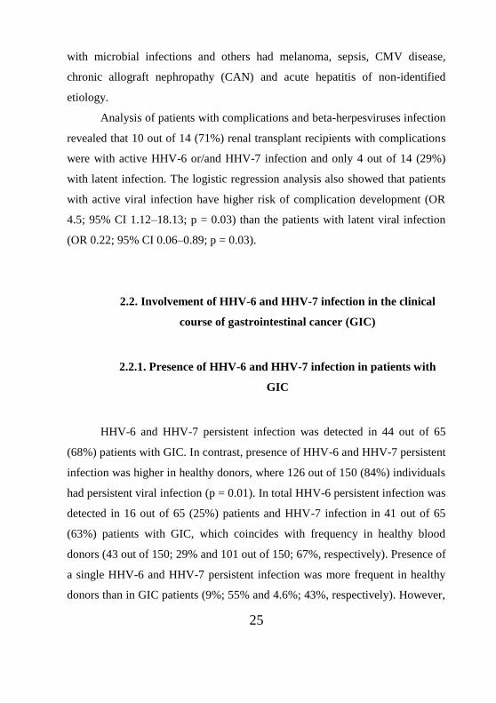
HHV-6 and HHV-7 persistent infection was detected in 44 out of 65
(68%) patients with GIC. In contrast, presence of HHV-6 and HHV-7 persistent
infection was higher in healthy donors, where 126 out of 150 (84%) individuals
had persistent viral infection (p = 0.01). In total HHV-6 persistent infection was
detected in 16 out of 65 (25%) patients and HHV-7 infection in 41 out of 65
(63%) patients with GIC, which coincides with frequency in healthy blood
donors (43 out of 150; 29% and 101 out of 150; 67%, respectively). Presence of
a single HHV-6 and HHV-7 persistent infection was more frequent in healthy
donors than in GIC patients (9%; 55% and 4.6%; 43%, respectively). However,

26
patients with GIC and healthy donors had the same level of persistent double
HHV-6 + HHV-7 infection frequency.
Presence rate of single HHV-6, HHV-7 and double HHV-6 + HHV-7
latent infection was higher in healthy blood donors than in patients with GIC.
However, single HHV-6 active infection was found only in patients with
GIC (one of two; 50%). Activation rate of single HHV-7 infection was 15% in
healthy blood donors, in contrast to 25% in patients with GIC. Presence of
double active viral infection was found only in 6 out of 7 (46%) patients with
GIC (Table 2.4).
Although, HHV-6 genomic sequence was detected in 16 out of 65 (25%)
of patients with GIC, anti- HHV-6 specific IgG class antibodies were found in
33 out of 65 (51%) patients. HHV-7 genomic sequence was detected in 63% of
patients by nPCR, however, presence of HHV-7 specific IgG class antibodies
was detected in 75%.
In all HHV-6 positive GIC patients HHV-6B was detected.
Table 2.4.
Presence of beta-herpesviruses infection in patients with GIC
HHV-6
persistent
(latent/
active)
infection
HHV-7
persistent
(latent/active)
infection
HHV-6 + HHV-7
persistent
(latent/active)
infection
Without
HHV-6 or
HHV-7
infection
Patients with
gastrointestinal
cancer (n = 65)
3 (4.6%)
(2/1)
28 (43%)
(21/7)
13 (20%)
(7/6) 21 (32%)
Blood donors
(n = 150)
13 (8.6%)
(13/0)
83 (55%)
(71/12)
30 (20%)
(30/0) 24 (16%)

27
2.2.2. Analysis of immunocompetent cell populations in patients
with GIC dependently on lymphocytes’ count
It was possible to divide patients with GIC into two groups according to
the lymphocytes count: group I – patients without lymphopenia (lymphocytes
> 1400 cells in 1mm3 of peripheral blood) and group II with lymphopenia
(lymphocytes < 1400 cells in 1mm3 of peripheral blood). Comparative analysis
of immunocompetent cells’ populations in both GIC patients groups showed
significant differences in almost all rate. The mean number of leukocytes in the
group I (7910 ± 1960 ×106/L) was significantly (p = 0.002) higher (27%) than
in the group II (5830 ± 2210 ×106/L). The mean absolute number of
lymphocytes in the group I was two times (50%) higher than in the
group II (2270 ± 700 ×106/L; 1140 ± 210 ×10
6/L, respectively; p = 0.0001).
Comparative analyses of lymphocytes’ subsets between the group I and II
(CD3+, CD4
+, CD8
+, CD38
+, CD16
+, CD19
+, CD25
+ and CD95
+) showed
significant decrease of immunological parameters in the group II –
approximately in two times in comparison with the group I (Table 2.5).

28
Table 2.5.
Absolute counts of immunocompetent cell populations in the I and II GIC
patients’ groups
Parameters Group I Ly > 1400 (n = 35) Group II Ly < 1400 (n = 30) p ≤
0.05 Absolute count
± SD
Count
%
Absolute count
± SD
Count %
Leu 7910 ± 1960 5830 ± 2210 0.002
Ly 2270 ± 700 28.83 1140 ± 210 19.88 0.0001
CD3+ 1600 ± 580 70.77 780 ± 180 70.77 0.0001
CD4+ 880 ± 330 39.29 450 ± 130 39.00 0.0001
CD8+ 680 ± 370 29.11 330 ± 120 31.08 0.0001
CD38+ 660 ± 300 29.03 320 ± 90 28.40 0.0001
CD16+ 460 ± 310 19.69 220 ± 130 19.4 0.0015
CD19+ 190 ± 110 8.03 110 ± 60 7.00 0.0018
CD95+ 1130 ± 370 50.89 520 ± 130 48.8 0.0001
CD25+ 180 ± 150 8.63 80 ± 50 7.50 0.047
CD4+/CD8+ 1.58 ± 0.85 1.18 ± 0.57
2.2.3. Presence of HHV-6 and HHV-7 infection in GIC patients
with and without lymphopenia
Frequency of single HHV-6 persistent infection was significantly
(p < 0.05) higher in patients with lymphopenia (2/35; 6.6%) than in patients
without it (1/30; 2.6%), however, presence of single HHV-7 persistent infection
was higher in patients from the group I (20/35; 57%) than from the group II
(8/30; 27%). Double persistent infection was more frequently detected in
patients with (9/30; 30%) than in patients without (4/35; 11%) lymphopenia.
There was significant difference in the presence of single HHV-6 latent
infection between two analysed groups, however, higher rate of single HHV-7

29
latent infection was found in the group I patients (17/20; 85%) comparing with
the group II patients (4/8; 50%). Frequency of double latent infection was
found more frequent in patients without lymphopenia (4/4; 100%) than in
patients with lymphopenia (3/9; 33%).
In the group of patients without lymphopenia significantly (p = 0.002)
lower incidence of active beta-herpesviruses infection was detected (3/25, 12%)
than in patients with lymphopenia (11/19, 58%). Active single HHV-6 infection
was detected only in one out of two patients with GIC from the group II.
Presence of single HHV-7 active infection was higher in (4/8; 50%) patients
from the group II comparing with patients from the group I (3/20; 15%).
Double HHV-6 + HHV-7 active infection was found only in patients with
lymphopenia (6/9; 64%) (Table 2.6).
Table 2.6.
Presence of latent and active beta-herpesviruses HHV-6 and HHV-7
infection in GIC patients group I and II
Patients HHV-6
persistent
(latent/active)
infection
HHV-7
persistent
(latent/active)
infection
HHV-6 + HHV-7
persistent
(latent/active)
infection
Without HHV-
6 or HHV-7
infection
Group I
(n=35)
1 (2.8%)
(1/0)
20 (57%)
(17/3)
4 (11%)
(4/0)
10 (29%)
Group II
(n=30)
2 (6.6%)
(1/1)
8 (27%)
(4/4)
9 (30%)
(3/6)
11 (37%)

30
2.2.4. Comparative analysis of immunocompetent cell populations
in GIC patients without and with lymphopenia dependently on
beta-herpesviruses infection
Comparative analysis of immunocompetent cell populations in the group
I and II was performed dependently on beta-herpeviruses infection. Each group
was subdivided into three subgroups: GIC patients without, with latent and with
active HHV-6 and/or HHV-7 infection.
The group I patients with active viral infection had tendency to increase
all cellular immunological parameters (leukocytes, monocytes, lymphocytes
and CD3+, CD4
+, CD8
+, CD38
+, CD16
+, CD19
+, CD25
+, CD95
+) comparing
with subgroups with and without latent HHV-6 and HHV-7 infection. The most
dramatic decrease was found in CD3+ and CD8
+ subpopulations (to 16% and to
21%, respectively) in the group II patients with active beta-herpesviruses
infection. Tendency to increase was detected in both patient groups’ B
lymphocytes (CD19+) during active viral infection. However, patients without
lymphopenia had increase to 105% of CD19+ subpopulation, but in patients
with lymphopenia only to 18%. The logistic regression analysis showed that
patients with active viral infection have higher risk of lymphopenia (OR 3.33;
95% CI 0.72–15.51; p = 0.0035) than patients with latent viral infection (OR
0.33; 95% CI 0.10–1.07; p = 0.0035).
2.2.5. Pro-inflammatory cytokines levels in GIC patients with and
without lymphopenia
There were no significant changes in the plasma levels of IL-6, IL-1β,
sIL-2R and TNF-alpha between GIC patients of the group I and II. However, in

31
the group I and II of patients with active viral infection the levels of IL-6 and
sIL-2R had tendency to increase, nonetheless, level of TNF-alpha in the group
II patients with active viral infection was lower than in patients with latent and
without viral infection, and also lower in comparison with the group I patients
with active viral infection (Table 2.7).
Table 2.7.
Levels of IL-1beta, IL-6, sIL2R and TNF-alpha in the patients with GIC
group I and II
Patients groups IL-1beta
(pg/ml)
N = < 5.0
IL-6 (U/ml)
N = 3.4
sIL2R (U/ml)
N = 223–710
TNF-alpha
(pg/ml)
N = 8.1
Group I (n = 10)
Without HHV-6
and/or HHV-7
< 5.0 4.08 ± 1.23 527 ± 159.58 15.13 ± 5.15
Group II (n = 11)
Without HHV-6
and/or HHV-7
< 5.0 5.15 ± 2.76 559.80 ± 379.10 15.58 ±5.66
Group I (n = 22)
HHV-6 and/or
HHV-7 latent
ifection
< 5.0 5.44 ± 2.99 604.78 ± 242.33 11.62±1.08
Group II (n = 8)
HHV-6 and/or
HHV-7 latent
infection
< 5.0 5.55 ± 1.91 617.00 ± 173.20 13.83±3.06
Group I (n = 3)
HHV-6 and/or
HHV-7 active
infection
< 5.0 7.20 ± 11.88 851 ± 528.55 14.40±5.31
Group II (n=11)
HHV-6 and/or
HHV-7 active
infection
<5.0 6.18±2.91 733.67±388.76 9.78±4.23

32
2.2.6. Gastrointestinal cancer clinical outcomes dependently on
beta-herpesviruses infection
Stages of gastrointestinal cancer were defined in accordance to TNM
evaluation (evaluation of the (primary) tumor 'T'; evaluation of the regional
lymph nodes 'N'; evaluation of distant metastasis 'M'). First stage was defined
only in 9% of patients. The majority of patients (43%) were with second stage
of cancer. Third stage was defined in 30% patient and the most severe – fourth
stage was defined in 18% patients with GIC. There was no association between
beta-herpesviruses infection and stages of cancer; however, fourth stage of
cancer was more prevalent in patients with HHV-6 and/or HHV-7 persistent
infection (23%) than in patients without it (6%) (Table 2.8). Also, no
association between lymphopenia and stages of cancer was found. However,
death rate was higher in patients with lymphopenia (53%) than in patients
without it (34%), but there was no statistical significance.
Table 2.8.
Stages of GIC frequency dependently on beta-herpesviruses infection

33
2.3. Involvement of HHV-6 and HHV-7 infection in the
development of autoimmune thyroid diseases (AIT)
2.3.1. Presence of HHV-6 and HHV-7 infection in patients with
AIT
Presence of beta-herpesviruses infection markers in blood samples were
found more frequently in patients with AIT than in healthy blood donors (93%
and 84%, respectively). HHV-6 persistent infection was detected in 12 out of
44 (27%) patients with AIT. The same infection frequency was detected in
healthy blood donors (43/150, 29%). HHV-7 persistent infection was revealed
in 40 out of 44 (91%) patients with AIT. Presence of HHV-7 was found to be
significantly lower (p = 0.03) in healthy individuals (only 75%).
Single HHV-6 and HHV-7 persistent infection was more frequently
observed in blood donors (8.6% and 55%, respectively) comparing to AIT
patients (2.3% and 36%, respectively). Frequency of double HHV-6+HHV-7
persistent infection was much higher in patients with AIT than in blood donors
(55% against 20%, respectively) (Table 2.9).
Single HHV-6 active infection was not found neither in patients with
AIT and in healthy blood donors. Activation rate of single HHV-7 infection
was significantly higher (p < 0.0001) in patients with AIT comparing with
blood donors (38% and 15%, respectively). Double HHV-6 + HHV-7 active
infection was found only in patients with AIT (13/44, 30%).

34
Table 2.9.
Presence of HHV-6 and HHV-7 infection in patients with AIT
HHV-6
persistent
(latent/active)
infection
HHV-7
persistent
(latent/active)
infection
HHV-6 + HHV-7
persistent
(latent/active)
infection
Without
HHV-6 or
HHV-7
infection
Patients
with AIT
(n = 44)
1 (2.3%)
(1/0)
16 (36%)
(10/6)
24 (55%)
(11/13)
3
(7%)
Blood
donors
(n = 150)
13 (8.6%)
(13/0)
83 (55%)
(71/12)
30 (20%)
(30/0)
24
(16%)
2.3.2. Analysis of immunocompetent cell populations in patients
with AIT dependently on viral infection
Comparative analysis of immunocompetent cell populations in the
patients with AIT was performed dependently on beta-herpesviruses infection.
Patients were subdivided into three subgroups: AIT patients without, with latent
and with active HHV-6 and/or HHV-7 infection. As in previous groups,
patients with latent HHV-6 and/or HHV-7 infection had increase almost in all
immunological parameters (Ly, CD3+, CD4
+, CD8
+, CD16
+, CD19
+ and
CD95+) comparing to patients without viral infection and patients with active
viral infection. In patients with active HHV-6 and/or HHV-7 infection decrease
in all immunocompetent cell populations was observed in comparison with the
patients with latent infection however statistical analysis showed no
significance (Table 2.10).

35
Table 2.10.
Analysis of cellular immune parameters in AIT patients dependently on
beta-herpesvirus infection
Parameters Without HHV-6 and/or
HHV-7 (n = 3)
Latent HHV-6 and/or
HHV-7 (n = 21)
Active HHV-6 and/or
HHV-7 (n = 20)
Leu 5833 ± 1401 6073 ± 1297 6252 ± 1597
Ly 1883 ± 188 2087 ± 732 1994 ± 427
CD3+ 1312 ± 131 1454 ± 510 1390 ± 297
CD4+ 497 ± 49 551 ± 193 527 ± 112
CD8+ 283 ± 28 314 ± 110 300 ± 64
CD16+ 376 ± 37 417 ± 146 399 ± 86
CD19+ 163 ± 16 181 ± 63 173 ± 37
CD95+ 1160 ± 115 1285 ± 450 1228 ± 263
CD4+/CD8+ 0.9 ± 0.3 1.33 ± 0.85 1.14 ± 0.56
2.3.3. Presence of HHV-6 and HHV-7 infection in thyroid gland
tissue
Beta-herpesviruses HHV-6 and HHV-7 persistent infection was detected
in 43 out of 44 (98%) of AIT patients’ thyroid tissue DNA samples and in 31
out of 41 (76%) control group autopsies’ DNA samples (p = 0.003). In 42
(95%) of the 44 patients with AIT in tissue DNA samples HHV-6 genomic
sequence was found, whereas in the control group HHV-6 genome sequence
was found in 25 out of 41 (61%) autopsy tissue DNA samples (p < 0.0001).
HHV-7-specific genomic sequence was presented in 33 (75%) out of 44
patients with AIT and in 16 (39%) out of 41 thyroid tissue DNA samples
extracted from control group autopsies (p = 0.001). There was no significant
difference in the presence of single HHV-6 and HHV-7 genomic sequences in
patients’ and control group thyroid tissue DNA, however, simultaneous

36
presence of HHV-6 and HHV-7 genomic sequences in tissue DNA samples was
significantly higher (p < 0.0001) in patients with AIT (Table 11).
Presence of HHV-6 genomic sequence in thyroid tissue DNA samples
was significantly more prevalent (p = 0.01) than the presence of HHV-7
genomic sequence (95% and 75%, respectively).
Table 2.11.
Presence of beta-herpesviruses (HHV-6 and HHV-7) infection in AIT
patients’ thyroid tissue DNA samples and autopsy materials
HHV6
HHV-7
HHV-6 + HHV-7
Without
AIT (n = 44) 10 (23%) 1 (2.3%) 32 (73%) 1 (2.3%)
Control group (n = 41) 15 (36%) 6 (15%) 10 (24%) 10 (24%)
2.3.4. Comparison of HHV-6 infection presence in thyroid gland
tissue and blood samples
In 43 (98%) of the 44 patients with AIT HHV-6 genomic sequence in
blood and/or thyroid tissue DNA samples was found: in 17 (39%) patients,
HHV-6 genomic sequence was detected in both tissue and blood DNA samples,
while in 25 (58%) patients viral genomic sequence was detected only in tissue
DNA samples. In contrast, HHV-6 genome sequence was found in 25 (61%)
DNA samples isolated from control groups’ autopsy materials: in 14 (34%)
cases in both blood and tissue DNA samples, and in 11 (27%) cases only in
tissue DNA samples. Presence of HHV-6 genomic sequence in thyroid gland

37
tissue only was significantly higher in patients with AIT comparing to the
control group (p = 0.008).
Table 2.12.
Presence of HHV-6 sequence in peripheral blood and thyroid tissue DNA
of patients with AIT and control group autopsy material
Distribution of HHV-6 genomic
sequence
Number of patients
with AIT (n = 44)
Number of control
group samples (n = 41)
Only blood DNA 1 0
Blood DNA + tissue DNA 17 14
Only tissue DNA 25 11
Blood DNA+plasma DNA 0 0
Tissue DNA+blood
DNA+plasma DNA 0 0
Without 1 16
2.3.5. HHV-6 load in blood and tissue DNA
Using real-time polymerase chain reaction HHV-6 load was determined
in DNA samples from patients’ whole blood, tissue and main lymphocyte
subpopulations (CD4+, CD8
+, CD16
+, CD19
+ and CD95
+) DNA samples. In
patients’ blood DNA samples the average viral load was 98 ± 44
copies/1×106cells while in the tissue DNA samples viral load in average was
2552 ± 2015 copies/1×106cells.
To ensure that detected HHV-6 sequence in DNA samples is due to the
involvement of thyroid gland tissue but not due to lymphocyte infiltration, the
main lymphocyte subpopulations DNA was also examined by real-time
polymerase chain reaction. Substantial HHV-6 viral load in lymphocyte

38
subpopulation was detected in two out of seven patients. In first patient, who
was positive for the presence of HHV-6 genomic sequence by nPCR in whole
blood and tissue DNA, HHV-6 load was detected in NK and CD95+ cells (24
and 100 copies/1×106cells, respectively). In this patient also lower HHV-6 load
was detected in whole blood DNA (27 copies/1×106cells) comparing to tissue
DNA, where HHV-6 load was 355 copies/1×106cells (Figure 2.1). In second
patient, who was positive for the presence of HHV-6 genomic sequence by
nPCR only in tissue DNA, HHV-6 load was detected only in CD95+ cells.
HHV-6 load in CD95+ cells was 11 copies/1×10
6cells (Figure 2.2). In other five
patients’ lymphocyte subpopulations (CD8+, CD16
+, and CD95
+) DNA
insignificant (according to the manufacturer’s protocol) HHV-6 load was
detected (range 14–20 copies/1×106cells).
46
0 0 0
24
100
355
0
0
50
100
150
200
250
300
350
400
co
pie
s/1
0*6
cell
s
Patient Nr. 1
whole blood DNA
CD4+
CD8+
CD19+
CD16+
CD95+
rihgt lobe DNA
left lobe DNA
Whole
blood DNA
Plasma
Thyroid
gland right
lobe
Thyroid
gland left
lobe
HHV-6 viral
sequence by nested
PCR
+
–
+
–
Figure 2.1. HHV-6 load in DNA samples of whole blood, tissue and main
lymphocyte subpopulations in Patient Nr.1; nested PCR results

39
0 0 0 0 0
40
132
0
0
20
40
60
80
100
120
140
co
pie
s/1
0*6
cell
s
Patient Nr. 2
whole blood DNA
CD4+
CD8+
CD19+
CD16+
CD95+
rihgt lobe DNA
left lobe DNA
Whole blood
DNA
Plasma
Thyroid gland
right lobe
Thyroid gland
left lobe
HHV-6 viral
sequence by
nested PCR
–
–
+
–
Figure 2.2. HHV-6 load in DNA samples of whole blood, tissue and main
lymphocyte subpopulations in Patient Nr.2; nested PCR results
2.4. Comparative analysis between patients with different immune
system dysfunctions
2.4.1. Comparison of beta-herpesviruses infection frequency
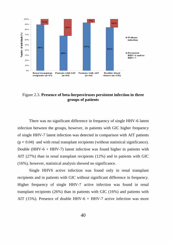
The highest frequency of HHV-6 and HHV-7 persistent infection was
detected in patients with AIT (93%) in comparison with other two groups. The
significantly lowest rate of beta-herpesviruses presence (68%) was detected in
patients with GIC (p < 0.05) comparing to the renal transplant recipients and
patients with AIT (Figure 2.3).
40
Figure 2.3. Presence of beta-herpesviruses persistent infection in three
groups of patients
There was no significant difference in frequency of single HHV-6 latent
infection between the groups, however, in patients with GIC higher frequency
of single HHV-7 latent infection was detected in comparison with AIT patients
(p = 0.04) and with renal transplant recipients (without statistical significance).
Double (HHV-6 + HHV-7) latent infection was found higher in patients with
AIT (27%) than in renal transplant recipients (12%) and in patients with GIC
(16%), however, statistical analysis showed no significance.
Single HHV6 active infection was found only in renal transplant
recipients and in patients with GIC without significant difference in frequency.
Higher frequency of single HHV-7 active infection was found in renal
transplant recipients (26%) than in patients with GIC (16%) and patients with
AIT (15%). Presence of double HHV-6 + HHV-7 active infection was more

41
frequently detected in patients with AIT (32%) comparing to renal transplant
recipients (19%) and patients with GIC (14%) (Figure 2.4).
Figure 2.4. Presence of latent and active beta-herpesviruses infection in
three groups of patients
2.4.2. Comparison of immunocompetent cell populations
dependently on beta-herpesviruses infection
Despite of more severe immunosuppression in renal transplant
recipients, in this group of patients’ level of leukocytes (Leu) was the highest
between all patients group. If in renal transplant recipients and patients with
GIC mean absolute count of Leu had increased during beta-herpesviruses latent
infection and had decreased during active infection, in patients with AIT
increase of Leu absolute count was observed during latent as well as active
infection.

42
Comparative analysis of immunocompetent cell populations
dependently on beta-herpesviruses infection in patients with AIT and two other
groups of patients with immunosuppression of different origin showed
remarkable differences in immunological parameter changes. During HHV-6
and/or HHV-7 latent infection in patients with AIT constant increase (to 11%)
in all immunocompetent cell populations (Ly, CD3+, CD4
+, CD8
+, CD16
+,
CD19+ and CD95
+) was observed and constant decrease (to 4%) during active
infection.
Increase of Ly count was detected in renal transplant recipients (to 13%)
and in patients with GIC (to 14%) in case of beta-herpesviruses latent infection.
However, during HHV-6 and/or HHV-7 active infection decrease in Ly number
was found in both groups (to 19% in renal transplant recipients and to 12% in
patients with GIC). Almost the same tendency was observed in level of CD3+
subpopulation (increase during latent infection to 18% in transplant recipients
and to 15% in patients with GIC, decrease during active infection – to 18% in
renal transplant recipients and to 12% in patients with GIC). Remarkable
increase of CD4+ number was detected in renal transplant recipients (to 41%)
during beta-herpesviruses latent infection comparing to GIC patients (to 19%)
and patients with AIT (to 11%). In all three groups of patients also increase in
CD8+ subpopulation (to 11% in all groups) was observed. However, increase in
CD16+ subpopulation was observed during latent infection only in patients with
AIT (to 11%).
Number of leukocytes and levels of another immunocompetent cell
subpopulations (CD3+, CD4
+, CD8
+, CD38
+, CD19
+, CD25
+ and CD95
+) were
increased in renal transplant recipients and patients with GIC during latent beta-
herpesvirus infection comparing to the subgroups without viral infection.
Comparison of both groups showed significant differences in numbers of CD4+,

43
CD16+, CD19
+ and CD25
+ subpopulations (p < 0.05) between patients with
latent and active beta-herpesvirus infection. In renal transplant recipients with
active viral infection mean absolute number of CD4+ lymphocytes was
decreased to 14% and in GIC patients to 19% in comparison with patients with
latent infection. The most dramatic decrease was detected in number of CD16+,
where in renal transplant recipients with active viral infection this
subpopulation was decreased to 24% and in the patients with GIC only to 11%
comparing both patients groups with latent viral infection. Another interesting
finding was revealed in patients’ groups with active beta-herpesvirus infection.
Number of B lymphocytes (CD19+) had tendency to decrease in renal
transplant recipients (to 10%) and in patients with AITD (to 4%), however in
GIC patients it had tendency to increase (to 26%).
By comparative analysis of immunocompetent cell subpopulations in all
three patients groups greater differences in cell counts were detected between
renal transplant recipients and patients with AIT. Mean absolute count of Ly
was significantly higher (p < 0.0001) in patients with AIT during latent (2087 ±
732) and active (1994 ± 427) HHV-6/7 infection comparing with transplant
recipients (1350 ± 700 and 1100 ± 630, respectively).
However, no significance was detected between patients with AIT and
patients with GIC. Also, in patients with AIT significantly higher (p < 0.0001)
mean absolute count of CD16+ and CD19
+ was found in comparison with renal
transplant recipients. Significantly (p < 0.005) higher amount of CD95+ was
detected in AIT patients comparing to renal transplant recipients and patients
with GIC. In its turn mean absolute number of CD8+ was significantly
(p < 0.0001) higher in GIC patients with latent and active beta-herpesviruses
infection comparing to patients with AIT (Figure 2.5).

44
Figure 2.5. Comparison of immunocompetent cell populations dependently
on beta-herpeviruses infection in three different patients groups

45
3. DISCUSSION
Today there are a lot of studies trying to find and evaluate a role of beta-
herpesviruses infection in etiopathogenesis of different kinds of chronic
diseases, but the final answer to this question is still not found. It could be due
their ubiquitous nature and different mechanisms of interference that these
viruses are using. But one common feature can unite all viruses – it is
disturbance in the immune system, especially immunosuppression, caused by
different origin that is a base for beta-herpesviruses reactivation. Beta-
herpesviruses in the host can function both - directly and indirectly, that means
the viruses infect cells involved in the cellular and humoral immune response
formation, and at the same time these viruses alter cell surface receptor
expression, as well as proinflammatory cytokine (IL-12, IL -1ß, IL-6, TNF-α,
IFN-γ) and chemokine expression levels, thereby contributing to a local
inflammation.
This work shows results obtained by comparison of beta-herpesviruses
infection and disease or complications development in three different groups of
patients with different types of immune system dysfunctions. Renal transplant
recipients have immunosuppression caused by medical treatment (immuno-
suppressants), GIC patients – immunosuppression caused by underlying disease
and patients with autoimmune thyroid disease (AIT) – immunosuppression
caused by immune system dysfunction.
Comparison of immunocompetent cell populations shows more severe
immunosuppression in renal transplant recipients (almost two fold decrease in
all parameters comparing with other two patient groups). This fact is crucial for
the patients with beta-herpesviruses active infection because this group has
showed more pronounced downfall of all Ly subsets than in other two groups

46
of patients and reactivation of these viruses can cause more severe
complications.
In this group also higher incidence of beta-herpesviruses infection
comparing to GIC patients (89% versus 68%) is found. Single HHV-7 active
infection is more prevalent in renal transplant recipients (26%) in comparison
with patients with GIC (16%) and patient with AIT (15%), however without
statistical significance. This finding indicates that HHV-7 important role in
immunosuppression is worsening the clinical outcome in these patients.
Comparative analysis of cellular immune parameters dependently on
beta-herpeviruses infection shows significantly lower level (14%, p < 0.0001)
of natural killer cells (CD16+) in renal transplant recipients with active viral
infection than in GIC patients (19.5%) and patients with AIT (20%). Decrease
of CD16+ number in this group could be caused by immunosuppressive therapy
and higher rate of HHV-7 activation.
Development of complications is detected in 30% of recipients and
significantly higher rate (p = 0.048) is found in renal transplant recipients with
active beta-herpesviruses infection (10/20, 50%) than in recipients with latent
infection (4/22, 18%). Also, logistic regression analysis shows higher risk of
complication development in patients with active viral infection than with
latent infection (OR 4.5; 95% CI 1.12–18.13; p = 0.03 and OR 0.22; 95% CI
0.06–0.89; p = 0.03, respectively). This is an evidence of the HHV-6 and
HHV-7 implication in complications development.
Concerning the patients with GIC, at present there is too little
information on the influence of beta-herpesviruses infection on the clinical
course of the disease. Gastrointestinal malignancies are associated with a
compromised immune system and viruses, such as immunotropic and immuno-
modulating HHV-6 and HHV-7 may be able to utilize cellular mechanisms

47
responsible for the immune response inhibition. Modulation of functional
properties of host immune factors is an important mechanism of evading the
immune response or creating an environment in which the virus can survive.
Analysis for immunocompetent cell populations shows that only 46% of
patients have lymphopenia (Ly < 1400 cells in 1mm3
peripheral blood), so it is
interesting to compare these patients with GIC patients without lymphopenia
(Ly > 1400 cells in 1mm3 peripheral blood).
These results show that in GIC patients group with lymphopenia the
activation of HHV-6 and HHV-7 infection is significantly more frequent
(p = 0.003). Moreover, comparative analysis of immunocompetent cell
subpopulations shows decrease of preferable beta-herpesviruses cell target
populations (absolute count of lymphocytes and CD3+, CD4
+, CD8
+ and CD38
+
subpopulations). Such difference in cell populations could be another evidence
of HHV-6 and HHV-7 involvement in this disease progression. It should be
pointed out that only in GIC patients without lymphopenia during active beta-
herpesviruses infection increase in absolute count of lymphocytes (to 9%),
CD3+ (to 13%), CD4
+ (to 19%) and CD8
+ (to 9%) subpopulations is identified;
however in all patients with lymphopenia these populations are decreased. This
fact can be explained by the effect of immunosuppression on more frequent
beta-herpesviruses activation which in turn subsequently affects the count of
immunocompetent cells (decreasing their count). In conformation of this
statement logistic regression analysis was done and it shows that patients with
active viral infection have higher risk of lymphopenia (OR 3.33;
95% CI 0.72–15.51; p = 0.0035) than patients with latent viral infection
(OR 0.33; 95% CI 0.10–1.07; p = 0.0035).
Although, development of the most sever (fourth) stage of GIC is more
prevalent in patients with persistent beta-herpesviruses infection (23%) than

48
without viral infection (6%), there is no significant association between cancer
stages development and beta-herpesviruses infection. However, higher death
rate (53%) is found between the patients with than without (34%) lymphopenia.
This fact indicates that lymphopenia is an important factor that impacts the
course of the disease, in its turn strong association of lymphopenia
development and active beta-herpesviruses infection is shown.
All these results show importance of HHV-6 and also HHV-7
involvement in immunosuppression in both mentioned patients groups,
however additional investigations are required to make general conclusion.
Viral infections have been frequently cited as important environmental
factors implicated in AIT, but no specific virus has yet been conclusively
associated to the disease. In particular, herpesviruses have been implicated in
this disease, with conflicting evidence. Case reports suggested a potential
association between herpesvirus infection and AIT, but in other report, when
thyroid specimens were analysed, no EBV, CMV or HSV-1 DNA is detected.
A recent study analyzed the presence of herpesvirus DNA in post-operative
thyroid specimens from tissue blocks, and HHV-6 was detected by single round
PCR in 2 out of 15 (13.3%) Hashimoto thyroiditis tissue specimens, whereas in
Grave's disease or multi nodular goitre tissues no HHV-6 DNA is detected
(Thomas et al., 2008).
In this work significantly (p=0.003) higher rate of HHV-6 and HHV-7
genomic sequence presence in DNA samples extracted from AIT patients
thyroid tissue (43/44, 98%) is shown in comparison to the control group (31/41,
76%). In 43/44 of AIT patients’ thyroid tissue DNA HHV-6 genomic sequence
is found. These results show a possible influence of HHV-6 on AIT
development. Interesting, that 25/44 (57%) of patients have HHV-6 genomic
presence only in thyroid tissue DNA samples, which evidences on HHV-6

49
latency in thyroid glands. However, presence of HHV-6 DNA in lymphocytes
infiltrates in tissue could not be excluded. Although, real-time PCR results
show that average HHV-6 viral load is higher in tissue DNA samples, rather
than in whole blood DNA, which could strengthen the evidence on HHV-6
latency in thyroid gland. These results are advocating to the involvement of
HHV-6 infection in AIT development.
The HHV-6B is detected in all DNA samples from patients with HHV-6
infection. This might suggest that either the HHV-6A is infrequent in these
patients or it might be limited to sites other than the peripheral blood. Also, one
of the causes of dominant HHV-6B presence in patients could be connected
with geographical distribution.
This work shows different ways of beta-herpesviruses interaction with
the host organism and their involvement in etiopathogenesis of chronic
diseases, complication development and clinical course worsening. In renal
transplant recipients beta-herpesviruses activity is associated with worsening of
immunosuppression during treatment. Also, substantial role in immuno-
suppression more likely plays also active HHV-7 infection in this group. On the
other hand, in patents with GIC lower beta-herpesviruses (HHV-6 and HHV-7)
distribution is found than in other two groups of patients – only in 68% of
patients. However, development of lymphopenia in immunocompromised
patients with GIC is strongly associated with the active beta-herpesviruses
infection. In case of AIT, especially HHV-6 is associated with this disease
development (because of its high frequency and higher viral load in thyroid
gland tissue in comparison with peripheral blood). HHV-6 and HHV-7
infection, possibly, could be a primary cause, by expressing antigens in thyroid
glands during the latent stage of infection or it could play secondary role by
infecting and destroying thyrocytes.

50
4. CONCLUSIONS
1. Renal transplant recipients have higher risk of immunosuppression
strengthening and complications development associated with HHV-6
and HHV-7 active infection.
2. Active beta-herpesviruses infection in patients with gastrointestinal
cancer is associated with decrease of lymphocytes’ total count,
increase of IL-6, sIL-2R and decrease of TNF-alpha expression levels
leading to the deepening of immunosuppression.
3. HHV-6 and HHV-7 persistent infection is not associated with
gastrointestinal cancer stage, at the same time activation of HHV-6
and HHV-7 leads to the worsening of clinical course and outcome of
underlying disease.
4. HHV-6 and HHV-7 genomic sequences are frequently detected in
thyroid tissue as well as in whole blood DNA samples of patients with
autoimmune thyroiditis therefore the association of viral infection with
the disease development could not be excluded.
5. Higher viral load in thyroid tissue of autoimmune thyroiditis patients
than in whole blood is additional evidence of HHV-6 involvement in
this disease development and indicates that thyroid gland is one of the
places of HHV-6 latency.
6. HHV-6B is revealed in renal transplant recipients, patients with
gastrointestinal cancer and autoimmune thyroiditis, which shows
dominant distribution of this species in Latvia.

51
5. RECOMMENDATIONS
1. Highly sensitive molecular and serological methods are required to
detect HHV-6 and HHV-7 infection, which allows distinguishing
latent and active phase of persistent infection.
2. HHV-6 and HHV-7 active infection in renal transplant recipients is
associated with immunosuppression caused by medication. Therefore,
patients with beta-herpesviruses persistent infection should be
monitored for the active phase to prevent development of
complications in time.
3. In 46% of patients with GIC association between active beta-
herpesviruses (HHV-6 and HHV-7) infection and lymphopenia is
found. Therefore, patients with beta-herpesviruses persistent infection
should be monitored for the active phase to avoid the deepening of
lymphopenia in time.
4. Taking into account that thyroid gland is one of the HHV-6 latency
places it is recommended to monitor the activity of beta-herpesviruses
infection in patients with the first signs of thyroid gland diseases to
prevent deepening of the autoimmune process.

52
6. ACKNOWLEDGMENTS
I would like to express my sincere of gratitude to my supervisors –
Assoc. Prof. Modra Murovska and Senior Researcher Svetlana Chapenko
for invaluable assistance, support and guidance. Also, I would like to
express gratitude to Prof. Sandra Lejniece for support and valuable advices.
I would like to express gratitude to Vice-Rector for Science, Prof.
Iveta Ozolanta and Scientific Secretary, Ingrida Kreile for support and
consultations during doctoral studies.
Finally, I would like to thank all my colleges for moral support.

53
7. THE LIST OF PUBLICATIONS
7.1. Publications
1. Sultanova A., Chistjakovs M., Chapenko S., Donina S., Murovska M.
Possible interference of human beta-herpesviruses-6 and -7 in
gastrointestinal cancer development // Experimental oncology, 2013;
35 (2): 93–96.
2. Sultanova A., Cistjakovs M., Capenko S., Donina S., Ziedina I.,
Murovska M. Frequency and activity phase of HHV-6 and HHV-7
persistent infection in renal transplant recipients and patients with
gastrointestinal cancer // RSU Research articles in medicine and
pharmacy, Collection of Scientific Papers 2013: 54–60.
3. Chapenko S., Ziedina I., Folkmane I., Sultanova A., Rozental R.,
Murovska M. The impact of beta-herpesviruses infection activation on
early complications development following renal transplantation //
Current Problems of Infectious Human Pathology, Vol. 4, 2011: 274–
278.
4. Gravelsina S., Nora-Krukle Z., Chapenko S., Sultanova A., Boka V.,
Cunskis E., Murovska M. Incidence of human herpesvirus 6, 7, and
parvovirus B 19 infection in patients with thyroid gland disorders //
Current Problems of Infectious Human Pathology, Vol. 4, 2011: 283–
287.
5. Čistjakovs M., Čapenko S., Sultanova A., Nora Z., Murovska M.
Cilvēka sestā herpesvīrusa (HHV-6) tipu sastopamība dažādu personu
grupās Latvijā // RSU Zinātniskie raksti 2008 Internā medicīna

54
Ķirurģija Medicīnas bāzes zinātnes Stomatoloģija Farmācija, 2009,
lpp. 317–322.
6. Ziediņa I., Čapenko S., Folkmane I., Sultanova A., Murovska M.,
Jušinskis J., Rozentāls R. Poliomas-BK vīrusa un beta-herpesvīrusu
reaktivācija pēc nieres transplantācijas // RSU Zinātniskie raksti 2008
Internā medicīna Ķirurģija Medicīnas bāzes zinātnes Stomatoloģija
Farmācija, 2009, lpp. 81–84.
7. Муровска M., Чапенко C., Козырева C., Султанова A., Дониня C.,
Фолкмане И., Круминя A., Лейниеце C., Лейниекс A. Бета-
герпесвирусы чеповека (ВГЧ-6, ВГЧ-7), их распространение и
ассоциация с патологическими процессами. Материалы
Международной научно-практической конференции Вирусные
инфекции: эпидемиология, клиника, лабораторная диагностика и
профилактика. Минск, Государственное учреждение «НИИ
эпидемиологии и микробиологии», 29-30 ноября 2007 г., стр. 93–
96.
8. Sultanova A., Čapenko S., Nora Z., Doniņa S., Murovska M.
Imūnsupresija kā β-herpesvīrusu aktivācijas faktors pacientiem ar
kuņģa-zarnu trakta onkoloģiskām slimībām. Latvijas Universitates
raksti. 2007, sēj.: Medicīna, lpp. 20–24.
9. Murovska M., Spuris K., Nora Z., Sultanova A., Čapenko S., Lejnieks
A. Limfotropo herpesvīrusu infekcija un vairogdziedzera slimības //
RSU Zinātniskie raksti 2003 Internā medicīna Ķirurģija Medicīnas
bāzes zinātnes Stomatoloģija Farmācija, 2003, lpp. 389–393.

55
7.2. Abstracts
1. Sultanova A., Cistjakovs M., Chapenko S., Gravelsina S., Nora-
Krukle Z., Donina S., Lejniece S., Murovska M. Human herpesvirus 6
and 7 infection as facilitator of chronic diseases and various
complications development. 10th
International Conference on New
Trends in Immunosuppression, Barcelona, Spain, March 11-12, 2013:
196
2. Nora-Krukle Z., Gravelsina S., Chapenko S., Sultanova A., Cunskis
E., Murovska M. Association between latent/persistent human
herpesvirus 6 (HHV-6) and 7 (HHV-7) infection and diseases of
thyroid gland. 8th
International Congress on Autoimmunity, Granada,
Spain, may 9-13, 2012.
3. Sultanova A., Cistjakovs M., Chapenko S., Donina S., Murovska M.
Frequency HHV-6 and HHV-7 infection in patients with different
stages of gastrointestinal cancer. 17th
International Symposium on
Infections in the Immunocompromised Host, Italy, Genova 24th
-27th
June, 2012: 65.
4. Fadeeva I., Sultanova A., Nora-Krukle Z., Murovska M. Human
herpesvirus-6 and -7 resctivation in thyroid gland tissues of patients
with thyroid gland disorders., XI International Congress of Medical
Sciences, Sofia, Bulgaria, 03-06 May, 2012: 148.
5. Gravelsina S., Nora-Krukle Z., Sultanova A., Chapenko S., Cunskis
E., Roga S., Murovska M. Human herpesvirus six and seven and
parvovirus B19 infections as possible risk factors for the development
of autoimmune and non-autoimmune thyroid diseases. School of

56
translational immunology, Belgrade, Serbia, 19-21 September 2012:
45.
6. Nora-Krukle Z., Gravelsina S., Sultanova A., Chapenko S., Rasa S.,
Roga S., Cunskis E., Murovska M. Prevalence of HHV-6, HHV-7 and
parvovirus B19 in thyroid tissue samples from patients with thyroid
gland disorders and subjects with unspecified encephalopathies. 22nd
Annual Meeting of the Society for Virology, Essen, Germany, March
14-17, 2012: 536.
7. Sultanova A., Chapenko S., Fadejeva I., Gravelsina S., Nora-Krukle
Z., Cunskis E., Murovska M. HHV-6 and HHV-7 Infection in Patients
with Struma nodosa III. RSU Zinātniskā konference, 2012. gada 29.
un 30. martā. Rīgā, lpp. 182.
8. Sultanova A., Chapenko S., Gravelsina S., Nora-Krukle Z., Cunskis
E., Murovska M. Genomic sequences of herpesvirus 6&7 and
parvovirus B 19 in DNA samples isolated from thyroid tissues. RSU
Zinātniskā konference, 2011. gada 14. un 15. aprīlī., Rīgā, lpp. 225.
9. Sultanova A., Nora-Krukle Z., Chapenko S., Cunskis E., Murovska M.
HHV-6 and HHV-7 in patients with thyroid gland disorders. 36th
International Herpesvirus Workshop, Gdansk 2011: 8.44
10. Nora-Krukle Z., Sultanova A., Gravelsina S., Chapenko S.,
Chistyakov M., Cunskis E., Murovska M. Presence and activity of
human herpesvirus-6 and 7 infections in patients with autoimmune
thyroid diseases. 7th
International Conference on HHV-6&7, Reston,
ASV, 2011: 55.
11. Chistjakovs M., Donina S., Chapenko S., Sultanova A., Murovska M.
Immunological parameters in colorectal cancer patients with latent and

57
active beta-herpesviruses infection. Acta Chirurgica Latviensis. 5th
Baltic Congress of Oncology. 2010: 19.
12. Sultanova A., Chapenko S., Chistjakovs M., Donina S., Murovska M.
Influence of HHV-6 and HHV-7 infection on proinflamotory
interleukines’ levels in colorectal cancer patients before and after
surgery. Acta Chirurgica Latviensis. 5th
Baltic Congress of Oncology.
2010: 25–26.
13. Ziediņa I., Folkmane I., Čapenko S., Murovska M., Sultanova A.,
Jušinskis J., Rozentāls R. Poliomas BK vīrusa un beta-herpesvīrusu
reaktivācija agrīnā periodā pēc transplantācijas. Rīgas Stradiņa
Universitāte, 2008.gada Zinātniskā konference. Tēzes, 2008: 122.
14. Chapenko S., Donina S., Sultanova A., Jaunalksne I., Kozireva S.,
Murovska M. Clinical relevance of HHV-6 and HHV-7 infection in
gastrointestinal cancer patients at different stages of the disease.
International Congress of Virology, 10-16 August 2008, Istanbul.
Abstract Book: 396.
15. Čistjakovs M., Čapenko S., Sultanova A., Nora Z., Murovska M.
Cilvēka sestā herpesvīrusa (HHV-6) tipu sastopamība dažādu personu
grupām Latvijā. RSU Zinātniskā konference, 2008. gada 13. un 14.
martā., Rīgā, lpp. 141.
16. Donina S., Chapenko S., Sultanova A., Jaunalksne I., Engele L.,
Murovska M. Influence of HHV-6 and HHV-7 activation on
immunological parameters in colorectal cancer patients. The 35th
Meeting of the International Society for Oncodevelopmental Biology
and Medicine, ISOBM 2007, September 15-19, 2007, Prague, Czech
Republic. Tumor Biology, 2007, 28 (suppl.), p. 121.

58
17. Chapenko S., Donina S., Sultanova A., Nora Z., Murovska M. Effect
of immunodepression on the activation of beta-herpesviruses in
colorectal cancer (CRC) patients. 12th
International Congress of
Immunology and 4th
Annual Conference of FOCIS, 2004, Montréal,
Canada. Clinical and Investigative Medicine, 2004, Vol. 27, No 4, p.
150B.

59
8. REFERENCES
1. Caiola D., Karras A., Flandre P., et al. Confirmation of the low clinical
effect of human herpesvirus-6 and -7 infections after renal
transplantation // Journal of medical virology, 2012; 84 (3): 450–456.
2. Caselli E., Zatelli M. C., Rizzo R., et al. Virologic and immunologic
evidence supporting an association between HHV-6 and Hashimoto's
thyroiditis // PLoS pathogens, 2012; 8 (10): e1002951.
3. Chan P. K., Li C. K., Chik K. W., et al. Risk factors and clinical
consequences of human herpesvirus 7 infection in paediatric
haematopoietic stem cell transplant recipients // Journal of medical
virology, 2004; 72 (4): 668–674.
4. Chapenko S., Folkmane I., Ziedina I., et al. Association of HHV-6 and
HHV-7 reactivation with the development of chronic allograft
nephropathy // Journal of clinical virology : the official publication of
the Pan American Society for Clinical Virology, 2009; 46 (1): 29–32.
5. Chapenko S., Folkmane I., Tomsone V., et al. Infection of β-
herpesviruses (CMV, HHV-6, HHV-7): role in postrenal
transplantation complication // Transplantation proceedings, 2001; 33
(4): 2463–2464.
6. Csoma E., Mészáros B., Gáll T., et al. Dominance of variant A in
human herpesvirus 6 viraemia after renal transplantation // Virology
journal, 2011; 8: 403.
7. Grima P., Chiavaroli R., Calabrese P., et al. Severe hepatitis with
autoimmune features following a HHV-6: a case report // Cases
journal, 2008; 1 (1): 110.

60
8. Helantera I., Loginov R., Koskinen P., Lautenschlager I. Demon-
stration of HHV-6 antigens in biopsies of kidney transplant recipients
with cytomegalovirus infection // Transplant international : official
journal of the European Society for Organ Transplantation, 2008; 21
(10): 980–984.
9. Holden S. R., Vas A. L. Severe encephalitis in a haematopoietic stem
cell transplant recipient caused by reactivation of human herpesvirus 6
and 7 // Journal of clinical virology : the official publication of the Pan
American Society for Clinical Virology, 2007; 40 (3): 245–247.
10. Lacroix A., Collot-Teixeira S., Mardivirin L., et al. Involvement of
human herpesvirus-6 variant B in classic Hodgkin's lymphoma via
DR7 oncoprotein // Clinical cancer research : an official journal of the
American Association for Cancer Research, 2010; 16 (19): 4711–
4721.
11. Lempinen M., Halme L., Arola J., et al. HHV-6B is frequently found
in the gastrointestinal tract in kidney transplantation patients //
Transplant international : official journal of the European Society for
Organ Transplantation, 2012; 25 (7): 776–782.
12. Lucas A., McFadden G. Secreted immunomodulatory viral proteins as
novel biotherapeutics // The Journal of immunology, 2004; 173 (8):
4765–4774.
13. Mocarski E. S. Jr. Immunomodulation by cytomegaloviruses:
manipulative strategies beyond evasion // Trends in microbiology,
2002; 10 (7): 332–339.
14. Ogata M., Satou T., Kawano R., et al. High incidence of
cytomegalovirus, human herpesvirus-6, and Epstein-Barr virus

61
reactivation in patients receiving cytotoxic chemotherapy for adult T
cell leukemia // Journal of medical virology, 2011; 83 (4): 702–709.
15. Puri R. K., Leland P., Razzaque A. Antigen(s)-specific tumour-
infiltrating lymphocytes from tumour induced by human herpes virus-
6 (HHV-6) DNA transfected NIH 3T3 transformants // Clinical and
experimental immunology, 1991; 83 (1): 96–101.
16. Siddon A., Lozovatsky L., Mohamed A., Hudnall S. D. Human
herpesvirus 6 positive Reed-Sternberg cells in nodular sclerosis
Hodgkin lymphoma // British journal of haematology, 2012; 158 (5):
635–643.
17. Sprengers D., Janssen H. L. Immunomodulatory therapy for chronic
hepatitis B virus infection // Fundamental & clinical pharmacology,
2005; 19 (1): 17–26.
18. Yagasaki H., Kato M., Shimizu N., et al. Autoimmune hemolytic
anemia and autoimmune neutropenia in a child with
erythroblastopenia of childhood (TEC) caused by human herpesvirus-
6 (HHV-6) // Annals of hematology, 2011; 90 (7): 851–852.
19. Zawilinska B., Kopec J., Szostek S., et al. Lymphotropic herpesvirus
DNA detection in patients with active CMV infection - a possible role
in the course of CMV infection after hematopoietic stem cell
transplantation // Medical science monitor : international medical
journal of experimental and clinical research, 2011; 17 (8): 432–441.