Embed Size (px)
Citation preview
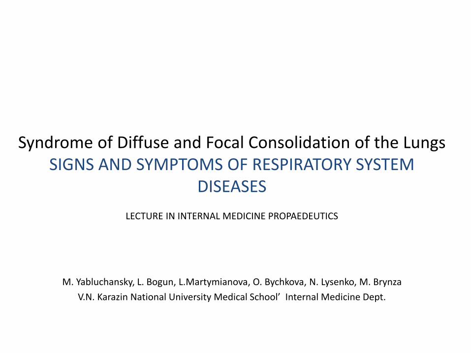
Syndrome of Diffuse and Focal Consolidation of the Lungs SIGNS AND SYMPTOMS OF RESPIRATORY SYSTEM
DISEASES
LECTURE IN INTERNAL MEDICINE PROPAEDEUTICS
M. Yabluchansky, L. Bogun, L.Martymianova, O. Bychkova, N. Lysenko, M. Brynza
V.N. Karazin National University Medical School’ Internal Medicine Dept.
USMLE STEP 1
Myeloperoxidase (MPO) is a heme-containing molecule that is found in the azurophilic granules of neutrophils. Upon release, the enzyme catalyzes hypochlorous acid production during the phagocytic response. In the setting of pneumonia, which of the following is the end result and clinical significance of this reaction?
1. Green color of sputum, 2. Cough, 3. Rust-tinged sputum, 4. Fever, 5. Shortness of breath
http://www.justforhearts.org/2013/08/for-how-long-a-person-can-survive-without-oxygen-water-food/ http://assets-s3.mensjournal.com/img/article/you-re-breathing-all-wrong/298_298_you-re-breathing-all-wrong.jpg

USMLE STEP 1
Correct answer 1: The reaction catalyzed by myeloperoxidase results in the characteristic green-colored sputum that is often found in respiratory infections.
Incorrect answers: 2 & 4: Cough and fever are often the result of a systemic inflammatory response found when a patient is sick. It is often the result of leukotriene release., 3: Usually secondary to a pneumococcal pneumonia infection., 5: Tachypnea is a physiologic response secondary to increased hypercarbia or hypoxemia, and is often secondary to lung pathology.
http://www.justforhearts.org/2013/08/for-how-long-a-person-can-survive-without-oxygen-water-food/ http://assets-s3.mensjournal.com/img/article/you-re-breathing-all-wrong/298_298_you-re-breathing-all-wrong.jpg
Preamble: the importance of the respiratory system
• Since our childhood we all are aware that food, water and oxygen are the basic necessities of life and we cannot survive without them
• An average person can live without food for 3-4 weeks
• We cannot survive without water for more that 3-5 days
• Oxygen is crucial to sustain life, and 3 minutes is the maximum time where person can stay alive without breathing
http://www.justforhearts.org/2013/08/for-how-long-a-person-can-survive-without-oxygen-water-food/
http://assets-s3.mensjournal.com/img/article/you-re-breathing-all-wrong/298_298_you-re-breathing-all-wrong.jpg
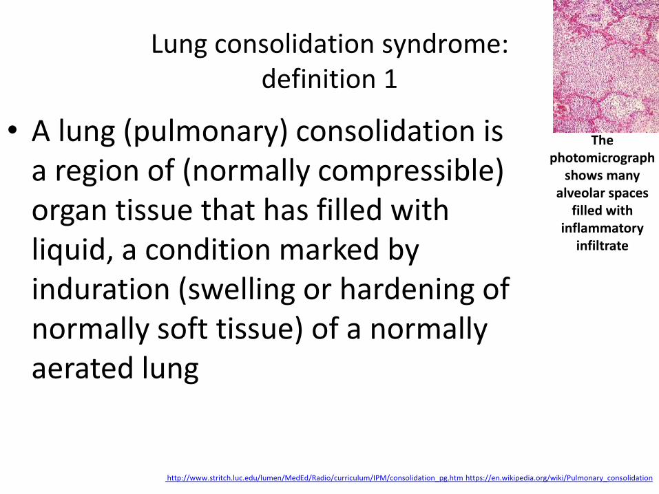
Lung consolidation syndrome: definition 1
• A lung (pulmonary) consolidation is a region of (normally compressible) organ tissue that has filled with liquid, a condition marked by induration (swelling or hardening of normally soft tissue) of a normally aerated lung
http://www.stritch.luc.edu/lumen/MedEd/Radio/curriculum/IPM/consolidation_pg.htm https://en.wikipedia.org/wiki/Pulmonary_consolidation
The photomicrograph
shows many alveolar spaces
filled with inflammatory
infiltrate
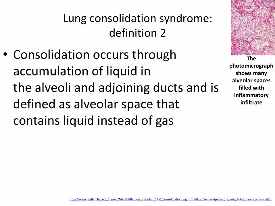
Lung consolidation syndrome: definition 2
• Consolidation occurs through accumulation of liquid in the alveoli and adjoining ducts and is defined as alveolar space that contains liquid instead of gas
http://www.stritch.luc.edu/lumen/MedEd/Radio/curriculum/IPM/consolidation_pg.htm https://en.wikipedia.org/wiki/Pulmonary_consolidation
The photomicrograph
shows many alveolar spaces
filled with inflammatory
infiltrate

Lung consolidation syndrome: definition 3
• The liquid can be pulmonary edema, inflammatory exudate, pus, inhaled water, or blood (from bronchial tree or hemorrhage from a pulmonary artery)
http://www.stritch.luc.edu/lumen/MedEd/Radio/curriculum/IPM/consolidation_pg.htm https://en.wikipedia.org/wiki/Pulmonary_consolidation
The photomicrograph
shows many alveolar spaces
filled with inflammatory
infiltrate

Lung consolidation syndrome: diseases
• Pneumonia
• Infections (lung): actinomycosis, ascariasis, aspergillosis (invasive/infection or allergic), blastomycosis, cryptococcosis, hydatid cyst, syphilis
• Pulmonary edema (fluid in lungs)
• Tumors of the lung
• Atelectasis (collapsed lung) http://www.healthhype.com/consolidation-in-lung-signs-symptoms-and-causes.html
The mediastinal lymphadenopathy and
lung consolidation
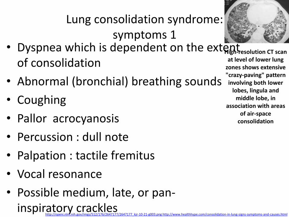
Lung consolidation syndrome: symptoms 1
• Dyspnea which is dependent on the extent of consolidation
• Abnormal (bronchial) breathing sounds
• Coughing
• Pallor acrocyanosis
• Percussion : dull note
• Palpation : tactile fremitus
• Vocal resonance
• Possible medium, late, or pan-inspiratory crackles
http://openi.nlm.nih.gov/imgs/512/176/2647177/2647177_kjr-10-21-g003.png http://www.healthhype.com/consolidation-in-lung-signs-symptoms-and-causes.html
High-resolution CT scan at level of lower lung
zones shows extensive "crazy-paving" pattern involving both lower
lobes, lingula and middle lobe, in
association with areas of air-space
consolidation
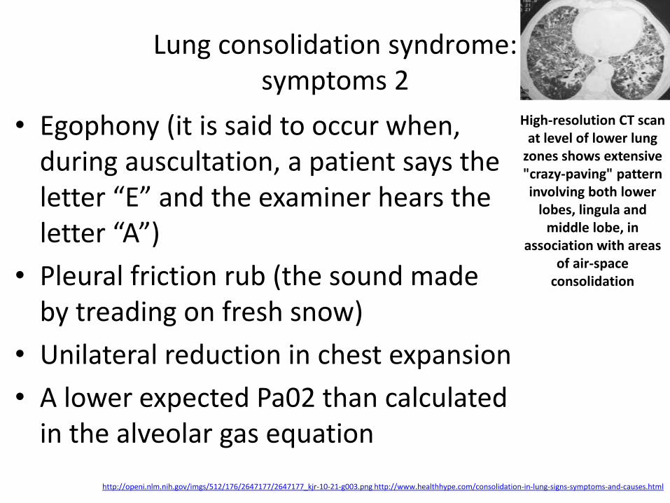
Lung consolidation syndrome: symptoms 2
• Egophony (it is said to occur when, during auscultation, a patient says the letter “E” and the examiner hears the letter “A”)
• Pleural friction rub (the sound made by treading on fresh snow)
• Unilateral reduction in chest expansion
• A lower expected Pa02 than calculated in the alveolar gas equation
http://openi.nlm.nih.gov/imgs/512/176/2647177/2647177_kjr-10-21-g003.png http://www.healthhype.com/consolidation-in-lung-signs-symptoms-and-causes.html
High-resolution CT scan at level of lower lung
zones shows extensive "crazy-paving" pattern involving both lower
lobes, lingula and middle lobe, in
association with areas of air-space
consolidation

USMLE STEP 1 A 21-year-old college student with a history of hypertrophic cardiomyopathy and a systolic murmur heard best at the left sternal border presents to the emergency room with a temperature of 102.3 F, worsening shortness of breath, chest pain and productive cough. He states that two days ago upon returning from Spring Break in Australia he drank a significant amount of alcohol and had an episode of vomiting. He recalls little about the evening. His electrocardiogram displays a regular sinus rhythm and his chest x-ray is shown in Figure A. Which of the following is the most likely etiology of his illness?
1. Long-distance travel, 2. Impaired cough reflex, 3. Hypertrophic cardiomyopathy, 4. Valvular disease, 5. Close living quarters
http://www.justforhearts.org/2013/08/for-how-long-a-person-can-survive-without-oxygen-water-food/ http://assets-s3.mensjournal.com/img/article/you-re-breathing-all-wrong/298_298_you-re-breathing-all-wrong.jpg
USMLE STEP 1
Correct answer 2: Aspiration pneumonia is associated with a diminished cough reflex and dense opacities in the dependent lung fields. Impaired cough reflex, as seen with alcohol ingestion, is a risk factor for aspiration pneumonia.
Incorrect answers: 1: Long distance travel is often associated with the formation of pulmonary embolism., 3: Hypertrophic cardiomyopathy is often asymptomatic and diagnosed incidentally by EKG. It can be a cause of sudden death., 4: Valvular heart disease can be associated with IV drug use with the resulting formation of pulmonary embolism., 5: Close living quarters such as army barracks or college dorm rooms are often associated with infectious diseases such as tuberculosis or meningococcemia.
http://www.justforhearts.org/2013/08/for-how-long-a-person-can-survive-without-oxygen-water-food/ http://assets-s3.mensjournal.com/img/article/you-re-breathing-all-wrong/298_298_you-re-breathing-all-wrong.jpg
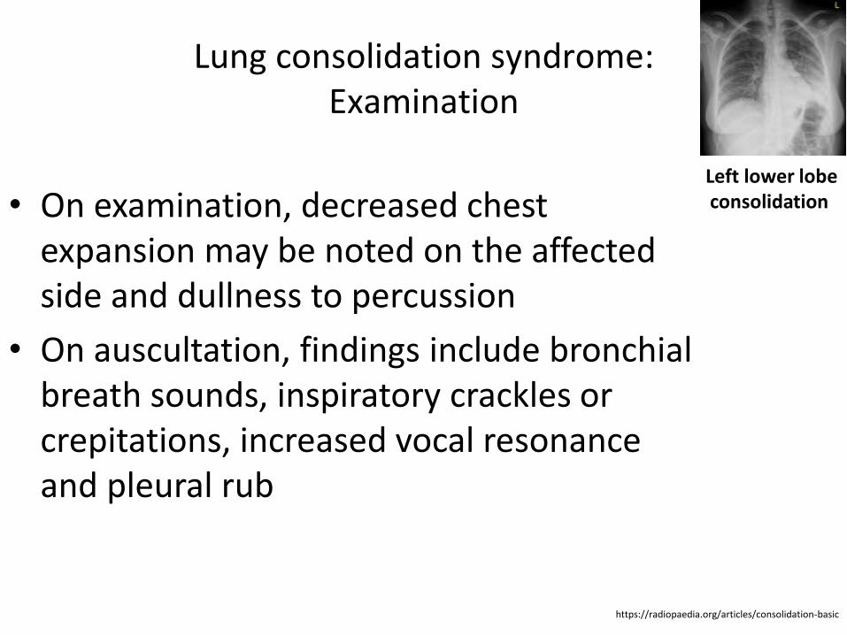
Lung consolidation syndrome: Examination
• On examination, decreased chest expansion may be noted on the affected side and dullness to percussion
• On auscultation, findings include bronchial breath sounds, inspiratory crackles or crepitations, increased vocal resonance and pleural rub
https://radiopaedia.org/articles/consolidation-basic
Left lower lobe consolidation
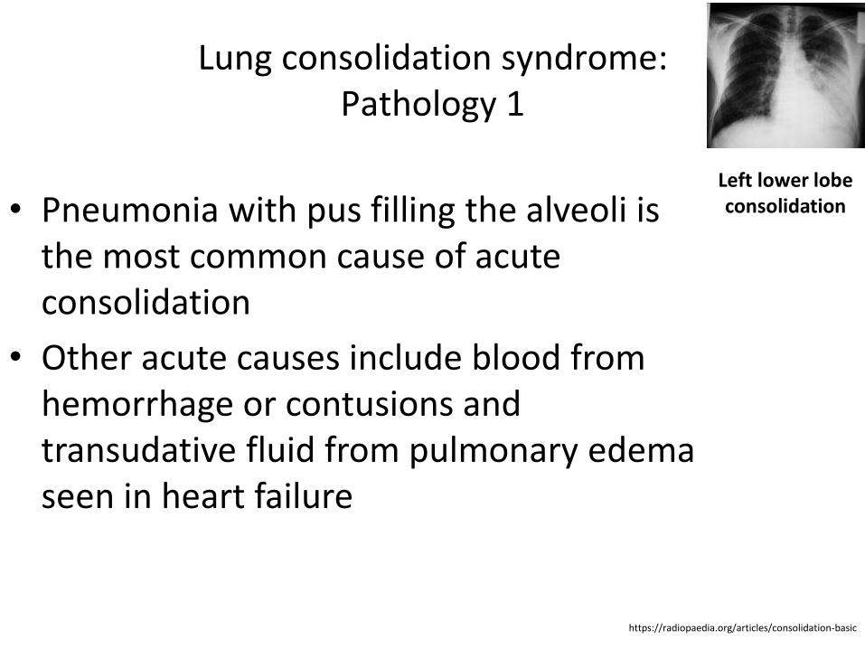
Lung consolidation syndrome: Pathology 1
• Pneumonia with pus filling the alveoli is the most common cause of acute consolidation
• Other acute causes include blood from hemorrhage or contusions and transudative fluid from pulmonary edema seen in heart failure
https://radiopaedia.org/articles/consolidation-basic
Left lower lobe consolidation
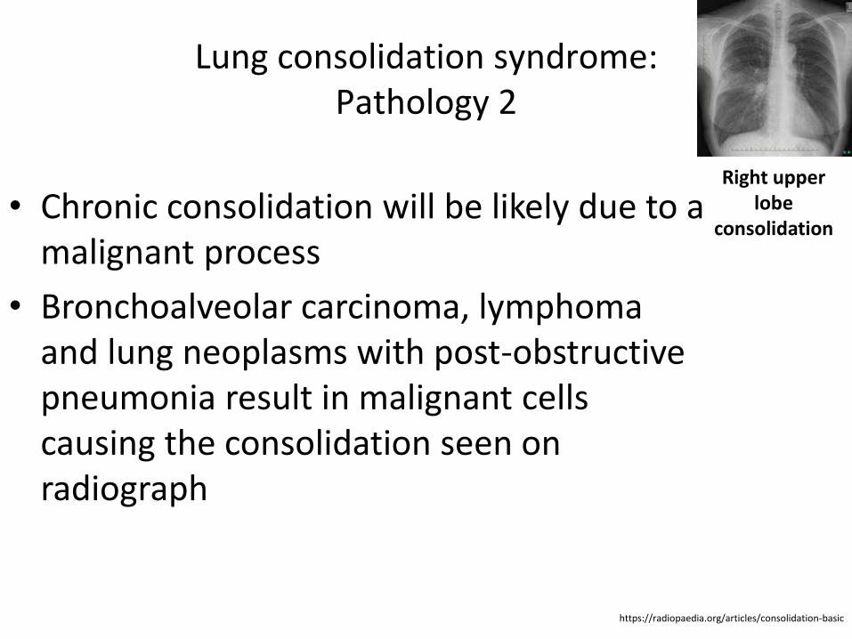
Lung consolidation syndrome: Pathology 2
• Chronic consolidation will be likely due to a malignant process
• Bronchoalveolar carcinoma, lymphoma and lung neoplasms with post-obstructive pneumonia result in malignant cells causing the consolidation seen on radiograph
https://radiopaedia.org/articles/consolidation-basic
Right upper lobe
consolidation

Lung consolidation syndrome: Pathology 3
• Chronic consolidation will be likely due to a malignant process
• Bronchoalveolar carcinoma, lymphoma and lung neoplasms with post-obstructive pneumonia result in malignant cells causing the consolidation seen on radiograph
https://radiopaedia.org/articles/consolidation-basic
Right sided consolidation (multi-lobar)
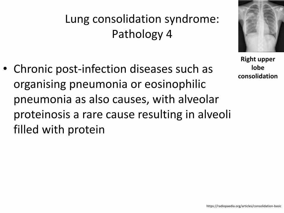
Lung consolidation syndrome: Pathology 4
• Chronic post-infection diseases such as organising pneumonia or eosinophilic pneumonia as also causes, with alveolar proteinosis a rare cause resulting in alveoli filled with protein
https://radiopaedia.org/articles/consolidation-basic
Right upper lobe
consolidation
Lung consolidation syndrome: Radiographic features
• Consolidated areas are radio opaque on chest radiograph and chest CT compared to normally air filled lung tissue
• The distribution pattern of consolidation can aid in narrowing the potential differential diagnosis
https://radiopaedia.org/articles/consolidation-basic
Lung consolidation syndrome: Lobar Consolidation
• Where increased density/opacity is seen in individual lung lobes
• Sharp delineation can be seen when consolidation reaches a fissure, since it does not cross
• Air bronchograms can also be seen due to bronchi becoming visible against the dense diseased tissue
• Volume loss is usually not seen
https://radiopaedia.org/articles/consolidation-basic
Lung consolidation syndrome: Diffuse Consolidation
• Most commonly due to heart failure, resulting in other signs such increased cardiac size, Kerley B-lines, redistribution on pulmonary blood flow and pleural fluid
• Other findings can include multiple ill defined opacities progressing to diffuse spread seen in bronchopneumonia and "white out" of a lung due to progressive consolidation from bronchoalveolar carcinoma
https://radiopaedia.org/articles/consolidation-basic
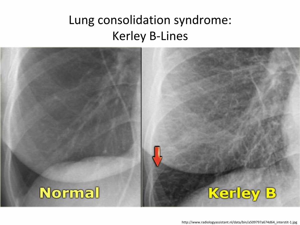
Lung consolidation syndrome: Kerley B-Lines
• These are short parallel lines at the lung periphery These lines represent interlobular septa, which are usually less than 1 cm in length and parallel to one another at right angles to the pleura
• They are located peripherally in contact with the pleura, but are generally absent along fissural surfaces
https://en.wikipedia.org/wiki/Kerley_lines
Lung consolidation syndrome: Kerley B-Lines
http://www.radiologyassistant.nl/data/bin/a509797a674d64_interstit-1.jpg
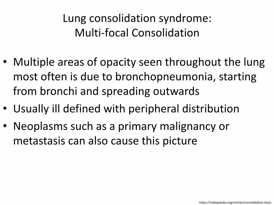
Lung consolidation syndrome: Multi-focal Consolidation
• Multiple areas of opacity seen throughout the lung most often is due to bronchopneumonia, starting from bronchi and spreading outwards
• Usually ill defined with peripheral distribution
• Neoplasms such as a primary malignancy or metastasis can also cause this picture
https://radiopaedia.org/articles/consolidation-basic
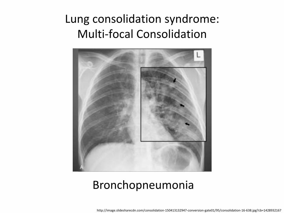
Lung consolidation syndrome: Multi-focal Consolidation
http://image.slidesharecdn.com/consolidation-150413132947-conversion-gate01/95/consolidation-16-638.jpg?cb=1428932167
Bronchopneumonia
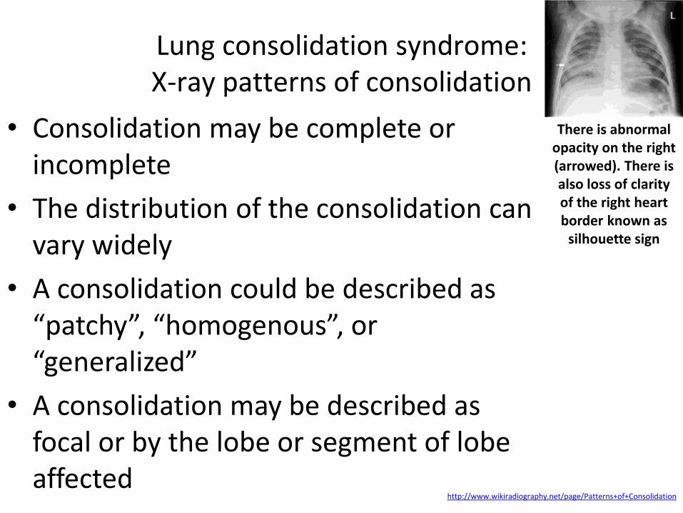
Lung consolidation syndrome: X-ray patterns of consolidation
• Consolidation may be complete or incomplete
• The distribution of the consolidation can vary widely
• A consolidation could be described as “patchy”, “homogenous”, or “generalized”
• A consolidation may be described as focal or by the lobe or segment of lobe affected
http://www.wikiradiography.net/page/Patterns+of+Consolidation
There is abnormal opacity on the right (arrowed). There is also loss of clarity of the right heart border known as
silhouette sign
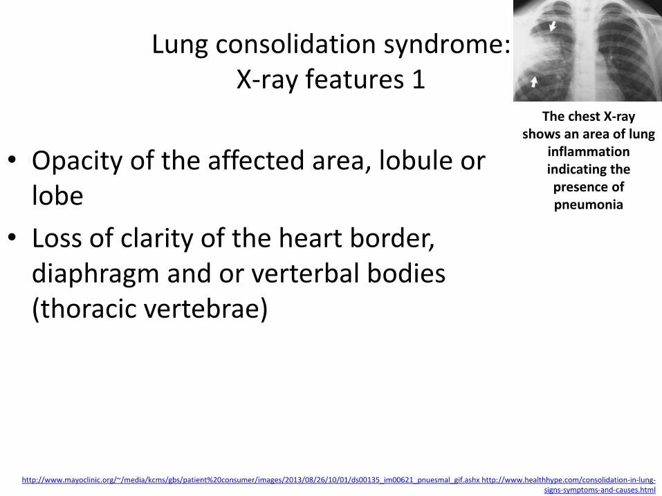
Lung consolidation syndrome: X-ray features 1
• Opacity of the affected area, lobule or lobe
• Loss of clarity of the heart border, diaphragm and or verterbal bodies (thoracic vertebrae)
http://www.mayoclinic.org/~/media/kcms/gbs/patient%20consumer/images/2013/08/26/10/01/ds00135_im00621_pnuesmal_gif.ashx http://www.healthhype.com/consolidation-in-lung-signs-symptoms-and-causes.html
The chest X-ray shows an area of lung
inflammation indicating the presence of pneumonia
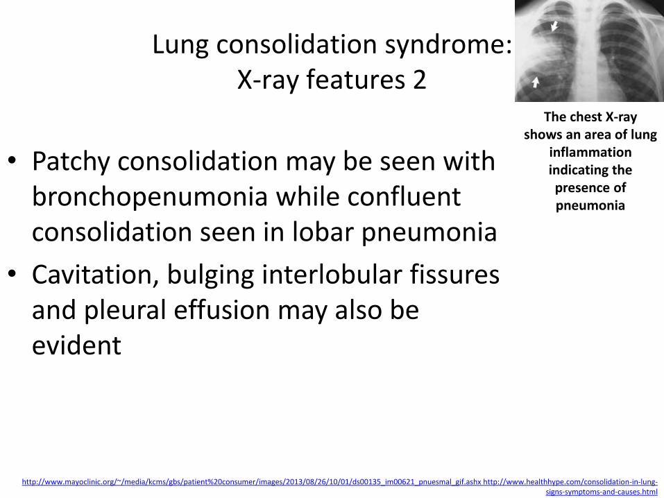
Lung consolidation syndrome: X-ray features 2
• Patchy consolidation may be seen with bronchopenumonia while confluent consolidation seen in lobar pneumonia
• Cavitation, bulging interlobular fissures and pleural effusion may also be evident
http://www.mayoclinic.org/~/media/kcms/gbs/patient%20consumer/images/2013/08/26/10/01/ds00135_im00621_pnuesmal_gif.ashx http://www.healthhype.com/consolidation-in-lung-signs-symptoms-and-causes.html
The chest X-ray shows an area of lung
inflammation indicating the presence of pneumonia

Lung consolidation syndrome: Right Upper Lobe (RUL) consolidation
http://www.wikiradiography.net/page/Patterns+of+Consolidation
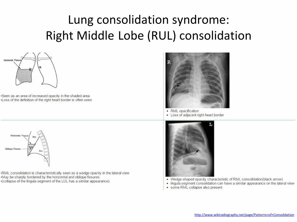
Lung consolidation syndrome: Right Middle Lobe (RUL) consolidation
http://www.wikiradiography.net/page/Patterns+of+Consolidation
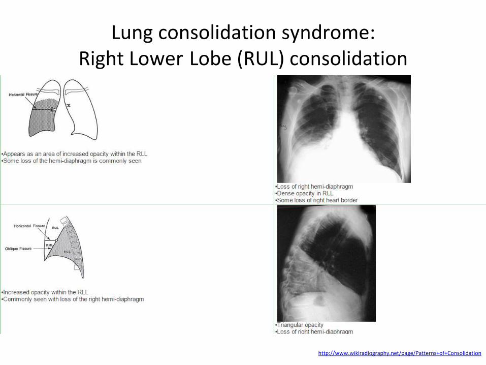
Lung consolidation syndrome: Right Lower Lobe (RUL) consolidation
http://www.wikiradiography.net/page/Patterns+of+Consolidation
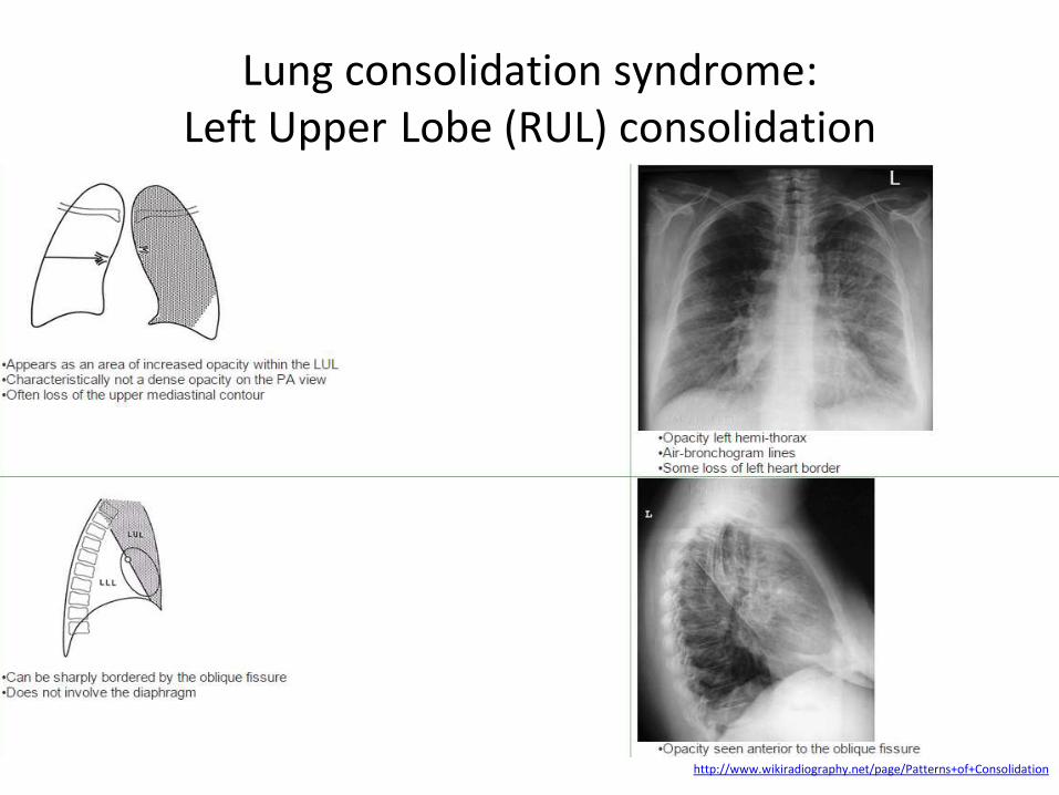
Lung consolidation syndrome: Left Upper Lobe (RUL) consolidation
http://www.wikiradiography.net/page/Patterns+of+Consolidation
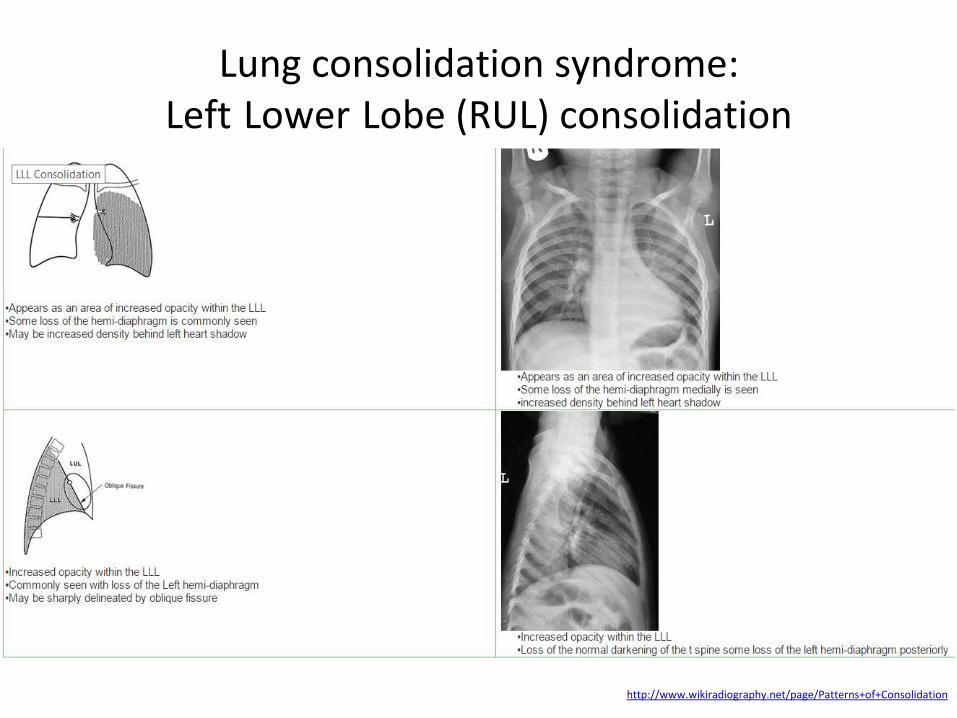
Lung consolidation syndrome: Left Lower Lobe (RUL) consolidation
http://www.wikiradiography.net/page/Patterns+of+Consolidation

Lung consolidation syndrome: lung ultrasound
• The consolidated lung is ‘hepatisised’ (looks similar to liver)
• Extensive consolidation (of a whole lobe) allows the opposite plural line to be seen (8-11cm deep) with mediastinum deeper and with the aorta or IVC still visible
• A fully consolidated lobe may be seen floating in a pleural effusion
http://www.icmteaching.com/ultrasound/lung%20ultrasound/alveolar%20syndrome/
USMLE STEP 1
A 48-year-old male dies in the intensive care unit following a severe Streptococcus pneumonia and septic shock. Autopsy of the lung reveals a red, firm left lower lobe. What would you most likely find on microscopic examination of the lung specimen?
1. Eosinophilia in the alveolar septa, 2. Vascular dilation and noncaseating granulomas, 3. Fragmented erythrocytes, 4. Alveolar exudate containing neutrophils, erythrocytes, and fibrin, 5. Collagen whorls
http://www.justforhearts.org/2013/08/for-how-long-a-person-can-survive-without-oxygen-water-food/ http://assets-s3.mensjournal.com/img/article/you-re-breathing-all-wrong/298_298_you-re-breathing-all-wrong.jpg
USMLE STEP 1 Correct answer 4: This patient died from lobar pneumonia. The red hepatization phase is grossly characterized by a red, firm lobe with a "liver-like" consistency, that corresponds microscopically to a massive confluent alveolar exudate containing neutrophils, RBCs, and fibrin. Incorrect answers: 1: Acute eosinophilic PNA is characterized by respiratory distress, eosinophilic infiltration in the lung, acute onset, resolution of symptoms with corticosteroids and the absence of relapse., 2: Vascular dilation is not a common manifestation of PNA. Noncaseating granulomas are often seen in sarcoidosis, not PNA., 3: Invasive Streptococcus PNA is an uncommon cause of hemolytic uremic syndrome which can cause fragmented erythrocytes. This would not be expected in a typical case of PNA. Fragmented red blood cells are also seen in grey hepatization., 5: Whorls of collagen in histological examination are highly indicative of silicosis.
http://www.justforhearts.org/2013/08/for-how-long-a-person-can-survive-without-oxygen-water-food/ http://assets-s3.mensjournal.com/img/article/you-re-breathing-all-wrong/298_298_you-re-breathing-all-wrong.jpg
Lung consolidation syndrome: pulmonary consolidation with fever is not always
pneumonia 1
• Microscopic polyangiitis (MPA) is defined as systemic necrotizing vasculitis with few or no immune deposits, affecting small vessels (capillaries, arterioles or venules)
• Although MPA can involve any organ, renal and pulmonary involvement predominate
• Pulmonary involvement can present from fleeting focal infiltrates to massive lung hemorrhage and hemoptysis secondary to alveolar capillaritis
http://www.sciencedirect.com/science/article/pii/S1755001708000328
Lung consolidation syndrome: pulmonary consolidation with fever is not always
pneumonia 2
• This is a case of a 39-year-old male who was admitted to hospital due to fever of unknown origin
• He was an ex-smoker (stopped 3 years ago) and his medical history is unremarkable
• Ten days back he was examined by his doctor due to fever, fatigue and dry cough
• He was diagnosed as having pneumonia
http://www.sciencedirect.com/science/article/pii/S1755001708000328
Lung consolidation syndrome: pulmonary consolidation with fever is not always
pneumonia 3 • The patient had normal breath sounds with normal
blood gases and his temperature was 38 °C
• Laboratory studies revealed white cell count: 13,9 (N:79, L:11.5, M:8.5, E:0.6) hemoglobulin 13.7 g/dl, the erythrocyte sedimentation rate 72 mm/h
• Renal, liver function and urinalysis were normal
• An arterial blood gas measurement while the patient was breathing room air demonstrated a PaO2 of 88 mmHg, a PaCO2 41 mmHg and pH=7.39
http://www.sciencedirect.com/science/article/pii/S1755001708000328
Lung consolidation syndrome: pulmonary consolidation with fever is not always
pneumonia 4
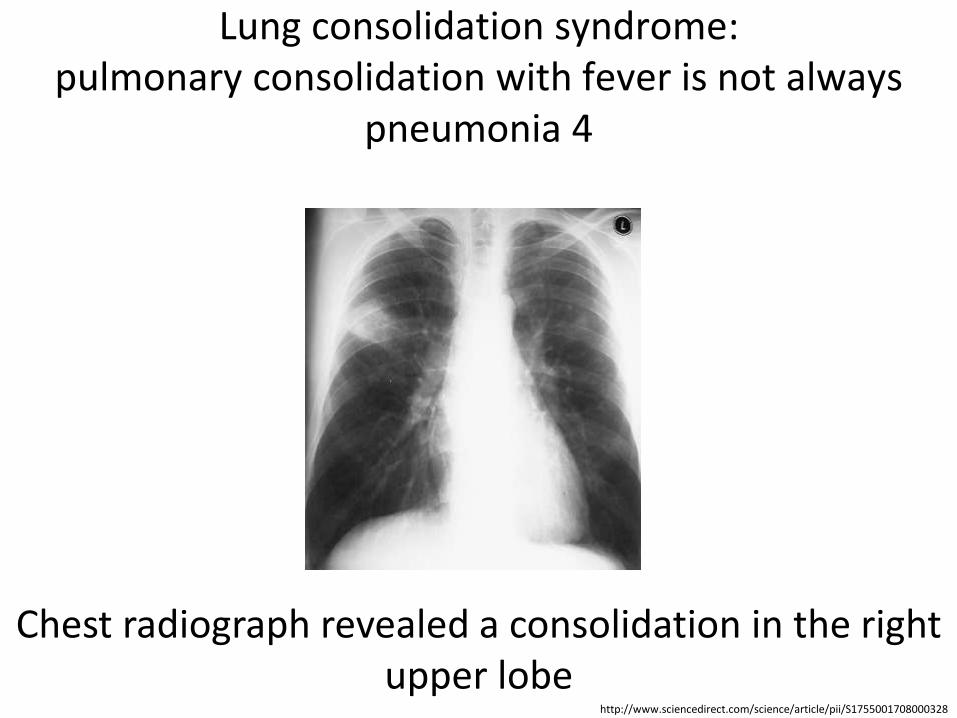
Chest radiograph revealed a consolidation in the right upper lobe
http://www.sciencedirect.com/science/article/pii/S1755001708000328
Lung consolidation syndrome: pulmonary consolidation with fever is not always
pneumonia 5 • The patient was admitted in hospital and began
treatment with moxifloxacin 400 mg once daily
• Sputum and blood cultures were obtained and serology tests were conducted to rule out bacterial or viral infection
• A new radiograph showed an increase of the consolidation
• Due to nonresolving of the shadowing on the X-ray, the treatment was switched to ticarcillin/ potassium plus teicoplanine
http://www.sciencedirect.com/science/article/pii/S1755001708000328
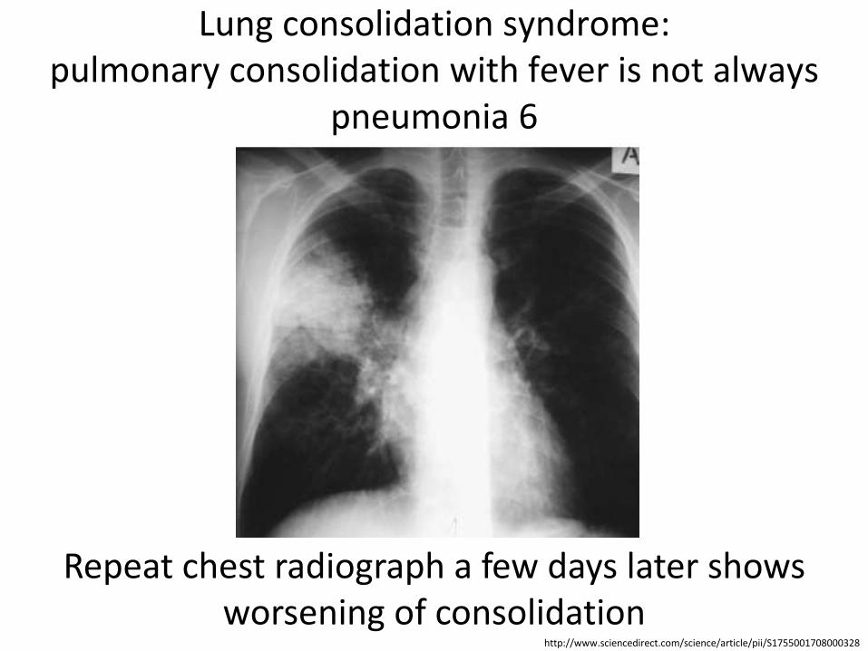
Lung consolidation syndrome: pulmonary consolidation with fever is not always
pneumonia 6
Repeat chest radiograph a few days later shows worsening of consolidation
http://www.sciencedirect.com/science/article/pii/S1755001708000328
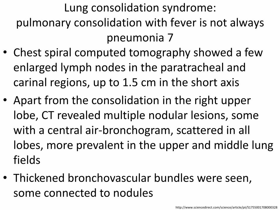
Lung consolidation syndrome: pulmonary consolidation with fever is not always
pneumonia 7 • Chest spiral computed tomography showed a few
enlarged lymph nodes in the paratracheal and carinal regions, up to 1.5 cm in the short axis
• Apart from the consolidation in the right upper lobe, CT revealed multiple nodular lesions, some with a central air-bronchogram, scattered in all lobes, more prevalent in the upper and middle lung fields
• Thickened bronchovascular bundles were seen, some connected to nodules
http://www.sciencedirect.com/science/article/pii/S1755001708000328
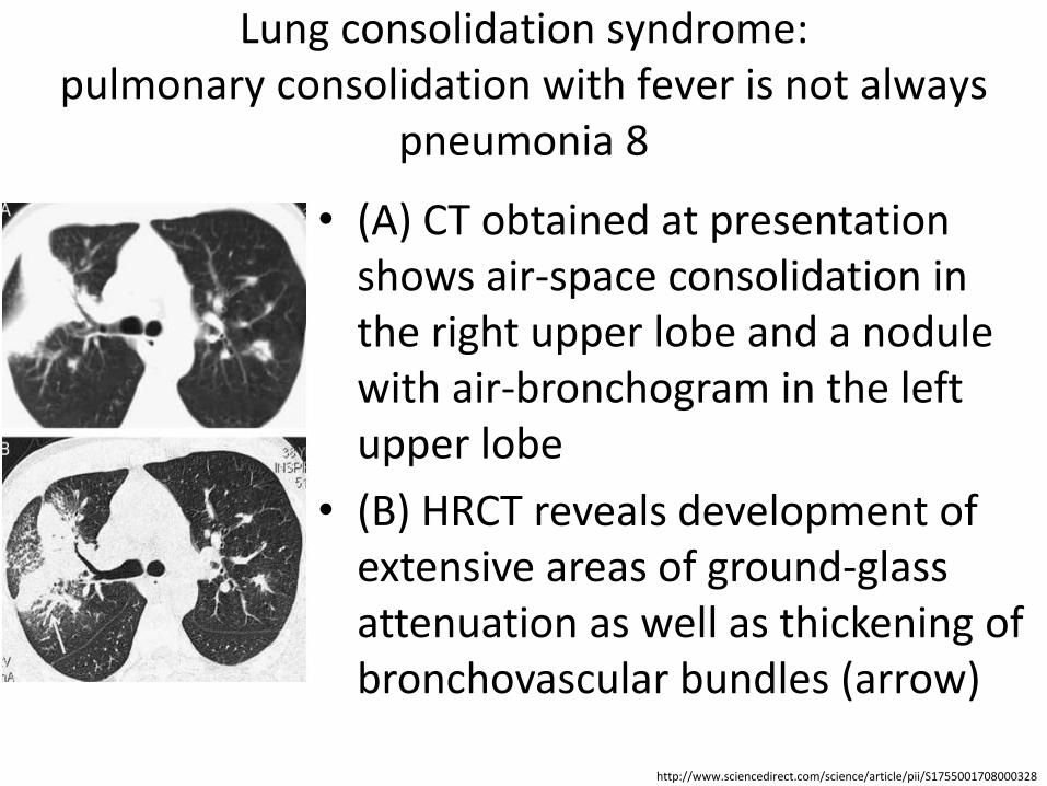
Lung consolidation syndrome: pulmonary consolidation with fever is not always
pneumonia 8
• (A) CT obtained at presentation shows air-space consolidation in the right upper lobe and a nodule with air-bronchogram in the left upper lobe
• (B) HRCT reveals development of extensive areas of ground-glass attenuation as well as thickening of bronchovascular bundles (arrow)
http://www.sciencedirect.com/science/article/pii/S1755001708000328

Lung consolidation syndrome: pulmonary consolidation with fever is not always
pneumonia 9 • (A) CT: multiple nodules in both
lungs connected to thickened bronchovascular bundles
• (B) HRCT: resolution of some nodular opacities in the right lung and patchy ground-glass opacities in both lung fields; newly developed ground-glass areas surround nodules in the left lung, a CT sign strongly indicative of hemorrhagic infiltration (arrows)
http://www.sciencedirect.com/science/article/pii/S1755001708000328
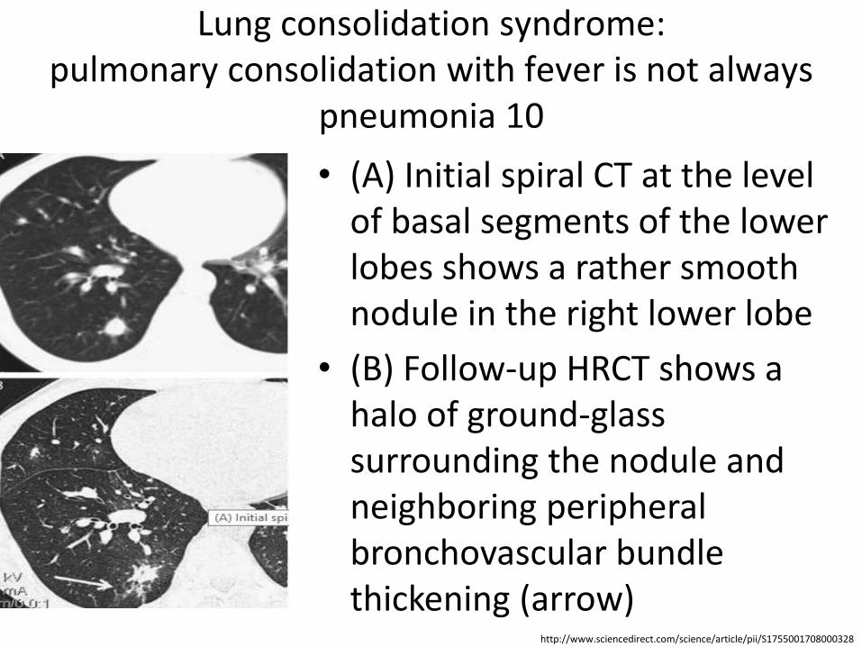
Lung consolidation syndrome: pulmonary consolidation with fever is not always
pneumonia 10 • (A) Initial spiral CT at the level
of basal segments of the lower lobes shows a rather smooth nodule in the right lower lobe
• (B) Follow-up HRCT shows a halo of ground-glass surrounding the nodule and neighboring peripheral bronchovascular bundle thickening (arrow)
http://www.sciencedirect.com/science/article/pii/S1755001708000328
Lung consolidation syndrome: pulmonary consolidation with fever is not always
pneumonia 11
• Taking into account the febrile patient and nonresolving “pneumonia”, the differential diagnosis was broadened widespread infections, tuberculosis, whereas thickened bronchovascular bundles connected with nodules and lymphadenopathy indicated Wegener granulomatosis and lymphoma
http://www.sciencedirect.com/science/article/pii/S1755001708000328
Lung consolidation syndrome: pulmonary consolidation with fever is not always
pneumonia 12
• Perinuclear anti-neutrophil cytoplasmic antibodies were positive and renal biopsy revealed focal pauci-immune necrotic glomerulonephritis
• The diagnosis of microscopic polyangiitis was established
• The patient began the treatment with methylprednisolone and
• Remission was achieved
http://www.sciencedirect.com/science/article/pii/S1755001708000328


![7 Catheter-associated Urinary Tract Infection (CAUTI) · UTI Urinary Tract Infection (Catheter-Associated Urinary Tract Infection [CAUTI] and Non-Catheter-Associated Urinary Tract](https://img.pdfslide.net/doc/110x75/5c40b88393f3c338af353b7f/7-catheter-associated-urinary-tract-infection-cauti-uti-urinary-tract-infection.jpg)