Embed Size (px)
Citation preview

7/27/2019 Silica Xerogel as an Implantable Carrier for Controlled Drug
http://slidepdf.com/reader/full/silica-xerogel-as-an-implantable-carrier-for-controlled-drug 1/6
*Corresponding author. Tel.: #358-2-2727690; fax: #358-2-
2727273.
E-mail address: pirjo.kortesuo@orion." (P. Kortesuo)ଝPresented in part at the 23rd Annual Meeting of the Society for
Biomaterials, New Orleans, 1997.
Biomaterials 21 (2000) 193}198
Silica xerogel as an implantable carrier for controlled drugdelivery* evaluation of drug distribution and tissue e! ects
after implantationଝ
Pirjo Kortesuo*, Manja Ahola, Stefan Karlsson, Ilkka Kangasniemi,Antti Yli-Urpo, Juha Kiesvaara
Orion Corporation, Orion Pharma, Pharmaceutical Development Department, P.O. Box 425, FIN-20101, Turku, Finland
Institute of Dentistry, University of Turku, Lemminka( isenkatu 2, FIN-20520, Turku, Finland
Received 30 September 1998; accepted 15 July 1999
Abstract
The purpose of the present study was to examine controlled delivery of toremifene citrate from subcutaneously implanted silica
xerogel carrier and to evaluate silica xerogel related tissue e! ects after implantation. Toremifene citrate was incorporated into
hydrolyzed silica sol in a room temperature process. Toremifene citrate treated silica xerogel implants were tested both in vitro and
in vivo using healthy mice. Silica xerogel with tritium-labelled toremifene was implanted subcutaneously in mice for 42 d. To
determine the amount of tritiated toremifene remaining in the silica discs at the implantation site, the discs were excised periodically
and radioactivity measured. The amount of tritiated toremifene in the implant after 42 d was still about 16% and the amount of silica
xerogel about 25%. In a histopathological study silica xerogel did not show any tissue irritation at the site of the implantation.
A "brotic capsule was formed around the implant. No silica xerogel related histological changes in liver, kidney, lymph nodes and
uterus were observed during the implantation period. The silica xerogel discs showed a sustained release of toremifene citrate over
42 d. Histologically, toremifene-related changes in the uterus were also detectable at all studied time points. These "ndings suggestthat silica xerogel is a promising carrier material for implantable controlled drug delivery system. 1999 Elsevier Science Ltd.
All rights reserved.
Keywords: Sol}gel processed silica xerogel; Implantation study; Drug delivery; Toremifene citrate; Tissue distribution
1. Introduction
Sol}gel processed porous silica glasses have a con-
siderable potential as carriers for controlled drug release
[1}4]. In recent years, processing factors controlling thedegradation rate of the matrix in vitro have been exten-
sively studied [1,3]. However, in vivo degradation and
drug delivery of low-temperature processed silica xerogel
are not well documented.
Parenterally implanted sintered sol}gel produced
glasses containing 85% silica and also pure silica glasses
are biocompatible [5,6]. Bioactive glasses in soft tissue or
bone enhance "broblast or osteoblast formation result-
ing in increased collagen formation, which in turn bonds
bioactive glass implants to bone or soft tissue [5,7].
However, crystalline silicon dioxide in the form of aero-
sol is known to cause a rapid in#ux of in#ammatory cellsand increased collagen deposition in lungs and histologi-
cal changes in the pulmonary lymph nodes [8].
Toremifene citrate, a non-steroidal antiestrogenic com-
pound, which has antitumor activity in breast and en-
dometrial cancer, was used as model drug in this study
[9]. Local therapy of antiestrogens after hormone-depen-
dent breast cancer surgery could give a targeted and
long-lasting disease control while minimizing systemic
distribution.
In this study toremifene citrate/ H-toremifene was in-
corporated into the silica gel matrix during a room-tem-
perature gelation process and silica gel was dried toa constant weight at 403C without sintering at a higher
temperature. The objectives of this work were: (i) to
0142-9612/00/$- see front matter 1999 Elsevier Science Ltd. All rights reserved.
PII: S 0 1 42 - 9 6 1 2 (9 9 ) 0 0 1 48 - 9

7/27/2019 Silica Xerogel as an Implantable Carrier for Controlled Drug
http://slidepdf.com/reader/full/silica-xerogel-as-an-implantable-carrier-for-controlled-drug 2/6
evaluate the capability of silica xerogel to deliver tor-
emifene in a sustained manner, (ii) to study tissue distri-
bution of toremifene and (iii) to evaluate histological
e! ects after implantation of the silica xerogel itself on the
uterus, liver, kidneys, lymph nodes and at the implanta-
tion site.
2. Materials and methods
2.1. Preparation of toremifene citrate incorporated
silica gels
Silica gel was prepared by the hydrolysis and polycon-
densation of tetraethoxysilane (TEOS) with polyethylene
glycol (PEG, M
4600), water and acetic acid in a
mole ratio of TEOS : PEG : H
O : CH
COOH"
1.0 : 0.0012 : 14.2 : 0.5 at room temperature. Toremifene
citrate (Orion Corporation, Turku, Finland) was dis-solved into the silica solution after 1 h hydrolysis at the
concentration of 33 mg/g. Tritiated toremifene was syn-
thetized as described earlier [10]. The speci"c activity of
the material was 16 Ci/mmol. It was added to the silica
sol to produce a "nal radioactivity of 16 Ci/g. Solution
was pipetted into round moulds (100 l/mould) for poly-
condensation and aging at 403C for 18 h. The poly-
merized silica gels were soaked in water to leach out
residual organic matter within the gel and dried at 403C
to a constant weight. The silica xerogel implants were
round with the diameter approximately 4.7 mm and
height 0.9 mm weighing about 15 mg.
2.2. In vitro dissolution test
The dissolution pro"les (each data point is the mean of
2 values) of H-toremifene and silica from test specimens
were studied using the USP XXIII dissolution apparatus
II (paddle method, Sotax AT6, Basel, Switzerland) at
a constant temperature (373C). Simulated body #uid
(SBF, pH 7.4) containing 0.5% (m/v) sodiumdodecylsul-
phate was used as dissolution medium. SBF was pre-
pared by dissolving reagent-grade NaCl (136.8 mM
),NaHCO
(4.2 mM), KCl (3.0 mM), K
HPO;3H
O
(1.0 mM), MgCl;6H
O (1.5 mM), CaCl
;2H
O
(2.5 mM) and Na
SO
(0.5 mM) in distilled water. The
solution was bu! ered at pH 7.4 with tris(hy-
droxymethyl)aminomethane (50 mM) and hydrochloric
acid.
The volume of dissolution medium was 250 ml and the
rotation speed 50 rpm. At each sample interval a 2 ml
sample was withdrawn and replaced with fresh medium.
To study the amount of tritiated toremifene released
from the matrix, samples of 0.5 ml were mixed with 10 ml
of scintillation liquid (Ready Safe2+, Beckmann, CA,USA) and measured by scintillation counting (Model
81 000, LKB-Wallac, Turku, Finland).
Degradation of silica xerogel matrix was determined
by measuring dissolved Si(OH)
spectrophotometrically
as a molybden blue complex at 820 nm [11].
2.3. Animal experiments
The animals were housed according to the recommen-
dations in DHEW pub. (NIH) 85-23 entitled &Guide for
the care and use of laboratory animals'. Thirty female
mice (C57Bl, Denmark) with an average weight of about
19.6 g (SD 1.2 g) received implants subcutaneously on the
back on either side of the backbone with tritium-labelled
toremifene loaded silica implants (a total of 2 implants
were placed in each mouse) and 30 mice with untreated
silica implants. The H-toremifene dose was about
80 Ci/kg (0.8 Ci/implant), toremifene citrate 350 mg/
kg (approx. 3.4 mg/implant) and silica xerogel about
1.53 g/kg body weight.
2.4. Histology
The animals were killed at 7, 14, 21, 28, 35 and 42 d
(5 mice per time period) after implantation. The silica
xerogel implants on the left side of the backbone were
removed together with the surrounding tissue and "xed
in 70% ethanol and embedded in PMMA (Technovit).
Sections of 20 m were stained with toluidine blue. Sam-
ples of liver, kidney, mesenterial lymph node and uterus
were "xed in bu! ered formaldehyde and embedded in
para$n. Sections of 6 m were stained with hema-toxylin}eosin. Samples were evaluated using light
microscopy. The samples of the implantation site were
studied from all interim kills but liver, kidney, lymph
node and uterus only from the animals treated 7, 28 and
42 d.
2.5. Drug release and silica xerogel erosion studies
The mice were killed, whereupon the silica xerogel
implants on the right side of the backbone were cut o!
from the surrounding "brous capsule and dried at room
temperature in a desiccator for 24 h. The percentage of implant remaining at each time point was calculated. To
determine the amount of toremifene remaining in the
implants, the dried implants were dissolved in 0.1 N
NaOH solution and the activity was measured in liquid
scintillation counter.
2.6. Organ radioactivity counting
After killing the mice, the tissue samples taken from
liver, kidney, uterus, lung, blood and application area
were burned in an oxidizer (Junitek, Kaarina, Finland)and the radioactivity was measured by scintillation
counting.
194 P. Kortesuo et al. / Biomaterials 21 (2000) 193}198

7/27/2019 Silica Xerogel as an Implantable Carrier for Controlled Drug
http://slidepdf.com/reader/full/silica-xerogel-as-an-implantable-carrier-for-controlled-drug 3/6
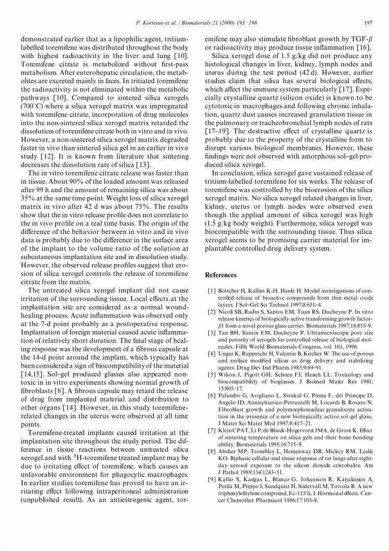
Fig. 1. In vitro release of H-toremifene and silica from silica xerogel
matrix.
Fig. 2. Percentage of remaining silica xerogel implant and H-tor-emifene radioactivity at di! erent time points in vivo.
Fig. 3. Biodistribution of H-toremifene radioactivity at di! erent time
points in some of the organs in mice.
3. Results
3.1. In vitro and in vivo release of tritium-labelled
toremifene and erosion of silica xerogel matrix
About 90% of the loaded tritium-labelled toremifene
was released after 99 h in vitro. The amount of silica
released in 99 h is equivalent to about 65% weight loss of silica xerogel matrix (Fig. 1).
Weight loss of silica gel matrix in vivo was about
75 wt% during 42 d. The erosion rate was fast during the
"rst 28 d and then decreased (Fig. 2). By an estimate from
the dissolution pro"le, the silica xerogel implant weigh-
ing about 15 mg is totally absorbed after 60}70 d.
The silica xerogel discs showed sustained release of
toremifene during the test period. About 40% of loaded
toremifene eluted during the "rst week. After that the
release of toremifene slowed down and 50% was released
in 3 weeks. The amount of
H-toremifene remaining inthe implant after 42 d was still about 16% (Fig. 2).
The radioactivity of tritiated toremifene was detected
upto 42 d in the liver, kidneys, and lung. The radioactiv-
ity reached a maximum value at 14 d and then remained
relatively constant until the end of the implantation test.
Radioactivity was not found in blood or uterus (Fig. 3).
Toremifene-induced uterine glandular hyperplasia, the
characteristic hormonal change seen in mice treated with
this drug, was observed at all studied time points.
3.2. E w ects after implantation
The local e! ect of the silica xerogel implant was re-
stricted to the close vicinity of the implant. A well-
organized "brous capsule was formed around the im-
plant from 14 d time point onward (Fig. 4A). The implant
did not cause necrosis and the in#ammatory response
observed was mainly due to a minor in"ltration of mac-
rophages at time points 14 d and later. Acute in#amma-
tion was observed only at the 7-d point. Phagocytic
macrophages were present close to the implant at 14 d or
later (Fig. 4B). No accumulation of dissolved material
occurred.
Toremifene released from the implant caused tissueirritation, necrosis and an unfavorable environment for
phagocytic macrophages causing an accumulation of
P. Kortesuo et al. / Biomaterials 21 (2000) 193}198 195

7/27/2019 Silica Xerogel as an Implantable Carrier for Controlled Drug
http://slidepdf.com/reader/full/silica-xerogel-as-an-implantable-carrier-for-controlled-drug 4/6
Fig. 4. (A) An untreated implant (S) in a mouse 35 d after implantation. The implant is surrounded by a "brotic capsule (arrow, original magni"cation
16;). (B) The surface (S) of an untreated implant 21 d after implantation. The "brous capsule (arrow) is well formed and phagocytic macrophages(open arrowheads) are abundant close to the surface (original magni"cation 100;).
dissolved silica inside the "brotic capsule. These "ndings
were present from the 14 d point onward.
No extensive silica xerogel related histological changes
could be observed in the liver, lymph nodes or kidney at
the 1.5 g/kg dose.
4. Discussion
The silica xerogel matrix gave sustained release of
toremifene citrate for more than six weeks. This was
con"rmed by erosion/release studies, activity measure-
ments of tritiated toremifene in tissue samples and by
histological studies. Histologically, toremifene-related
changes in the uterus were detectable at all studied time
points. About 40% of loaded toremifene was eluted from
silica xerogel matrix during the "rst week. After that the
release rate slowed down and about 16% of the loaded
amount was still remaining after 42 d. Therefore, radio-
activity in tissue samples reached a maximum valueat 14 d and remained relatively constant in the liver,
lung and kidneys throughout the test period. It was
196 P. Kortesuo et al. / Biomaterials 21 (2000) 193}198
7/27/2019 Silica Xerogel as an Implantable Carrier for Controlled Drug
http://slidepdf.com/reader/full/silica-xerogel-as-an-implantable-carrier-for-controlled-drug 5/6
demonstrated earlier that as a lipophilic agent, tritium-
labelled toremifene was distributed throughout the body
with highest radioactivity in the liver and lung [10].
Toremifene citrate is metabolized without "rst-pass
metabolism. After enterohepatic circulation, the metab-
olites are excreted mainly in feces. In tritiated toremifene
the radioactivity is not eliminated within the metabolic
pathways [10]. Compared to sintered silica xerogels(7003C) where a silica xerogel matrix was impregnated
with toremifene citrate, incorporation of drug molecules
into the non-sintered silica xerogel matrix retarded the
dissolution of toremifene citrate both in vitro and in vivo.
However, a non-sintered silica xerogel matrix degraded
faster in vivo than sintered silica gel in an earlier in vivo
study [12]. It is known from literature that sintering
decreases the dissolution rate of silica [13].
The in vitro toremifene citrate release was faster than
in tissue. About 90% of the loaded amount was released
after 99 h and the amount of remaining silica was about35% at the same time point. Weight loss of silica xerogel
matrix in vivo after 42 d was about 75%. The results
show that the in vitro release pro"le does not correlate to
the in vivo pro"le on a real time basis. The origin of the
di! erence of the behavior between in vitro and in vivo
data is probably due to the di! erence in the surface area
of the implant to the volume ratio of the solution at
subcutaneous implantation site and in dissolution study.
However, the observed release pro"les suggest that ero-
sion of silica xerogel controls the release of toremifene
citrate from the matrix.
The untreated silica xerogel implant did not causeirritation of the surrounding tissue. Local e! ects at the
implantation site are considered as a normal wound-
healing process. Acute in#ammation was observed only
at the 7-d point probably as a postoperative response.
Implantation of foreign material caused acute in#amma-
tion of relatively short duration. The "nal stage of heal-
ing response was the development of a "brous capsule at
the 14-d point around the implant, which typically has
been considered a sign of biocompatibility of the material
[14,15]. Sol}gel produced glasses also appeared non-
toxic in in vitro experiments showing normal growth of
"broblasts [6]. A "brous capsule may retard the release
of drug from implanted material and distribution to
other organs [14]. However, in this study toremifene-
related changes in the uterus were observed at all time
points.
Toremifene-treated implants caused irritation at the
implantation site throughout the study period. The dif-
ference in tissue reactions between untreated silica
xerogel and with H-toremifene treated implant may be
due to irritating e! ect of toremifene, which causes an
unfavorable environment for phagocytic macrophages.
In earlier studies toremifene has proved to have an ir-ritating e! ect following intraperitoneal administration
(unpublished result). As an antiestrogenic agent, tor-
emifene may also stimulate "broblast growth by TGF-
or radioactivity may produce tissue in#ammation [16].
Silica xerogel dose of 1.5 g/kg did not produce any
histological changes in liver, kidney, lymph nodes and
uterus during the test period (42 d). However, earlier
studies claim that silica has several biological e! ects,
which a! ect the immune system particularly [17]. Espe-
cially crystalline quartz (silicon oxide) is known to becytotoxic in macrophages and following chronic inhala-
tion, quartz dust causes increased granulation tissue in
the pulmonary or tracheobronchial lymph nodes of rats
[17}19]. The destructive e! ect of crystalline quartz is
probably due to the property of the crystalline form to
disrupt various biological membranes. However, these
"ndings were not observed with amorphous sol}gel pro-
duced silica xerogel.
In conclusion, silica xerogel gave sustained release of
tritium-labelled toremifene for six weeks. The release of
toremifene was controlled by the bioerosion of the silicaxerogel matrix. No silica xerogel related changes in liver,
kidney, uterus or lymph nodes were observed even
though the applied amount of silica xerogel was high
(1.5 g/kg body weight). Furthermore, silica xerogel was
biocompatible with the surrounding tissue. Thus silica
xerogel seems to be promising carrier material for im-
plantable controlled drug delivery system.
References
[1] Bo K ttcher H, Kallies K-H, Haufe H. Model investigations of con-trolled release of bioactive compounds from thin metal oxide
layers. J Sol}Gel Sci Technol 1997;8:651}4.
[2] Nicoll SB, Radin S, Santos EM, Tuan RS, Ducheyne P. In vitro
release kinetics of biologically active transforming growth factor-
1 from a novel porous glass carrier. Biomaterials 1997;18:853}9.
[3] Tan BH, Santos EM, Ducheyne P. Ultramicroscopic pore size
and porosity of xerogels for controlled release of biological mol-
ecules. Fifth World Biomaterials Congress, vol. 161, 1996.
[4] Unger K, Rupprecht H, Valentin B, Kircher W. The use of porous
and surface modi"ed silicas as drug delivery and stabilizing
agents. Drug Dev Ind Pharm 1983;9:69}91.
[5] Wilson J, Pigott GH, Schoen FJ, Hench LL. Toxicology and
biocompatibility of bioglasses. J Biomed Mater Res 1981;
15:805}17.
[6] Palumbo G, Avigliano L, Strukul G, Pinna F, del Principe D,
Angelo ID, Annicchiarico-Petruzzelli M, Locardi B, Rosato N.
Fibroblast growth and polymorphonuclear granulocyte activa-
tion in the presence of a new biologically active sol}gel glass.
J Mater Sci Mater Med 1997;8:417}21.
[7] Klein CPAT, Li P, de Blieck-Hogervorst JMA, de Groot K. E! ect
of sintering temperature on silica gels and their bone bonding
ability. Biomaterials 1995;16:715}9.
[8] Absher MP, Trombley L, Hemenway DR, Mickey RM, Leslie
KO. Biphasic cellular and tissue response of rat lungs after eight-
day aerosol exposure to the silicon dioxide cristobalite. Am
J Pathol 1989;134:1243}51.
[9] Kallio S, Kangas L, Blanco G, Johansson R, Karjalainen A,
Perila K M, Piippo I, Sundquist H, So Kdervall M, Toivola R. A new
triphenylethylene compound, Fc-1157a. I. Hormonal e! ects. Can-
cer Chemother Pharmacol 1986;17:103}8.
P. Kortesuo et al. / Biomaterials 21 (2000) 193}198 197

7/27/2019 Silica Xerogel as an Implantable Carrier for Controlled Drug
http://slidepdf.com/reader/full/silica-xerogel-as-an-implantable-carrier-for-controlled-drug 6/6
[10] Kangas L, Haaparanta M, Paul R, Dirk R, Sipila K H. Biodistribu-
tion and scintigraphy of C-toremifene in rats bearing DMBA-
induced mammary carcinoma. Pharmacol Toxicol 1989;64:373}7.
[11] Koch OG, Koch-Dedic GA, Handbuch der Spurenanalyse.
Berlin: Springer, 1974. p. 1105.
[12] Kortesuo P, Ahola M, Karlsson S, Kangasniemi I, Kiesvaara J,
Yli-Urpo A. Sol}gel-processed sintered silica xerogel as a carrier
in controlled drug delivery. J Biomed Mater Res 1999;44:162}7.
[13] Li P, Ohtsuki C, Kokubo T, Nakanishi K, Soga N. Apatiteformation induced by silica gel in a simulated body #uid. J Am
Ceram Soc 1992;75:2094}7.
[14] Anderson JM, Niven H, Pelagalli J, Olano! LS, Jones RD. The role
of the "brous capsule in the function of implanted drug-polymer
sustained release systems. J Biomed Mater Res 1981;15: 889}902.
[15] Anderson JM. In vivo biocompatibility of implantable deliv-
ery systems and biomaterials. Eur J Pharm Biopharm 1994;
40:1}8.
[16] Border WA, Noble NA. Targeting of TGF- for treatment of
disease. Nature Med 1995;1:1000}1.
[17] Burns CA, Zarkower A, Ferguson FG. Murine immunological
and histological changes in response to chronic silica exposure.
Environ Res 1980;21:298}307.
[18] Allison AC, Harrington JS, Bibeck M. An examination of thecytotoxic e! ects of silica on macrophages. J Exp Med 1966;124:
141}61.
[19] Gopinath C, Prentice DE, Lewis DJ. Atlas of experimental tox-
icological pathology. Norwell, MA, USA: MTP press, 1987.
198 P. Kortesuo et al. / Biomaterials 21 (2000) 193}198