Embed Size (px)
Citation preview
© 2016 Aldag et al. This work is published and licensed by Dove Medical Press Limited. The full terms of this license are available at https://www.dovepress.com/terms. php and incorporate the Creative Commons Attribution – Non Commercial (unported, v3.0) License (http://creativecommons.org/licenses/by-nc/3.0/). By accessing the work
you hereby accept the Terms. Non-commercial uses of the work are permitted without any further permission from Dove Medical Press Limited, provided the work is properly attributed. For permission for commercial use of this work, please see paragraphs 4.2 and 5 of our Terms (https://www.dovepress.com/terms.php).
Clinical, Cosmetic and Investigational Dermatology 2016:9 411–419
Clinical, Cosmetic and Investigational Dermatology Dovepress
submit your manuscript | www.dovepress.com
Dovepress 411
R E V I E W
open access to scientific and medical research
Open Access Full Text Article
http://dx.doi.org/10.2147/CCID.S116158
Skin rejuvenation using cosmetic products containing growth factors, cytokines, and matrikines: a review of the literature
Caroline Aldag1,*Diana Nogueira Teixeira1,*Phillip S Leventhal2
1Merz Pharmaceuticals GmbH, Frankfurt am Main, Germany; 24Clinics, Paris, France
*These authors contributed equally to this work
Abstract: Skin aging is primarily due to alterations in the dermal extracellular matrix,
especially a decrease in collagen I content, fragmentation of collagen fibrils, and accumula-
tion of amorphous elastin material, also known as elastosis. Growth factors and cytokines
are included in several cosmetic products intended for skin rejuvenation because of their
ability to promote collagen synthesis. Matrikines and matrikine-like peptides offer the
advantage of growth factor-like activities but better skin penetration due to their much
smaller molecular size. In this review, we summarize the commercially available products
containing growth factors, cytokines, and matrikines for which there is evidence that they
promote skin rejuvenation.
Keywords: cosmetics, skin, aging, growth factor, cytokine, matrikine
IntroductionSkin aging is a natural process caused by both intrinsic changes and extrinsic dam-
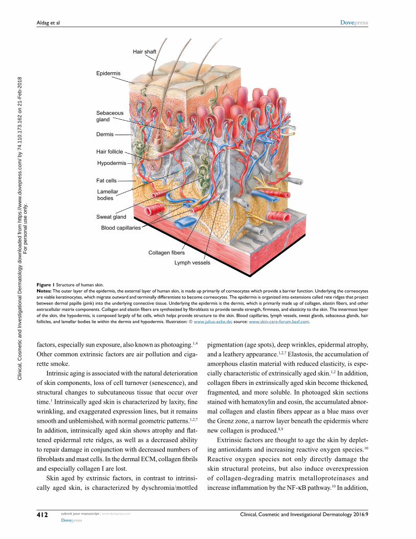
age.1,2 Much of the change occurs in the dermis, which is mostly composed of a dense,
collagen-rich extracellular matrix (ECM) that provides structure and support for the skin
cells and confers tensile strength and firmness to the skin (Figure 1).1,2 Elastic fibers,
which are made up of a cross-linked elastin core within fibrillin-based microfibrils,
are key secondary components of the dermis that provide elasticity, resilience, and
added tensile strength.3 Important changes also occur in the epidermis, most notably
the accumulation of corneocytes, which causes the skin to take on a rough and dull
appearance.1,4 In addition, reduced skin vasculature and structural changes to the
subcutaneous tissue participate in skin aging.
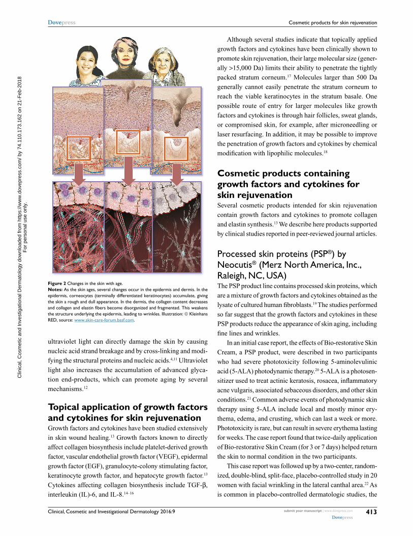
Within the dermal ECM, aging is associated with a thickening of collagen fibrils
and disorganization of total collagen content, mainly due to decreased collagen I syn-
thesis and increased fibril fragmentation.1,2,5 In addition, skin aging is associated with
increased levels of matrix metalloproteinases, which can break down collagen and
elastin fibers, combined with impaired transforming growth factor (TGF)-β signaling,
which may reduce collagen deposition (Figure 2).1,2,6
Intrinsic versus extrinsic skin agingAlthough all aged skin shares common structural changes, intrinsically and extrinsi-
cally aged skin differs in several ways. Intrinsic aging occurs as a result of the natural
chronological aging process, whereas extrinsic aging is brought about by environmental
Correspondence: Caroline AldagMerz Pharmaceuticals GmbH, Eckenheimer Landstraße 100, 60318 Frankfurt am Main, GermanyTel +49 69 1503 4524Fax +49 69 1503 1394Email [email protected].
Journal name: Clinical, Cosmetic and Investigational DermatologyArticle Designation: REVIEWYear: 2016Volume: 9Running head verso: Aldag et alRunning head recto: Cosmetic products for skin rejuvenationDOI: http://dx.doi.org/10.2147/CCID.S116158
C
linic
al, C
osm
etic
and
Inve
stig
atio
nal D
erm
atol
ogy
dow
nloa
ded
from
http
s://w
ww
.dov
epre
ss.c
om/ b
y 74
.110
.173
.162
on
21-F
eb-2
018
For
per
sona
l use
onl
y.
Powered by TCPDF (www.tcpdf.org)
1 / 1
Clinical, Cosmetic and Investigational Dermatology 2016:9submit your manuscript | www.dovepress.com
Dovepress
Dovepress
412
Aldag et al
factors, especially sun exposure, also known as photoaging.1,4
Other common extrinsic factors are air pollution and ciga-
rette smoke.
Intrinsic aging is associated with the natural deterioration
of skin components, loss of cell turnover (senescence), and
structural changes to subcutaneous tissue that occur over
time.1 Intrinsically aged skin is characterized by laxity, fine
wrinkling, and exaggerated expression lines, but it remains
smooth and unblemished, with normal geometric patterns.1,2,7
In addition, intrinsically aged skin shows atrophy and flat-
tened epidermal rete ridges, as well as a decreased ability
to repair damage in conjunction with decreased numbers of
fibroblasts and mast cells. In the dermal ECM, collagen fibrils
and especially collagen I are lost.
Skin aged by extrinsic factors, in contrast to intrinsi-
cally aged skin, is characterized by dyschromia/mottled
pigmentation (age spots), deep wrinkles, epidermal atrophy,
and a leathery appearance.1,2,7 Elastosis, the accumulation of
amorphous elastin material with reduced elasticity, is espe-
cially characteristic of extrinsically aged skin.1,2 In addition,
collagen fibers in extrinsically aged skin become thickened,
fragmented, and more soluble. In photoaged skin sections
stained with hematoxylin and eosin, the accumulated abnor-
mal collagen and elastin fibers appear as a blue mass over
the Grenz zone, a narrow layer beneath the epidermis where
new collagen is produced.8,9
Extrinsic factors are thought to age the skin by deplet-
ing antioxidants and increasing reactive oxygen species.10
Reactive oxygen species not only directly damage the
skin structural proteins, but also induce overexpression
of collagen-degrading matrix metalloproteinases and
increase inflammation by the NF-κB pathway.10 In addition,
Epidermis
Dermis
Hair follicle
Hypodermis
Fat cells
Sweat gland
Blood capillaries
Collagen fibers
Lymph vessels
Lamellarbodies
Sebaceousgland
Hair shaft
Figure 1 Structure of human skin.Notes: The outer layer of the epidermis, the external layer of human skin, is made up primarily of corneocytes which provide a barrier function. Underlying the corneocytes are viable keratinocytes, which migrate outward and terminally differentiate to become corneocytes. The epidermis is organized into extensions called rete ridges that project between dermal papille (pink) into the underlying connective tissue. Underlying the epidermis is the dermis, which is primarily made up of collagen, elastin fibers, and other extracellular matrix components. Collagen and elastin fibers are synthesized by fibroblasts to provide tensile strength, firmness, and elasticity to the skin. The innermost layer of the skin, the hypodermis, is composed largely of fat cells, which helps provide structure to the skin. Blood capillaries, lymph vessels, sweat glands, sebaceous glands, hair follicles, and lamellar bodies lie within the dermis and hypodermis. Illustration: © www.julius-ecke.de; source: www.skin-care-forum.basf.com.
C
linic
al, C
osm
etic
and
Inve
stig
atio
nal D
erm
atol
ogy
dow
nloa
ded
from
http
s://w
ww
.dov
epre
ss.c
om/ b
y 74
.110
.173
.162
on
21-F
eb-2
018
For
per
sona
l use
onl
y.
Powered by TCPDF (www.tcpdf.org)
1 / 1
Clinical, Cosmetic and Investigational Dermatology 2016:9 submit your manuscript | www.dovepress.com
Dovepress
Dovepress
413
Cosmetic products for skin rejuvenation
ultraviolet light can directly damage the skin by causing
nucleic acid strand breakage and by cross-linking and modi-
fying the structural proteins and nucleic acids.4,11 Ultraviolet
light also increases the accumulation of advanced glyca-
tion end-products, which can promote aging by several
mechanisms.12
Topical application of growth factors and cytokines for skin rejuvenationGrowth factors and cytokines have been studied extensively
in skin wound healing.13 Growth factors known to directly
affect collagen biosynthesis include platelet-derived growth
factor, vascular endothelial growth factor (VEGF), epidermal
growth factor (EGF), granulocyte-colony stimulating factor,
keratinocyte growth factor, and hepatocyte growth factor.13
Cytokines affecting collagen biosynthesis include TGF-β,
interleukin (IL)-6, and IL-8.14–16
Although several studies indicate that topically applied
growth factors and cytokines have been clinically shown to
promote skin rejuvenation, their large molecular size (gener-
ally >15,000 Da) limits their ability to penetrate the tightly
packed stratum corneum.17 Molecules larger than 500 Da
generally cannot easily penetrate the stratum corneum to
reach the viable keratinocytes in the stratum basale. One
possible route of entry for larger molecules like growth
factors and cytokines is through hair follicles, sweat glands,
or compromised skin, for example, after microneedling or
laser resurfacing. In addition, it may be possible to improve
the penetration of growth factors and cytokines by chemical
modification with lipophilic molecules.18
Cosmetic products containing growth factors and cytokines for skin rejuvenationSeveral cosmetic products intended for skin rejuvenation
contain growth factors and cytokines to promote collagen
and elastin synthesis.13 We describe here products supported
by clinical studies reported in peer-reviewed journal articles.
Processed skin proteins (PSP®) by Neocutis® (Merz North America, Inc., Raleigh, NC, USA)The PSP product line contains processed skin proteins, which
are a mixture of growth factors and cytokines obtained as the
lysate of cultured human fibroblasts.19 The studies performed
so far suggest that the growth factors and cytokines in these
PSP products reduce the appearance of skin aging, including
fine lines and wrinkles.
In an initial case report, the effects of Bio-restorative Skin
Cream, a PSP product, were described in two participants
who had severe phototoxicity following 5-aminolevulinic
acid (5-ALA) photodynamic therapy.20 5-ALA is a photosen-
sitizer used to treat actinic keratosis, rosacea, inflammatory
acne vulgaris, associated sebaceous disorders, and other skin
conditions.21 Common adverse events of photodynamic skin
therapy using 5-ALA include local and mostly minor ery-
thema, edema, and crusting, which can last a week or more.
Phototoxicity is rare, but can result in severe erythema lasting
for weeks. The case report found that twice-daily application
of Bio-restorative Skin Cream (for 3 or 7 days) helped return
the skin to normal condition in the two participants.
This case report was followed up by a two-center, random-
ized, double-blind, split-face, placebo-controlled study in 20
women with facial wrinkling in the lateral canthal area.22 As
is common in placebo-controlled dermatologic studies, the
Figure 2 Changes in the skin with age.Notes: As the skin ages, several changes occur in the epidermis and dermis. In the epidermis, corneocytes (terminally differentiated keratinocytes) accumulate, giving the skin a rough and dull appearance. In the dermis, the collagen content decreases and collagen and elastin fibers become disorganized and fragmented. This weakens the structure underlying the epidermis, leading to wrinkles. Illustration: © Kleinhans RED, source: www.skin-care-forum.basf.com.
C
linic
al, C
osm
etic
and
Inve
stig
atio
nal D
erm
atol
ogy
dow
nloa
ded
from
http
s://w
ww
.dov
epre
ss.c
om/ b
y 74
.110
.173
.162
on
21-F
eb-2
018
For
per
sona
l use
onl
y.
Powered by TCPDF (www.tcpdf.org)
1 / 1
Clinical, Cosmetic and Investigational Dermatology 2016:9submit your manuscript | www.dovepress.com
Dovepress
Dovepress
414
Aldag et al
vehicle (cream base) used to formulate the product was used
as the control. Participants in the study were randomized
to twice-daily application for 2 months of Bio-restorative
Skin Cream or vehicle on the respective face halves. Three-
dimensional in vivo skin imaging was used to measure aver-
age roughness, mean roughness depth, maximum roughness
depth, base roughness depth, maximum base roughness
depth, and 10-point average roughness. In addition, inves-
tigators assessed wrinkles using the Rao–Goldman 5-point
scale, which scores the wrinkles from 1 for no wrinkle to 5
for very deep wrinkles with redundant folds.23 Differences
in roughness parameters and clinical assessments were com-
pared by one-way analysis of variance (ANOVA) and were
considered significant at p≤0.05. As measured by skin imag-
ing, after 2 months, all roughness parameters significantly
improved versus baseline with Bio-restorative Skin Cream,
and three of six roughness parameters were significantly
better with Bio-restorative Skin Cream than with vehicle.
The investigators’ clinical assessment of wrinkles, however,
did not significantly differ between the Bio-restorative Skin
Cream and the vehicle. In a separate publication based on the
same study, according to investigators’ visual assessments,
periorbital and perioral wrinkles improved significantly at day
30 and day 60 versus baseline.24 At day 60, periorbital wrinkle
scores decreased by 17% and perioral wrinkle scores by 13%.
Also, chin texture improved significantly by 17% at day 60.
Finally, participants felt that Bio-restorative Skin Cream
had improved their skin quality and appearance of wrinkles.
Further details of the effects of Bio-restorative Skin
Cream were investigated in a single-center prospective study
in 12 women with facial wrinkling in the lateral canthal
area.25 Bio-restorative Skin Cream was applied twice daily
for 6 months. Investigators assessed fine wrinkles in the
periorbital and perioral areas on a scale of 0 for absent to 4
for very deep with redundant folds. They also took punch
biopsies for microscopic analysis. According to the investiga-
tors, after 6 months, periorbital wrinkles decreased by 33%
and perioral wrinkles by 25%. Also, 55% of participants
reported improved facial lines and wrinkles, 64% reported
improved facial skin texture, and 73% reported improved skin
hydration. Light microscopy revealed a mild increase versus
baseline in dermal fibroplasia and an associated reduction
in elastosis, as well as increased numbers of fibrocytes and
fibroblasts in 64% of participants and increased epidermal
thickness in 55%.
The effects of Lumiere® Bio-restorative Eye Cream,
another PSP-containing product, were examined in a multi-
center, open-label prospective study in 40 women with fine
or deep wrinkles, visible dark areas, and slightly coarse or
grainy lower eyelids.26 Investigators evaluated the skin color,
texture, sagging, and wrinkles using 4-point scales. In addi-
tion, the participants answered a 10-item questionnaire about
the quality of their periorbital skin, in which the answers were
reported on 5-point scales. Differences in investigator and
participant evaluations were assessed by repeated-measures
ANOVA, with a p-value ≤0.05 indicating a significant dif-
ference. Following twice-daily application of the product for
6 weeks, the investigators’ clinical scores improved signifi-
cantly versus baseline for skin texture (28%), skin sagging
(26%), dark circles (15%), and periorbital wrinkles (14%).
The participants’ scores also improved significantly versus
baseline for nine of ten parameters.
TNS® (SkinMedica, Carlsbad, CA, USA)The TNS product line includes TNS Recovery Complex® and
TNS Essential Serum®. TNS products contain conditioned
medium obtained from neonatal foreskin fibroblast cul-
ture.27,28 The conditioned medium from these cells includes
growth factors and cytokines that can promote angiogenesis
(VEGF and hepatocyte growth factor), modulate inflamma-
tion (IL-6 and IL-8), and enhance ECM deposition (TGF-β1
and platelet-derived growth factor-A).9,27,28 The currently
available data indicate that the growth factors and cytokines in
TNS may help improve the clinical appearance of aged skin.
A pilot study in 14 participants examined the effects
of twice-daily application of TNS Recovery Complex for
60 days on photodamaged skin.9 Physicians measured
photodamage on a 9-point scale. In addition, silicone
impressions were made for optical profilometry, and punch
biopsies were taken to assess Grenz zone and epidermal
thickness. Differences were compared using two-tailed
t-tests, with p-values <0.05 considered to indicate statisti-
cal significance. On the basis of investigators’ evaluations,
11/14 participants in the study showed improvement in at
least one facial area. The periorbital region, in particular,
showed a statistically significant improvement in clinically
assessed photodamage (p=0.0003). As shown by optical
profilometry, the depth and number of textural irregulari-
ties or fine lines were found to have decreased significantly
from baseline (p=0.0075), and ultrastructural evaluation
indicated new collagen formation in the Grenz zone and
thickening of the epidermis. In addition, eight of the par-
ticipants felt that their wrinkles had improved, while 12
felt that their skin texture had improved.
TNS Recovery Complex was further studied in a sin-
gle-center, randomized, double-blind, placebo-controlled
study in 60 participants with mild-to-severe facial photo-
damage.28 In this study, the participants used a basic skin
C
linic
al, C
osm
etic
and
Inve
stig
atio
nal D
erm
atol
ogy
dow
nloa
ded
from
http
s://w
ww
.dov
epre
ss.c
om/ b
y 74
.110
.173
.162
on
21-F
eb-2
018
For
per
sona
l use
onl
y.
Powered by TCPDF (www.tcpdf.org)
1 / 1
Clinical, Cosmetic and Investigational Dermatology 2016:9 submit your manuscript | www.dovepress.com
Dovepress
Dovepress
415
Cosmetic products for skin rejuvenation
care regimen consisting of a cleanser and a moisturizer
combined with twice-daily TNS Recovery Complex or a
vehicle control for 6 months. Photodamage was assessed
by optical profilometry using silicone impressions. Also,
investigators assessed fine wrinkling, tactile roughness,
telangiectasia, mottled pigmentation, and sallowness on
a scale of 0 (absence of photodamage) to 4 (most severe
photodamage). Differences were compared by paired
t-test for profilometry and by two-sided Fisher’s exact test
for clinical assessments, with a p-value ≤0.05 considered
to indicate statistical significance. Optical profilometry
revealed significantly improved facial lines and wrinkles
at month 3 versus baseline in participants using TNS
Recovery Complex. In addition, according to investiga-
tors’ clinical assessments, the appearance of fine lines was
significantly improved versus baseline for TNS Recovery
Complex (p=0.012) but not for the vehicle control. How-
ever, measures of photodamage were not significantly
different between TNS Recovery Complex and the vehicle
control at month 6, and only fine lines and texture shadows
were significantly different at month 3.
ReGenica® (Histogen Aesthetics, San Diego, CA, USA)ReGenica is a third product line containing growth fac-
tors and cytokines from conditioned medium of fibroblasts
(MRCx™). The fibroblasts used to generate MRCx are grown
on beads under hypoxic conditions to simulate the early
embryonic environment prior to angiogenesis.29,30 MRCx
contains VEGF, keratinocyte growth factor, and IL-8, but
not TGF-β.
In a proof-of-concept study including 49 participants who
had been treated by laser resurfacing, a lotion containing
MRCx produced a greater reduction in erythema and bet-
ter re-epithelialization of the perioral and periocular areas
than a placebo lotion.29 In a following split-face study in
42 participants, investigators assessed the effect of MRCx,
formulated as a gel, on transepidermal water loss, measured
by Vapometer, and on erythema, edema, dryness, and peeling
using a scale of 0 (none) to 4 (severe).30 Erythema, edema,
and crusting were also assessed by blinded observers using
photographs and on the same 4-point scale. A p-value ≤0.05
was considered to indicate statistical significance, although
the statistical tests used were not described. The study found
that MRCx, formulated as a gel, produced a dose-dependent
improvement in crusting and significantly improved bar-
rier function as measured by transepidermal water loss.30
In addition, biopsies, taken from three randomly selected
subjects from each treatment group, showed that skin areas
treated with the active gel had less leukocyte infiltration as
well as more normal stratum germinativum and more normal
organization of epidermal layers than the biopsies taken from
skin areas treated with the vehicle control.
The effect of a 3-month application of ReGenica
Replenishing Crème, applied every evening, combined
with ReGenica Renew SPF 15, applied every morning, was
examined in a study of 21 women with mild-to-moderate
photodamage, fine lines, and wrinkles.31 ReGenica Replen-
ishing Crème is a moisturizing cream containing MRCx,
and ReGenica Renew SPF 15 is a cream containing sun-
screens and MRCx. Investigators assessed tactile roughness,
visual texture, wrinkles, and blotchiness from 0 (none) to 4
(severe) and skin tone evenness, radiance, and translucency
from 0 (worst possible results) to 4 (best possible results).
Participants also completed questionnaires assessing overall
satisfaction with the product and on a range of skin proper-
ties. Differences in investigator and participant assessments
were compared by paired t-tests. Investigators’ assessments
of tactile roughness, visual texture, wrinkles, blotchiness,
skin tone evenness, radiance, and translucency progressively
and significantly improved at weeks 4, 8, and 12 versus
baseline (p<0.05). Participants also reported a similar sta-
tistically significant improvement in skin qualities at week 4
(p=0.034), week 8 (p=0.004), and week 12 (p<0.001) versus
the first post-baseline visit (at week 2). On the basis of these
findings, the authors concluded that the topically applied
growth factors and cytokines in MRCx promote recovery,
improve skin barrier function, and may reduce inflammation
in sun-damaged skin.
The effects of another MRCx product, ReGenica Revital-
izing Eye Crème, were examined in a multicenter, open-label
pilot study in 39 women of age ≥35 years with wrinkles,
uneven skin texture, puffiness, and lack of skin firmness in
the lateral canthal and infraorbital areas.32 Using 10-point
scales, investigators assessed wrinkles, sagging, under-eye
dark circles, sallowness, telangiectasia, and crepiness of the
infraorbital area; wrinkles, sagging, dyschromia/mottled
pigmentation, and crepiness of the lateral canthal area; and
smooth texture, tightness, tone, brightness, and moistness
of the infraorbital and lateral canthal areas. After applying
the product twice daily for 60 days, participants reported
improvements in infraorbital brightness, moistness, wrinkles,
sallowness, crepiness, smooth texture, skin tightness, and skin
tone. Investigators also found improvement in lateral can-
thal wrinkles, dyschromia/mottled pigmentation, skin tone,
overall brightness, and moistness and that approximately
C
linic
al, C
osm
etic
and
Inve
stig
atio
nal D
erm
atol
ogy
dow
nloa
ded
from
http
s://w
ww
.dov
epre
ss.c
om/ b
y 74
.110
.173
.162
on
21-F
eb-2
018
For
per
sona
l use
onl
y.
Powered by TCPDF (www.tcpdf.org)
1 / 1
Clinical, Cosmetic and Investigational Dermatology 2016:9submit your manuscript | www.dovepress.com
Dovepress
Dovepress
416
Aldag et al
two-thirds of participants were improved or much improved
at days 14 and 60 compared to baseline.
EGF serum (Bioeffect, Kopavogur, Iceland)EGF is a 6 kDa polypeptide growth factor that has been of
particular interest because of its ability to promote wound
and ulcer healing when applied topically.33 A serum contain-
ing recombinant EGF produced in barley was examined in
an open-label trial in 29 participants of age 30 years with
photoaging.34 The participants completed a survey rating
improvements in brown spots, red spots, age spots, and
skin smoothness on a 5-point scale, and differences were
compared by paired t-tests. The study found that twice-
daily application of EGF serum for 3 months significantly
improved brown spotting, skin texture, pore size, red
spotting, and wrinkles versus baseline (p<0.0002 for all
assessments).
CRS with growth factor (Topix Pharmaceuticals, Amityville, NY, USA)Citrix® CRS Cell Rejuvenation System contains recombi-
nant TGF-β1, l-ascorbic acid, and extract from the plant
Cimicifuga racemosa. This product was examined in a
two-part, split-face, randomized study in women with facial
wrinkling.35 In the first part of the study, facial halves of 11
women were randomized to twice-daily application of CRS
or a similar cream lacking TGF-β1 (VitC) for 3 months. In
the second part, facial halves of 20 women were random-
ized to twice-daily application of TNS Recovery Complex
or CRS for another 3 months. Blinded experts assessed
photographs for wrinkles using a 5-point scale, and the par-
ticipants completed questionnaires about changes in facial
photoaging and satisfaction with the treatments. Differences
were compared using two-tailed tests. The treatment was
considered successful if the mean half-face wrinkle score
showed a positive change or no worsening between baseline
and the end of the study. According to investigator assess-
ments, wrinkles showed “successful treatment” in 27 of 31
(87.1%) participants who applied CRS, 14 of 19 (73.7%)
who applied TNS Recovery Complex, and seven of 12
(58.3%) who applied VitC. In addition, 18 of 31 (58.1%)
participants who applied CRS, nine of 19 (47.4%) who
applied TNS Recovery Complex, and four of 12 (33.3%)
who applied VitC reported subjective improvement. The
study also found significant improvements versus baseline
in clinically assessed wrinkling (p=0.03), but it did not
report comparisons between regimens, so the effect of the
cytokine TGF-β1 was not clear.
Cosmetic products containing matrikines and matrikine-like peptides for skin rejuvenationMatrikines are short peptides generated by proteolysis of
ECM macromolecules that can modulate cell proliferation,
migration, and apoptosis.36,37 Matrikines are widespread and,
like growth factors, appear to have a variety of activities. For
example, matrikines have been identified as markers of lung
inflammation.38 In addition, like many growth factors, some
matrikines can promote tumorigenesis, while others inhibit
it.37,39–42 Matrikines have been of particular interest in skin
rejuvenation because of their role in wound healing.37
Given their small molecular size, matrikines offer
the possibility of better skin penetration than traditional
growth factors and cytokines. Matrikine-like peptides are
now included in several commercially available cosmetic
formulations, although the effect of individual products has
rarely been reported in the peer-reviewed literature. We,
therefore, describe the existing clinical evidence for indi-
vidual products and the in vitro and clinical evidence for
single molecules included in currently marketed matrikine-
containing products.
Glycine–histidine–lysine tripeptide (GHK)The tripeptide GHK is a matrikine that can complex with
copper and, in skin, enhances ECM synthesis.36,43 GHK can
penetrate the stratum corneum and accumulate to relevant
concentrations in the skin.43 Application of a cream contain-
ing Cu-GHK, a complex of copper and GHK, for 1 month was
reported to increase collagen levels in the skin, as measured
by immunohistochemistry.44
Glycine–glutamate–lysine–glycine tetrapeptide (GEKG)GEKG, also known as tetrapeptide-21, is a matrikine whose
activity was investigated in a series of in vitro studies and
clinical assessments in participants with facial wrinkles.45 In
the in vitro studies, the peptide increased procollagen secre-
tion and collagen mRNA levels in human foreskin fibroblasts.
In the first clinical assessment, which was a double-blind,
randomized, placebo-controlled study in 10 healthy volun-
teers (>35 years), daily application of a cream containing
50 ppm GEKG for 8 weeks significantly increased type I
procollagen mRNA levels in the skin (p=0.02 by Wilcoxon
test). Further histochemical analysis revealed that GEKG,
but not placebo, increased skin procollagen, hyaluronic acid,
and fibronectin levels. GEKG improved point estimates of
C
linic
al, C
osm
etic
and
Inve
stig
atio
nal D
erm
atol
ogy
dow
nloa
ded
from
http
s://w
ww
.dov
epre
ss.c
om/ b
y 74
.110
.173
.162
on
21-F
eb-2
018
For
per
sona
l use
onl
y.
Powered by TCPDF (www.tcpdf.org)
1 / 1
Clinical, Cosmetic and Investigational Dermatology 2016:9 submit your manuscript | www.dovepress.com
Dovepress
Dovepress
417
Cosmetic products for skin rejuvenation
resilient distension and other elasticity assessments, although
differences were not significant, possibly due to the small
sample size. In a subsequent clinical assessment in the same
report, 60 volunteers received twice-daily applications of 10
or 100 ppm GEKG for 8 weeks. This portion of the study
showed improved roughness parameters versus baseline at
both 10 and 100 ppm GEKG. In a final clinical assessment in
30 volunteers, twice-daily application of 50 ppm GEKG for
4 or 8 weeks, but not a vehicle cream, improved periorbital
wrinkling versus baseline. Collectively, the results indicated
improvements in the skin quality, lines, and wrinkles, along
with increased procollagen production.
Lysine–threonine–threonine–lysine–serine pentapeptide (KTTKS)KTTKS is a matrikine derived from the proteolytic hydrolysis
of collagen.46 This peptide promotes the production of ECM
and the expression of fibronectin and collagen types I and III
in vitro.46 It also stabilizes mRNAs that upregulate TGF-β,
which may increase collagen production.47 A palmitoylated
and more stable form of KTTKS (pal-KTTKS), also known
as Matrixyl® or palmitoyl pentapeptide-3, has been reported
to penetrate the stratum corneum48 and enhance collagen I
secretion by fibroblasts in vitro.49 Pal-KTTKS has also been
reported to significantly increase procollagen secretion and
upregulate type I collagen and hyaluronic acid synthase-1
expression by human fibroblasts in vitro.45
The clinical outcome of an oil-in-water moisturizer
containing pal-KTTKS was assessed in a double-blind,
placebo-controlled, randomized, split-face study in 94 healthy
Caucasian women with facial wrinkles.50 Total length of fine
lines and wrinkles was assessed from digital photographs
by computer algorithms. Also, blinded experts assessed
improvements in skin texture, fine lines/wrinkles, age spots,
dark circles under the eyes, and skin firmness using a scale
of −4 (much worse) to +4 (much better). Differences in
adjusted treatment means were compared by ANOVA using
a mixed model, with a p-value ≤0.10 indicating a significant
difference. After twice-daily application for 8 and 12 weeks,
fine line/wrinkle length was significantly lower with the pal-
KTTKS cream than with a placebo cream, and clinically
assessed fine lines and wrinkles were significantly better
with the pal-KTTKS cream at weeks 8 and 12. In addition,
skin texture at weeks 4 and 8 and age spots and dark circles
at week 12 were significantly better with the pal-KTTKS
cream. However, using a more standard definition of p≤0.05
to indicate statistical significance, only skin texture at 4
weeks and age spots at 12 weeks were significantly better.
No differences were found in skin texture or barrier function
as measured by transepidermal water loss. Thus, although
the in vitro studies suggest that KTTKS and pal-KTTKS
increase collagen production, evidence of clinical relevance
remains limited.
Micro-protein complex (MPC™; Merz North America, Inc., Raleigh, NC, USA)MPC is a mixture of N-octanoyl-carnosine (an antioxidant)
and the matrikines GEKG and palmitoyl-GHK. A recent
report described a randomized, investigator-blinded study in
which 133 healthy women were randomized 1:1:1 to MPC,
PSP, or conditioned media growth factor.51 After a 2-week
conditioning phase, during which the study participants
washed their faces twice daily with a gentle cleanser and
applied sun protection, the test products were applied for
6 months. An independent expert scored the severity of peri-
orbital and perioral wrinkles using the Rao–Goldman 5-point
visual scoring system and assessed the appearance of pores,
tactile skin roughness, and additional indicators of skin aging
using a 9-point visual analog scale. Skin elasticity and firm-
ness were assessed by cutometry. In addition, facial wrinkle
count, length, and area were analyzed in digital photos using
a computer algorithm. Differences were analyzed by ANOVA,
with p-values of ≤0.05 considered to indicate statistical sig-
nificance. According to the independent expert, periorbital
wrinkles improved from baseline by an average of 23% at
month 3 (p<0.0001) and 33% at month 6 (p<0.0001) in the
MPC group. In addition, perioral wrinkles improved from
baseline by an average 22% at month 3 (p<0.001) and 31%
at month 6 (p<0.001). Similar improvements were observed
in the PSP and conditioned media growth factor groups, and
improvements were not statistically significantly different
between the three groups. Mean periorbital wrinkle count
significantly improved at each time point versus baseline
for MPC and PSP (p<0.05). The evaluator also judged that
tactile skin roughness improved significantly from baseline
by 51% at month 3 and 59% at month 6 (p<0.001 for both)
in the MPC group, and that pore appearance improved sig-
nificantly from baseline at months 2, 3, and 6 (p≤0.001). For
MPC users, quantitatively measured skin elasticity improved
from baseline by an average of 20% at month 2 (p<0.001),
13% at month 3 (p=0.018), and 16% at month 6 (p<0.001).
In contrast, no significant improvements in skin elasticity
were observed at any point in time with PSP or conditioned
media growth factor. MPC also improved skin firmness by
an average of 11% at month 3 (p≤0.008) and 22% at month
6 (p<0.001).
C
linic
al, C
osm
etic
and
Inve
stig
atio
nal D
erm
atol
ogy
dow
nloa
ded
from
http
s://w
ww
.dov
epre
ss.c
om/ b
y 74
.110
.173
.162
on
21-F
eb-2
018
For
per
sona
l use
onl
y.
Powered by TCPDF (www.tcpdf.org)
1 / 1
Clinical, Cosmetic and Investigational Dermatology 2016:9submit your manuscript | www.dovepress.com
Dovepress
Dovepress
418
Aldag et al
ConclusionThe results of in vitro and clinical studies suggest that
cosmetic products containing growth factors, cytokines,
matrikines, or matrikine-like peptides can enhance the pro-
duction of collagen and other ECM molecules and promote
skin rejuvenation. Due to their small size, matrikines and
matrikine-like peptides offer the promise of growth factor-
like activities with improved skin penetration. However, data
are limited. These products need to be further evaluated in
well-designed, randomized trials to be able to draw firm
conclusions about their clinical effects and mechanisms
of action.
AcknowledgmentsThe authors would like to thank Frank Dreher and Uwe
Aßmus for critically reading the manuscript. This work
was funded by Merz Pharmaceuticals GmbH (Frankfurt am
Main, Germany).
DisclosureCA and DNT are employees of Merz Pharmaceuticals GmbH,
which sells products mentioned in this review. PSL is an
employee of 4Clinics, which was paid for medical writing by
Merz Pharmaceuticals GmbH. The authors report no other
conflicts of interest in this work.
References 1. Baumann L. Skin ageing and its treatment. J Pathol. 2007;211(2):241–251. 2. Quan T, Fisher GJ. Role of age-associated alterations of the dermal
extracellular matrix microenvironment in human skin aging: a mini-review. Gerontology. 2015;61(5):427–434.
3. Sherratt MJ. Tissue elasticity and the ageing elastic fibre. Age (Dordr). 2009;31(4):305–325.
4. Rittie L, Fisher GJ. Natural and sun-induced aging of human skin. Cold Spring Harb Perspect Med. 2015;5(1):a015370.
5. Uitto J. The role of elastin and collagen in cutaneous aging: intrinsic aging versus photoexposure. J Drugs Dermatol. 2008;7(2 Suppl): S12–S16.
6. Sardy M. Role of matrix metalloproteinases in skin ageing. Connect Tissue Res. 2009;50(2):132–138.
7. Khavkin J, Ellis DA. Aging skin: histology, physiology, and pathology. Facial Plast Surg Clin North Am. 2011;19(2):229–234.
8. Montagna W, Kirchner S, Carlisle K. Histology of sun-damaged human skin. J Am Acad Dermatol. 1989;21(5 Pt 1):907–918.
9. Fitzpatrick RE, Rostan EF. Reversal of photodamage with topical growth factors: a pilot study. J Cosmet Laser Ther. 2003;5(1):25–34.
10. Kammeyer A, Luiten RM. Oxidation events and skin aging. Ageing Res Rev. 2015;21:16–29.
11. Thurstan SA, Gibbs NK, Langton AK, Griffiths CE, Watson RE, Sherratt MJ. Chemical consequences of cutaneous photoageing. Chem Cent J. 2012;6(1):34.
12. Gkogkolou P, Bohm M. Advanced glycation end products: key players in skin aging? Dermatoendocrinol. 2012;4(3):259–270.
13. Fabi S, Sundaram H. The potential of topical and injectable growth factors and cytokines for skin rejuvenation. Facial Plast Surg. 2014;30(2):157–171.
14. Sproul EP, Argraves WS. A cytokine axis regulates elastin formation and degradation. Matrix Biol. 2013;32(2):86–94.
15. Uitto J, Kouba D. Cytokine modulation of extracellular matrix gene expression: relevance to fibrotic skin diseases. J Dermatol Sci. 2000;24(Suppl 1):S60–S69.
16. Verrecchia F, Mauviel A. Transforming growth factor-beta and fibrosis. World J Gastroenterol. 2007;13(22):3056–3062.
17. Mehta RC, Fitzpatrick RE. Endogenous growth factors as cosmeceu-ticals. Dermatol Ther. 2007;20(5):350–359.
18. Schaefer H, Lademann J. The role of follicular penetration. A differential view. Skin Pharmacol Appl Skin Physiol. 2001;14(Suppl 1):23–27.
19. De Buys Roessingh AS, Hohlfeld J, Scaletta C, et al. Development, characterization, and use of a fetal skin cell bank for tissue engineering in wound healing. Cell Transplant. 2006;15(8–9):823–834.
20. Gold MH, Biron J. A novel skin cream containing a mixture of human growth factors and cytokines for the treatment of adverse events associ-ated with photodynamic therapy. J Drugs Dermatol. 2006;5(8):796–798.
21. Nestor MS, Gold MH, Kauvar AN, et al. The use of photodynamic therapy in dermatology: results of a consensus conference. J Drugs Dermatol. 2006;5(2):140–154.
22. Gold MH, Goldman MP, Biron J. Human growth factor and cytokine skin cream for facial skin rejuvenation as assessed by 3D in vivo optical skin imaging. J Drugs Dermatol. 2007;6(10):1018–1023.
23. Rao J, Ehrlich M, Goldman MP. Facial skin rejeuvenation with a novel topical compound containing transforming growth factor b1 and vitamin C. Cosmet Dermatol. 2004;17:705–713.
24. Gold MH, Goldman MP, Biron J. Efficacy of novel skin cream containing mixture of human growth factors and cytokines for skin rejuvenation. J Drugs Dermatol. 2007;6(2):197–201.
25. Hussain M, Phelps R, Goldberg DJ. Clinical, histologic, and ultra-structural changes after use of human growth factor and cytokine skin cream for the treatment of skin rejuvenation. J Cosmet Laser Ther. 2008;10(2):104–109.
26. Lupo ML, Cohen JL, Rendon MI. Novel eye cream containing a mixture of human growth factors and cytokines for periorbital skin rejuvenation. J Drugs Dermatol. 2007;6(7):725–729.
27. Knighton DR, Ciresi K, Fiegel VD, Schumerth S, Butler E, Cerra F. Stimulation of repair in chronic, nonhealing, cutaneous ulcers using platelet-derived wound healing formula. Surg Gynecol Obstet. 1990;170(1):56–60.
28. Mehta RC, Smith SR, Grove GL, et al. Reduction in facial pho-todamage by a topical growth factor product. J Drugs Dermatol. 2008;7(9):864–871.
29. Kellar RS, Hubka M, Rheins LA, Fisher G, Naughton GK. Hypoxic con-ditioned culture medium from fibroblasts grown under embryonic-like conditions supports healing following post-laser resurfacing. J Cosmet Dermatol. 2009;8(3):190–196.
30. Zimber MP, Mansbridge JN, Taylor M, et al. Human cell-condi-tioned media produced under embryonic-like conditions result in improved healing time after laser resurfacing. Aesthetic Plast Surg. 2012;36(2):431–437.
31. Bruce S, Karnik J, Dryer L, Burkholder D. Anti-aging proof of concept study: results and summary. J Drugs Dermatol. 2014;13(9):1074–1081.
32. Sundaram H, Gold M, Waldorf H, Lupo M, Nguyen VL, Karnik J. Pilot, multicenter, open-label evaluation of safety, tolerability and efficacy of a novel, topical multipotent growth factor formulation for the periorbital region. J Drugs Dermatol. 2015;14(12):1410–1417.
33. Hardwicke J, Schmaljohann D, Boyce D, Thomas D. Epidermal growth factor therapy and wound healing – past, present and future perspectives. Surgeon. 2008;6(3):172–177.
34. Schouest JM, Luu TK, Moy RL. Improved texture and appearance of human facial skin after daily topical application of barley produced, synthetic, human-like epidermal growth factor (EGF) serum. J Drugs Dermatol. 2012;11(5):613–620.
35. Ehrlich M, Rao J, Pabby A, Goldman MP. Improvement in the appear-ance of wrinkles with topical transforming growth factor beta(1) and l-ascorbic acid. Dermatol Surg. 2006;32(5):618–625.
C
linic
al, C
osm
etic
and
Inve
stig
atio
nal D
erm
atol
ogy
dow
nloa
ded
from
http
s://w
ww
.dov
epre
ss.c
om/ b
y 74
.110
.173
.162
on
21-F
eb-2
018
For
per
sona
l use
onl
y.
Powered by TCPDF (www.tcpdf.org)
1 / 1
Clinical, Cosmetic and Investigational Dermatology 2016:9 submit your manuscript | www.dovepress.com
Dovepress
Dovepress
Clinical, Cosmetic and Investigational Dermatology
Publish your work in this journal
Submit your manuscript here: https://www.dovepress.com/clinical-cosmetic-and-investigational-dermatology-journal
Clinical, Cosmetic and Investigational Dermatology is an interna-tional, peer-reviewed, open access, online journal that focuses on the latest clinical and experimental research in all aspects of skin disease and cosmetic interventions. This journal is included on PubMed. The manuscript management system is completely online
and includes a very quick and fair peer-review system, which is all easy to use. Visit http://www.dovepress.com/testimonials.php to read real quotes from published authors
Dovepress
419
Cosmetic products for skin rejuvenation
36. Maquart FX, Pasco S, Ramont L, Hornebeck W, Monboisse JC. An introduction to matrikines: extracellular matrix-derived peptides which regulate cell activity. Implication in tumor invasion. Crit Rev Oncol Hematol. 2004;49(3):199–202.
37. Tran KT, Lamb P, Deng JS. Matrikines and matricryptins: implications for cutaneous cancers and skin repair. J Dermatol Sci. 2005;40(1):11–20.
38. Abdul Roda M, Fernstrand AM, Redegeld FA, Blalock JE, Gaggar A, Folkerts G. The matrikine PGP as a potential biomarker in COPD. Am J Physiol Lung Cell Mol Physiol. 2015;308(11):L1095–L1101.
39. Monboisse JC, Oudart JB, Ramont L, Brassart-Pasco S, Maquart FX. Matrikines from basement membrane collagens: a new anti-cancer strategy. Biochim Biophys Acta. 2014;1840(8):2589–2598.
40. Brassart-Pasco S, Senechal K, Thevenard J, et al. Tetrastatin, the NC1 domain of the alpha4(IV) collagen chain: a novel potent anti-tumor matrikine. PLoS One. 2012;7(4):e29587.
41. Williams KE, Fulford LA, Albig AR. Lumican reduces tumor growth via induction of fas-mediated endothelial cell apoptosis. Cancer Micro-environ. 2010;4(1):115–126.
42. Ramont L, Brassart-Pasco S, Thevenard J, et al. The NC1 domain of type XIX collagen inhibits in vivo melanoma growth. Mol Cancer Ther. 2007;6(2):506–514.
43. Pickart L, Vasquez-Soltero JM, Margolina A. The human tripeptide GHK-Cu in prevention of oxidative stress and degenerative conditions of aging: implications for cognitive health. Oxid Med Cell Longev. 2012;2012:324832.
44. Abdulghani AA, Sherr A, Shirin S, et al. Effects of topical creams containing vitamin C, a copper-binding peptide cream and melatonin compared with tretinoin on the ultrastructure of normal skin – a pilot clinical, histologic, and ultrastructural study. Dis Manag Clin Outcome. 1998;1(4):136–141.
45. Farwick M, Grether-Beck S, Marini A, et al. Bioactive tetrapeptide GEKG boosts extracellular matrix formation: in vitro and in vivo molecular and clinical proof. Exp Dermatol. 2011;20(7):602–604.
46. Abu Samah NH, Heard CM. Topically applied KTTKS: a review. Int J Cosmet Sci. 2011;33(6):483–490.
47. Tsai WC, Hsu CC, Chung CY, Lin MS, Li SL, Pang JH. The pen-tapeptide KTTKS promoting the expressions of type I collagen and transforming growth factor-beta of tendon cells. J Orthop Res. 2007;25(12):1629–1634.
48. Choi YL, Park EJ, Kim E, Na DH, Shin YH. Dermal stability and in vitro skin permeation of collagen pentapeptides (KTTKS and palmitoyl-KTTKS). Biomol Ther (Seoul). 2014;22(4):321–327.
49. Jones RR, Castelletto V, Connon CJ, Hamley IW. Collagen stimulating effect of peptide amphiphile C16-KTTKS on human fibroblasts. Mol Pharm. 2013;10(3):1063–1069.
50. Robinson LR, Fitzgerald NC, Doughty DG, Dawes NC, Berge CA, Bissett DL. Topical palmitoyl pentapeptide provides improvement in photoaged human facial skin. Int J Cosmet Sci. 2005;27(3):155–160.
51. Dreher F. A novel matrikine-like micro-protein complex (MPC) technol-ogy for topical skin rejuvenation. J Drugs Dermatol. 2016;15(4):611–618.
C
linic
al, C
osm
etic
and
Inve
stig
atio
nal D
erm
atol
ogy
dow
nloa
ded
from
http
s://w
ww
.dov
epre
ss.c
om/ b
y 74
.110
.173
.162
on
21-F
eb-2
018
For
per
sona
l use
onl
y.
Powered by TCPDF (www.tcpdf.org)
1 / 1