Embed Size (px)
Citation preview
639
CHAPTER 121 DIARRHEA
Daniel Z. Hume, DVM, DACVIM (Internal Medicine), DACVECC
KEY POINTS
• Diarrheaisacommonclinicalfindingincriticallyillanimals.• Diarrheacanleadtoabnormalitiesinnutrient,acid-base,and
electrolytebalance.• Diarrheamayresultfromiatrogeniccauses,primary
gastrointestinaldiseases,orotherdiseaseprocesses.
Diarrhea is a common clinical sign observed in critically ill canine and feline patients. Diarrhea is defined as an increase in fecal mass caused by an increase in fecal water or solid content. This usually is associated with an increase in frequency, fluidity, or volume of feces. In a 20-kg dog, approximately 2.5 L of fluid enters the duodenum each day, and about 98% of the fluid entering the intestine is absorbed.1 Diarrhea in the critical care setting often is overlooked and overshadowed by the primary disease process. However, diarrhea can lead to severe aberrations in nutrient, acid-base, fluid, and elec-trolyte balance. Without proper attention it can lead to deterioration of the patient’s condition. Diarrhea may be associated with patient discomfort, local dermatitis, catheter or catheter site infections, and potentially bacterial translocation if the integrity of the intestinal mucosa is altered. Consideration of the most likely cause is important because it allows the clinician to decide which diagnostic modalities are indicated for proper workup of the diarrhea. Three broad etio-logic categories may be used when considering the potential cause of diarrhea in a given patient: iatrogenic causes, primary gastrointesti-nal (GI) causes, and other diseases secondarily causing diarrhea.
PATHOPHYSIOLOGIC MECHANISMS OF DIARRHEA
The several categorization schemes for diarrhea have great overlap among the classifications. One of the most commonly used classifica-tion schemes arranges the pathophysiologic mechanisms underlying diarrhea as follows: osmotic diarrhea, secretory diarrhea, diarrhea resulting from altered permeability, and diarrhea resulting from deranged motility.
Osmotic diarrhea is caused by the presence of excess luminal osmoles, drawing fluid into the intestinal lumen. Most causes of a diarrhea have an osmotic component.
Secretory diarrhea is caused by a net increase in intestinal fluid secretion. This results from either an absolute increase in intestinal secretion or a relative increase caused by a decrease in intestinal absorption.
Normal intestinal physiology and systemic health depend on the semipermeable nature of the intestinal mucosa. Nutrients, electro-lytes, and fluid are absorbed and secreted, and the mucosa and immune system of the intestine inhibit translocation of bacteria and bacterial toxins. However, microscopic and macroscopic damage to either the epithelial cells or epithelial cell junctions can lead to altered
intestinal permeability. Vital substances are lost into the intestinal lumen, and the altered permeability leaves the intestine vulnerable to translocation of potentially fatal bacteria and their products.
Alterations in intestinal motility are probably the least under-stood of the causes of diarrhea. Motility alterations leading to diar-rhea include either increased peristaltic contractions or decreased segmental contractions.1 Even within this classification scheme, significant overlap occurs among the groups.
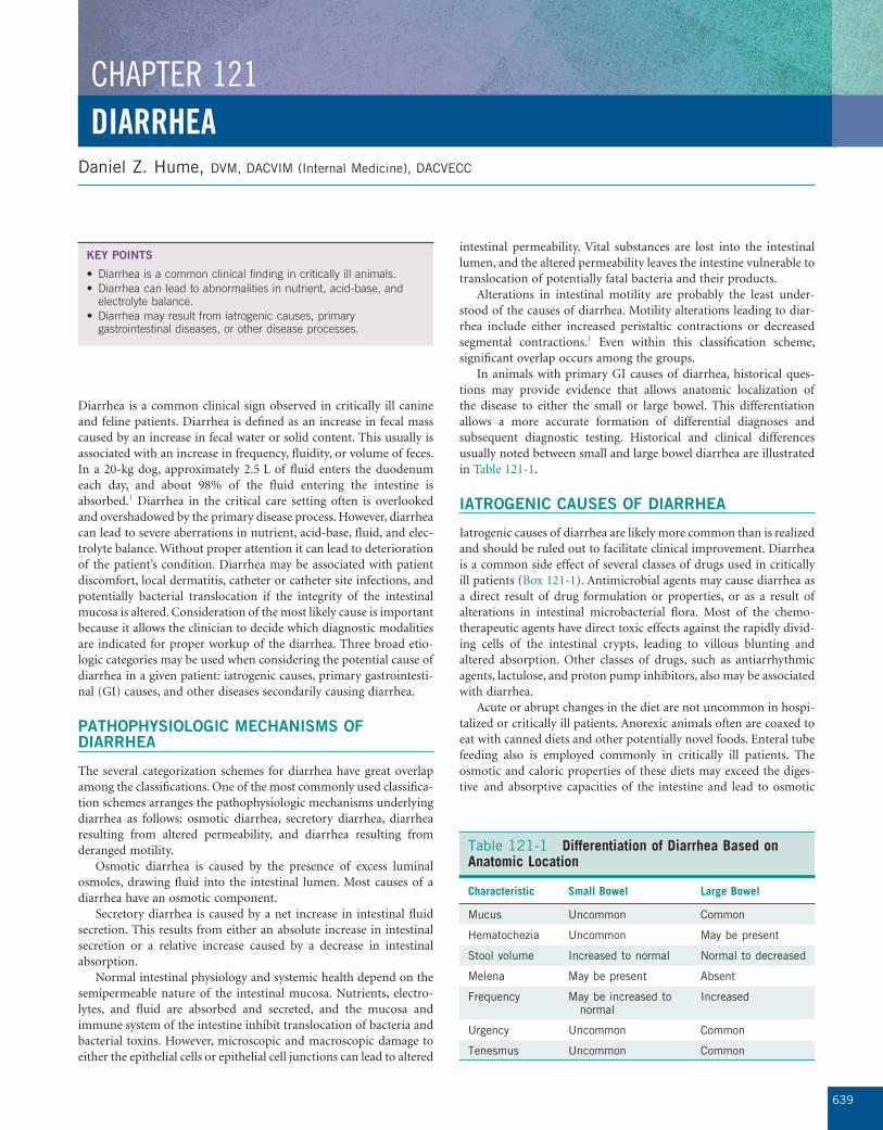
In animals with primary GI causes of diarrhea, historical ques-tions may provide evidence that allows anatomic localization of the disease to either the small or large bowel. This differentiation allows a more accurate formation of differential diagnoses and subsequent diagnostic testing. Historical and clinical differences usually noted between small and large bowel diarrhea are illustrated in Table 121-1.
IATROGENIC CAUSES OF DIARRHEA
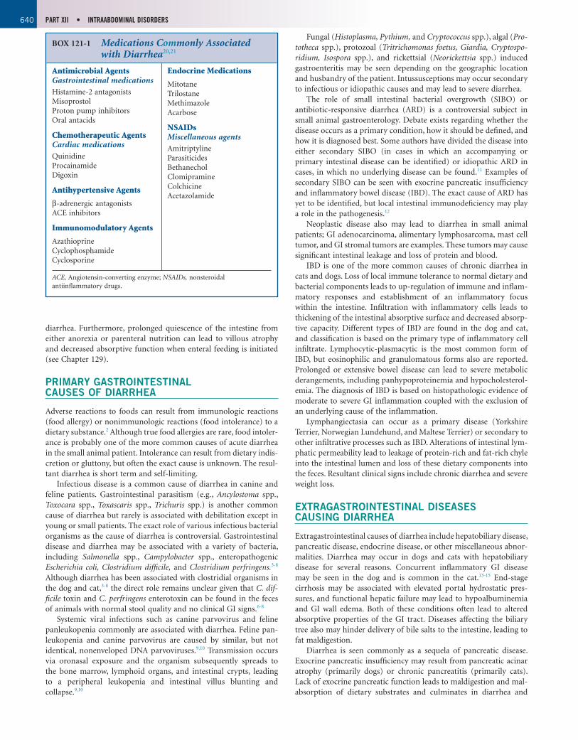
Iatrogenic causes of diarrhea are likely more common than is realized and should be ruled out to facilitate clinical improvement. Diarrhea is a common side effect of several classes of drugs used in critically ill patients (Box 121-1). Antimicrobial agents may cause diarrhea as a direct result of drug formulation or properties, or as a result of alterations in intestinal microbacterial flora. Most of the chemo-therapeutic agents have direct toxic effects against the rapidly divid-ing cells of the intestinal crypts, leading to villous blunting and altered absorption. Other classes of drugs, such as antiarrhythmic agents, lactulose, and proton pump inhibitors, also may be associated with diarrhea.
Acute or abrupt changes in the diet are not uncommon in hospi-talized or critically ill patients. Anorexic animals often are coaxed to eat with canned diets and other potentially novel foods. Enteral tube feeding also is employed commonly in critically ill patients. The osmotic and caloric properties of these diets may exceed the diges-tive and absorptive capacities of the intestine and lead to osmotic
Table 121-1 Differentiation of Diarrhea Based on Anatomic Location
Characteristic Small Bowel Large Bowel
Mucus Uncommon Common
Hematochezia Uncommon May be present
Stool volume Increased to normal Normal to decreased
Melena May be present Absent
Frequency May be increased to normal
Increased
Urgency Uncommon Common
Tenesmus Uncommon Common
640 PART XII • INTRAABDOMINAL DISORDERS
Fungal (Histoplasma, Pythium, and Cryptococcus spp.), algal (Pro-totheca spp.), protozoal (Tritrichomonas foetus, Giardia, Cryptospo-ridium, Isospora spp.), and rickettsial (Neorickettsia spp.) induced gastroenteritis may be seen depending on the geographic location and husbandry of the patient. Intussusceptions may occur secondary to infectious or idiopathic causes and may lead to severe diarrhea.
The role of small intestinal bacterial overgrowth (SIBO) or antibiotic-responsive diarrhea (ARD) is a controversial subject in small animal gastroenterology. Debate exists regarding whether the disease occurs as a primary condition, how it should be defined, and how it is diagnosed best. Some authors have divided the disease into either secondary SIBO (in cases in which an accompanying or primary intestinal disease can be identified) or idiopathic ARD in cases, in which no underlying disease can be found.11 Examples of secondary SIBO can be seen with exocrine pancreatic insufficiency and inflammatory bowel disease (IBD). The exact cause of ARD has yet to be identified, but local intestinal immunodeficiency may play a role in the pathogenesis.12
Neoplastic disease also may lead to diarrhea in small animal patients; GI adenocarcinoma, alimentary lymphosarcoma, mast cell tumor, and GI stromal tumors are examples. These tumors may cause significant intestinal leakage and loss of protein and blood.
IBD is one of the more common causes of chronic diarrhea in cats and dogs. Loss of local immune tolerance to normal dietary and bacterial components leads to up-regulation of immune and inflam-matory responses and establishment of an inflammatory focus within the intestine. Infiltration with inflammatory cells leads to thickening of the intestinal absorptive surface and decreased absorp-tive capacity. Different types of IBD are found in the dog and cat, and classification is based on the primary type of inflammatory cell infiltrate. Lymphocytic-plasmacytic is the most common form of IBD, but eosinophilic and granulomatous forms also are reported. Prolonged or extensive bowel disease can lead to severe metabolic derangements, including panhypoproteinemia and hypocholesterol-emia. The diagnosis of IBD is based on histopathologic evidence of moderate to severe GI inflammation coupled with the exclusion of an underlying cause of the inflammation.
Lymphangiectasia can occur as a primary disease (Yorkshire Terrier, Norwegian Lundehund, and Maltese Terrier) or secondary to other infiltrative processes such as IBD. Alterations of intestinal lym-phatic permeability lead to leakage of protein-rich and fat-rich chyle into the intestinal lumen and loss of these dietary components into the feces. Resultant clinical signs include chronic diarrhea and severe weight loss.
EXTRAGASTROINTESTINAL DISEASES CAUSING DIARRHEA
Extragastrointestinal causes of diarrhea include hepatobiliary disease, pancreatic disease, endocrine disease, or other miscellaneous abnor-malities. Diarrhea may occur in dogs and cats with hepatobiliary disease for several reasons. Concurrent inflammatory GI disease may be seen in the dog and is common in the cat.13-15 End-stage cirrhosis may be associated with elevated portal hydrostatic pres-sures, and functional hepatic failure may lead to hypoalbuminemia and GI wall edema. Both of these conditions often lead to altered absorptive properties of the GI tract. Diseases affecting the biliary tree also may hinder delivery of bile salts to the intestine, leading to fat maldigestion.
Diarrhea is seen commonly as a sequela of pancreatic disease. Exocrine pancreatic insufficiency may result from pancreatic acinar atrophy (primarily dogs) or chronic pancreatitis (primarily cats). Lack of exocrine pancreatic function leads to maldigestion and mal-absorption of dietary substrates and culminates in diarrhea and
diarrhea. Furthermore, prolonged quiescence of the intestine from either anorexia or parenteral nutrition can lead to villous atrophy and decreased absorptive function when enteral feeding is initiated (see Chapter 129).
PRIMARY GASTROINTESTINAL CAUSES OF DIARRHEA
Adverse reactions to foods can result from immunologic reactions (food allergy) or nonimmunologic reactions (food intolerance) to a dietary substance.2 Although true food allergies are rare, food intoler-ance is probably one of the more common causes of acute diarrhea in the small animal patient. Intolerance can result from dietary indis-cretion or gluttony, but often the exact cause is unknown. The resul-tant diarrhea is short term and self-limiting.
Infectious disease is a common cause of diarrhea in canine and feline patients. Gastrointestinal parasitism (e.g., Ancylostoma spp., Toxocara spp., Toxascaris spp., Trichuris spp.) is another common cause of diarrhea but rarely is associated with debilitation except in young or small patients. The exact role of various infectious bacterial organisms as the cause of diarrhea is controversial. Gastrointestinal disease and diarrhea may be associated with a variety of bacteria, including Salmonella spp., Campylobacter spp., enteropathogenic Escherichia coli, Clostridium difficile, and Clostridium perfringens.3-8 Although diarrhea has been associated with clostridial organisms in the dog and cat,3-8 the direct role remains unclear given that C. dif-ficile toxin and C. perfringens enterotoxin can be found in the feces of animals with normal stool quality and no clinical GI signs.6-8
Systemic viral infections such as canine parvovirus and feline panleukopenia commonly are associated with diarrhea. Feline pan-leukopenia and canine parvovirus are caused by similar, but not identical, nonenveloped DNA parvoviruses.9,10 Transmission occurs via oronasal exposure and the organism subsequently spreads to the bone marrow, lymphoid organs, and intestinal crypts, leading to a peripheral leukopenia and intestinal villus blunting and collapse.9,10
BOX 121-1 Medications Commonly Associated with Diarrhea20,21
Antimicrobial AgentsGastrointestinal medicationsHistamine-2 antagonistsMisoprostolProton pump inhibitorsOral antacids
Chemotherapeutic AgentsCardiac medicationsQuinidineProcainamideDigoxin
Antihypertensive Agents
β-adrenergic antagonistsACE inhibitors
Immunomodulatory Agents
AzathioprineCyclophosphamideCyclosporine
Endocrine Medications
MitotaneTrilostaneMethimazoleAcarbose
NSAIDsMiscellaneous agentsAmitriptylineParasiticidesBethanecholClomipramineColchicineAcetazolamide
ACE, Angiotensin-converting enzyme; NSAIDs, nonsteroidal antiinflammatory drugs.
CHAPTER 121 • DIARRHEA 641
be helpful in animals with suspected SIBO. Although abdominal radiographs are of limited value in animals affected primarily with diarrhea, abdominal ultrasound often is indicated and useful for assessing integrity, architecture, and thickness of the GI system and other abdominal organs. Last, GI endoscopy or exploratory lapa-rotomy often is needed for direct visualization of the intestinal tract and procurement of diagnostic samples.
TREATMENT
Iatrogenic causes of diarrhea should be considered in all patients, especially those in which diarrhea was not part of the presenting complaint. If the diarrhea is severe, current medications may have to be discontinued or modified. Although diarrhea is associated com-monly with enteral feeding, the diet formulation may require altera-tion if the diarrhea is severe or adversely affecting the patient’s quality of life.
Treatment of diarrhea associated with primary GI diseases or diseases secondarily causing diarrhea is achieved best after careful diagnostic evaluation of the underlying cause. Once a definitive diag-nosis has been achieved, direct treatment can be initiated. Rarely, medications directed toward symptomatic treatment of the diarrhea are used (see Chapter 161). Intestinal transit time is effectively a result of the balance between propulsive peristalsis and segmental contractions.1 Contrary to historical belief, diarrhea rarely results from increased peristalsis but more commonly is the result of decreased segmental contractions.1 Anticholinergic agents generally are contraindicated because they decrease propulsive peristalsis and segmental contractions and predispose the patient to ileus. Con-versely, opioid-containing medications such as loperamide, diphenox-ylate, and opium tincture can decrease propulsive contractions and increase segmental contractions; water and fluid absorption also is augmented.1 These medications may be indicated in some cases of diarrhea in which infectious causes have been excluded. Kaolin, pectin, and bismuth subsalicylate are used occasionally for symptom-atic treatment.1 However, this rarely is indicated because treatment of the primary disease process provides the best means for eliminat-ing diarrhea. Indications for symptomatic therapy include diarrhea that adversely affects the patient’s quality of life, causes severe fecal scalding of the skin, or predisposes to secondary infection (e.g., urinary or intravenous catheter infections in recumbent animals).
In animals with IBD, treatment often is tailored to the individual patient based on the severity of the clinical signs and histopathologic lesions. Dogs and cats with intermittent clinical signs, good body condition, and mild histologic lesions may respond to dietary therapy alone. This may be due to a loss of immunologic tolerance to normal dietary proteins and subsequent GI inflammation in patients with IBD. Dietary therapy for pets with IBD usually relies on either a novel protein diet or a diet with a hydrolyzed protein source. Animals with some degree of lymphangiectasia may benefit from a low-fat diet.
However, most animals with moderate to severe disease need some degree of immunomodulation to obtain clinical remission. Glucocorticoids (prednisone, prednisolone, and dexamethasone) are the mainstay of immunomodulatory therapy. The locally active steroid, budesonide, undergoes significant first-pass metabolism, thereby limiting systemic absorption and potentially lessening the side effects compared with glucocorticoids. Azathioprine, chloram-bucil, or other immunomodulating agents may be necessary for dogs with refractory disease or those unable to tolerate glucocorticoid therapy. Aminosalicylates (sulfasalazine, mesalamine) can be pre-scribed for dogs with primarily large bowel disease. Metronidazole often is used for its antimicrobial and antiinflammatory effects.
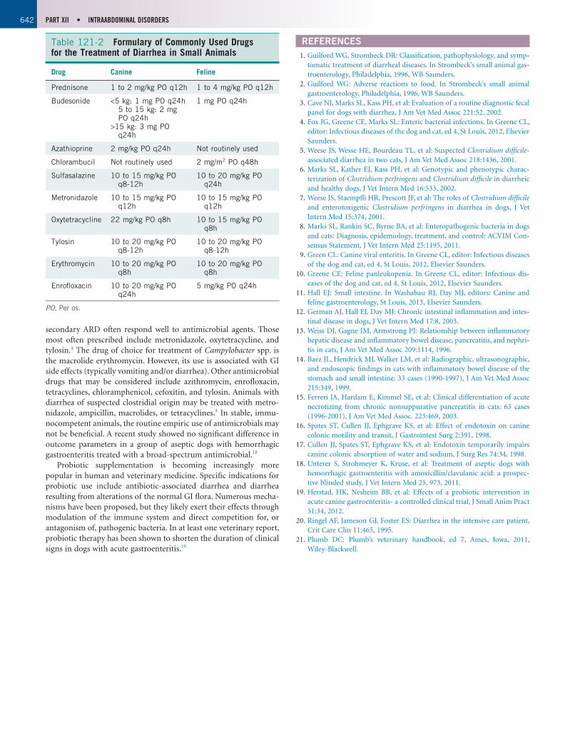
Antimicrobial therapy may be useful in many other diarrheal diseases of dogs and cats (Table 121-2). Primary (idiopathic) or
weight loss. Acute and chronic pancreatitis also may lead to diarrhea. Pancreatic inflammation may cause local inflammation of the duo-denum and colon, interfere with pancreatic acinar secretion, and result in decreased bile salt delivery to the small intestine via obstruc-tion of biliary flow.
Congestive heart failure, particularly right-sided failure, can lead to intestinal and hepatic venous congestion and ascites. Congestion of the splanchnic vasculature may cause alteration in the absorptive capacities of the intestine.
Several endocrine disorders may be associated with diarrhea. Diarrhea is noted in some cats with hyperthyroidism. The diarrhea in these cases may be a result of increased food intake as well as intestinal hypermotility. Waxing and waning GI signs are seen fre-quently in dogs and less commonly in cats with hypoadrenocorti-cism. Cortisol is vital for maintenance of normal GI function, motility, and integrity, as well as vascular tone and subsequent perfu-sion. The lack of mineralocorticoids may be associated with altera-tions in electrolyte balance, leading to altered GI motility and absorption. Diarrhea is an uncommonly reported clinical sign associ-ated with hypothyroidism.
Various other diseases may be associated with diarrhea. Idiopathic noncirrhotic portal hypertension may interfere with absorption within the intestinal tract. The role of hypoalbuminemia as a direct cause of diarrhea is debated. The decreased oncotic draw resulting from hypoalbuminemia leads to alterations in Starling forces and decreased absorption of fluid across the intestinal lumen. Hemor-rhagic diarrhea is seen commonly in critically ill patients suffering from, or after resuscitation from, various causes of cardiovascular shock (e.g., heat-induced illness). GI complications are common in animals with acute and chronic renal disease, but diarrhea is not reported commonly. Systemic infections (including sepsis) may affect secondarily the GI tract and cause diarrhea. Experimental canine studies have shown that bacterial endotoxin impairs colonic water and sodium absorption and increases small and large intestinal motility, at least partially explaining the diarrhea noted in septic patients.16,17
DIAGNOSTIC EVALUATION
The diagnostic evaluation of patients with diarrhea is guided best by the historical, clinicopathologic, and physical examination findings. The physical condition of the patient and the duration and clinical course of the diarrhea help determine how aggressive the clinician should be in attempting to find a cause.
Results of a complete blood count, serum chemistry profile, and urinalysis are indicated in all critically ill patients and often help to differentiate between GI and non-GI causes of diarrhea. Based on these findings, additional tests may be needed to screen for hyper-thyroidism, hypoadrenocorticism, or occult liver disease. Fecal flota-tion (including zinc sulfate for Giardia spp.) and direct cytologic examination of the feces is recommended in most cases. Although cytologic examination of the feces may help indirectly to point toward a particular pathogen, isolation or amplification of toxin or enterotoxin may provide a more specific diagnosis in the case of clostridial infection. Bacterial culture (Salmonella spp., Campylo-bacter spp., enteropathogenic E. coli), enterotoxin screening (Clos-tridium spp.), and enzyme-linked immunosorbent assay (ELISA) (parvovirus) of the feces may be indicated when an infectious cause is suspected. Specific tests for other infectious agents also may prove helpful depending on the geographic location and husbandry of the patient. Exfoliative rectal cytology may be useful in diagnosing fungal, algal, inflammatory, and neoplastic diseases. Trypsin-like immunoreactivity testing is indicated in any patient with suspected exocrine pancreatic insufficiency. Folate and cobalamin testing may
642 PART XII • INTRAABDOMINAL DISORDERS
REFERENCES1. Guilford WG, Strombeck DR: Classification, pathophysiology, and symp-
tomatic treatment of diarrheal diseases. In Strombeck’s small animal gas-troenterology, Philadelphia, 1996, WB Saunders.
2. Guilford WG: Adverse reactions to food. In Strombeck’s small animal gastroenterology, Philadelphia, 1996, WB Saunders.
3. Cave NJ, Marks SL, Kass PH, et al: Evaluation of a routine diagnostic fecal panel for dogs with diarrhea, J Am Vet Med Assoc 221:52, 2002.
4. Fox JG, Greene CE, Marks SL: Enteric bacterial infections. In Greene CL, editor: Infectious diseases of the dog and cat, ed 4, St Louis, 2012, Elsevier Saunders.
5. Weese JS, Wesse HE, Bourdeau TL, et al: Suspected Clostridium difficile-associated diarrhea in two cats, J Am Vet Med Assoc 218:1436, 2001.
6. Marks SL, Kather EJ, Kass PH, et al: Genotypic and phenotypic charac-terization of Clostridium perfringens and Clostridium difficile in diarrheic and healthy dogs, J Vet Intern Med 16:533, 2002.
7. Weese JS, Staempfli HR, Prescott JF, et al: The roles of Clostridium difficile and enterotoxigenic Clostridium perfringens in diarrhea in dogs, J Vet Intern Med 15:374, 2001.
8. Marks SL, Rankin SC, Byrne BA, et al: Enteropathogenic bacteria in dogs and cats: Diagnosis, epidemiology, treatment, and control: ACVIM Con-sensus Statement, J Vet Intern Med 25:1195, 2011.
9. Green CL: Canine viral enteritis. In Greene CL, editor: Infectious diseases of the dog and cat, ed 4, St Louis, 2012, Elsevier Saunders.
10. Greene CE: Feline panleukopenia. In Greene CL, editor: Infectious dis-eases of the dog and cat, ed 4, St Louis, 2012, Elsevier Saunders.
11. Hall EJ: Small intestine. In Washabau RJ, Day MJ, editors: Canine and feline gastroenterology, St Louis, 2013, Elsevier Saunders.
12. German AJ, Hall EJ, Day MJ: Chronic intestinal inflammation and intes-tinal disease in dogs, J Vet Intern Med 17:8, 2003.
13. Weiss DJ, Gagne JM, Armstrong PJ: Relationship between inflammatory hepatic disease and inflammatory bowel disease, pancreatitis, and nephri-tis in cats, J Am Vet Med Assoc 209:1114, 1996.
14. Baez JL, Hendrick MJ, Walker LM, et al: Radiographic, ultrasonographic, and endoscopic findings in cats with inflammatory bowel disease of the stomach and small intestine: 33 cases (1990-1997), J Am Vet Med Assoc 215:349, 1999.
15. Ferreri JA, Hardam E, Kimmel SE, et al: Clinical differentiation of acute necrotizing from chronic nonsuppurative pancreatitis in cats: 63 cases (1996-2001), J Am Vet Med Assoc. 223:469, 2003.
16. Spates ST, Cullen JJ, Ephgrave KS, et al: Effect of endotoxin on canine colonic motility and transit, J Gastrointest Surg 2:391, 1998.
17. Cullen JJ, Spates ST, Ephgrave KS, et al: Endotoxin temporarily impairs canine colonic absorption of water and sodium, J Surg Res 74:34, 1998.
18. Unterer S, Strohmeyer K, Kruse, et al: Treatment of aseptic dogs with hemorrhagic gastroenteritis with amoxicillin/clavulanic acid: a prospec-tive blinded study, J Vet Intern Med 25, 973, 2011.
19. Herstad, HK, Nesheim BB, et al: Effects of a probiotic intervention in acute canine gastroenteritis- a controlled clinical trial, J Small Anim Pract 51:34, 2012.
20. Ringel AF, Jameson GJ, Foster ES: Diarrhea in the intensive care patient, Crit Care Clin 11:465, 1995.
21. Plumb DC: Plumb’s veterinary handbook, ed 7, Ames, Iowa, 2011, Wiley-Blackwell.
secondary ARD often respond well to antimicrobial agents. Those most often prescribed include metronidazole, oxytetracycline, and tylosin.4 The drug of choice for treatment of Campylobacter spp. is the macrolide erythromycin. However, its use is associated with GI side effects (typically vomiting and/or diarrhea). Other antimicrobial drugs that may be considered include azithromycin, enrofloxacin, tetracyclines, chloramphenicol, cefoxitin, and tylosin. Animals with diarrhea of suspected clostridial origin may be treated with metro-nidazole, ampicillin, macrolides, or tetracyclines.4 In stable, immu-nocompetent animals, the routine empiric use of antimicrobials may not be beneficial. A recent study showed no significant difference in outcome parameters in a group of aseptic dogs with hemorrhagic gastroenteritis treated with a broad-spectrum antimicrobial.18
Probiotic supplementation is becoming increasingly more popular in human and veterinary medicine. Specific indications for probiotic use include antibiotic-associated diarrhea and diarrhea resulting from alterations of the normal GI flora. Numerous mecha-nisms have been proposed, but they likely exert their effects through modulation of the immune system and direct competition for, or antagonism of, pathogenic bacteria. In at least one veterinary report, probiotic therapy has been shown to shorten the duration of clinical signs in dogs with acute gastroenteritis.19
Table 121-2 Formulary of Commonly Used Drugs for the Treatment of Diarrhea in Small Animals
Drug Canine Feline
Prednisone 1 to 2 mg/kg PO q12h 1 to 4 mg/kg PO q12h
Budesonide <5 kg: 1 mg PO q24h 5 to 15 kg: 2 mg PO q24h
>15 kg: 3 mg PO q24h
1 mg PO q24h
Azathioprine 2 mg/kg PO q24h Not routinely used
Chlorambucil Not routinely used 2 mg/m2 PO q48h
Sulfasalazine 10 to 15 mg/kg PO q8-12h
10 to 20 mg/kg PO q24h
Metronidazole 10 to 15 mg/kg PO q12h
10 to 15 mg/kg PO q12h
Oxytetracycline 22 mg/kg PO q8h 10 to 15 mg/kg PO q8h
Tylosin 10 to 20 mg/kg PO q8-12h
10 to 20 mg/kg PO q8-12h
Erythromycin 10 to 20 mg/kg PO q8h
10 to 20 mg/kg PO q8h
Enrofloxacin 10 to 20 mg/kg PO q24h
5 mg/kg PO q24h
PO,Peros.