Embed Size (px)
Citation preview

1
ANNOUNCEMENTS
Monday, Oct. 25th Dr. Rebecca Levy
Interested in Lunch with Levy?
Join the Luncheon Lottery
Review Session Every Friday at 12:20
Lab Practical Coming up! October 26th, 27th
Motor Neuron
Nerve Action Potential
Synapse
Neuromuscular Junction
Neurotransmitter (ACh)
Receptors (AChR)
Muscle Action Potential
Transverse Tubules
Sarcoplasmic Reticulum
Calcium release
Smooth Muscle •! Spindle shaped (fusiform) •! Nonstriated (smooth)
- Contractile proteins are less organized •! Contractile proteins anchored at densities
- Contractile proteins bind to Dense Bodies
•! Gap Junctions between cells

2
Smooth Muscle

3
Smooth Muscle Contraction
- Actin filaments are associated with tropomyosin but Not Troponin
Smooth Muscle Contraction -Myosin interacts with actin only when the
myosin light chains are phosphorylated
http://www.ncbi.nlm.nih.gov/bookshelf/br.fcgi?book=cell&part=A4335
Smooth Muscle Excitation-Contraction
1) Excitation=> increased [Ca++]intracellular
2) Ca++ binds Calmodulin
3) Ca++-Calmodulin activates myosin light chain kinase (MLCK )
4) MLCK phosphorylates myosin light chains
5) Myosin binds actin => contraction
Regulation of Smooth Muscle Contraction
Nonneural regulation
Hormonal: Oxytocin- uterine contraction
Nitric Oxide (NO): Produced by endothelial cells of arterioles Relaxes smooth muscle
Mice have been produced whose eNOS (endothelial cell NO synthase) genes been "knocked out”. Predict the blood pressure levels of these mice.

4
Nitric Oxide ----> increased [cGMP]
activates a kinase
inhibits Ca++ influx into smooth muscle cell
decreased calcium-calmodulin stimulation of MLCK
decreased phosphorylation of myosin light chains
decreased smooth muscle tension development
vasodilation (expansion of vessel lumen)
Predict the effect of drugs that inhibit
breakdown of cGMP ?
Drugs that inhibit the breakdown of cGMP potentiate (increase) the effects of NO
actions on target cells.
EXAMPLE: Viagra and other inhibitors of cGMP-dependent phosphodiesterase
Nervous System
Functions: –!Input from sensory receptors
–!Integration and processing of signals
–!Output to effector cells
–!Behavior
Neurons
5
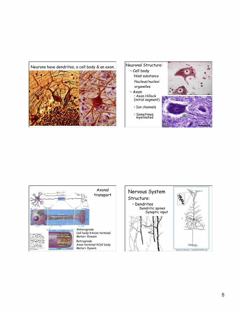
Neurons have dendrites, a cell body & an axon. Neuronal Structure: –!Cell body
Nissl substance
Nucleus/nucleoi
organelles
–!Axon •!Axon Hillock (initial segment)
•!Ion channels
•!Sometimes myelinated
Anterograde Cell body!Axon terminal Motor= Kinesin
Retrograde Axon terminal!Cell body Motor= Dynein
Axonal transport
Nervous System Structure:
–!Dendrites Dendritic spines
Synaptic input
www.neuro.duke.edu/.../ media/3381036764_t.gif
6
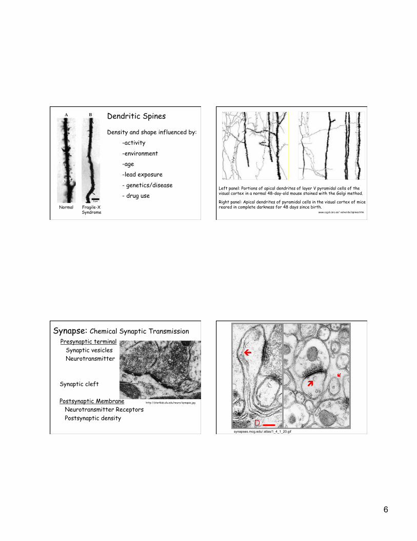
Normal Fragile-X Syndrome
Dendritic Spines
Density and shape influenced by:
-activity
-environment
-age
-lead exposure
- genetics/disease
- drug use Left panel: Portions of apical dendrites of layer V pyramidal cells of the visual cortex in a normal 48-day-old mouse stained with the Golgi method.
Right panel: Apical dendrites of pyramidal cells in the visual cortex of mice reared in complete darkness for 48 days since birth.
www.cajal.csic.es/ valverde/spines.htm
Synapse: Chemical Synaptic Transmission
Presynaptic terminal
Synaptic vesicles
Neurotransmitter
Synaptic cleft
Postsynaptic Membrane
Neurotransmitter Receptors
Postsynaptic density
http://starklab.slu.edu/neuro/synapse.jpg
synapses.mcg.edu/ atlas/1_4_1_20.gif

7
Electrical Synaptic Transmission
NO SYNAPTIC VESICLES
Gap Junction (Connexons)
Chemical Transmission
Electrical Transmission
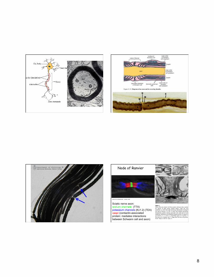
Myelin sheath Function: increase speed of action potential
propagation (saltatory conduction)
Produced by Glial cells •! Schwann cells- motor & sensory neurons •!Oligodendrocytes- brain & spinal cord
Nodes of Ranvier
8
Node of Ranvier
Sciatic nerve axon:
sodium channels (TTX)
potassium channels (Kv1.2) (TEA) caspr (contactin-associated
protein: mediates interactions
between Schwann cell and axon)
www.urmc.rochester.edu/. ../sciatic_node.gif

9
Peripheral Nerve: Schwann cells wrap axons
- MYELINATED Myelin sheath 1 axon/Schwann cell
-UNMYELINATED No myelin sheath Multiple axons/Schwann cell
Oligodendrocytes form myelin in CNS
-! glial cell
-! myelinates CNS axons
-! multiple axons/ oligodendrocyte
-! NOT associated with unmyelinated axons
Multiple Sclerosis
gamma-interferon

10
What are the consequences of cutting the axon of a neuron?
Consider: Transmission of action potential Composition of synapses Structure of damaged neuron
Axonal damage influences target and input cells.!
Muscle Atrophy

11
CNS: Limited Regeneration
Unlike PNS axons, after CNS axon damage: •!neurons die •!growth of regenerating axons is inhibited •!lack of environmental growth factors
Glial Scar Formation: Astrocyte and microglia
Myelin contains growth-inhibiting proteins: Nogo-A MAG (myelin-associated glycoprotein) Bind to Nogo receptor (NgR)
Approaches to improve CNS regeneration:
1) Prevent neuronal death -!Limit inflammatory response
2) Induce CNS axon growth -!Implant Schwann cells or PNS nerve -!Implant Olfactory ensheathing cells
3) Overcome inhibitory environmental factors -! prevent or impede scar formation -! block activity of Nogo-A and MAG
RELEASE OF SYNAPTIC VESICLES
Botulinum toxin:
from Clostridium botulinum
prevents neurotransmitter release by proteolytically cleaving proteins required for vesicle release
RELEASE OF SYNAPTIC VESICLES

12
A: A patient with blepharospasm, forceful involuntary closure of the eyelids, is unable to open her eyes due to abnormal muscle contractions.
B: Pre-injection, she uses her fingers to keep her eyes open.
C: After Botox injection, her eyes stay open without difficulty. (Photos courtesy Joseph Jankovic, M.D., professor of neurology, Baylor College of Medicine, Houston, Texas)
A B C
users.rcn.com/.../ BiologyPages/P/PNS.html
Nervous System Organization

13
Brain, Spinal cord
users.rcn.com/.../ BiologyPages/P/PNS.html
Peripheral Nervous System (PNS)
Autonomic PNS Somatic PNS Internal stimuli External stimuli parasympathetic/sympathetic (sensory-motor)
Groups of Neuronal Cell Bodies
CNS --> Nucleus (Gray matter) -functionally related
PNS --> Ganglion
Bundles of Axons
CNS --> Tracts (White matter)
PNS --> Nerves

14
MENINGES
3 connective tissue membranes Cover brain and spinal cord
1)! Dura Mater: dense CT
3)!Arachnoid: web-like arachnoid trabeculae loose CT
subarachnoid space cerebral spinal fluid
3) Pia Mater ensheathes blood vessels
Meninges

15
Cerebral Spinal Fluid (CSF) -! Clear fluid
- Circulates through ventricles & subarachnoid space
- Produced by choroid plexus •!modified ependymal cells •!lining ventricles
-!Functions: -!“shock absorber” -!transport nutrients / waste -!compensate for changes in brain blood volume
Choroid Plexus Cuboidal-Columnar Ependymal Cells neighbor capillaries to form the choroid plexus that produces the cerebral spinal fluid
Production of CSF = Absorption of CSF into blood
Blocked CSF flow or overproduction of CSF results in hydrocephalus.
Hydrocephalus: excess CSF accumulation in brain
Hydrocephalus: excess CSF accumulation in brain
Treatment: shunting of CSF into abdominal cavity

16
MENINGITIS
-! infection / inflammation of meninges and CSF
Infectious Agents: Viruses Bacteria pneumococcus: blood->meninges meningococcus: respiratory->blood
(college epidemics)
Signs or symptoms: * High fever * Severe headache * Stiff neck * Sensitivity to light * Seizures
DORSAL ROOT GANGION
AUTONOMIC NERVOUS SYSTEM
17
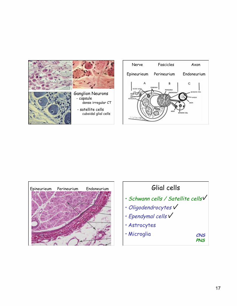
Ganglion Neurons
- capsule
dense irregular CT
- satellite cells
cuboidal glial cells
Nerve Fascicles Axon
Epineurieum Perineurium Endoneurium
Epineurieum Perineurium Endoneurium Glial cells •!Schwann cells / Satellite cells
•!Oligodendrocytes
•!Ependymal cells
•!Astrocytes
•!Microglia CNS
PNS
18
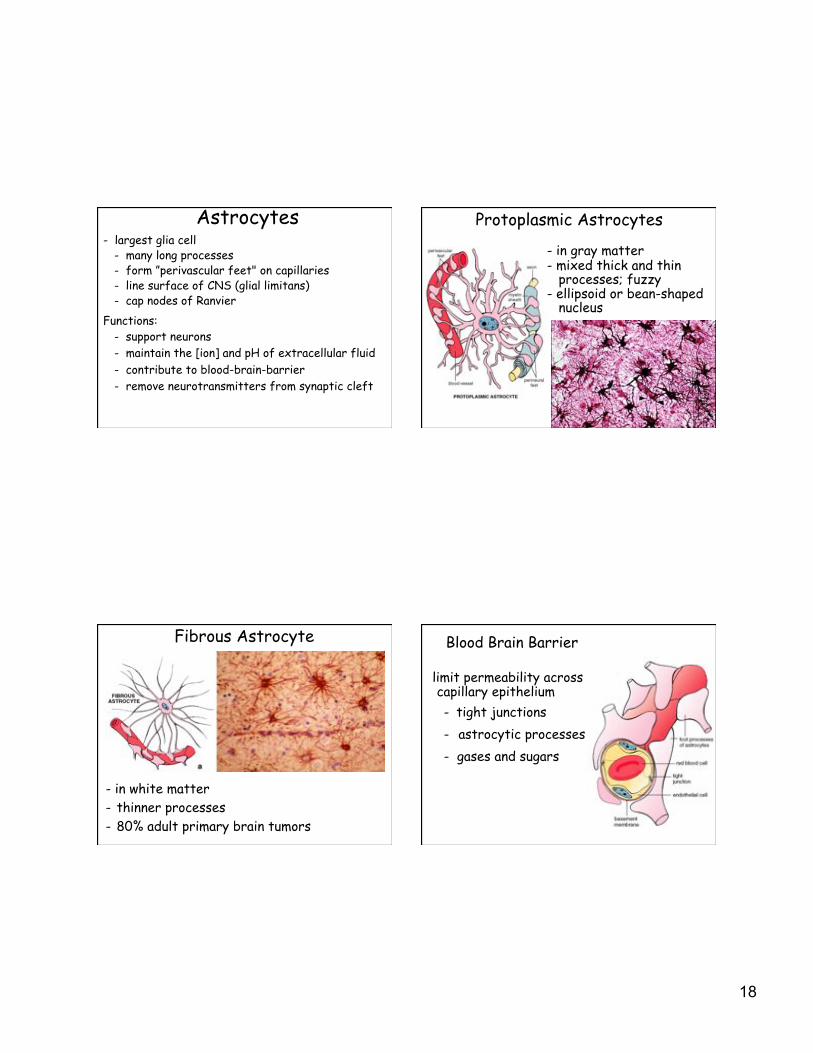
Astrocytes - largest glia cell - many long processes
- form ”perivascular feet" on capillaries - line surface of CNS (glial limitans) - cap nodes of Ranvier
Functions:
- support neurons
- maintain the [ion] and pH of extracellular fluid
- contribute to blood-brain-barrier
- remove neurotransmitters from synaptic cleft
Protoplasmic Astrocytes
-! in gray matter -! mixed thick and thin processes; fuzzy -! ellipsoid or bean-shaped nucleus
Fibrous Astrocyte
- in white matter
-! thinner processes
-! 80% adult primary brain tumors
Blood Brain Barrier
limit permeability across capillary epithelium
-! tight junctions
-! astrocytic processes
-! gases and sugars

19
Challenge: limited access of drugs and therapeutic molecules
Solution: Selectively open barrier (cancer treatment) Couple transport with permeable compound
Microglia
-! smallest, infrequent glial cell
-! short twisted processes
-! act as macrophages, remove debris
-! proliferate & become phagocytic after damage