Embed Size (px)
Citation preview
Sn-rich tourmaline from the Land’s End granite, SW England
Kristian Drivenes
Na
Ca
Mg
Fe
Sn
Ti
Al
F
10a
10b
10c
10
10a 10b 10c
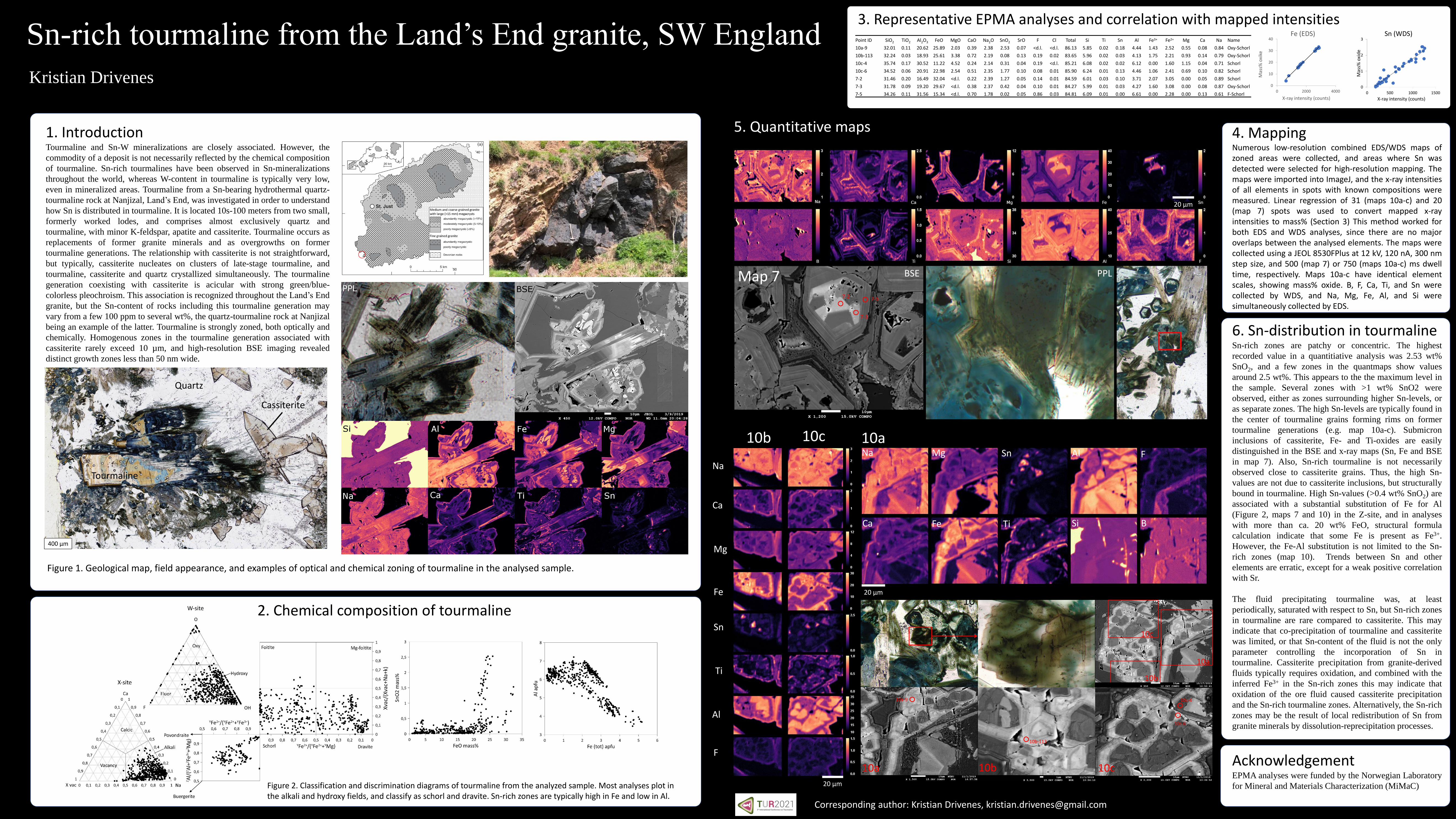
1. IntroductionTourmaline and Sn-W mineralizations are closely associated. However, the
commodity of a deposit is not necessarily reflected by the chemical composition
of tourmaline. Sn-rich tourmalines have been observed in Sn-mineralizations
throughout the world, whereas W-content in tourmaline is typically very low,
even in mineralized areas. Tourmaline from a Sn-bearing hydrothermal quartz-
tourmaline rock at Nanjizal, Land’s End, was investigated in order to understand
how Sn is distributed in tourmaline. It is located 10s-100 meters from two small,
formerly worked lodes, and comprises almost exclusively quartz and
tourmaline, with minor K-feldspar, apatite and cassiterite. Tourmaline occurs as
replacements of former granite minerals and as overgrowths on former
tourmaline generations. The relationship with cassiterite is not straightforward,
but typically, cassiterite nucleates on clusters of late-stage tourmaline, and
tourmaline, cassiterite and quartz crystallized simultaneously. The tourmaline
generation coexisting with cassiterite is acicular with strong green/blue-
colorless pleochroism. This association is recognized throughout the Land’s End
granite, but the Sn-content of rocks including this tourmaline generation may
vary from a few 100 ppm to several wt%, the quartz-tourmaline rock at Nanjizal
being an example of the latter. Tourmaline is strongly zoned, both optically and
chemically. Homogenous zones in the tourmaline generation associated with
cassiterite rarely exceed 10 µm, and high-resolution BSE imaging revealed
distinct growth zones less than 50 nm wide.
6. Sn-distribution in tourmalineSn-rich zones are patchy or concentric. The highest
recorded value in a quantitiative analysis was 2.53 wt%
SnO2, and a few zones in the quantmaps show values
around 2.5 wt%. This appears to the the maximum level in
the sample. Several zones with >1 wt% SnO2 were
observed, either as zones surrounding higher Sn-levels, or
as separate zones. The high Sn-levels are typically found in
the center of tourmaline grains forming rims on former
tourmaline generations (e.g. map 10a-c). Submicron
inclusions of cassiterite, Fe- and Ti-oxides are easily
distinguished in the BSE and x-ray maps (Sn, Fe and BSE
in map 7). Also, Sn-rich tourmaline is not necessarily
observed close to cassiterite grains. Thus, the high Sn-
values are not due to cassiterite inclusions, but structurally
bound in tourmaline. High Sn-values (>0.4 wt% SnO2) are
associated with a substantial substitution of Fe for Al
(Figure 2, maps 7 and 10) in the Z-site, and in analyses
with more than ca. 20 wt% FeO, structural formula
calculation indicate that some Fe is present as Fe3+.
However, the Fe-Al substitution is not limited to the Sn-
rich zones (map 10). Trends between Sn and other
elements are erratic, except for a weak positive correlation
with Sr.
The fluid precipitating tourmaline was, at least
periodically, saturated with respect to Sn, but Sn-rich zones
in tourmaline are rare compared to cassiterite. This may
indicate that co-precipitation of tourmaline and cassiterite
was limited, or that Sn-content of the fluid is not the only
parameter controlling the incorporation of Sn in
tourmaline. Cassiterite precipitation from granite-derived
fluids typically requires oxidation, and combined with the
inferred Fe3+ in the Sn-rich zones this may indicate that
oxidation of the ore fluid caused cassiterite precipitation
and the Sn-rich tourmaline zones. Alternatively, the Sn-rich
zones may be the result of local redistribution of Sn from
granite minerals by dissolution-reprecipitation processes.
4. MappingNumerous low-resolution combined EDS/WDS maps ofzoned areas were collected, and areas where Sn wasdetected were selected for high-resolution mapping. Themaps were imported into ImageJ, and the x-ray intensitiesof all elements in spots with known compositions weremeasured. Linear regression of 31 (maps 10a-c) and 20(map 7) spots was used to convert mapped x-rayintensities to mass% (Section 3) This method worked forboth EDS and WDS analyses, since there are no majoroverlaps between the analysed elements. The maps werecollected using a JEOL 8530FPlus at 12 kV, 120 nA, 300 nmstep size, and 500 (map 7) or 750 (maps 10a-c) ms dwelltime, respectively. Maps 10a-c have identical elementscales, showing mass% oxide. B, F, Ca, Ti, and Sn werecollected by WDS, and Na, Mg, Fe, Al, and Si weresimultaneously collected by EDS.
0
10
20
30
40
0 2000 4000
Mas
s% o
xike
X-ray intensity (counts)
Fe (EDS)
0
1
2
3
0 500 1000 1500
Mas
s% o
xid
e
X-ray intensity (counts)
Sn (WDS)Point ID SiO2 TiO2 Al2O3 FeO MgO CaO Na2O SnO2 SrO F Cl Total Si Ti Sn Al Fe3+ Fe2+ Mg Ca Na Name
10a-9 32.01 0.11 20.62 25.89 2.03 0.39 2.38 2.53 0.07 <d.l. <d.l. 86.13 5.85 0.02 0.18 4.44 1.43 2.52 0.55 0.08 0.84 Oxy-Schorl
10b-113 32.24 0.03 18.93 25.61 3.38 0.72 2.19 0.08 0.13 0.19 0.02 83.65 5.96 0.02 0.03 4.13 1.75 2.21 0.93 0.14 0.79 Oxy-Schorl
10c-4 35.74 0.17 30.52 11.22 4.52 0.24 2.14 0.31 0.04 0.19 <d.l. 85.21 6.08 0.02 0.02 6.12 0.00 1.60 1.15 0.04 0.71 Schorl
10c-6 34.52 0.06 20.91 22.98 2.54 0.51 2.35 1.77 0.10 0.08 0.01 85.90 6.24 0.01 0.13 4.46 1.06 2.41 0.69 0.10 0.82 Schorl
7-2 31.46 0.20 16.49 32.04 <d.l. 0.22 2.39 1.27 0.05 0.14 0.01 84.59 6.01 0.03 0.10 3.71 2.07 3.05 0.00 0.05 0.89 Schorl
7-3 31.78 0.09 19.20 29.67 <d.l. 0.38 2.37 0.42 0.04 0.10 0.01 84.27 5.99 0.01 0.03 4.27 1.60 3.08 0.00 0.08 0.87 Oxy-Schorl
7-5 34.26 0.11 31.56 15.34 <d.l. 0.70 1.78 0.02 0.05 0.86 0.03 84.81 6.09 0.01 0.00 6.61 0.00 2.28 0.00 0.13 0.61 F-Schorl
3. Representative EPMA analyses and correlation with mapped intensities
2. Chemical composition of tourmaline
5. Quantitative maps
Map 7
10b 10c 10aNa
Ca
Mg
Fe
Sn
Ti
Al F
BSi
BSE PPL
Figure 2. Classification and discrimination diagrams of tourmaline from the analyzed sample. Most analyses plot in the alkali and hydroxy fields, and classify as schorl and dravite. Sn-rich zones are typically high in Fe and low in Al.
Figure 1. Geological map, field appearance, and examples of optical and chemical zoning of tourmaline in the analysed sample.
Quartz
Cassiterite
Tourmaline
AcknowledgementEPMA analyses were funded by the Norwegian Laboratory
for Mineral and Materials Characterization (MiMaC)
10a-9
10b-113
10c-6
10c-4
7-2
7-3
7-5
400 µm
20 µm
20 µm
20 µm
Corresponding author: Kristian Drivenes, [email protected]