Embed Size (px)
Citation preview
1
Management of Acute Traumatic
Spinal Cord Injury
Karen A. McQuillan, RN, MS, CNS-BC, CCRN, CNRN, FAAN Past President, American Association of Critical Care Nurses
Clinical Nurse Specialist R Adams Cowley Shock Trauma Center, UMMC
Incidence of Traumatic Spinal Cord Injury in the United States
• 40 new SCI/ 1 million/ year
• ~ 12,500 new SCI/ year (Deaths at scene not included)
• 240,000 – 337,000 living with SCI in U.S.
• Average age 42 years (National Spinal Cord Injury Statistical Center, 2015)
Neurologic Level of Spinal Injuries
55%
15%
15%
15%
Soft Tissue Injury
2
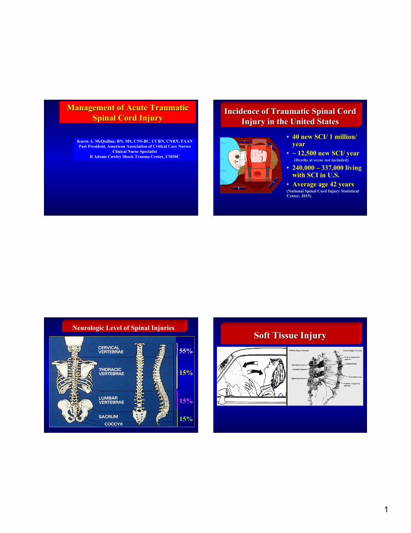
Wedge Fracture Burst Fracture Teardrop Fracture
Vertebral Fractures Dislocation
Spinal Cord Injury With Out Radiographic Abnormality
(SCIWORA) • No boney abnormality seen on x-rays • Spinal cord injury may be seen on MRI • Has been attributed to ligamentous injury,
disc prolapse, and cervical spondylosis • Common in children < 9 years old, but also
seen in middle aged and elderly
3
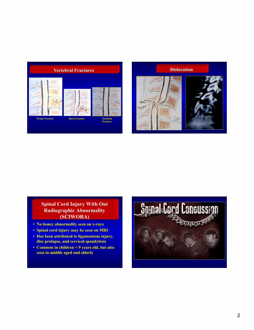
Contusion
Hemorrhage and edema of the
cord
Laceration Complete transection is rare!
Hemorrhage Vascular damage

4
Complete vs. Incomplete Spinal Cord Injury
• Complete – No preservation of
voluntary motor function or sensation below the lesion
• Incomplete – Varied motor and sensory
loss below the level of the lesion due to sparing of some tracts
Posterior Columns (position sense)
Leg Trunk Arm
Leg Trunk Arm
Lateral Corticospinal Tract (motor)
Lateral Spinothalamic Tract (pain & temperature on the opposite side of body) Anterior Spinal Artery
Central Cord Syndrome Anterior Cord Syndrome

5
Brown-Sequard Syndrome
Dorsal Column Syndrome
Apoptosis
6
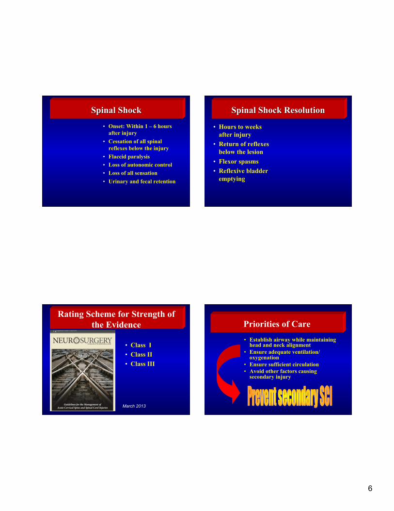
Spinal Shock
• Onset: Within 1 – 6 hours after injury
• Cessation of all spinal reflexes below the injury
• Flaccid paralysis • Loss of autonomic control • Loss of all sensation • Urinary and fecal retention
Spinal Shock Resolution
• Hours to weeks after injury
• Return of reflexes below the lesion
• Flexor spasms • Reflexive bladder
emptying
Rating Scheme for Strength of the Evidence
• Class I • Class II • Class III
March 2013
Priorities of Care • Establish airway while maintaining
head and neck alignment • Ensure adequate ventilation/
oxygenation • Ensure sufficient circulation • Avoid other factors causing
secondary injury
7
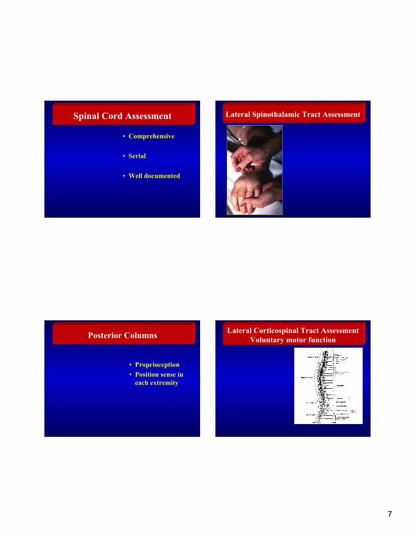
Spinal Cord Assessment
• Comprehensive • Serial • Well documented
Lateral Spinothalamic Tract Assessment
Posterior Columns
• Proprioception • Position sense in
each extremity
Lateral Corticospinal Tract Assessment Voluntary motor function

8
ASIA Tool
Check for rectal sparing! • Light touch /
pinprick sensation S3-4
• Deep anal pressure • Voluntary anal
contraction • More initial sacral
sparing à greater recovery potential
• More gained à greater chance of motor recovery
Kirshblum, et al. Arch Phys Med Rehab. 2016;97:1647.
Deep Tendon Reflexes Correlated with Level of Spinal Cord
Innervations Biceps C5 – C6 Triceps C7 – C8 Quadriceps L2 – L4 Achilles S1 – S2
Priapism
Sustained reflexive erection of the penis
due to unopposed parasympathetic stimulation and increased arterial inflow

9
Vital Sign Alterations
• Bradycardia • Hypotension • Hypoventilation • Loss of
Thermoregulation
Cervical Spinal Radiographs
• Not necessary in awake, alert, non-intoxicated patient without neck pain or tenderness or other significant injuries
• If CT unavailable, obtain 3 views (Level 1) – Lateral – Anterior-posterior – Odontoid
• Must view C1 through T1
Magnetic Resonance Imaging

10
Myelogram
• Contrast medium is injected into the subarachnoid space • Recognizes obstruction of contrast medium flow
Somatosensory Evoked Potentials
Vertebral Artery Injury (VAI) Diagnosis After Blunt Cervical Trauma
• CTA to screen selected pts who meet the Denver Screening Criteria for cerebral artery injury (Level I)
Vertebral Artery Injury (VAI) Diagnosis After Blunt Cervical Trauma
• Conventional angio for selected patients to diagnose VAI – Concurrent endovascular
therapy – CTA unavailable (Level III)
• MRI to diagnose VAI – Complete SCI – Vertebral subluxation (Level III)
• Rule out VAI with comminuted fracture of axis body (Level III)

11
Specialized referral center (Parent et al. J Neurotrauma, August 2011)
• Early transfer to a specialized center should be done promptly to decrease overall length of stay
• Early transfer to an
integrated multidisciplinary specialized center decreases overall mortality and the number and severity of complications
What’s wrong with this picture? Maintain spine alignment until definitive treatment
or rule out spinal injury Recommended Collar Removal
• Awake with neck pain or tenderness with normal films – Normal dynamic flexion/extension
films – Normal MRI within 48 hrs – Physician Discretion
• Obtunded with normal films – Normal MRI within 48 hrs – At physicians discretion – Normal flexion/extension under
fluoroscopy NOT recommended (Level III)

12
Initial Closed Reduction of the Cervical Spine (Level III)
• Early closed reduction of cervical fracture/dislocation with traction recommended in awake patients
• NOT recommended in pts with other rostral injury • MRI recommended for patients with cervical
fracture/dislocation if they can not be examined during closed reduction or before anterior or posterior procedures when closed reduction failed
• If more contemporary treatment options unavailable, treatment of subaxial cervical fracture or dislocation with prolonged bedrest in traction recommended
Braces to Immobilize the Spine
• Halo Vest Yale Brace
13
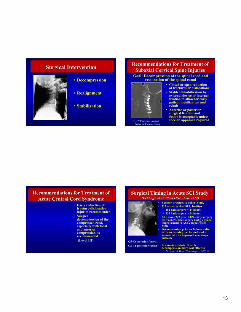
Surgical Intervention
• Decompression
• Realignment
• Stabilization
Recommendations for Treatment of Subaxial Cervical Spine Injuries
• Closed or open reduction of fractures or dislocations
• Stable immobilization by external device or internal fixation to allow for early patient mobilization and rehab
• Anterior or posterior surgical fixation and fusion is acceptable unless specific approach required
C1-C7 Posterior surgical fusion and laminectomy
Goal: Decompression of the spinal cord and restoration of the spinal canal
Recommendations for Treatment of Acute Central Cord Syndrome
• Early reduction of fracture-dislocation injuries recommended
• Surgical decompression of the compressed cord, especially with focal and anterior compression, is recommended
(Level III)
Surgical Timing in Acute SCI Study (Fehlings, et al. PLoS ONE, Feb. 2012)
• 6 center prospective cohort study • 313 acute cervical SCI, 16-80yo
– 182 had surgery < 24 hours – 131 had surgery > 24 hours
• At 6 mos. (222 pts) 19.8% early surgery pts vs. 8.8% late surgery had > 2 grade improvement in ASIA Impairment Scale
• Decompression prior to 24 hours after SCI can be safely performed and is associated with improved neurologic outcome
• Economic analysis à early
decompression more cost effective Furlan et al, World Neurosurgery. 2016;88
C5-C6 anterior fusion; C3-T1 posterior fusion

14
Surgical Timing in Acute SCI Study (Bourassa-Moreaus, et al. J Neurotrauma, Feb. 2016)
• Observational study • 53 complete SCI – 33 thoracolumbar; 20
cervical – 38 had surgery < 24 hours; younger – 15 had surgery > 24 hours
• At rehabilitation discharge – Overall, 34% early surgery patients vs. 13% > 24
hours had improvement in ASIA Impairment Scale score
– Of cervical SCI, 64% of early surgery patients vs. none > 24 hours had improvement in ASIA Impairment Scale score
Recommendations for Pharmacologic Use
• Administration of methylprednisolone is NOT recommended – No Class I or II medical
evidence to support benefit – Scattered Class III evidence
claims inconsistent effects – Class I, II and III data
associate methylprednisolone with harmful side effects
Incidence of and Risk Factors for Organ Dysfunction and Failure
• 40 pts with isolated cervical SCI; in ICU >24hr • 75% had at least 1 organ fail per Multiple Organ
Dysfunction Score; 85% per Sequential Organ Failure Assessment
• Common failure: CV and Respiratory • Respiratory, CV, neurologic, renal, hepatic and
hematologic dysfunction were common • Organ dysfunction and failures correlated:
– Strongly with ASIA motor index score and ASIA impairment scale
– Poorly with level of injury
(Stein, et al, Neurocritical Care, 2010) Pulmonary Complications
Most common acute systemic adverse event following SCI Most common cause of death and morbidity following SCI
ASIA Impairment Scale grade A or B was the fundamental
clinical entity predicting pulmonary complications
(Aarabi, et al, J Neurosurg, 2012)

15
Innervation of Respiratory Muscles
• C4 and above – no patient-initiated respirations likely • C5-6-7-8 – Phrenic nerve intact without the intercostals • T1à6 – Phrenic nerve intact with some intercostal function • T1 – 11 - Internal intercostals • T7 – 11 - Abdominal • Below T12 – No interference with respiration
Cough
Respiratory Function with Cervical SCI
• During spinal shock – Intercostals are flaccid – As diaphragm contracts and descends à Chest
wall contracts rather than expands à Loss of ventilation function (20-50% of expected)
• After spinal shock resolves – Intercostals become spastic – Chest wall becomes rigid and no longer collapses
during inspiration à Ventilation function improves (60% of expected)
Impaired Cough
• Denervated muscles – T1 – 11 Internal intercostals – T7 – 11 Abdominal
• Sympathetic denervation – Increased bronchial tone – Increased mucus production
Respiratory fatigue

16
Pulmonary Complications
• Atelectasis • Pulmonary
infection • Pulmonary edema • Adult respiratory
distress syndrome • Pulmonary emboli
Monitor respiratory parameters to recognize need for treatment
• Vital Capacity < 1 liter à ? need mechanical ventilation
• Negative Inspiratory Force < -20 mmHg à ? Need intubation to provide adequate airway clearance
• Tidal Volume <5ml/kg
• Maintain head and neck alignment • Use manual in-line cervical traction • Indirect methods may cause less movement
than direct laryngoscopy • Avoid succinylcholine as a NMB after 48 hours • Tracheostomy often needed with C5 or above SCI
Pulmonary Care
• Positive pressure ventilation
• Bronchodilator • Positioning • Abdominal
binder
17
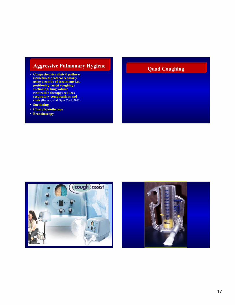
Aggressive Pulmonary Hygiene • Comprehensive clinical pathway
(structured protocol regularly using a combo of treatments i.e., positioning; assist coughing /suctioning; lung volume restoration therapy) reduces respiratory complications and costs (Berney, et al. Spin Cord, 2011)
• Suctioning • Chest physiotherapy • Bronchoscopy
Quad Coughing
18
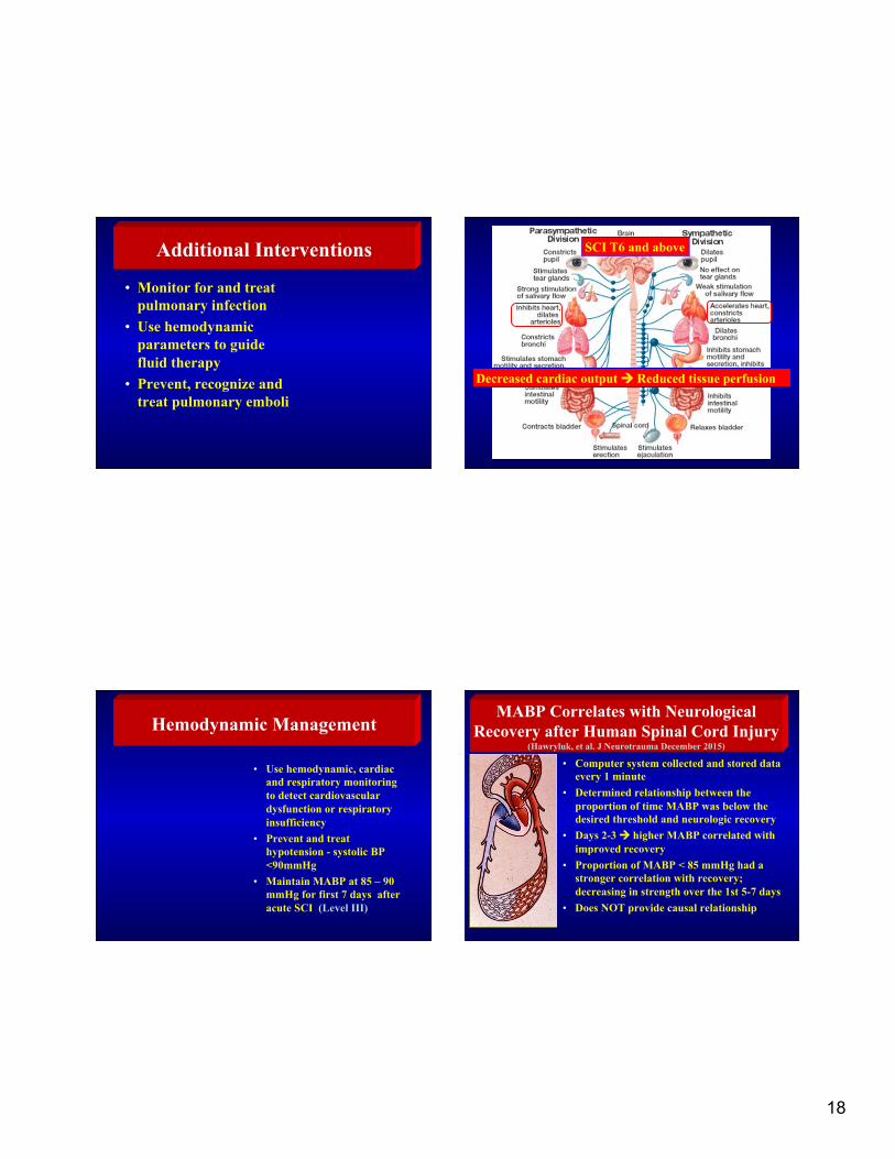
Additional Interventions
• Monitor for and treat pulmonary infection
• Use hemodynamic parameters to guide fluid therapy
• Prevent, recognize and treat pulmonary emboli
Decreased cardiac output à Reduced tissue perfusion
SCI T6 and above
Hemodynamic Management
• Use hemodynamic, cardiac and respiratory monitoring to detect cardiovascular dysfunction or respiratory insufficiency
• Prevent and treat hypotension - systolic BP <90mmHg
• Maintain MABP at 85 – 90 mmHg for first 7 days after acute SCI (Level III)
MABP Correlates with Neurological Recovery after Human Spinal Cord Injury
(Hawryluk, et al. J Neurotrauma December 2015) • Computer system collected and stored data
every 1 minute • Determined relationship between the
proportion of time MABP was below the desired threshold and neurologic recovery
• Days 2-3 à higher MABP correlated with improved recovery
• Proportion of MABP < 85 mmHg had a stronger correlation with recovery; decreasing in strength over the 1st 5-7 days
• Does NOT provide causal relationship

19
Hemodynamic Management
• Rule out other causes for hypotension • Early appropriate fluid resuscitation
• Isotonic crystalloids • Blood products, if appropriate
• Use hemodynamic parameters and indicators of tissue perfusion (e.g., lactate, base deficit) to guide fluid replacement
• Inotropic or vasoactives if necessary
Vasovagal Response
Sympathetic cardiac
acceleration is disrupted
Intact vagal nerve
slows the heart
Prevent Vasovagal Reflex
• Prevention – Avoid hypoxia – Avoid rapid position change – Avoid the valsalva maneuver
• Treatment – Increase HR – Atropine – Other drugs – Pacemaker
Orthostatic Hypotension • Wrap legs before
raising head • Abdominal binder • Gradually raise the
head of the bed • Medication
– Midodrine (alpha adrenergic agonist -à increases vascular tone and BP)
20
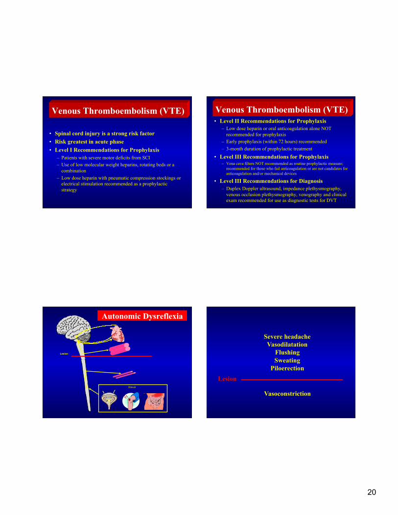
Venous Thromboembolism (VTE)
• Spinal cord injury is a strong risk factor • Risk greatest in acute phase • Level I Recommendations for Prophylaxis
– Patients with severe motor deficits from SCI – Use of low molecular weight heparins, rotating beds or a
combination – Low dose heparin with pneumatic compression stockings or
electrical stimulation recommended as a prophylactic strategy
Venous Thromboembolism (VTE) • Level II Recommendations for Prophylaxis
– Low dose heparin or oral anticoagulation alone NOT recommended for prophylaxis
– Early prophylaxis (within 72 hours) recommended – 3-month duration of prophylactic treatment
• Level III Recommendations for Prophylaxis – Vena cava filters NOT recommended as routine prophylactic measure;
recommended for those who fail anticoagulation or are not candidates for anticoagulation and/or mechanical devices
• Level III Recommendations for Diagnosis – Duplex Doppler ultrasound, impedance plethysmography,
venous occlusion plethysmography, venography and clinical exam recommended for use as diagnostic tests for DVT
Stimuli
Lesion
Autonomic Dysreflexia
Lesion
Severe headache Vasodilatation
Flushing Sweating
Piloerection
Vasoconstriction

21
Other Signs and Symptoms
• Hypertension • Nasal congestion • Chest pain • Nausea • Bradycardia
Treatment of Autonomic Dysreflexia
• Sit patient up • Loosen tight clothing • Monitor BP closely • REMOVE NOXIOUS
STIMULI! • Antihypertensive (e.g.,
Nifedipine, NTG) • Alpha – adrenergic
blocking agents
Poikilothermia
• Loss of thermoregulation • Hypothermia OR may have fever
– Neurogenic incidence reportedly 2.6 - 28% – Pathogenesis not clear (Savage, et al. Global Spin J. 2016;6:607)
• Caused by disruption of the sympathetic pathways
• Must monitor temperature closely • Take measures to maintain
normothermia
Types of Spinal Cord Injury Pain
Nociceptive Neuropathic
• Analgesics • Nonsteroidal anti-
inflammatory agents • Neuropathic
• Gabapentin • Pregabalin Be cautious not to impair
respiratory status!

22
Ileus
• Common with increased incidence with complete lesions
• Usually seen first 2-3 days • Lasts 3 – 7 days • Caused by disruption of
the autonomic nervous system
Risk for GI Ulceration
• Autonomic nervous system disruption
• Stress • Systemic
hypotension and hypoxia
• No gastric intake
Nutrition
• Ensure adequate nutrition to meet the patient’s determined metabolic needs
• Energy expenditure is best determined by indirect calorimetry since equation estimates tend to be inaccurate
• Maintain normoglycemia • Evaluate swallowing
Upper Motor Neuron Bowel • Hyper-reflexive after spinal
shock resolves • No sensation or control of
defecation • Spastic external sphincter • Unable to consciously defecate • Allows reflexive stool
propulsion • UMN goal: soft-formed stool
readily evacuated with rectal stimulation

23
Lower Motor Neuron Bowel
• Sacral level injury (e.g., cauda equina, conus medullaris)
• Areflexive bowel • Slow colonic propulsion • Hypotonic sphincter à More
frequent defecation/ incontinence • LMN goal: firm-formed stool
retained between bowel care episodes and is easy to evacuate
Bowel Program • Initiate ASAP! • Perform at least once every 1-2 days; 30
minutes after meal • Ensure diet with sufficient fiber and fluid • Stool softeners/ bulk-forming agents • Manual stimulation • Chemical stimulants (suppositories, small
enemas) • Assistive techniques (eg. abdominal
massage) ???
Genitourinary dysfunction • Spinal shock à Urine retention • Reflexive neurogenic bladder
– Injury above the conus – Spontaneous voiding at low
volumes • Areflexive neurogenic bladder
– Injury at sacral level – Urine retention with overflow
• Continuous catheterization à Fluid intake of < 3 liters and no need to monitor I & Oà Intermittent catheterization
Urinary Tract Infection
• Prevention – Remove catheter ASAP – Keep bladder empty
• Treatment – Only symptomatic
bacteruria – Prophylactic treatment
increases antibiotic resistant infection risk
24
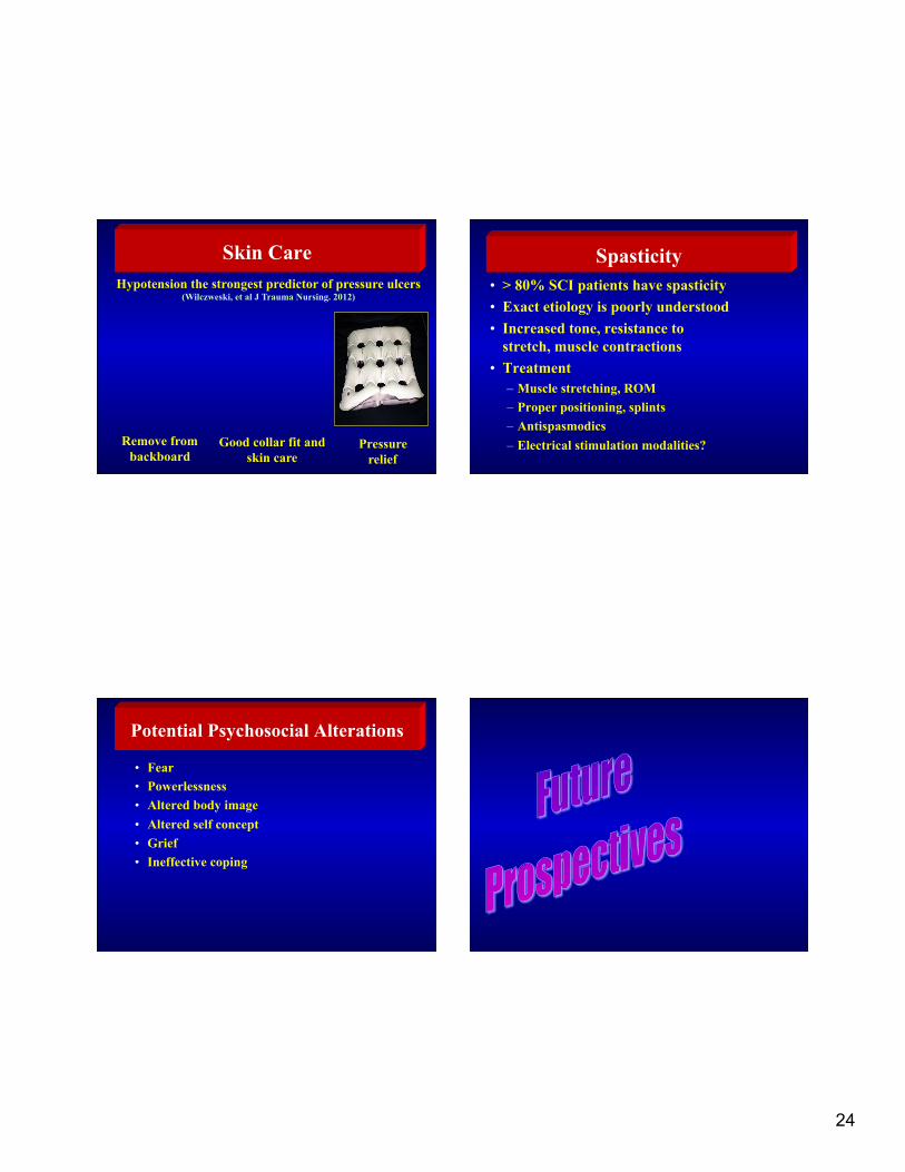
Skin Care
Remove from backboard
Good collar fit and skin care
Pressure relief
Hypotension the strongest predictor of pressure ulcers (Wilczweski, et al J Trauma Nursing. 2012)
Spasticity • > 80% SCI patients have spasticity • Exact etiology is poorly understood • Increased tone, resistance to
stretch, muscle contractions • Treatment
– Muscle stretching, ROM – Proper positioning, splints – Antispasmodics – Electrical stimulation modalities?
Potential Psychosocial Alterations
• Fear • Powerlessness • Altered body image • Altered self concept • Grief • Ineffective coping

25
Advanced MRI Strategies
Functional MRI
Diffusion Tensor Imaging
Improved Management of Systemic Complications
Riluzole • Approved for
amyotrophic lateral sclerosis
• Neuroprotective after SCI? – Blocking sodium channels – Decreasing glutamate
release • Multicenter randomized,
placebo controlled, Phase II/III trial in humans with acute traumatic SCI to evaluate efficacy and safety underway

26
Monitoring and Treating Spinal Cord Perfusion Pressure
Papadopoulos, et al. Saint George’s, University of London
Spinal cord perfusion pressure = MABP – Intraspinal pressure
Typically optimal about 90 mmHg Saadoun, Papadopoulos. Critical Care. 2016: 20:308
Neurotropic Factors
• Support neuronal survival
• Induce sprouting of neurites
• Facilitate guidance of neurons to their proper target sites
Cell Transplantation • Extensive preclinical literature
suggests stem cell based therapies may offer promise (Antonic , et al, PLOS Biology. Dec. 2013)
• Intralesion transplantation of autologous mesenchymal stem cells in chronic, complete SCI is safe, feasible and may promote neuro recovery (Mendonca et al. Stem cell res. 2014)
• Human stem cell trial underway – 10-15 U.S. sites to enroll 52 patient – C5-7, At least 12 weeks after injury
Hypothermia • Neuroprotective • Mixed results with local and
systemic hypothermia in animals • Local hypothermia in humans
– Not published since 1984 – Lacked statistical power
• Systemic hypothermia safely used in humans (Dietrich, et al . Neurotherapeutics 4/2011)
• Larger randomized controlled trials needed to establish efficacy
• Prospective trial underway • at University of Miami