Embed Size (px)
Citation preview

International Journal of Scientific and Research Publications, Volume 5, Issue 8, August 2015 1 ISSN 2250-3153
www.ijsrp.org
Soft Tissue metastasis in Carcinoma Breast: a rare presentation
Dr Meher Lakshmi Konatam, Dr Triveni. B, Dr Md Shoiab Zeesham
Assistant Professor, MNJ Institute of Oncology and & Regional Cancer Centre, Red Hills, Lakadikapul, Hyderabad-500004, Telangana, India
Abstract- Soft tissue metastasis is rare in carcinoma breast and can mimic soft tissue sarcoma. We present a rare case of a young lady presenting with forearm swelling. On examination, lump is found in the breast. Histopathological examination of breast lump revealed invasive ductal carcinoma and similar histology in forearm swelling also suggesting carcinoma breast metastasizing to forearm. Index Terms- Breast cancer, soft tissue, metastasis
I. INTRODUCTION etastatic (stage IV) breast cancer is defined by tumor spread beyond the breast, chest wall, and ipsilateral
regional lymph nodes. The most common sites for breast cancer metastasis include the bone, lung, liver, lymph nodes, chest wall, and brain. Soft tissue is not the usual site of distant dissemination of breast cancer. The clinical distinction between metastasis to
soft tissue and primary soft tissue sarcoma is important as treatment for both differs. Only a few small series and isolated cases of soft tissue metastasis from breast cancer have been reported.1, 2 We report a rare case of forearm metastasis from breast cancer mimicking soft tissue sarcoma.
II. CASE REPORT A 26 year old lady presented with swelling in the right forearm of 3 months duration, progressively increasing in size. The swelling is not associated with pain but caused restriction of movements of fingers. There is no history of systemic symptoms. On examination, Performance status: 1, 15x7cm nontender firm to hard diffuse swelling is present on flexor aspect of right forearm, fixed to underlying muscle. Skin over the swelling is stretched , shiny and pinchable (figure 1).
M
International Journal of Scientific and Research Publications, Volume 5, Issue 8, August 2015 2 ISSN 2250-3153
www.ijsrp.org
Figure 1: Swelling on the forearm. Trucut biopsy site is also seen.
On systemic examination, 4x5cm non tender firm to hard swelling is present in upper outer quadrant of right breast. No palpable axillary nodes. Skin over the swelling is normal.

International Journal of Scientific and Research Publications, Volume 5, Issue 8, August 2015 3 ISSN 2250-3153
www.ijsrp.org
Trucut biopsy from breast lump is suggestive of infiltrating duct cell carcinoma. ER, PR, Her2 neu negative. Trucut biopsy from forearm swelling is suggestive of deposits of duct cell carcinoma (figures 2,3and 4).
Figure 2: scanner view: H & E sections showing linear cores of tissue exhibiting tumor tissue
International Journal of Scientific and Research Publications, Volume 5, Issue 8, August 2015 4 ISSN 2250-3153
www.ijsrp.org
Figure 3: low power view (20X): tumor cells arranged in loosely cohesive clusters and sheets infiltrating fibrocollagenous tissue.
International Journal of Scientific and Research Publications, Volume 5, Issue 8, August 2015 5 ISSN 2250-3153
www.ijsrp.org
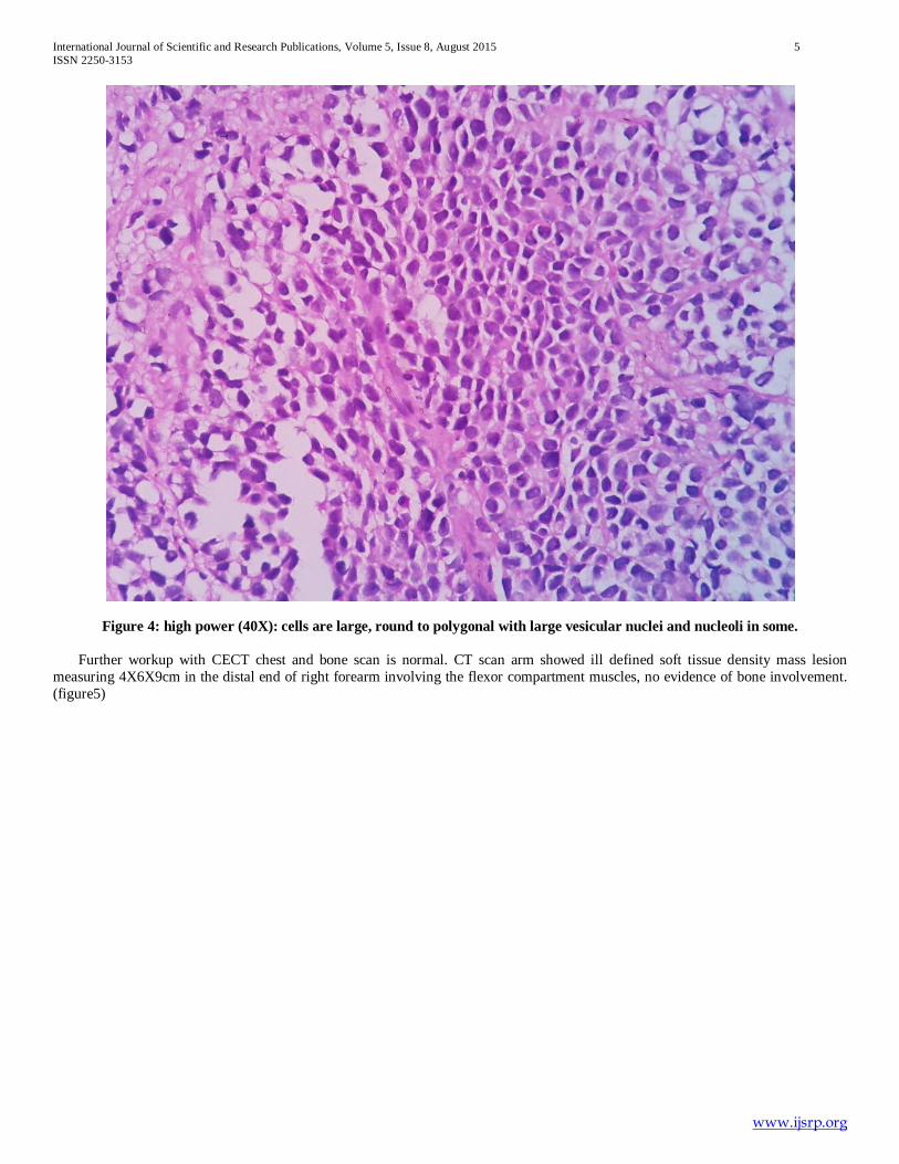
Figure 4: high power (40X): cells are large, round to polygonal with large vesicular nuclei and nucleoli in some. Further workup with CECT chest and bone scan is normal. CT scan arm showed ill defined soft tissue density mass lesion measuring 4X6X9cm in the distal end of right forearm involving the flexor compartment muscles, no evidence of bone involvement. (figure5)

International Journal of Scientific and Research Publications, Volume 5, Issue 8, August 2015 6 ISSN 2250-3153
www.ijsrp.org
Figure 5: CT scan arm showing ill defined mass in flexor aspect of forearm. She is finally diagnosed as stage 4 carcinoma breast, the only site of metastasis being forearm muscles. She is started on chemotherapy with doxorubicin and cyclophosphomide. She completed one cycle and is doing well.
III. DISCUSSION Breast Cancer metastasis to soft tissue is rare though case reports have documented breast cancer dissemination to almost every organ in the body. Hormone receptor–positive tumors are more likely to spread to bone as the initial site of metastasis; hormone receptor–negative and/orHER2+ tumors are more likely to recur initially in viscera.3Lobular (as opposed to ductal) cancers are more often associated with serosal metastases to the pleura and abdomen. Most women with metastatic disease will have been initially diagnosed with early-stage breast cancer, treated with curative intent, and then experience metastatic recurrence. Only about 10% of patients with newly diagnosed breast cancer in the United States have metastatic disease at presentation; this proportion is far higher in areas where screening programs are not available. Plaza JA et al., (2008) had a series of 118 patients with metastasis to soft tissue and out of those only 13 cases were of metastasis from breast cancer.4
The treatment goals in women with advanced breast cancer include prolongation of life, control of tumor burden, reduction in cancer-related symptoms or complications, and maintenance of quality of life and function. Therapy is not generally considered curative. Some patients with breast cancer will have limited sites of metastatic disease, such as isolated pulmonary nodules, isolated contralateral lymph node recurrence, or bone lesions. Single-institutional experience from the MD Anderson Cancer Center suggests that a fraction of such patients may be treated “aggressively” with curative intent, with favorable long-term results.5 The treatment of the primary tumor in the breast in women who present with metastatic disease is another area of controversy. An analysis of 16,023 patients presenting with stage IV disease and an intact primary tumor compared outcomes between patients having surgery of the primary tumor to negative margins or no surgery. In a multivariate analysis adjusting for known prognostic factors, surgery reduced the HR for death to 0.61 (95% CI = 0.58 to 0.65).6
Our patient presented with metastasis to forearm only with no evidence of disease elsewhere other than that in primary site. Hence she is planned for chemotherapy upfront and surgery to primary and local RT according to her response.

International Journal of Scientific and Research Publications, Volume 5, Issue 8, August 2015 7 ISSN 2250-3153
www.ijsrp.org
IV. CONCLUSIONS Soft tissue metastasis from solid tumors should be borne in mind when evaluating soft tissue tumors and thorough clinical examination is mandatory for accurate diagnosis and treatment.
REFERENCES [1] Leinung S et al. Histopathological outcome of 597 islated soft tissue tumors
suspected of soft tissue sarcoma: a single centre 12 year experience. Eur J Surg Oncol. 2006:33:508-511
[2] Damron TA et al. Distant soft tissue metastasis: a series of 30 new patients and 91 cases from the literature. Ann Surg Oncol. 2007:7:526-534.
[3] Lin Y, Yin W, Yan T, et al. Site-specific relapse pattern of the triple negative tumors in Chinese breast cancer patients. BMC Cancer 2009; 9:342.
[4] Plaza JA et al, Metastasis to soft tissue: a review of 118 cases over a 30 year period. Cancer 2008: 112: 193-203.
[5] Rivera E, Holmes FA, Buzdar AU, et al. Fluorouracil, doxorubicin, and cyclophosphamide followed by tamoxifen as adjuvant treatment for patients with stage IV breast cancer with no evidence of disease.Breast J 2002;8:2–9.
[6] Khan SA, Stewart AK, Morrow M. Does aggressive local therapy improve survival in metastatic breast cancer? Surgery 2002; 132:620–626, discussion 626–627.
AUTHORS First author: Dr Meher Lakshmi Konatam MD DM, Assistant professor, Department of Medical Oncology, MNJ Institute of Oncology and Regional Cancer Centre, Red Hills, Lakdikapul, Telangana, India. Email: [email protected] Second author: Dr Triveni. B MD, Professor and Head of the Department, Department of Pathology, MNJ Institute of Oncology and Regional Cancer Centre, Red Hills, Lakdikapul, Telangana, India Third author: Dr Md Shoiab Zeesham, postgraduate in pathology, MNJ Institute of Oncology and Regional Cancer Centre, Red Hills, Lakdikapul, Telangana, India