Embed Size (px)
Citation preview
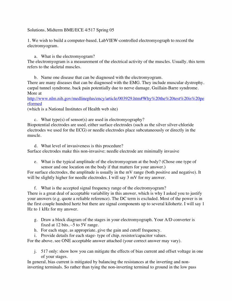
Solutions, Midterm BME/ECE 4/517 Spring 05
1. We wish to build a computer-based, LabVIEW-controlled electromyograph to record theelectromyogram.
a. What is the electromyogram?The electromyogram is a measurement of the electrical activity of the muscles. Usually, this termrefers to the skeletal muscles.
b. Name one disease that can be diagnosed with the electromyogram.There are many diseases that can be diagnosed with the EMG. They include muscular dystrophy,carpal tunnel syndrome, back pain potentially due to nerve damage, Guillain-Barre syndrome.More athttp://www.nlm.nih.gov/medlineplus/ency/article/003929.htm#Why%20the%20test%20is%20performed(which is a National Institutes of Health web site)
c. What type(s) of sensor(s) are used in electromyography?Biopotential electrodes are used, either surface electrodes (such as the silver silver-chlorideelectrodes we used for the ECG) or needle electrodes place subcutaneously or directly in themuscle.
d. What level of invasiveness is this procedure?Surface electrodes make this non-invasive; needle electrode are minimally invasive
e. What is the typical amplitude of the electromyogram at the body? (Chose one type ofsensor and one location on the body if that matters for your answer.)
For surface electrodes, the amplitude is usually in the mV range (both positive and negative). Itwill be slightly higher for needle electrodes. I will say 3 mV for my answer.
f. What is the accepted signal frequency range of the electromyogram?There is a great deal of acceptable variability in this answer, which is why I asked you to justifyyour answers (e.g. quote a reliable reference). The DC term is excluded. Most of the power is inthe first couple hundred hertz but there are signal components up to several kilohertz. I will say 1Hz to 1 kHz for my answer.
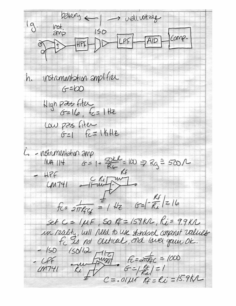
g. Draw a block diagram of the stages in your electromyograph. Your A/D converter isfixed at 12 bits, –5 to 5V range.
h. For each stage, as appropriate, give the gain and cutoff frequency.i. Provide details for each stage- type of chip, resistor/capacitor values.
For the above, see ONE acceptable answer attached (your correct answer may vary).
j. 517 only: show how you can mitigate the effects of bias current and offset voltage in oneof your stages.
In general, bias current is mitigated by balancing the resistances at the inverting and non-inverting terminals. So rather than tying the non-inverting terminal to ground in the low pass
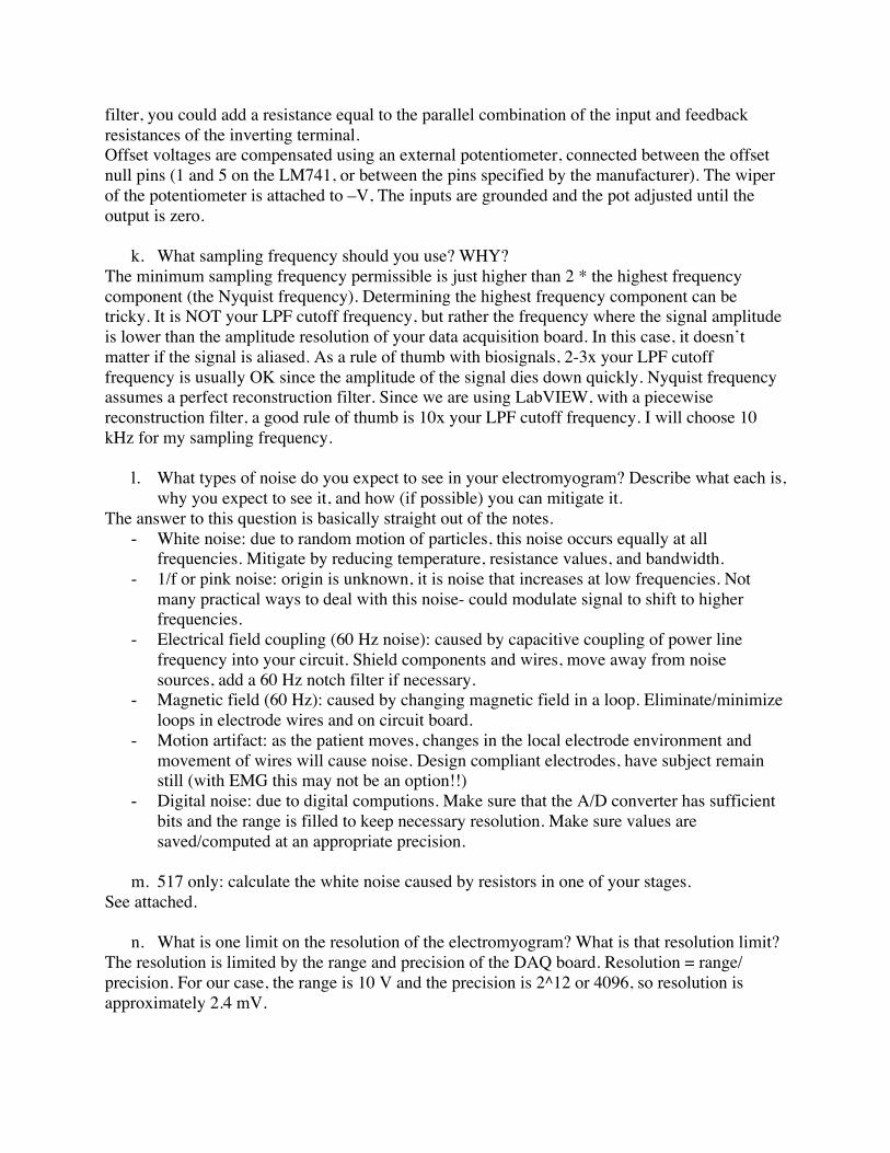
filter, you could add a resistance equal to the parallel combination of the input and feedbackresistances of the inverting terminal.Offset voltages are compensated using an external potentiometer, connected between the offsetnull pins (1 and 5 on the LM741, or between the pins specified by the manufacturer). The wiperof the potentiometer is attached to –V, The inputs are grounded and the pot adjusted until theoutput is zero.
k. What sampling frequency should you use? WHY?The minimum sampling frequency permissible is just higher than 2 * the highest frequencycomponent (the Nyquist frequency). Determining the highest frequency component can betricky. It is NOT your LPF cutoff frequency, but rather the frequency where the signal amplitudeis lower than the amplitude resolution of your data acquisition board. In this case, it doesn’tmatter if the signal is aliased. As a rule of thumb with biosignals, 2-3x your LPF cutofffrequency is usually OK since the amplitude of the signal dies down quickly. Nyquist frequencyassumes a perfect reconstruction filter. Since we are using LabVIEW, with a piecewisereconstruction filter, a good rule of thumb is 10x your LPF cutoff frequency. I will choose 10kHz for my sampling frequency.
l. What types of noise do you expect to see in your electromyogram? Describe what each is,why you expect to see it, and how (if possible) you can mitigate it.
The answer to this question is basically straight out of the notes.- White noise: due to random motion of particles, this noise occurs equally at all
frequencies. Mitigate by reducing temperature, resistance values, and bandwidth.- 1/f or pink noise: origin is unknown, it is noise that increases at low frequencies. Not
many practical ways to deal with this noise- could modulate signal to shift to higherfrequencies.
- Electrical field coupling (60 Hz noise): caused by capacitive coupling of power linefrequency into your circuit. Shield components and wires, move away from noisesources, add a 60 Hz notch filter if necessary.
- Magnetic field (60 Hz): caused by changing magnetic field in a loop. Eliminate/minimizeloops in electrode wires and on circuit board.
- Motion artifact: as the patient moves, changes in the local electrode environment andmovement of wires will cause noise. Design compliant electrodes, have subject remainstill (with EMG this may not be an option!!)
- Digital noise: due to digital computions. Make sure that the A/D converter has sufficientbits and the range is filled to keep necessary resolution. Make sure values aresaved/computed at an appropriate precision.
m. 517 only: calculate the white noise caused by resistors in one of your stages.See attached.
n. What is one limit on the resolution of the electromyogram? What is that resolution limit?The resolution is limited by the range and precision of the DAQ board. Resolution = range/precision. For our case, the range is 10 V and the precision is 2^12 or 4096, so resolution isapproximately 2.4 mV.
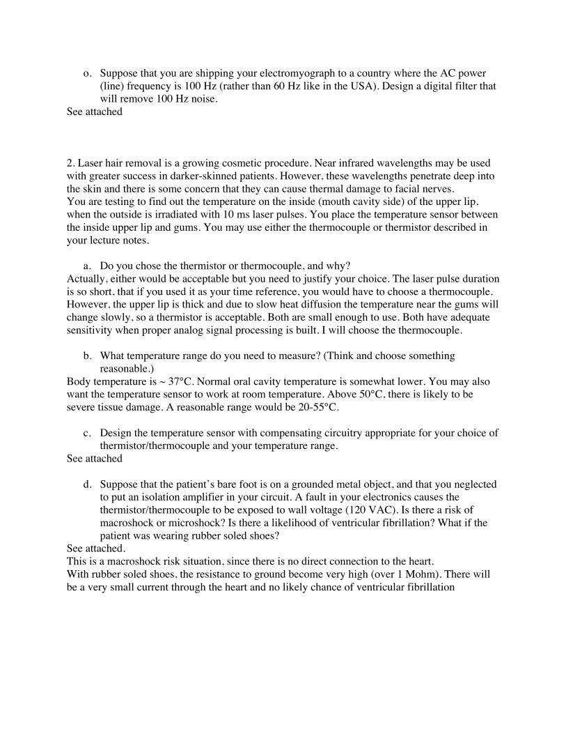
o. Suppose that you are shipping your electromyograph to a country where the AC power(line) frequency is 100 Hz (rather than 60 Hz like in the USA). Design a digital filter thatwill remove 100 Hz noise.
See attached
2. Laser hair removal is a growing cosmetic procedure. Near infrared wavelengths may be usedwith greater success in darker-skinned patients. However, these wavelengths penetrate deep intothe skin and there is some concern that they can cause thermal damage to facial nerves.You are testing to find out the temperature on the inside (mouth cavity side) of the upper lip,when the outside is irradiated with 10 ms laser pulses. You place the temperature sensor betweenthe inside upper lip and gums. You may use either the thermocouple or thermistor described inyour lecture notes.
a. Do you chose the thermistor or thermocouple, and why?Actually, either would be acceptable but you need to justify your choice. The laser pulse durationis so short, that if you used it as your time reference, you would have to choose a thermocouple.However, the upper lip is thick and due to slow heat diffusion the temperature near the gums willchange slowly, so a thermistor is acceptable. Both are small enough to use. Both have adequatesensitivity when proper analog signal processing is built. I will choose the thermocouple.
b. What temperature range do you need to measure? (Think and choose somethingreasonable.)
Body temperature is ~ 37°C. Normal oral cavity temperature is somewhat lower. You may alsowant the temperature sensor to work at room temperature. Above 50°C, there is likely to besevere tissue damage. A reasonable range would be 20-55°C.
c. Design the temperature sensor with compensating circuitry appropriate for your choice ofthermistor/thermocouple and your temperature range.
See attached
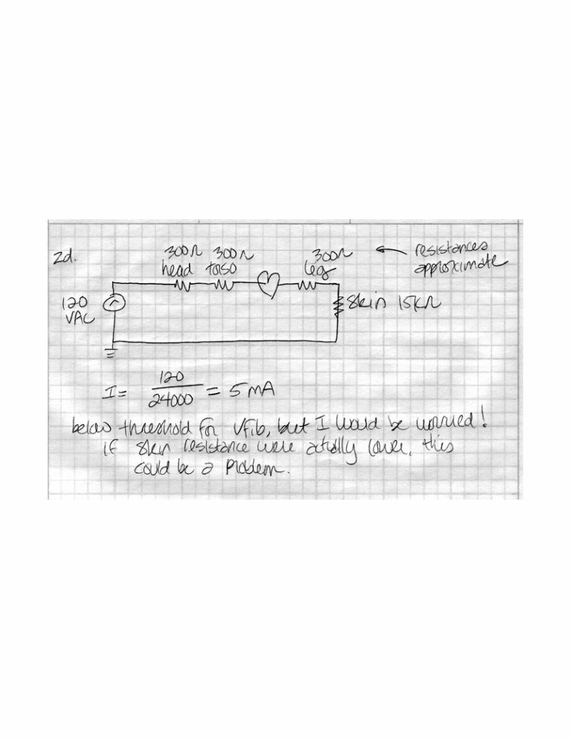
d. Suppose that the patient’s bare foot is on a grounded metal object, and that you neglectedto put an isolation amplifier in your circuit. A fault in your electronics causes thethermistor/thermocouple to be exposed to wall voltage (120 VAC). Is there a risk ofmacroshock or microshock? Is there a likelihood of ventricular fibrillation? What if thepatient was wearing rubber soled shoes?
See attached.This is a macroshock risk situation, since there is no direct connection to the heart.With rubber soled shoes, the resistance to ground become very high (over 1 Mohm). There willbe a very small current through the heart and no likely chance of ventricular fibrillation