Embed Size (px)
Citation preview

s
SOMATOTOPIC ORGANIZATION OF THE FACIAL NUCLEUS IN THEト10NKEY AND
THE RABBIT
Ph.D. Thesis
by
TAKAHIRO SATODA, D.D.S.
Hiroshima University School of Dentistry
1989
The majority of data used in this thesis have been published
in the Neuroscience Letters, 78: 283-287, 1987 and the
Anatomischer Anzeiger, 165: 83-90, 1988.
I. Representation of the main branches of the facial nerve
within the facial nucleus of the Japanese monkey (Macaca
fuscata)
Takahiro Satoda, Osamu Takahashi, Takashi Tashiro, Ryotaro
Matsushima, Masanon Uemura-Sumi, and Noboru Mizuno
Neuroscience Letters 78: 283-287, 1987.
II. Somatotopic organization of facial nucleus of rabbit, with
particular reference to intranuclear representation of
penoral branches of the facial nerve
Takahiro Satoda, Osamu Takahashi, Takashi Tashiro, Ryotaro
Matsushima, Masanori Uemura-Sumi, and Noboru Mizuno
Anatomischer Anzeiger 165: 83-90, 1988.
- 2.-
CONTENTS
I.川TRODUCTION
II. MATERIALS AND METHODS
1. Application of horseradish peroxidase (HRP)
1) Monkey
2) Rabbit
2. Per fusion
3. Histoiogical procedures
4. Observation of the facial nerve
5. Observation of HRP-labeled neurons
- 3 -
10
10
Ill
ll

III. RESULTS
1. Monkey
2. Rabbit
IV. DISCUSSION
1. Methodological consideration
2. So汀Iatotopic organization of the facial nuc一eus
V. SUMMARY
VI. ACKNOWLEDGEMENTS
VII. REFERENCES
VIII. FIGURES
- 4 -
12
12
15
19
19
21
25
28
29
34

I. INTRODUCTION
Somatotopic organization of the facial nucleus has been
studied in a variety of mammals by the morphological and
electrophysiological techniques (Yagita, 1910; Szentagothai,
1948; Kitai et al., 1972; Dom et al., 1973; Dom and Zieiinsky,
1977; Kume et al., 1978; Shohara and Sakai, 1983; Uemura-Sumi et
al., 1986. for further reviews,, cf. Papez, 1927; Vraa-Jensen,
1942; Courville, 1966; Martin and Lodge, 1977; Dom, 1982;
Hinnchsen and Watson, 1984, Baisden et al., 1987). The previous
morphological studies usually used transection of individual
facial nerve branches and retrograde degeneration techniques for
identification of facial motoneurons which send their axons to
the facial nerve branches (for reviews, cf. Yagita, 1910; Papez,
1927; Vraa-JenseI一, 1942; Courv川e, 1966). More recently, the
retrograde HRP (horseradish peroxidase) method has been developed
(for review, cf. Mesuiam, 1982). Since this method was shown to
enable an easier and clearer identification of subgroups of
motoneurons to be made (Mizuno et al., 1975), a group of studies
have been carried out on the myotopic organization of the facial
nucleus after injection of HRP into the facial muscles in the
opossum (Dom and Zieiinsky, 1977; Provis, 1977), bat (Friauf and
Herbert, 1985), mouse (Ashweil, 1982; Komiyama et al., 1984), rat
(Watson et al., 1982; Hinrichsen and Watson, 1984; Friauf and
- 5 -

Herbert, 1985), kitten (Radpour, 1977), cat (K叩e et a!., 1978;
Parnes et al., 1982), and rhesus and cynomolgus monkeys (Parnes
et 尋1., 1982). Correlation of the subdivisions of the facia一
nuc一eus with the peripheral branches of the facia一 nerve has also
been examined after applying HRP to the facial nerve branches in
the opossum (Dom, 1982), rat (Semba, 1984), guinea pig
(Uemura-Sumi et al., 1986), and rabbit (Baisden et al., 1987).
The results of the previous studies indicate that there more or
less exist species differences in the somatotopic organization of
the facial nucleus.
The somatotopie organization of the facial nucleus of the
macaque monkeys has not been systematically studied by the HRP
method. The present study, therefore, was attempted to examine
the correlation of the main peripheral branches of the facial
nerve with cytoarchitectonic divisions of the facial nucleus in
the Japanese monkey (Macaca fuscata) to shed more light on the
general organization of the facial nucleus in mammalian species.
For the purpose of comparison, the study was also performed in
the rabbit as a representative of the most common laboratory
animals, although a study in the rabbit has already been reported
(Baisden et al., 1987).
- 6 I

II. MATERIALS AND METHODS
The present reports are based on the results which were
obtained from 18 Japanese monkeys (Macaca fuscata) weighing 3 -
12 kg, and 35 adult rabbits weighing 1.8 - 3.0 kg. The
experiments were performed according to the NIH Guide for the
Care and Use of Laboratory Animals (National Institute of Health
Publications No. 85-83, Revised 1985). Horseradish peroxidase
(HRP) was purchased from Toyobo, Japan (HRP, Grade-1-C: RZ= 3.3
or3.4).
1. Application of HRP
1) monkey
The monkeys were anesthetized with an intraperitoneal
injection of sodium pentobarbital (30 - 35 mg/kg body weight).
Supplementary doses were given as necessary to maintain a deep
level of anesthesia during long surgical procedures.
In 12 monkeys, the main branches of the facial nerve
supplying the superficial facial muscles were exposed
urnlaterally with an aid of an operation microscope. They were
cut, and both proximal and distal cut-ends were ligated (Fig.
la). Subsequently, at the sites close to the proximal cut-end,
- 7 -

mu一tiple injections of the total of 0.1 - 0.5 jjl of 30% HRP
dissolved in 0.9% saline were made manually by pressure through a
glass micropipette (tip diameter: 40 - 60 〃m) coupled to a lO-^ii
Hamilton microsyringe; the main branches other than the target
nerve branch had been cut and ligated with thin thread before the
HRP injection into the target nerve branch. The proximal cut-end
of the target nerve branch injected with HRP was further inserted
into a piece of polyethyiene tube which was filled with 30% HRP
dissolved in sterile 0.9% saline. The open end of the tube was
then sealed with vaseline. Subsequently, the tube was fixed to
the neighboring region with a quick set adhesive (Alon Al fa;
alpha-cyanoacrylate).
In 3 monkeys, the HRP solution was applied to the trunk of
the facial nerve at the site close to the styiomastoid foramen.
In additiona一 3 monkeys, 5 jjl of 30% HRP disso一ved in 0.9%
saline was injected into the orbicularis oculi musele after
severing and ligating nerve branches supplying the superficial
facial muscles in the anterior auricular and the superior labial
regions.
2) Rabbit
The rabbits were anesthetized with an intraperitoneal
injection of.urethane (1 g/kg body weight), followed with an
intravenous injection of sodium pentobarbital (25 - 3牀mg/kg body
ー 8 -

weight). Supplementary doses of sodium pentobarbitai were given
when necessary throughout the duration of the experiments.
In 17 rabbits, the main peripheral branches of the facial
nerve were exposed unilaterally under an operation microscope
(Fig. 2a). They were cut, and both proximal and distal cut-ends
werとIigated. Subsequently, at the sites close to the proximal
cut-end, a sing一e mam branch of the facia一 nerve was injected
with 0.1 - 0.5 /jI of 30% HRP dissolved in sterile 0.9% saline
manually by pressure through a glass micropipette (tip diameter:
40 - 60 pi) coupled to a 10-jul Hamilton microsyringe. The
proximal cut-end of the target nerve branch injected with HRP was
further inserted into早 piece of polyethylene tube filled with
30% HRP dissolved in sterile 0.9% saline. The open end of the
tube was then sealed with vaseline, and the tube was fixed to
the neighboring region by Alon Al fa.
In 2 rabbits, the application of the HRP solution was
performed to the trunk of the facial nerve at the sites close to
the styiomastoid foramen (Fig. 2a).
In each of- 12 rabbits, the application of the HRP solution
was done to one of the 4 periol-al branchlets arising from the
superior labial branch, and/or the inferior 'labial branch (Fig.
3a).
In 4 rabbits, the HRP solution was injected unilaterally
into the superficial facial muscles in the periorbital region
- 9 -

after cutting and ligating the superior labial and the inferior
labial branches of the facial nerve.
2. Per fusion
After the application of HRP to the facial nerve or muscles,
the animals were allowed to survive for 36-48 hours, and then
re-anesthetized deeply with an overdose of the anesthesia, and
were per fused through the ascending aorta with 9% formalin in 0.1
M phosphate buffer (pH 7.3) (1 I/kg body weight), followed by the
same buffer containing lO% sucros`e (0.5 "I/kg body weight).
3. Histological procedures
After the transcardial per fusion, the brainstems were
removed and placed into 30% sucrose in 0.1 M phosphate buffer (pH
7.3) at 4-C. when the brainstems were saturated with the
so一ution, they were cut into transverse serial sections of 60 〃m
thickness on a freezing microtome. For the histochemical
demonstration of HRP, the sections were treated with
- 10 -

tetramethylbenzidine (TMB) according to Mesulam (1978). Then the
sections were mounted on glass slides coated with a chrome
alum-gelatine (Pappas, 1971), and counterstained with l% neutral
red.
4. Observation of the facial nerve
After the per fusion and removal of the brainstem, the
pattern of branching of the facial nerve supplying the
superficial facial muscles were examined macroscopicaTly with an
aid of an operation microscope.
5. Observation of HRP-labeled neurons
The serial sections were examined microscopically in
bright-field and dark-field illumination. The distribution of
neuronal cell bodies labeled retrogradely with HRP was charted
onto enlarged outline drawings of representative sections that
were enlarged by using a projection apparatus.
r ifl ニ
III. Results
1. Monkey
In al一 monkeys examined in the present study, 5 main
peripheral branches of the facial nerve were identified, although
the pattern of branching of the facial nerve was variant from
animal to animal (Fig. la). The 5 main peripheral branches were
the cervical (Ce), the posterior auricular (PA), the
auriculo-zygomatico-orbitai (AZO),, the superior labial (SL), and
the inferior labial (IL) branches. The auriculo-zygomatic-orbital
(AZO) branch was composed of several nerve branch!ets supplying
the anterior auricular (AA) and zygomatic0-orbitai (ZO) regions.
In 3 monkeys, HRP was applied to the trunk of the facial
nerve. In these monkeys, the facial nucleus ipsilateral to the
HRP app一ication was filled with neuronal cell bodies which were
labeled retrogradely with HRP (Fig. 4a). No HRP-labeled neuronal
cell bodies were found on the side contralateral to the HRP
application. The facial nucleus filled with HRP-labeled
motoneurons was divided into 5 divisions; into the ventral (V),
medial (M), intermediate (I), dorsal (D), and lateral (L)
divisions (Figs, lb-d and 4a). Facial motoneurons in the ventral
division were clustered into 2 subgroups at the level of the
- 12 -
caudal two thirds of the facial nucleus (Figs, lc, d and 4b).
In 2 monkeys which were applied with HRP to the cervical
(Ce) branch of the facial nerve, HRP-Iabeled neuronal cell bodies
were seen ipsiiaterally in the ventral (V) division of the facial
nucleus ipsiiateral to the HRP application. Both of the 2
subgroups of facial motoneurons in the ventral division were
labeled with HRP (Figs, lb-d and 4b).
In 2 monkeys which were applied with HRP to the posterior
auricular (PA) branch of the facial nerve, neuronal cell bodies
labeled with HRP were observed ipsilaterally in the medial (M)
division of the facia一 nucleus (Figs, lb-d and 4c).
In 3 monkeys which were applied with HRP to the
auriculo-zygomatico-orbital (AZO) branch of the facial nerve,
HRP-labeled neuronai cell bodies were seen lpsilaterally in the
intermediate (I) and dorsal (D) divisions of the facial nucleus.
In one of the 3 monkeys, a few HRP-Iabeled neurons were
distributed in the dorsolaterai aspects of the medial (M)
division of the facial nucleus (Fig. lb-d).
In 3 monkeys which were app一ied with HRP to the superior
一abial (SL) branch of the facial nerve,トIRP-labeled neuronal cell
bodies were seen ipsilaterally in the dorsal (D) and lateral (L)
divisions of the facia一 nucl占us (Figs. Ib-d and 4d).
In 2 monkeys which were applied with HRP to the inferior
labial (IL) branch of the facial nerve, HRP-labeled neuronal cell
- 13 -

bodies were seen ipsilateraily in the lateral (L) division of the
facial nucleus (Fig. lb-d).
In 3 monkeys which were injected with HRP into the
or.biculans oculi muscles, HRP-labeled neuronal cell bodies were
distributed in the dorsal (D) division of the facial nucleus
ipsilateral to the HRP injection.
The results which were obtained from the 18 monkeys
described above are summarized in Fig. 1, which shows the
representation of the 5 main peripheral branches of the facial
nerve within the facial nucleus of the Japanese monkey.
- 14 -

2. Rabbit
In all rabbits examined in the present, study, 6 main
peripheral branches of the facial nerve were identified. These
were the cervical (Ce), posterior auricular (PA), anterior
auricular (AA), zygomatico-orbital (ZO), superior labial (SL),
inferior labial (IL) branches of the facial nerve (Fig. 2a). The
zygomatic0-orbital branches (ZO) were composed of several
branchlets distributing the perioral region. The periorai region
was usually supplied with 4 branchlets of the facial nerve
arising from the superior labial (SL) and/or the inferior labial
(IL) branch of the facial nerve (Figs. 2a and 3a).
In 2 rabbits in which HRP was applied to the trunk of the
facial nerve, the facial nucleus was filled with neuronal cell
bodies labeled retrogradely with HRP on the side ipsilateral to
the HRP application (Fig. 5a-c). No HRP-labeled neurons were
found in the facial nucleus on the side contraiateral to the HRP
application. The facial nucleus filled with HRP-labeled
motoneurons was divided into 5 divisions; into the ventromedial
(V), medial (M), dorsal (D), lateral (L), and intermediate (I)
divisions (Figs. 2, 3, and 5).
The HRP application was、高ade to the cervical (Ce) branch of
the facial nerve in 4 rabbits. HRP-Iabeled facial motoneurons
were localized in the ventromedial (V) division of the facial
- 15 -

nucleus ipsilateral to the HRP application (Figs. 2b and 6a).
In other 4 rabbits which were injected with HRP into the
superficial facial muscles around the.periorbital region after
cutting and ligating the superior labial (SL) and the inferior
labial (IL) branches, HRP-labeled neuronai cell bodies in the
facial nucleus ipsilateral to the HRP injection were located in
the dorsal (D) division of the facial nucleus (Figs. 2b and 6b).
In 4 rabbits which were applied with HRP to the anterior
auricular (AA) branch of the facial nerve, HRP-labeled neuronal
cell bodies were seen in the dorsal (D) division of the facial
nucleus, and in the dorsal part of media一 (M) division of the
facial nucleus ipsilateral to the HRP application (Figs. 2b and
6c).
In 3 rabbits which were applied with HRP to the posterior
auncular (PA) branch of the facial nerve, HRP-labeled neuronal
ceil bodies were observed in the medial (M) division of the
facial nucleus ipsilateral to the HRP application (Fig. 2b and
6d).
In other 3 rabbits which were applied with HRP to,the
superior labial (SL) branch of the facial nerve after cutting and
ligating the zygomatico-orbital (ZO) and the inferior labial (IL)
branches of the facial nerve, HRP-labeled neuronal cell bodies
were distributed in the lateral (L) division, and in the dorsal
aspect of the intermediate (I) division of the facia一 nucleus
- 16 -

ipsilateral to the HRP application (Figs. 2b and 6e).
In 3 rabbits which were applied with HRP to the inferior
labial (IL) branch of the facial nerve after cutting and Tigating
the superior labial (SL) and the cervical (Ce) branches of the
facial nerve, HRP-labeled neuronal cell bodies were seen in the
lateral (L) and the intermediate (I) divisions of.the facial
nucleus ipsilateral to the HRP application (Figs. 2b and 6f).
The results described above indicated that the area of the
nuc一ear representation of the superior labial (SL) branch of the
facial nerve highly overlapped with that of the inferior labial
(IL) branch of the facial nerve (Fig. 2b. compare Fig. 6e with
Fig. 6f). In fact, the perioral regions were usually supplied
with 4 nerve branchlets arising from the superior labial (SL)
and/or the inferior labial (IL) branches of the facial nerve
(Figs. 2a and 3a). The 4 perioral nerve branchlets mainly
supplied to the superior labial, oral angular, inferior labial
and submandibular regions, respectively (Figs. 2a and 3a).
In additional 12 rabbits, the HRP application was further
attempted to each of the 4 periorai nerve branchlets of the
facial nerve. The 12 rabbits were divided into 4 groups and the
rabbits of each group was applied with HRP to one of the perioral
nerve branchlets 1, 2, 3, and 4 after cutting and ligating the
other 3 periorai branchlets of the facial nerve (Fig. 3a). Facia一
motoneurons which were retrogradely labeled with HRP applied to
- 17 -

the perioral nerve branch!et 1, 2, 3 or 4 of the facial nerve
were distributed mainly in the lateral (L) division, in the
dorsal part of the intermediate (I) division, intermediate part
of the intermediate (I) division, or in the ventral part of the
intermediate (I) division of the facial nucleus, respectively
(Figs. 3 and 7).
The results obtained from the 35 rabbits described above are
summarized in Figs. 2 and 3, which show the representation of
peripheral nerve branches of the facial nerve within the facial
nuc一eus.
- 18 -

IV. DISCUSSION
1. Methodological consideration
The myotopical arrangement of facial motoneurons in the
facial nucleus has been studied by the retrograde HRP 甲ethod
after injecting HRP into the superficial facial muscles in the
opossum (Dom and Zieiinsky, 1977; Provis, 1977), bat (Friauf aヮd
Herbert, 1985), mouse (Aschwell, 1982; Komiyama et ah, 1984),
rat (Watson et al., 1982; Hinrichs。en and Watson, 1984; Friauf and
Herbert, 1985), kitten (Radpour et ai., 1977), cat (Kume et al
1978; Parnes et al., 1982), and rhesus and cynomoigus monkeys
(Parnes et al., 1982). HRP injection into the muscle, however,
has the inherent problem of leaking out of injected HRP into the
surroundings. In order to avoid this inherent problem, HRP has
been applled to the proximal cut-end of the peripheral branches
of the facial nerve, and the representation of the main
peripheral branches of the facial nerve within the facial nucleus
has been investigated in the opossum (Dom, 1982), rat (Semba,
1984), guinea pig (Uemura-Sumi et al 1986), and rabbit (Baisden
et a!., 1987). In the presdnt, study, the representation of the
mam peripheral branches of the facial nerve within the facial
nucleus of the Japanese monkey and the rabbit was examined after
- 19 -

application of HRP to the proximal cut-end of the target branch
of the facial nerve; when HRP was injected into the superficial
facia一 muscles, HRP was injected in the target muscle only after
cutting and ligating the nerve branches supplying the superficial
facial muscles adjacent to the target muscle.
The fixation of tissue in the HRP method is usually
performed with a mixture of 1% parafo.rmaldehyde and 1.25%
giutaraidehyde (cf. Mesuiam, 1982). In the present study,
however, the fixation of tissue was done with 9% formalin in 0.1
M phosphate buffer (pH 7.3). The tissue sections fixed with 9%
formalin could be counterstained more satisfactorily than those
fixed with a mixture of 1% 、paraformaldehyde and 1.25%
glutaraldehyde (cf. Itoh and Mizun0, 1977); 9% formalin in 0.1 M
phosphate buffer (pH 7.3) did not seem to depress the HRP
reaction,.if the per fusion with 9% formalin in 0.1 M phosphate
buffer (pH 7.3) is followed the per fusion with 10% sucrose in the
same buffer.
- 20 -
2. Somatotopic organization of the facial nucleus
A correlation of the main peripheral branches of the facial
nerve with cytoarchitectonic divisions of the facial nucleus was
clearly shown by the retrograde HRP method firstly in the cat
(Kume et al., 1978). Kume et a!. (1978) correlated the 6 main
peripheral branches of the facial nerve with the 6
cytoarchitectonic divisions of the facial nucleus. In the present
study, a correlation of the 5 main peripheral branches of the
facial nerve with the 5 cytoarchitectonic divisions of the facial
nucleus was revealed in the Japanese monkey (Fig. 1), although it
was not so clear-cut as reported in the cat by Kume e.t ai.
(1978): The inferior labial, the cervical, or the posterior
auricular branch of the facial nerve was represented in the
lateral, the ventral, or the medial division of the facial
nucleus, respectively. The auriculo-zygomatico-orbital branch of
the facial nerve was represented in the intermediate and the
dorsal divisions of the facial nucleus. The superior labial
branch of the facial nerve was represented in the dorsal and
lateral division of the facial nucleus.
Thus, it was indicated in the Japanese monkey that the
ventral, the medial, or the intermediate division of the facial
nucleus supplied the cervical, the posterior auricular, or the
auricul0-zygomatico-orbital branch of the facial nerve,
- 21 -

respect!vely; the dorsal division of the facial nucleus supplied
the aunculo-zygomatico-orbitai and the superior labial branches
of the facial nerve; the lateral division of the facial nucleus
supplied the superior labial and the inferior labial branches of
the facial nerve.
The present results obtained from the rabbits confirmed and
exte.nded those reported by Baisden et al. (1987). In the rabbit,
6 main peripheral branches of the facial nerve, and 5
cytoarchitectonic divisions of the facial nucleus were
identified (Figs. 2 and 3). The cervical, the posterior
auricular, or the zygomatico-orbital branch of the facial nerve
was represented in the ventromedial, the medial, or the dorsal
division of the facial nucleus, respectively. The anterior
auricular branch of the facial nerve was represented in the
medial and the dorsal divisions of the facial nucleus. Both the
superior 一abial and the inferior labial branches of the facial
nerve were represented in the lateral and the intermediate
divisions of the facial nucleus.
Thus, it was indicated in the rabbit that the ventromedial
division of the facial nucleus supplied the cervical branch of
the facial nerve; the medial division of the facial nucleus
supplied the anterior auricular and the posterior auricular
branches of the facial nerve; both the intermediate and the
lateral divisions of the facial nucleus supplied the superior
- 22 -

labial and the inferior labial branches of the facial nerve; the
dorsal division of the facial nucleus supplied the anterior
auricu一ar and the zygomatic0-orbital branches of the facial
nerve.
The correlation of cytoarchitectonic divisions of the facial
nucleus with the main periphera一 branches of the facial nerve was
not so clear-cut in the rabbit as reported in the cat by Kume
et ai. (1978). Overlapping of the origins of the main peripheral
branches of the facial nerve within a cytoarchitectonic division
of the facial nucleus as seen in the present study has also been
reported in the rat (Semba 1984), guinea pig (Uemura-Sumi et al.,
1986), and rabbit (Baisden et al., 1987).
The area of distribution of facial motoneurons supplying the
penoral region appeared to occupy a larger proportion of the
nuclear region in the rabbit than in the monkey. On the other
hand, the area of distribution of facial motoneurons supplying
the periorbital region appeared to occupy a smaller proportion of
the nuclear region in the rabbit than in the monkey.
The present results, in combination with those of the
previous studies in a variety of mammals (for references, cf.
INTRODUCTION), suggest that species-differences are more or less
present in both branching pattern of the facial nerve and
cytoarchitecture of the facial nucleus. On the other hand, it is
a一so indicated that there exists a common principle in the
- 23 -
somatotopic organization of the mammalian facial nucleus; the
cervical, auricular, periorbital, or perioral region of the face
is represented in the ventral, medial, dorsal, or lateral aspect
of the facial nucleus, respectively.
- 24 -

V. SUMMARY
In an attempt to throw more light on the general plan of
somatotopic organization of the facial nucleus in mammalian
species, correlation of the main peripheral branches of the
facial nerve with the cytoarchitectonic divisions of the facial
nucleus was examined in the Japanese monkey蝣(Macaca fuscata) and
the rabbit by the tracing technique of retrograde axonal
transport of horseradish peroxidase (HRP). The results are
summarized diagrammatically in Figs. 1, 2, and 3.
In the monkey, 5 main peripheral branches of the facial
nerve were identified; they were the cervical (Ce), posterior
auricular (PA), auricufo-zygomムtic0-orbital (AZO), superior
labial (SL), and inferior labial (IL) branches. The facial
nucleus was divided cytoarchitectonica.lly into 5 divisions; they
were the ventral (V), medial (M), intermediate (I), dorsal (D),
and lateral (L) divisions. When HRP was applied to the cervical
(Ce), posterior auricular (PA), or inferior labial (IL) branch of
the facial nerve, neuronai cell bodies which were retrogradely
labeled with HRP were seen respectively in the ventral (V),
medial (M), or lateral (L) division of the facial nucleus
ipsilateral to the HRP application. When HRP was applied to the
aunculo-zygomatico-orbital (AZO) branch of the facial nerve,
HRP-labeled neuronai cell bodies were observed mainly in the
- 25 -

intermediate (I) and dorsal (D) divisions of the facial nucleus
ipsilateral to the HRP application. When HRP was applied to the
superior labial (SL) branch of the facial nerve, HRP-Iabeled
neuronal cell bodies were seen in the dorsal (D) and lateral (L)
divisions of the facial nucleus ipsilateral to the HRP
application.
Thus, it was indicated that the ventral (V), medial (M), or
intermediate (I) division of the facial nucleus contained
motoneurons supp一ying the cervical (Ce), posterior auricular
(PA), or auriculo-zygomatico-orbital (AZO) branch of the facial
nerve, respectively; the dorsal (D) division of the facial
nucleus were distributed with motoneurons supplying the
auricuio-zygomatico-orbital (AZO) and superior labia一 (SL)
branches of the facial nerve; the lateral (L) division of the
facial nucleus contained motoneurons supplying the superior
labial (SL) and inferior labial (IL) branches of the facial
nerve.
In the rabbit, 6 main peripheral branches of the facial
nerve were identified; they were the cervical (Ce), posterior
auricular (PA), anterior auricular (AA), zygomatico-orbital (ZO),
S'upenor 一abial (SL), and inferior labial (IL) branches of the
facial nerve. The facial nucleus was divided into 5
cytoarchitectonic divisions; they were the ventromedial (V),
medial (M), intermediate (I), dorsal (D), and lateral (L)
- 26 -

divisions. The results obtained by the retrograde HRP method
indicated that the ventromedial division of the facial nucleus
contained motoneurons supplying the cervical branch of the facial
nerve; the medial division of the facial nucleus supplied the
anterior auricular and the posterior auricular branch of the
facial nerve; the dorsal division of the facial nucleus was
relatively small and contained motoneurons innervating the
superficial facial muscles in the periorbital region through the
anterior auricular and the zygomatico-orbital branches of the
facial nerve; the intermediate and the lateral divisions of the
facial nuc一eus were distributed with many motoneurons innervating
the superficial facial muscles in the penoral region through the
superior labial and the inferior labial branches of the facial
nerve.
The present results, in combination with those of the
previous studies, suggest that in spite of the presence of
species-differences in the branching pattern of the facial nerve
and the cytoarcrntectonic organization of the facial nucleus, a
common principle exists in the somatotopic organization of the
mammalian facial nucleus; the cervical, auricular, periorbital
or perioral region of the face is represented in the ventral,
medial, dorsal, or lateral aspect of the facial nucleus,
respectively.
- 27 -
VI. ACKNOWLEDGEMENTS
I wish to express my sincere gratitude to:
my tutor professor Dr. Ryotaro Matsusrnma for his excellent
guidance with profound scientific knowledge and encouragement.
Professor Dr. Yoshio Srngenaga and Professor Dr. Hiromasa Nikai
for reading the manuscipt and for their constructive criticism.
Members of the staff of the Department of Oral Anatomy (2nd
Division), Hiroshima University School of Dentistry; Associate
Professor Dr. Takashi Tashiro, Instructor Dr. Osamu Takahashi,
Miss. Masam「Sasaki, Lecturer (Part-Tjme) Dr. Noboru Mizuno, and
Lecturer (Part-Time) Dr. Masanori Uemura-Sumi for their kind and
wonderful cooperation and stimulating discussions.
- 28 -

VII. REFERENCES
Ashwell, K. W. (1982) The adult mouse facial nerve nucleus:
Morphology and muscuiotopic organization. J. Anat., 135:
53ト538.
Baisden, R. H., M. L. Woodruff, D. L. Whittington, D. C. Baker,
and A. E. Benson (1987) Cells of origin of the branches of the
facial nerve: A retrograde HRP study in the rabbit. Am. J.
Anat., 178: 175-184.
Courville, J. (1966) The nucleus of the facial nerve: The
relation between cellular groups and peripheral branches of
the nerve. Brain Res., 1: 338-354.
Dom, R. M. (1982) Topographical representation of the peripheral
nerve branches of the facial nucleus of the opossum: A study
utilizing horseradish peroxidase. Brain Res., 246: 281-284.
Dom, R. M., and X. J. Zietinski (1977) Major subdivisions of the
facial nucleus of the pouch young opossum, Pidelphis
marsupiふIis virginiana. Anat. Rec, 187: 567 (abstract).
Dom, R. M., W. Falls, and G. F. Martin (1973) The motor nucleus
of the facial nerve in the opossum (Dideiphis marsupialis
virginiana). Its organization and connections. J. Comp.
Neurol., 152: 373-402.
- 29 -

Friauf, E., and H. Herbert (1985) Topographic organization of
facial motoneurons to individual pinna muscles in rat (Rattus
rattus) and bat (Rousettus
240: 161-170.
Hinrichsen, C. F. L., and C. D. Watson (1984) The facial nucleus
of the rat: Representation of facial muscles revealed by
retrograde transport of horseradish peroxidase. Anat. Rec,
209: 407-415.
Itoh.'K., and N. Mizuno (1977) Topographical arrangement of
thalamocortical neurons in the centrolateral nucleus (CL) of
the cat, with special reference to a spino-thaiamo-motor
cortical path through the CL. Exp. Brain Res., 30: 471-480.
Kitai, S. T., T. Tanaka, N. Tsukahara, and H. Yu (1972) The
facial nucleus of cat: Antidromic and synaptic activation and
peripheral nerve representation. Exp. Brain Res., 16:
161-183.
Komiyama, M., H. Shibata, and T. Suzuki (1984) Somatotopic
representation of facial muscles within the facial nucleus of
the mouse. A study using the retrograde horseradish peroxidase
and cell degeneration techniques. Brain Behav. Evol., 24:
144-151.
- 30 -

Kume, M., M. Uemura, K. Matsuda, R. Matsushima, and N. Mizuno
(1978) Topographical representation of peri,pherai branches of
the facial nerve within the facial nucleus: A HRP study in the
cat. Neurosci. Lett., 8: 5-8.
Martin, M. R., and D. Lodge (1977) Morphology of the facia一
nucleus of the rat. Brain Res., 123: 1-12.
Mesuiam, M-M. (1978) Tetramethyl benzidine for horseradish
peroxidase neurohjstochemistry: A non-carcinogenic blue
reaction-product with superior sensitivity for visualizing
neural afferehts and efferents. J. Histochem. Cytochem.
26: 106-117.
Mesulam, M-M. (1982) Princip一es of horseradish peroxidase
neurohistochemistry and their applications for tracing neural
pathways - Axonal transport, enzyme histochemistry and 一ight
microscopic analysis. In: Mesulam, M-M. (ed.) Tracing Neural
Connections with Horseradish Peroxidase. Wiley, New York, pp.
1-151.
Mizuno, N., A. Konishi, and M. Sato (1975) Localization of
masticatory motoneurons in the cat and rat by means of
retrograde axonal transport of horseradish peroxidase. J.
Comp. Neurol., 164: 105-116.
Papez, J. W. (1927) Subdivisions of the facial nucleus. J. Comp.
Neurol., 43: 159-191.
- 31 -

Pappas, P. W. (1971) The use of a chrome alum-gelatin (subbing)
solution as a general adhesive for paraffin sections. Stain
Techno!., 46: 12ト124.
Parries, S. M., N. Strominger, S. Silver, and J. C. Goldstein
(1982) Alternate innervations of facial musculature. Arch.
Otolaryngol., 108: 418-421.
Provis, J. (1977) The organization of the facial nucleus of the
brush-tailed possum (Trichosurus vulpecuia). j. Comp.
Neurol., 172: 177-188.
Radpour, S. (1977) Organization of the facial nerve nucleus in
the cat. Laryngoscope 87: 557.-574.
Semba, K. (1984) Topographical representation of peripheral
branches of the facial nerve in the facial motor nucleus
revealed by horseradish peroxidase transport in the rat.
Anat. Rec, 208: 162A-163A (abstract).
Shohara, E., and A. Sakai (1983) Localization of motoneurons
innervating deep and superficial facial muscles in the rat: A
horseradish peroxidase and electrophysiologic study. Exp.
Neurol., 81: 14-33.
Szentagothai, J. (1948) The representation of facial and scalp
muscles in the facial nucleus. J. Comp. Neurol., 88: 207-220.
- 32 -
Uemura-Sumi, M., Y. Manabe, R. Matsushima, and N. Mizuno (1986)
Correlation of the main peripheral branches of the facial
nerve with the cytoarchitectonic subdivisions of the facial
nucleus in the guinea pig. Anat. Embryo!., 174: 161-166.
Vraa-Jensen, G. (1942) The Motor Nucleus of the Facial Nerve.
With a Survey of the Efferent Innervation of the Facial
Muscles. A Normal-Anatomical Study. Munksgaard, Saabye &
Chnstensen, Copenhagen, pp. 157.
Watson, C. R. R., S. Sakai, and W. Armstrong (1982) Organization
of the facial nucleus in the rat. Brain Behav. Evol., 20:
19-28.
Yagita, K. (1910) Experimentelle Untersuchungen liber den Ursprung
des Nervus facialis. Anat. Anz., 38: 195-218.
- 33 -

Fig. 1. The pattern of branching of the facial nerve (a), and
the representation of the 5 main peripheral branches of the
facial nerve within the facial nucleus (b-d) in the Japanese
monkey.
a: The sites of HRP application are indicated with double
一ines crossing the cervical (Ce), posterior auricular (PA),
auricuio-zygomatico-orbital (AZO), superior labial (SL), and
inferior labial (IL) branches of the facial nerve. AA, nerve
branchlets supplying the anterior auricular region; ZO, nerve
branchlets innervating the zygomatic0-orbital region.
b-d: Within 3 cross-sections through a rostral (b), a middle
(c), and a caudal (C) level of the facial nucleus, the pattern of
distribution of facial motoneurons which were labeled with HRP
applied to the main peripheral branches of the facial nerve is
shown diagrammaticaTly. Facial motoneurons supplying the Ce, PA,
AZO, SL, or IL branch of the facial nerve are indicated with
filled squares, asterisks, filled triangles, crosses, or filled
circ一es, respectively. The facial nucleus is divided into the
ventral (V), medial (M), intermediate (I), dorsal (D), and
lateral (L) divisions. The ventral (V) division is further
divided into 2 subdivisions at the middle (c) and caudal (d)
levels of the facia一 nucleus.

Middle
Ce PA
Rostral
Lat一●-
Caudal
AZO . SL ・
-34-
lL

Fig. 2. The pattern of branching of the 6 main peripheral
bra-nches of the facia一 nerve (a), and the representation of them
within the facial nucleus (b) in the rabbit.
a: The sites of HRP application are indicated with double
lines crossing the cervical (Ce), anterior auricular (AA),
posterior auricular (PA), superior labial (SL), and inferior
labial (IL) branches of the facial nerve. ZO, zygomatico-orbital
branch of the facial nerve.
b: Within 3 cross-sections through a rostral, a middle, and a
caudal level of the facial nucleus, the pattern of distribution
of facial motoneurons which were labeled with HRP applied to the
main peripheral branches of the facial nerve is shown
diagrammatically. Facial motoneuron云 supplying the Ce, ZO, AA,
PA, SL, or IL branch of the facial nerve are indicated with
fi一led squares, filled triangles, open triang一es, fined stars,
crosses, or filled circles, respectively. The facial nucleus is
divided into the ventromedial (V), medial (M), dorsal (D),
lateral (L), and intermediate (I) divisions. Dor, dorsal; Lat,
lateral.

Ce ■
ZO ▲
wァI
PA ★
SL ・
lL ●
-35-

Fig. 3. Thd pattern of representation of the 4 perioral
branch!ets of the facial n、erve within the facial nucleus of the
rabbit.
a: The sites of HRP application are indicated with the double
lines crossing the 4 perioral branchlets (1-4) of the facial
nerve.
b: Within a cross-section through a middle 一evel of the facial
nucleus, the pattern of distribution of facial motoneurons which
were labe一ed with HRP applied to th、e 4 perioral branchlets of the
facia一 nerve is shown diagrammaticaliy. Facial motoneurons
supplying the branchiet 1, 2, 3, or 4 are indicated with crosses,
open triangles, fil一ed circles, or filled stars, respectively.
Abbreviations are as in Fig. 2.

-36-
M

Fig. 4. Photomicrographs of 4 cross-sections through the midd一e
levels of the facial nucleus of the Japanese monkey, showing
distribution of facial motoneurons which were labeled with HRP
applied to the trunk of the facial nerve (a), cervical branch
(b), posterior auricular branch (c), or superior labial branch
(d) of the facial nerve. HRP-labeied neurons are seen in the al一
divisions (a), ventral division(b), media一 division (c), or
dorsal and lateral divisions (d) of the facial nucleus.
HRP-labeled neurons in the ventral division are clustered into 2
subgroups (b). Scale = 200pn. Abbreviations are as in Fig. 1.

-37-
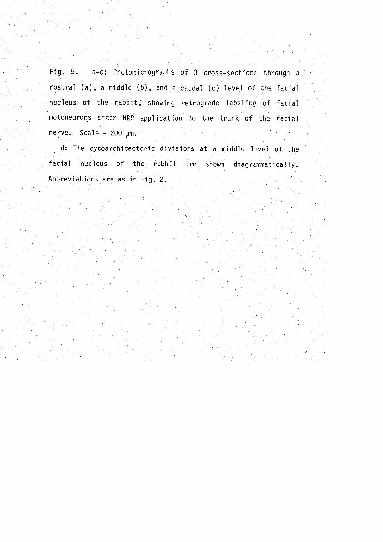
Fig. 5. a-c: Photomicrographs of 3 cross-sections through a
rostral (a), a middle (b), and a caudal (c) level of the facial
n-ucleus of the rabbit, showing retrograde labeling of facial
motoneurons after HRP application to the trunk of the facial
nerve. Scale = 200 〃m・
d: The cytoarchitectonic divisions at a middle level of the
facia一 nucleus of the rabbit are shown diagrammatically.
Abbreviations are as in Fig. 2.

-38-
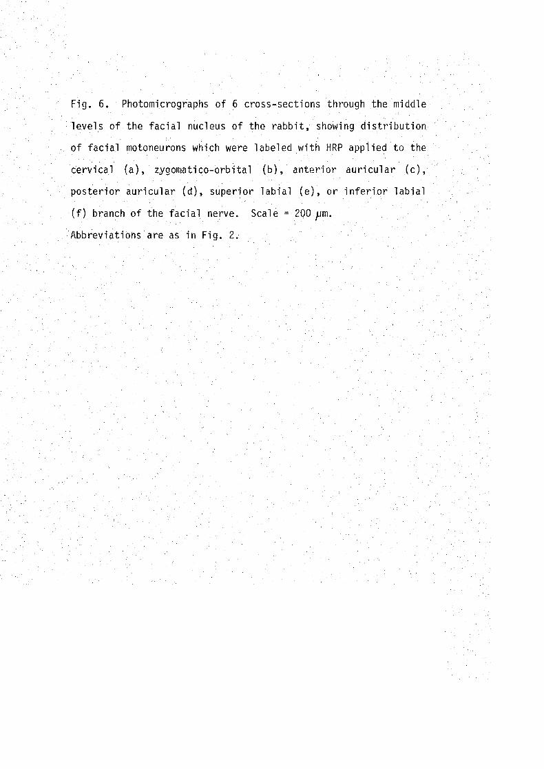
Fig. 6. Photomicrographs of 6 cross-sections through the middle
le,vels of the facial nucleus of the rabbit, showing distribution
of facial motoneurons which were labeled with HRP applied to the
cervical (a), zygomatico-orbital (b), anterior auricular (c)8
posterior auricular (d), superior 一abial (e), or inferior labial
(f) b一anch of the facial nerve. Scale.- 200〃m・
Abbreviations are as in Fig. 2.

-39-

Fig. 7. Photomicrographs of 4 cross-sections through the middle
levels of the facial nucleus of the rabbit, showing the
distribution of facial motoneurons which were labeled with HRP
applied to the perioral branchlet 1 (a), 2 (b), 3 (c), or 4 (d)
of the facial ner心e (cf. Fig. 3). Seale = 200^m. Abbreviations
are as in Fig. 2.

-40-


![Pope Molecular Pain ... · in the dorsal motor nucleus of the vagus and nucleus ambiguus [9] and a similar increase is found in the facial nucleus following facial nerve axotomy [10]](https://img.pdfslide.net/doc/110x75/5e30a53081ee76193945b62f/pope-molecular-pain-in-the-dorsal-motor-nucleus-of-the-vagus-and-nucleus-ambiguus.jpg)


![Pope Molecular Pain … · 2017-08-26 · in the dorsal motor nucleus of the vagus and nucleus ambiguus [9] and a similar increase is found in the facial nucleus following facial](https://img.pdfslide.net/doc/110x75/5e30a53181ee76193945b634/pope-molecular-pain-2017-08-26-in-the-dorsal-motor-nucleus-of-the-vagus-and-nucleus.jpg)