Embed Size (px)
Citation preview
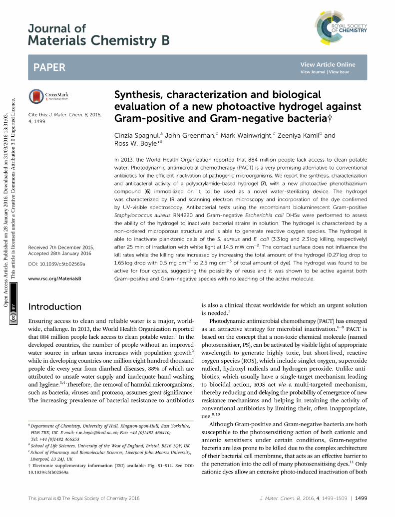
Spagnul, C., Greenman, J., Wainwright, M., Kamil, Z. and Boyle, R.W. (2016) Synthesis, characterization and biological evaluation of anew photoactive hydrogel against Gram-positive and Gram-negativebacteria. Journal of Materials Chemistry B, 4 (8). pp. 1499-1509.ISSN 2050-750X Available from: http://eprints.uwe.ac.uk/28575
We recommend you cite the published version.The publisher’s URL is:http://dx.doi.org/10.1039/c5tb02569a
Refereed: Yes
(no note)
Disclaimer
UWE has obtained warranties from all depositors as to their title in the materialdeposited and as to their right to deposit such material.
UWE makes no representation or warranties of commercial utility, title, or fit-ness for a particular purpose or any other warranty, express or implied in respectof any material deposited.
UWE makes no representation that the use of the materials will not infringeany patent, copyright, trademark or other property or proprietary rights.
UWE accepts no liability for any infringement of intellectual property rightsin any material deposited but will remove such material from public view pend-ing investigation in the event of an allegation of any such infringement.
PLEASE SCROLL DOWN FOR TEXT.
This journal is©The Royal Society of Chemistry 2016 J. Mater. Chem. B, 2016, 4, 1499--1509 | 1499
Cite this: J.Mater. Chem. B, 2016,
4, 1499
Synthesis, characterization and biologicalevaluation of a new photoactive hydrogel againstGram-positive and Gram-negative bacteria†
Cinzia Spagnul,a John Greenman,b Mark Wainwright,c Zeeniya Kamilb andRoss W. Boyle*a
In 2013, the World Health Organization reported that 884 million people lack access to clean potable
water. Photodynamic antimicrobial chemotherapy (PACT) is a very promising alternative to conventional
antibiotics for the efficient inactivation of pathogenic microorganisms. We report the synthesis, characterization
and antibacterial activity of a polyacrylamide-based hydrogel (7), with a new photoactive phenothiazinium
compound (6) immobilized on it, to be used as a novel water-sterilizing device. The hydrogel
was characterized by IR and scanning electron microscopy and incorporation of the dye confirmed
by UV-visible spectroscopy. Antibacterial tests using the recombinant bioluminescent Gram-positive
Staphylococcus aureus RN4220 and Gram-negative Escherichia coli DH5a were performed to assess
the ability of the hydrogel to inactivate bacterial strains in solution. The hydrogel is characterized by a
non-ordered microporous structure and is able to generate reactive oxygen species. The hydrogel is
able to inactivate planktonic cells of the S. aureus and E. coli (3.3 log and 2.3 log killing, respectively)
after 25 min of irradiation with white light at 14.5 mW cm�2. The contact surface does not influence the
kill rates while the killing rate increased by increasing the total amount of the hydrogel (0.27 log drop to
1.65 log drop with 0.5 mg cm�3 to 2.5 mg cm�3 of total amount of dye). The hydrogel was found to be
active for four cycles, suggesting the possibility of reuse and it was shown to be active against both
Gram-positive and Gram-negative species with no leaching of the active molecule.
Introduction
Ensuring access to clean and reliable water is a major, world-wide, challenge. In 2013, the World Health Organization reportedthat 884 million people lack access to clean potable water.1 In thedeveloped countries, the number of people without an improvedwater source in urban areas increases with population growth2
while in developing countries one million eight hundred thousandpeople die every year from diarrheal diseases, 88% of which areattributed to unsafe water supply and inadequate hand washingand hygiene.3,4 Therefore, the removal of harmful microorganisms,such as bacteria, viruses and protozoa, assumes great significance.The increasing prevalence of bacterial resistance to antibiotics
is also a clinical threat worldwide for which an urgent solutionis needed.5
Photodynamic antimicrobial chemotherapy (PACT) has emergedas an attractive strategy for microbial inactivation.6–8 PACT isbased on the concept that a non-toxic chemical molecule (namedphotosensitiser, PS), can be activated by visible light of appropriatewavelength to generate highly toxic, but short-lived, reactiveoxygen species (ROS), which include singlet oxygen, superoxideradical, hydroxyl radicals and hydrogen peroxide. Unlike anti-biotics, which usually have a single-target mechanism leadingto biocidal action, ROS act via a multi-targeted mechanism,thereby reducing and delaying the probability of emergence of newresistance mechanisms and helping in retaining the activity ofconventional antibiotics by limiting their, often inappropriate,use.9,10
Although Gram-positive and Gram-negative bacteria are bothsusceptible to the photosensitising action of both cationic andanionic sensitisers under certain conditions, Gram-negativebacteria are less prone to be killed due to the complex architectureof their bacterial cell membrane, that acts as an effective barrier tothe penetration into the cell of many photosensitising dyes.11 Onlycationic dyes allow an extensive photo-induced inactivation of both
a Department of Chemistry, University of Hull, Kingston-upon-Hull, East Yorkshire,
HU6 7RX, UK. E-mail: [email protected]; Fax: +44 (0)1482 466410;
Tel: +44 (0)1482 466353b School of Life Sciences, University of the West of England, Bristol, BS16 1QY, UKc School of Pharmacy and Biomolecular Sciences, Liverpool John Moores University,
Liverpool, L3 2AJ, UK
† Electronic supplementary information (ESI) available: Fig. S1–S11. See DOI:10.1039/c5tb02569a
Received 7th December 2015,Accepted 28th January 2016
DOI: 10.1039/c5tb02569a
www.rsc.org/MaterialsB
Journal ofMaterials Chemistry B
PAPER
Ope
n A
cces
s A
rtic
le. P
ublis
hed
on 2
8 Ja
nuar
y 20
16. D
ownl
oade
d on
31/
03/2
016
13:3
1:03
. T
his
artic
le is
lice
nsed
und
er a
Cre
ativ
e C
omm
ons
Attr
ibut
ion
3.0
Unp
orte
d L
icen
ce.
View Article OnlineView Journal | View Issue
1500 | J. Mater. Chem. B, 2016, 4, 1499--1509 This journal is©The Royal Society of Chemistry 2016
types. The selection of an appropriate photosensitiser is therefore acritical step in PACT.
Different photosensitisers, both cationic and anionic, havebeen widely tested, against Gram-positive and Gram-negativebacteria both in solution12 or immobilized onto a surface.13
Cationic phenothiazines, such as methylene blue (MB) andtoluidine blue (TBO) have a well-documented killing abilitytowards viruses and, bacteria in blood fractions14–16 and for plasmasterilization. They are active both in solution and incorporated intopolymeric materials, such as silicone,17–20 polysiloxane polymers,21
polyurethane,22,23 polyethylene,24 and cellulose acetate.25–27
These antimicrobial polymers showed significant antimicrobialactivity against Staphylococcus aureus (MRSA),20,23,25,26 Escherichiacoli,18,19,23,24,26 Staphylococcus epidermidis,17–19 Staphylococcusaureus,22,24,26,27 and Pseudomonas aeruginosa.25
Immobilization onto an inert support offers many advantages.An effective attachment of the photosensitiser allows for itscomplete removal from the treated water, thus eliminating anyphotosensitiser contamination of the water, which would notbe acceptable for water disinfection.
Secondly, immobilization may enable the re-use of the activegel over several cycles of use, thus increasing its renewabilityand friendliness towards the environment. Other possible benefitsinclude the re-use of the photosensitiser, which consequentlyreduces the cost of the treatment, and the gradual photobleachingof the dyes by solar light, which prevents their accumulation inthe environment.
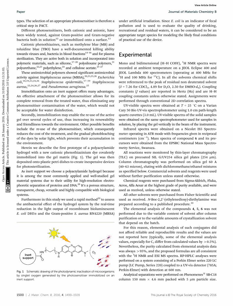
Herein we describe the first prototype of a polyacrylamidehydrogel with a new cationic phenothiazinium dye covalentlyimmobilized into the gel matrix (Fig. 1). The gel was thendeposited onto plastic petri dishes to create inexpensive devicesfor photosterilization.
As inert support we choose a polyacrylamide hydrogel becauseit is among the most commonly applied and well-studied gelmaterial systems due to their utility for high-resolution electro-phoretic separation of proteins and DNA.28 It’s a porous structure,transparent, cheap, versatile and highly compatible with biologicalsystems.
Furthermore in this study we used a rapid method29 to assessthe antibacterial effect of the hydrogel system by the real-timereduction in the light output of recombinant bioluminescentE. coli DH5a and the Gram-positive S. aureus RN4220 (MRSA)
under artificial irradiation. Since E. coli is an indicator of fecalpollution and is used to evaluate the quality of drinking,recreational and residual waters, it can be considered to be anappropriate target species for modeling the likely final conditionsof practical use of the device.
Experimental
Mono and bidimensional (H–H COSY), 1H NMR spectra wererecorded at ambient temperature on a JEOL Eclipse 400 andJEOL Lambda 400 spectrometers (operating at 400 MHz for1H and 100 MHz for 13C). In all the solvents chemical shiftswere referenced to the peak of residual non deuterated solvent(d = 7.26 for CDCl3, 4.89 for D2O, 2.50 for DMSO-d6). Couplingconstants (J values) are reported in Hertz (Hz) and are H–Hcoupling constants unless otherwise stated. Assignments wereperformed through conventional 2D correlation spectra.
UV-visible spectra were obtained at T = 25 1C on a VarianCary 50 Bio UV-vis spectrophotometer using 1.0 cm path-lengthquartz cuvettes (3.0 mL). UV-visible spectra of the solid sampleswere obtained on the same spectrophotometer used for samples insolution, by placing the gel vertically in the beam of the instrument.
Infrared spectra were obtained on a Nicolet IS5 Spectro-meter operating in ATR mode with frequencies given in reciprocalcentimeters (cm�1). Mass spectra of compound 6 and its pre-cursors were obtained from the EPSRC National Mass Spectro-metry Service, Swansea.
All reactions were monitored by thin-layer chromatography(TLC) on precoated SIL G/UV254 silica gel plates (254 mm).Column chromatography was performed on silica gel 60 A(40–63 micron), eluting with dichloromethane/ethanol mixturesas specified below. Commercial solvents and reagents were usedwithout further purification unless stated otherwise.
Chemical reagents were purchased from Sigma-Aldrich, Fluka,Acros, Alfa Aesar at the highest grade of purity available, and wereused as received, unless otherwise stated.
All other solvents were purchased from Fisher Scientific andused as received. N-Boc-2,20-(ethylenedioxy)-diethylamine wasprepared according to a published procedure.30
The elemental analysis of the compounds 4, 5, 6 was notperformed due to the variable content of solvent after columnpurification or to the variable amounts of crystallization solventthat depend on the batch.
For this reason, elemental analysis of such conjugates didnot afford reliable and reproducible results and the values arenot reported here (typically, some of the elemental analysisvalues, especially for C, differ from calculated values by 40.5%).Nevertheless, the purity calculated from elemental analysis datawas always 495%, and the proposed formulas are all consistentwith the 1H NMR and ESI MS spectra. RP-HPLC analyses wereperformed on a system consisting of a Perkin Elmer series 220 LCpump (LC Pump, Series 220) coupled to a UV-vis detector (785A;Perkin-Elmer) with detection at 600 nm.
Analytical separations were performed on Phenomenexs SB-C18column 150 mm � 4.6 mm packed with 5 mm particle size.
Fig. 1 Schematic drawing of the photodynamic inactivation of microorganismsby singlet oxygen generated by the photosensitiser immobilized on aninert support.
Paper Journal of Materials Chemistry B
Ope
n A
cces
s A
rtic
le. P
ublis
hed
on 2
8 Ja
nuar
y 20
16. D
ownl
oade
d on
31/
03/2
016
13:3
1:03
. T
his
artic
le is
lice
nsed
und
er a
Cre
ativ
e C
omm
ons
Attr
ibut
ion
3.0
Unp
orte
d L
icen
ce.
View Article Online
This journal is©The Royal Society of Chemistry 2016 J. Mater. Chem. B, 2016, 4, 1499--1509 | 1501
The mobile phase consisted of two solutions namely A and B.Solution A was made from 0.1 M ammonium acetate and aceticacid (pH 5.3), whereas solution B was acetonitrile. The gradientelution was 5% A and 95% B in 15 min with a flow rate of0.8 mL min�1 and the injection volume was 100 ml.
Synthesis and characterization of compounds
Phenothiazin-5-ium tetraiodide hydrate (1). A proceduresimilar to that described in the literature was used31 with thefollowing parameters: a solution of iodine (8.0 g, 31.52 mmol,3 equiv.) in chloroform (200 mL) was added to a stirred solutionof phenothiazine (2.0 g, 10.03 mmol, 1 equiv.) in chloroform(45 mL) within 1 h at room temperature. The reaction mixturewas then stirred for a further 5 h and it was monitored by thin-layer chromatography on silica gel (CH2Cl2/MeOH 90 : 10). Theresultant precipitate was filtered, washed with chloroform(2 � 50 mL), diethyl ether (2 � 50 mL) and then dried undervacuum at room temperature to give the phenothiazine tetraiodide(6.79 g, 93%) as a black solid.
Found C, 20.42; H, 1.14; N, 1.98; S, 4.54. Calcd forC12H8I4NS: C, 19.91; H 1.39; N 1.93; 4.43%.
lmax(DMSO, 25 1C)/nm 643 (e/dm3 mol�1 cm�1 5690).IR (neat): n (cm�1) = 1558, 1142, 1380, 1314, 1240, 1153,
1132, 1072, 1027, 744.1H (400 MHz; DMSO-d6) d: 7.61 (1 H, t, J = 7.5 Hz), 7.72 (1H,
t, J = 7.5 Hz), 7.91 (1H, d, J = 7.8 Hz), 8.06 (1H, d, J = 7.8 Hz), 8.10(2H, m), 8.48 (2H, m).
13C (100 MHz; DMSO-d6) d: 152.8, 130.2, 129.8, 129.1, 127.4.ESI-MS (m/z) (MeOH + NH4OAc): 198.04 found (M + H+):
199.04.3-(Dimethylamino)phenothiazin-5-ium Triiodide (2). To a
solution of phenothiazin-5-ium tetraiodide hydrate (1.00 g,1.38 mmol, 1 equiv.) dissolved in dichloromethane (30 mL),methanol (1 mL) and acetone (1 mL) was added solution ofdimethylamine (40 wt% in H2O) in methanol (2 M, 1.03 mL,2.07 mmol, 1.5 equiv.) dropwise over 6 h under Argon. Theformation of the product was monitored by thin-layer chromato-graphy on silica gel (CH2Cl2/CH3OH 90 : 10).
The reaction mixture was diluted with dichloromethane(100 mL) and washed with water. The organic layer was driedover anhydrous Na2SO4, filtered and concentrated underreduced pressure. The product was washed with diethyl etherto give the compound as dark-blue solid (0.30 g, 35%).
Found C, 27.23; H, 1.86; N, 4.50; S, 5.25%. Calcd forC14H13I3N2S: C, 27.03; H, 2.11; N, 4.50; S, 5.15%.
lmax(DMSO, 25 1C)/nm 580 (e/dm3 mol�1 cm�1 7424), 415(6473) and 301 (13766).
IR (neat): n (cm�1) = 2923, 2849, 1616, 1589,1560, 1496, 1461,1428, 1311, 1251, 1118, 1075, 881, 831, 769, 742.
1H (400 MHz, DMSO-d6) d: 8.23 (dd, J = 7.9 Hz, J = 1.5 Hz,1H, H-9), 8.18 (dd, J = 8.0 Hz, J = 1.5 Hz, 1H, H-6), 8.10 (d,J = 10.0 Hz, 1H, H-1), 8.06 (d, J = 2.6 Hz, 1H, H-2), 8.02 (dd,J = 7.7 Hz, J = 2.6 Hz, 1H, H-4), 7.81–7.91 (2H, m, J = 9.6 Hz,7.6 Hz, 1.6 Hz, H-7, H-8), 3.66 (s, 3H, NCH3), 3.60 (s, 3H, NCH3).
13C (100 MHz, DMSO-d6) d: 140.4, 137.9, 135.1, 133.8, 130.4,130.0, 129.0, 128.5, 128.0, 126.9, 126.7, 110.3, 43.9, 43.4.
ESI-MS (m/z) (MeOH + NH4OAc): calcd for C14H13N2S:241.0794 found (M+): 241.0793.
N-{2-[2-(2-tert-Butoxycarbonylaminoethoxy)ethoxy]ethyl}acryl-amide (4). N-Boc-2,20-(ethylenedioxy)-diethylamine (3) (1.0 g,4.03 mmol, 1 equiv.) and diisopropylethylamine (1.40 mL,8.05 mmol, 2 equiv.) were dissolved in dry dichloromethane(10 mL) in an argon atmosphere. Acryloyl chloride (327 ml,8.05 mmol, 2 equiv.) was added dropwise over 3 hours at 0 1C.The reaction mixture was stirred for 4 h, while being allowed toreach room temperature, then it was diluted with dichloro-methane, and washed with 5% aqueous solution of citric acidand brine. The organic fraction was concentrated, dried withMgSO4, concentrated under reduced pressure and the crudeproduct was purified by column chromatography (CH2Cl2/MeOH95 : 5) to give 0.85 g of 4 as a pure pale yellow oil in 70% yield.
1H (400 MHz, CDCl3) d: 6.29 (dd, Jtrans = 17.0 Hz, Jgem =1.6 Hz, 1H, CH2CHCO), 6.12 (dd, Jtrans = 17.0 Hz, Jcis = 10.2 Hz,1H, CH2CHCO), 5.63 (dd, Jcis = 10.2 Hz, Jgem = 1.6 Hz, 1H,CH2CHCO), 5.00 (brs, 1H, NHCO), 3.71–3.39 (m, 10H, CH2peg),3.31 (m, 2H, CH2NHCOBoc), 1.43 (s, 9H, CH3Boc).
13C (100 MHz, CDCl3) d: 167.48, 157.34, 130.9, 126.5, 80.6,70.4, 70.3, 70.2, 69.8, 40.4, 39.4, 28.5.
ESI-MS (m/z): calcd for C14H26N2O5: 302.18; found: (M +Na)+: 325.17.
N-{2-[2-(2-aminoethoxy)ethoxy]ethyl}acrylamide (5). N-{2-[2-(2-tert-Butoxycarbonylaminoethoxy)ethoxy]ethyl}acrylamide (2)(1.27 g, 4.20 mmol, 1 equiv.) was treated with 4 M HCl in1,4-dioxane (2.52 mL, 10.08 mmol, 2.4 equiv.). The resultingsolution was stirred at room temperature for 1 h, and thesolvent was evaporated. The residue was triturated with diethylether and thoroughly dried in vacuo to afford 0.82 g of 5 as ayellow oil in 92% yield.
1H (400 MHz, D2O) d: 6.26 (dd, Jtrans = 17.1 Hz, Jcis = 10.0 Hz,1H, CH2CHCO), 6.17 (dd, Jtrans = 17.1 Hz, Jgem = 1.6 Hz, 1H,CH2CHCO), 5.75 (dd, Jcis = 10.0 Hz, Jgem = 1.6 Hz, 1H,CH2CHCO), 3.80–3.57 (m, 10H, CH2peg), 3.47 (t, 2H, J = 5.4 Hz,CH2NHCOCH), 3.19 (m, 2H, CH2NH2).
13C (400 MHz, D2O) d: 129.9, 127.5, 69.6, 68.8, 66.4,39.1, 38.9.
ESI-MS (m/z) (MeOH + NH4OAc): calcd for C9H19N2O3:202.1387; found (M + H+): 203.1387.
3-(2-(2-(2-Acrylamidoethoxy)ethoxy)ethylamino)-7-(dimethyl-amino)phenothiazin-5-ium chloride (6). A procedure from theliterature32 was modified as follows: to a solution of 3-dialkyl-aminophenothiazinium triiodide (100 mg, 0.16 mmol, 1 equiv.)in anhydrous methanol (20 mL) was added dropwise underargon the alkyl amine 5 (49 mg, 0.24 mmol, 1.5 equiv.) previouslydissolved in 12.5 mL of anhydrous methanol. The reaction wasallowed to stir at room temperature for 2 hours and the formationof the product was monitored by thin-layer chromatography onsilica gel (CH2Cl2/CH3OH 90 : 10).
The product was isolated by evaporation of the methanol,redissolution in dichloromethane (50 mL) and extraction withwater (2 � 100 mL).
The water extracts were concentrated under reduced pressure(5 mL) and passed though a small column (height 5 cm,
Journal of Materials Chemistry B Paper
Ope
n A
cces
s A
rtic
le. P
ublis
hed
on 2
8 Ja
nuar
y 20
16. D
ownl
oade
d on
31/
03/2
016
13:3
1:03
. T
his
artic
le is
lice
nsed
und
er a
Cre
ativ
e C
omm
ons
Attr
ibut
ion
3.0
Unp
orte
d L
icen
ce.
View Article Online
1502 | J. Mater. Chem. B, 2016, 4, 1499--1509 This journal is©The Royal Society of Chemistry 2016
diameter 2 cm) packed with ion exchange resin (Amberlite IRA-400) previously rinsed with acidic sodium chloride solution(10% aqueous NaCl cont. 0.1% HCl, 100 mL) and conditionedwith dilute hydrochloric acid (0.1%) eluting with double distilledwater (40 mL). The aqueous solution was evaporated undervacuum to give the product as dark blue solid. Compounds stillimpure by thin-layer chromatography at this stage were chromato-graphed on silica gel using gradient elution in dichloromethane/ethanol 99 : 5. The product was obtained as a dark blue solid(0.39 g, 76%).
lmax(water, 25 1C)/nm 627 (e/dm3 mol�1 cm�1 7463), 584(9953) and 421 (8211).
IR (neat): n (cm�1) = 2923, 2349, 1657, 1624, 1551, 1496,1435, 1314, 1250, 1121, 1095, 747, 724.
1H (400 MHz, DMSO-d6) (as the iodide salt) d: 8.24 (t, J = 5.0Hz, 1H, CH2NHAr), 8.14–7.78 (m, 6H, H aromatic), 6.26 (dd,Jtrans = 17.1, Jcis = 10.1 Hz, 1H, CH2CHCO), 6.08 (dd, Jtrans = 17.1Hz, Jgem = 3.3 Hz, 1H, CH2CHCO), 5.58 (dd, Jcis = 10.1 Hz,Jgem = 3.3 Hz, 1H, CH2CHCO), 3.70–3.54 (m, 10H, CH2peg +N(CH3)2), 3.47–3.44 (t, 1H, CONH), 3.29 (m, 2H, CH2CONH),3.03–2.86 (m, 2H, CH2NH2).
1H (400 MHz, DMSO-d6) (as chloride salt) d: 8.33 (t, J =5.0 Hz, 1H, CH2NHAr), 8.27–8.01 (m, 6H, H aromatic), 6.28 (dd,Jtrans = 17.1, Jcis = 10.1 Hz, 1H, CH2CHCO), 6.07 (dd, Jtrans =17.1 Hz, Jgem = 3.3 Hz, 1H, CH2CHCO), 5.57 (dd, Jcis = 10.1 Hz,Jgem = 3.3 Hz, 1H, CH2CHCO), 3.72–3.57 (m, 16H, CH2peg +N(CH3)2), 3.47–3.44 (t, 1H, CONH) 3.29 (m, 2H, CH2CONH),3.03–2.86 (m, 2H, CH2NH).
13C (100 MHz, DMSO-d6) d: 140.4, 139.8, 135.1, 133.4, 132.4,130.4, 128.4, 127.9, 126.9, 126.9, 125.6, 110.3, 70.2, 70.0, 69.5,67.1, 40.5, 40.7, 39.1, 38.9.
ESI-MS (m/z) (MeOH + NH4OAc): 439.1796 found (M+)439.1798.
HPLC: tR: 5.92 min.Synthesis of the photoantimicrobial hydrogel (7). 1.91 g of
acrylamide (26.87 mmol), 66 mg of N,N0-methylenebisacryl-amide (0.43 mmol) were dissolved in 6.6 mL of water. 100 mlof a 10% solution of sodium dodecylsulphate (SDS) (9.95 mg,0.035 mmol, 1.5 equiv.) and 6 (11 mg, 0.023 mmol, 1 equiv.)previously dissolved in 3.3 mL of double distilled water weresubsequently added the solution was gently stirred. 100 ml of a10% solution of APS and 20 ml of TEMED were subsequentlyadded and the solution was poured into a petri dish (3.5 cmdiameter) to polymerize and kept away from sunlight for aperiod of 10–15 minutes.
After the polymerization was complete, the resultant bluecylindrical gels were taken out of the petri dishes, washed withdistilled water and then kept moist or dried in a dust-freechamber until the gels were dried completely.
lmax(solid, 25 1C)/nm 626 (e/dm3 mol�1 cm�1 4729), 589(6018) and 418 (7087).
IR (neat): n (cm�1) = 2923, 2349, 1658, 1624, 1551, 1496,1436, 1314, 1250, 1121, 1095, 747, 724.
Synthesis of the control hydrogel (8). 1.91 g of acrylamide(26.87 mmol), 66 mg of N,N0-methylenebisacrylamide (0.43 mmol)were dissolved in 6.6 mL of water. 100 ml of a 10% solution of SDS
(9.95 mg, 0.035 mmol) and 3.3 mL of double distilled water weresubsequently added and the solution was gently stirred.
100 ml of a 10% solution of APS and 20 ml of TEMED wereadded and the solution was poured into a petri dish, (3.5 cmdiameter) to polymerize, and kept away from sunlight for aperiod of 10 minutes.
After the polymerization was over, the resultant transparentcylindrical gels were taken out of the petri dishes, washed withdistilled water and then kept moist, or dried in a dust-freechamber.
IR (neat): n (cm�1) = 1672, 1612, 1425, 1355, 1355, 1280,1134, 988, 960, 841, 817.
Characterization of the hydrogels by scanning electronmicroscopy (SEM)
The morphology of the hydrogels was characterized by field-emission scanning electron microscopy.
Scanning electron microscope (SEM) images of the hydrogelalone (8) or in the presence of the phenothiazinium compound(7) before and after irradiation with visible light were obtainedusing a EVO60 scanning electron microscope (Zeiss) fitted witha cryo-preparation system-model: PP3010T, manufacturer: QuorumTechnologies.
After plunge-freezing in liquid nitrogen the sample wastransferred (under vacuum) to the cryo system preparationchamber.
Most of the water-ice was removed by sublimation by increasingthe temperature of the sample from �140 1C to �60 1C at apressure of 5 � 10�5 mbar for 10 minutes. After this, thetemperature of the sample was reduced to �140 1C and apressure of approximately 5 � 10�7 mbar.
The sample was then sputter coated with about 2 nm ofplatinum and then transferred to the SEM for examination.
The SEM electron beam accelerating voltage used was 15 kVat a probe current of 20–35 pA.
The diameters of the gel pores were determined using theImage J program.
Detection of singlet oxygen generation. Detection of singletoxygen was determined by photobleaching of the chemicalprobe, 9,10-anthracenediyl-bis(methylene)dimalonic acid (ABDA)according to a previously published protocol.33
ABDA is a water-soluble derivative of anthracene that can bephotobleached by singlet oxygen to its corresponding endoperoxide.
150 mmol L�1 of ABDA in PBS solution (pH = 7.0) containingthe hydrogels 7 or 8 (1 mg cm�3 previously cut in 4 squares) wasirradiated in a 1 cm path length spectrofluorimetric cuvette orin a petri dish (3 mL).
The photobleaching of ABDA was carried out using a CHF-XM500 M mercury lamp as light source, in combination with a550 nm cut off filter. The ABDA absorbance was recorded in the300–550 nm wavelength range. The kinetic of ABDA photooxidationwas monitored spectrophotometrically following the decreaseof the absorbance at lmax = 380 nm (lmax of ABDA) in differentirradiation periods in a 1 cm path length spectrofluorimetriccuvette.
Paper Journal of Materials Chemistry B
Ope
n A
cces
s A
rtic
le. P
ublis
hed
on 2
8 Ja
nuar
y 20
16. D
ownl
oade
d on
31/
03/2
016
13:3
1:03
. T
his
artic
le is
lice
nsed
und
er a
Cre
ativ
e C
omm
ons
Attr
ibut
ion
3.0
Unp
orte
d L
icen
ce.
View Article Online

This journal is©The Royal Society of Chemistry 2016 J. Mater. Chem. B, 2016, 4, 1499--1509 | 1503
Antibacterial activity of the hydrogels
E. coli strain DH5a contains the plasmid pGLITE, a derivative ofpBBR1MCS-2 containing the lux CDABE operon of Photorhabdusluminescens34 and bioluminescent S. aureus strain RN422035
were maintained from frozen stock on nutrient agar (Oxoid Ltd,Basingstoke, UK) and were grown as broth cultures usingReinforced Clostridial Medium (RCM; Oxoid Ltd Basingstoke,UK) with addition of kanamycin (10 mg l�1) or erythromycin(5 mg l�1) for E. coli or S. aureus respectively, to selectivelymaintain the lux plasmids.
For experiments on photodynamic killing, microbial cellswere obtained by inoculation of test species in 10 mL volumesof appropriate liquid medium and incubated in a shakingincubator (model S 150, Stuart shakers, UK) at 37 1C for 4 to6 hours to obtain mid exponential phase cultures.
A total of 100 mL of bacterial suspension was appropriatelydiluted in 1 mL PBS (pH = 7.0) to obtain approximately 106
colony forming units (cfu) mL�1.The photoantimicrobial hydrogel (7) was cut into four
squares and equilibrated with PBS (pH = 7.0) for 20 minutes.Then the media was discarded, the gel was washed with PBS ofthe same pH and placed into the diluted bacterial suspensionin borosilicate glass tubes (12 by 75 mm; Fisher Scientific,Loughborough, UK) using the same tube for both light irradia-tion and measurement of bioluminescence light output. Thesamples were irradiated with a white light at a fluence rate of14.5 mW cm�2 for 20 or 25 minutes (total light dose was either17.4 or 21.8 J cm�2).
The illumination was performed using a fiber optic cable(F = 1 cm) and lamp (Fiber Illuminator, OSL1-EC, ThorLabsInc,Ely, Cambridgeshire) with a 150 W halogen lamp. Luminescencelight output was measured by quickly inserting the tube into aFB12 luminometer (Berthold Detection Systems, Germany) toquantify the light output as relative light units (RLU).
A borosilicate tube was similarly treated, but not exposed tolight and used as a reference for the dark toxicity under thesame experimental conditions.
The hydrogel alone (8) was tested following the same protocol.Control experiments on E. coli and S. aureus suspensions
irradiated and in the dark indicated that light doses alone up to21.8 J cm�2 cause no evident bacterial damage. All experimentswere conducted in duplicate.
Results and discussion1. Synthesis and characterization of compounds
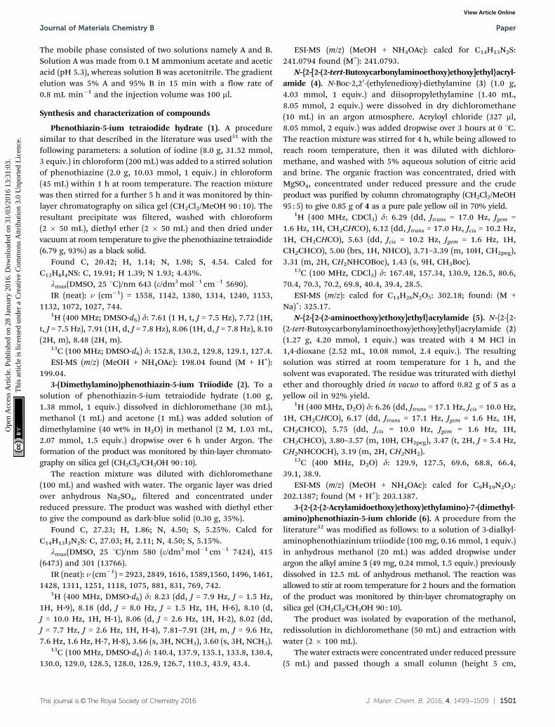
Phenothiazinium tetraiodide (1) has been proven to be a goodstarting material to obtain the final asymmetric phenothiaziniumderivative 6 as can be seen in Fig. 2. 1 was successfully synthesizedthrough the oxidation of the commercially available phenothiazine.Phenothiazinium tetraiodide was obtained in almost a quantitativeyield using a relatively straightforward process, as the procedureis well established within the literature.31 3-Dimethylamino-phenothiazin-5-ium triiodide (2) was subsequently obtained bysubstitution at C3 of the phenothiazinium ring in dichloro-methane/acetone/methanol mixture under argon with dimethyl-amine 2 M in methanol in 35% yield.
Compound 2 was obtained in a reasonable yield (35%) withoutcomplex and expensive separation protocols, involving a waterextraction and a subsequent precipitation in diethyl ether.
The target molecule and intermediates were characterizedusing NMR spectroscopy and Electrospray Mass Spectrometry.The mass spectrum of 2 provides evidence that the molecularpeak is at 241.07 as (M)+ (Fig. S1, ESI†).
The 1H NMR spectrum of 2 (Fig. S2, ESI†) shows in thedownfield region the two double doublets for the protons at d8.22 (H9, 1H) and d 8.19 (H6, 1H), the multiplet for the protonsat d 8.24 (2H), and two resolved resonances attributed to 4 (2H)
Fig. 2 Synthetic route to 3-(2-(2-(2-acrylamidoethoxy)ethoxy)ethylamino)-7-(dimethylamino)phenothiazin-5-ium chloride (6). Reactions and conditions: (a) I2,CH2Cl2, 5 h, rt, 93% yield. (b) NH(CH3)2, CH2Cl2/MeOH/acetone mixture, 6 h, Ar, rt, 35% yield. (c) acryloyl chloride, DIPEA, CH2Cl2, Ar, 7 h, 0 1C-rt, 70% yield.(d) HCl in 1,4-dioxane, rt, 1 h, 92% yield. (e) Alkyl amine, dry MeOH, Ar, 2 h, rt, 76% yield.
Journal of Materials Chemistry B Paper
Ope
n A
cces
s A
rtic
le. P
ublis
hed
on 2
8 Ja
nuar
y 20
16. D
ownl
oade
d on
31/
03/2
016
13:3
1:03
. T
his
artic
le is
lice
nsed
und
er a
Cre
ativ
e C
omm
ons
Attr
ibut
ion
3.0
Unp
orte
d L
icen
ce.
View Article Online
1504 | J. Mater. Chem. B, 2016, 4, 1499--1509 This journal is©The Royal Society of Chemistry 2016
and 1–2 (4H) at d 8.06 and d 8.09 respectively. In the upfieldregion, the spectrum shows only two sharp singlets, at d 3.62and d 3.64 for the N(CH3)2 (3H each).
To insert an acryloyl function on the PS, useful for the latercovalent immobilization of the photosensitising unit on apolymeric support, 3 was proven to be a suitable starting material.Preparation of the commercially unavailable N-2-[2-(2-aminoethoxy)-ethoxy]ethylacrylamide (5) was carried out via standard reactionsas previously reported36,37 by reaction of 3 with acryloyl chloridefollowing HCl-mediated Boc-deprotection (Fig. 2).
The mass spectrum of 5 provides evidence that the molecularpeak is at 203.1387 as (M + H+) (Fig. S3, ESI†).
The 1H NMR spectrum of 5 (Fig. S4, ESI†) shows thecharacteristic resonances of the acryloyl group at d 6.26(CH2QCHCO), d 6.17 for the CH2QCHCO trans and at d 5.75for the CH2CHQCO cis. In the upfield region, the spectrumshows the multiplet of the alkylic chain (d = 3.80–3.57), thetriplet at d 3.47 and the multiplet d 3.19 of the CH2NHCOCHand CH2NH2 respectively.
Compound 2 was converted to the desired product 6 in 75%yield using an excess of 5 previously synthesized using mildconditions in methanol, at room temperature.
After purification, the counter ion was exchanged for chlorideusing Amberlite IRA 400, an ion exchange resin.
1H NMR and 13C NMR clearly demonstrated that 6 is anasymmetric structure. It was difficult to obtain good NMRspectra of 6 as iodide, indeed some papers report the synthesisbut not the NMR spectra of similar phenothiazinium photo-sensitisers31,38,39 but after the ion exchange with the IRA-400(Cl) the NMR spectra of phenothiazinium chlorides were easilyobtained in deuterated dmso.
Furthermore, iodide ions can react with singlet oxygen beingproduced from the photosensitiser, which consequently decreasesthe overall efficiency. The correlation H–H COSY spectrum of 6(Fig. S5, ESI†) displays, besides the expected cross peaks, also twolong range weak correlation peaks between the aromatic protonsand the CH2 of the C1 of the alkylic chain and between the CH2
proton of the C8 of the alkylic chain and the NH amide proton thatallowed their assignment unambiguously.
Interestingly, all of the protons that belong to the aromaticpart, sharp for the precursor 2, now appear quite broad,probably as effect of the aggregation.
Compound 6 gives a single HPLC peak with a retention timedistinctly lower than the parent commercially available Azure B(rt = 5.92 and rt = 6.37 respectively, Fig. S6 and S7, ESI†).
It was not possible to compare the retention time of 6 withits precursor 2 due to the low solubility of the latter in themobile phase.
The final molecule 6 dissolves well in polar solvents such asmethanol and ethanol as well as in water and PBS which allowsit to be used with a range of different polymer gelificationsystems where water solubility is often an advantage.
2. Synthesis and characterization of the hydrogels
We incorporated our photosensitiser into a polyacrylamide hydrogelbecause, due to its high porosity, good biocompatibility, its high
water content and permeability,40 it is widely used in medicine ascarrier of immobilized biologically active substances.41,42
The hydrogels 7 and 8 were obtained through the usual freeradical copolymerization of acrylamide (Am) and N,N0-methylene-bisacrylamide in a ratio of 19 : 1 (w/v%) using ammoniumpersulfate (APS) as the redox initiator and N,N,N0,N0-tetramethyl-ethylenediamine (TEMED) as the catalyst.
11 mg of 6 was dissolved in 3.3 mL of water and added tothe monomers solution at room temperature (total monomerconcentration in the final solution: 20 wt%). SDS was added tothe gel mixture in order to disrupt phenothiazinium dimerization.Dimerization of phenothiazinium produce a blue-shifted absorptionband with respect to the absorption band of the correspondingphenothiazine monomer and decreases the singlet oxygen yield.43,44
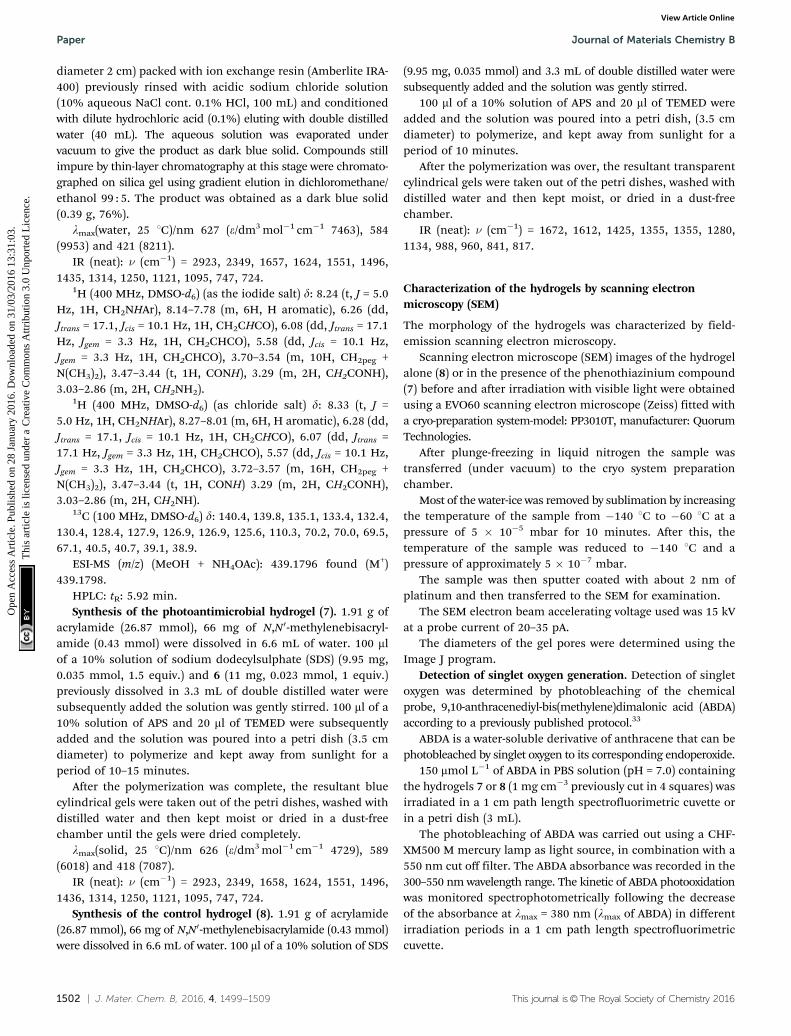
After the addition of the ammonium persulfate (APS) andthe tetramethylethylenediamine (TEMED), the homogeneousgel was obtained after 10–15 minutes (Fig. 3, right). Each cm3 ofthe photoantimicrobial hydrogel (7) contains 1 mg of 6. Thecontrol hydrogel 8 was obtained in a similar way, adding 3.3 mLof water without the PS (Fig. 3, left).
The gels were then hydrated in deionized water for at least24 h with the water being changed three times to remove anyunreacted reagents.
This system showed no appreciable leaching of the PS inwater. This makes it promising for real-world applications,where leaching of the dye from the material has been observedsometimes,13,21,22 limiting the applicability of the material tested.
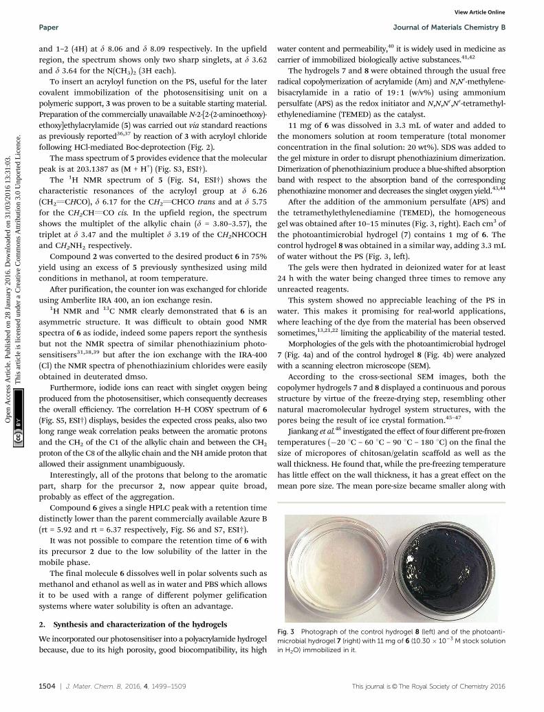
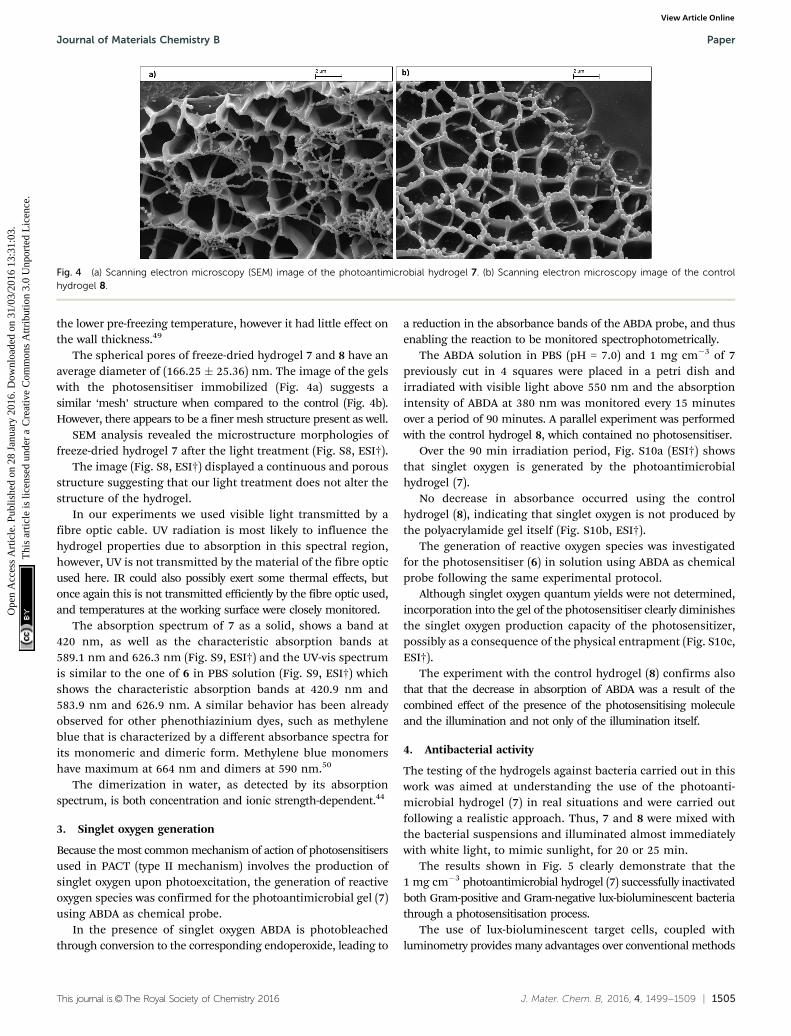
Morphologies of the gels with the photoantimicrobial hydrogel7 (Fig. 4a) and of the control hydrogel 8 (Fig. 4b) were analyzedwith a scanning electron microscope (SEM).
According to the cross-sectional SEM images, both thecopolymer hydrogels 7 and 8 displayed a continuous and porousstructure by virtue of the freeze-drying step, resembling othernatural macromolecular hydrogel system structures, with thepores being the result of ice crystal formation.45–47
Jiankang et al.48 investigated the effect of four different pre-frozentemperatures (�20 1C – 60 1C – 90 1C – 180 1C) on the final thesize of micropores of chitosan/gelatin scaffold as well as thewall thickness. He found that, while the pre-freezing temperaturehas little effect on the wall thickness, it has a great effect on themean pore size. The mean pore-size became smaller along with
Fig. 3 Photograph of the control hydrogel 8 (left) and of the photoanti-microbial hydrogel 7 (right) with 11 mg of 6 (10.30 � 10�3 M stock solutionin H2O) immobilized in it.
Paper Journal of Materials Chemistry B
Ope
n A
cces
s A
rtic
le. P
ublis
hed
on 2
8 Ja
nuar
y 20
16. D
ownl
oade
d on
31/
03/2
016
13:3
1:03
. T
his
artic
le is
lice
nsed
und
er a
Cre
ativ
e C
omm
ons
Attr
ibut
ion
3.0
Unp
orte
d L
icen
ce.
View Article Online
This journal is©The Royal Society of Chemistry 2016 J. Mater. Chem. B, 2016, 4, 1499--1509 | 1505
the lower pre-freezing temperature, however it had little effect onthe wall thickness.49
The spherical pores of freeze-dried hydrogel 7 and 8 have anaverage diameter of (166.25 � 25.36) nm. The image of the gelswith the photosensitiser immobilized (Fig. 4a) suggests asimilar ‘mesh’ structure when compared to the control (Fig. 4b).However, there appears to be a finer mesh structure present as well.
SEM analysis revealed the microstructure morphologies offreeze-dried hydrogel 7 after the light treatment (Fig. S8, ESI†).
The image (Fig. S8, ESI†) displayed a continuous and porousstructure suggesting that our light treatment does not alter thestructure of the hydrogel.
In our experiments we used visible light transmitted by afibre optic cable. UV radiation is most likely to influence thehydrogel properties due to absorption in this spectral region,however, UV is not transmitted by the material of the fibre opticused here. IR could also possibly exert some thermal effects, butonce again this is not transmitted efficiently by the fibre optic used,and temperatures at the working surface were closely monitored.
The absorption spectrum of 7 as a solid, shows a band at420 nm, as well as the characteristic absorption bands at589.1 nm and 626.3 nm (Fig. S9, ESI†) and the UV-vis spectrumis similar to the one of 6 in PBS solution (Fig. S9, ESI†) whichshows the characteristic absorption bands at 420.9 nm and583.9 nm and 626.9 nm. A similar behavior has been alreadyobserved for other phenothiazinium dyes, such as methyleneblue that is characterized by a different absorbance spectra forits monomeric and dimeric form. Methylene blue monomershave maximum at 664 nm and dimers at 590 nm.50
The dimerization in water, as detected by its absorptionspectrum, is both concentration and ionic strength-dependent.44
3. Singlet oxygen generation
Because the most common mechanism of action of photosensitisersused in PACT (type II mechanism) involves the production ofsinglet oxygen upon photoexcitation, the generation of reactiveoxygen species was confirmed for the photoantimicrobial gel (7)using ABDA as chemical probe.
In the presence of singlet oxygen ABDA is photobleachedthrough conversion to the corresponding endoperoxide, leading to
a reduction in the absorbance bands of the ABDA probe, and thusenabling the reaction to be monitored spectrophotometrically.
The ABDA solution in PBS (pH = 7.0) and 1 mg cm�3 of 7previously cut in 4 squares were placed in a petri dish andirradiated with visible light above 550 nm and the absorptionintensity of ABDA at 380 nm was monitored every 15 minutesover a period of 90 minutes. A parallel experiment was performedwith the control hydrogel 8, which contained no photosensitiser.
Over the 90 min irradiation period, Fig. S10a (ESI†) showsthat singlet oxygen is generated by the photoantimicrobialhydrogel (7).
No decrease in absorbance occurred using the controlhydrogel (8), indicating that singlet oxygen is not produced bythe polyacrylamide gel itself (Fig. S10b, ESI†).
The generation of reactive oxygen species was investigatedfor the photosensitiser (6) in solution using ABDA as chemicalprobe following the same experimental protocol.
Although singlet oxygen quantum yields were not determined,incorporation into the gel of the photosensitiser clearly diminishesthe singlet oxygen production capacity of the photosensitizer,possibly as a consequence of the physical entrapment (Fig. S10c,ESI†).
The experiment with the control hydrogel (8) confirms alsothat that the decrease in absorption of ABDA was a result of thecombined effect of the presence of the photosensitising moleculeand the illumination and not only of the illumination itself.
4. Antibacterial activity
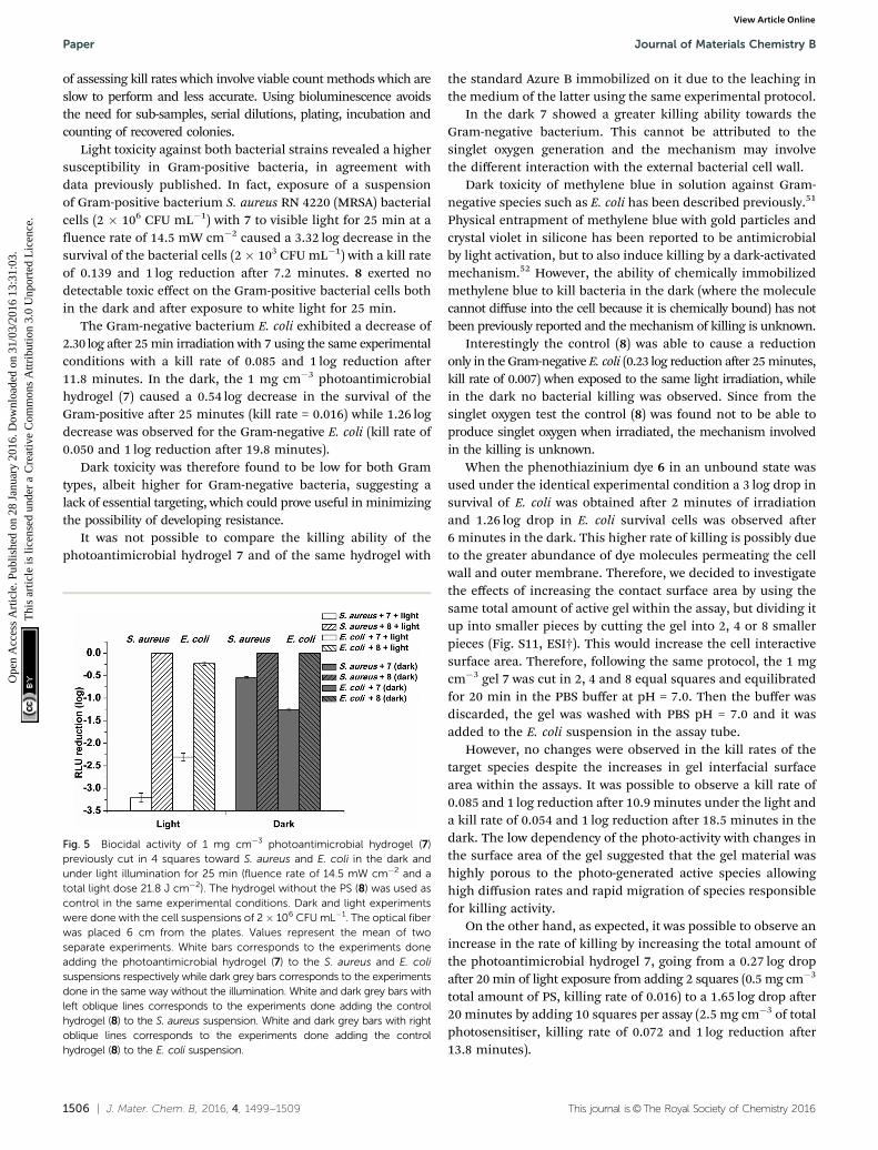
The testing of the hydrogels against bacteria carried out in thiswork was aimed at understanding the use of the photoanti-microbial hydrogel (7) in real situations and were carried outfollowing a realistic approach. Thus, 7 and 8 were mixed withthe bacterial suspensions and illuminated almost immediatelywith white light, to mimic sunlight, for 20 or 25 min.
The results shown in Fig. 5 clearly demonstrate that the1 mg cm�3 photoantimicrobial hydrogel (7) successfully inactivatedboth Gram-positive and Gram-negative lux-bioluminescent bacteriathrough a photosensitisation process.
The use of lux-bioluminescent target cells, coupled withluminometry provides many advantages over conventional methods
Fig. 4 (a) Scanning electron microscopy (SEM) image of the photoantimicrobial hydrogel 7. (b) Scanning electron microscopy image of the controlhydrogel 8.
Journal of Materials Chemistry B Paper
Ope
n A
cces
s A
rtic
le. P
ublis
hed
on 2
8 Ja
nuar
y 20
16. D
ownl
oade
d on
31/
03/2
016
13:3
1:03
. T
his
artic
le is
lice
nsed
und
er a
Cre
ativ
e C
omm
ons
Attr
ibut
ion
3.0
Unp
orte
d L
icen
ce.
View Article Online
1506 | J. Mater. Chem. B, 2016, 4, 1499--1509 This journal is©The Royal Society of Chemistry 2016
of assessing kill rates which involve viable count methods which areslow to perform and less accurate. Using bioluminescence avoidsthe need for sub-samples, serial dilutions, plating, incubation andcounting of recovered colonies.
Light toxicity against both bacterial strains revealed a highersusceptibility in Gram-positive bacteria, in agreement withdata previously published. In fact, exposure of a suspensionof Gram-positive bacterium S. aureus RN 4220 (MRSA) bacterialcells (2 � 106 CFU mL�1) with 7 to visible light for 25 min at afluence rate of 14.5 mW cm�2 caused a 3.32 log decrease in thesurvival of the bacterial cells (2 � 103 CFU mL�1) with a kill rateof 0.139 and 1 log reduction after 7.2 minutes. 8 exerted nodetectable toxic effect on the Gram-positive bacterial cells bothin the dark and after exposure to white light for 25 min.
The Gram-negative bacterium E. coli exhibited a decrease of2.30 log after 25 min irradiation with 7 using the same experimentalconditions with a kill rate of 0.085 and 1 log reduction after11.8 minutes. In the dark, the 1 mg cm�3 photoantimicrobialhydrogel (7) caused a 0.54 log decrease in the survival of theGram-positive after 25 minutes (kill rate = 0.016) while 1.26 logdecrease was observed for the Gram-negative E. coli (kill rate of0.050 and 1 log reduction after 19.8 minutes).
Dark toxicity was therefore found to be low for both Gramtypes, albeit higher for Gram-negative bacteria, suggesting alack of essential targeting, which could prove useful in minimizingthe possibility of developing resistance.
It was not possible to compare the killing ability of thephotoantimicrobial hydrogel 7 and of the same hydrogel with
the standard Azure B immobilized on it due to the leaching inthe medium of the latter using the same experimental protocol.
In the dark 7 showed a greater killing ability towards theGram-negative bacterium. This cannot be attributed to thesinglet oxygen generation and the mechanism may involvethe different interaction with the external bacterial cell wall.
Dark toxicity of methylene blue in solution against Gram-negative species such as E. coli has been described previously.51
Physical entrapment of methylene blue with gold particles andcrystal violet in silicone has been reported to be antimicrobialby light activation, but to also induce killing by a dark-activatedmechanism.52 However, the ability of chemically immobilizedmethylene blue to kill bacteria in the dark (where the moleculecannot diffuse into the cell because it is chemically bound) has notbeen previously reported and the mechanism of killing is unknown.
Interestingly the control (8) was able to cause a reductiononly in the Gram-negative E. coli (0.23 log reduction after 25 minutes,kill rate of 0.007) when exposed to the same light irradiation, whilein the dark no bacterial killing was observed. Since from thesinglet oxygen test the control (8) was found not to be able toproduce singlet oxygen when irradiated, the mechanism involvedin the killing is unknown.
When the phenothiazinium dye 6 in an unbound state wasused under the identical experimental condition a 3 log drop insurvival of E. coli was obtained after 2 minutes of irradiationand 1.26 log drop in E. coli survival cells was observed after6 minutes in the dark. This higher rate of killing is possibly dueto the greater abundance of dye molecules permeating the cellwall and outer membrane. Therefore, we decided to investigatethe effects of increasing the contact surface area by using thesame total amount of active gel within the assay, but dividing itup into smaller pieces by cutting the gel into 2, 4 or 8 smallerpieces (Fig. S11, ESI†). This would increase the cell interactivesurface area. Therefore, following the same protocol, the 1 mgcm�3 gel 7 was cut in 2, 4 and 8 equal squares and equilibratedfor 20 min in the PBS buffer at pH = 7.0. Then the buffer wasdiscarded, the gel was washed with PBS pH = 7.0 and it wasadded to the E. coli suspension in the assay tube.
However, no changes were observed in the kill rates of thetarget species despite the increases in gel interfacial surfacearea within the assays. It was possible to observe a kill rate of0.085 and 1 log reduction after 10.9 minutes under the light anda kill rate of 0.054 and 1 log reduction after 18.5 minutes in thedark. The low dependency of the photo-activity with changes inthe surface area of the gel suggested that the gel material washighly porous to the photo-generated active species allowinghigh diffusion rates and rapid migration of species responsiblefor killing activity.
On the other hand, as expected, it was possible to observe anincrease in the rate of killing by increasing the total amount ofthe photoantimicrobial hydrogel 7, going from a 0.27 log dropafter 20 min of light exposure from adding 2 squares (0.5 mg cm�3
total amount of PS, killing rate of 0.016) to a 1.65 log drop after20 minutes by adding 10 squares per assay (2.5 mg cm�3 of totalphotosensitiser, killing rate of 0.072 and 1 log reduction after13.8 minutes).
Fig. 5 Biocidal activity of 1 mg cm�3 photoantimicrobial hydrogel (7)previously cut in 4 squares toward S. aureus and E. coli in the dark andunder light illumination for 25 min (fluence rate of 14.5 mW cm�2 and atotal light dose 21.8 J cm�2). The hydrogel without the PS (8) was used ascontrol in the same experimental conditions. Dark and light experimentswere done with the cell suspensions of 2� 106 CFU mL�1. The optical fiberwas placed 6 cm from the plates. Values represent the mean of twoseparate experiments. White bars corresponds to the experiments doneadding the photoantimicrobial hydrogel (7) to the S. aureus and E. colisuspensions respectively while dark grey bars corresponds to the experimentsdone in the same way without the illumination. White and dark grey bars withleft oblique lines corresponds to the experiments done adding the controlhydrogel (8) to the S. aureus suspension. White and dark grey bars with rightoblique lines corresponds to the experiments done adding the controlhydrogel (8) to the E. coli suspension.
Paper Journal of Materials Chemistry B
Ope
n A
cces
s A
rtic
le. P
ublis
hed
on 2
8 Ja
nuar
y 20
16. D
ownl
oade
d on
31/
03/2
016
13:3
1:03
. T
his
artic
le is
lice
nsed
und
er a
Cre
ativ
e C
omm
ons
Attr
ibut
ion
3.0
Unp
orte
d L
icen
ce.
View Article Online
This journal is©The Royal Society of Chemistry 2016 J. Mater. Chem. B, 2016, 4, 1499--1509 | 1507
Again in the dark, the gel 7 exerted a similar behavior, goingfrom a 0.13 log drop with 2 squares (killing rate of 0.009) to a1.07 log drop after 20 minutes with 10 squares added to the assay(killing rate of 0.057 and 1 log reduction after 17.5 minutes).
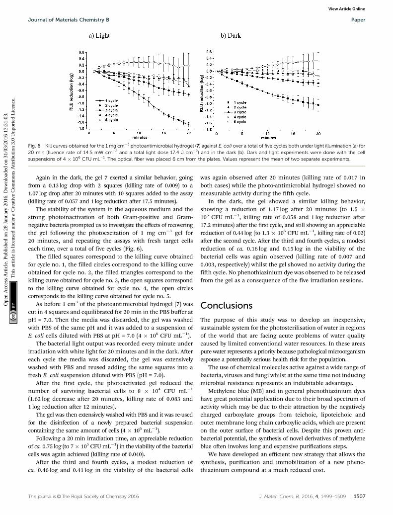
The stability of the system in the aqueous medium and thestrong photoinactivation of both Gram-positive and Gram-negative bacteria prompted us to investigate the effects of recoveringthe gel following the photoexcitation of 1 mg cm�3 gel for20 minutes, and repeating the assays with fresh target cellseach time, over a total of five cycles (Fig. 6).
The filled squares correspond to the killing curve obtainedfor cycle no. 1, the filled circles correspond to the killing curveobtained for cycle no. 2, the filled triangles correspond to thekilling curve obtained for cycle no. 3, the open squares correspondto the killing curve obtained for cycle no. 4, the open circlescorresponds to the killing curve obtained for cycle no. 5.
As before 1 cm3 of the photoantimicrobial hydrogel (7) wascut in 4 squares and equilibrated for 20 min in the PBS buffer atpH = 7.0. Then the media was discarded, the gel was washedwith PBS of the same pH and it was added to a suspension ofE. coli cells diluted with PBS at pH = 7.0 (4 � 106 CFU mL�1).
The bacterial light output was recorded every minute underirradiation with white light for 20 minutes and in the dark. Aftereach cycle the media was discarded, the gel was extensivelywashed with PBS and reused adding the same squares into afresh E. coli suspension diluted with PBS (pH = 7.0).
After the first cycle, the photoactivated gel reduced thenumber of surviving bacterial cells to 8 � 104 CFU mL�1
(1.62 log decrease after 20 minutes, killing rate of 0.083 and1 log reduction after 12 minutes).
The gel was then extensively washed with PBS and it was re-usedfor the disinfection of a newly prepared bacterial suspensioncontaining the same amount of cells (4 � 106 mL�1).
Following a 20 min irradiation time, an appreciable reductionof ca. 0.75 log (to 7� 105 CFU mL�1) in the viability of the bacterialcells was again achieved (killing rate of 0.040).
After the third and fourth cycles, a modest reduction ofca. 0.46 log and 0.41 log in the viability of the bacterial cells
was again observed after 20 minutes (killing rate of 0.017 inboth cases) while the photo-antimicrobial hydrogel showed nomeasurable activity during the fifth cycle.
In the dark, the gel showed a similar killing behavior,showing a reduction of 1.17 log after 20 minutes (to 1.5 �105 CFU mL�1, killing rate of 0.058 and 1 log reduction after17.2 minutes) after the first cycle, and still showing an appreciablereduction of 0.44 log (to 1.3 � 106 CFU mL�1, killing rate of 0.02)after the second cycle. After the third and fourth cycles, a modestreduction of ca. 0.16 log and 0.15 log in the viability of thebacterial cells was again observed (killing rate of 0.007 and0.003, respectively) whilst the gel showed no activity during thefifth cycle. No phenothiazinium dye was observed to be releasedfrom the gel as a consequence of the five irradiation sessions.
Conclusions
The purpose of this study was to develop an inexpensive,sustainable system for the photosterilisation of water in regionsof the world that are facing acute problems of water qualitycaused by limited conventional water resources. In these areaspure water represents a priority because pathological microorganismespouse a potentially serious health risk for the population.
The use of chemical molecules active against a wide range ofbacteria, viruses and fungi whilst at the same time not inducingmicrobial resistance represents an indubitable advantage.
Methylene blue (MB) and in general phenothiazinium dyeshave great potential application due to their broad spectrum ofactivity which may be due to their attraction by the negativelycharged carboxylate groups from teichoic, lipoteichoic andouter membrane long chain carboxylic acids, which are presenton the outer surface of bacterial cells. Despite this proven anti-bacterial potential, the synthesis of novel derivatives of methyleneblue often involves long and expensive purifications steps.
We have developed an efficient new strategy that allows thesynthesis, purification and immobilization of a new pheno-thiazinium compound at a much reduced cost.
Fig. 6 Kill curves obtained for the 1 mg cm�3 photoantimicrobial hydrogel (7) against E. coli over a total of five cycles both under light illumination (a) for20 min (fluence rate of 14.5 mW cm�2 and a total light dose 17.4 J cm�2) and in the dark (b). Dark and light experiments were done with the cellsuspensions of 4 � 106 CFU mL�1. The optical fiber was placed 6 cm from the plates. Values represent the mean of two separate experiments.
Journal of Materials Chemistry B Paper
Ope
n A
cces
s A
rtic
le. P
ublis
hed
on 2
8 Ja
nuar
y 20
16. D
ownl
oade
d on
31/
03/2
016
13:3
1:03
. T
his
artic
le is
lice
nsed
und
er a
Cre
ativ
e C
omm
ons
Attr
ibut
ion
3.0
Unp
orte
d L
icen
ce.
View Article Online
1508 | J. Mater. Chem. B, 2016, 4, 1499--1509 This journal is©The Royal Society of Chemistry 2016
An optically transparent polyacrylamide hydrogel was preparedby free radical polymerization of acrylamide (Am) and N,N0-methylenebisacrylamide using ammonium persulfate (APS) andN,N,N0,N0-tetramethylethylenediamine (TEMED) as the redoxinitiator and the catalyst respectively.
The photosensitiser bearing a terminal acryloyl group, suitablefor the later polymerization and incorporation into a hydrogel,was successfully incorporated into the matrix resulting in anhomogeneous and stable conjugate with no observable leachingeven after one week in water.
The hydrogel was characterized by IR and scanning electronmicroscopy and incorporation of the dye confirmed by UV-visiblespectroscopy.
SEM pictures confirmed that both the hydrogels with, andwithout, the PS are porous structures, allowing a large surfacearea and thus increasing contact between the gel and the bacteria.
The photoactive gel (7) successfully killed S. aureus andE. coli and the use of bioluminescent target species allowedmany variables to be tested such as the effects of dividing thegel to see the effects of increasing the interactive surface areabetween target cells and immobilized gel and the recovery andfurther testing of gel through five cycles of fresh challenge.
Further tests using more bacterial strains will be required tounderstand the applicability of the gel in real conditions, wherethe water can be infected by many different species of bacteriaas well as viruses and fungi.
The synthesized gels meet the intention to use these materialsas inexpensive practical systems of water disinfection suitable inremote regions of the world, where healthcare facilities are minimal.
Furthermore, the incorporation of the phenothiaziniumchromophore in this hydrogel does not require any specializedcondition (e.g. N2, glove box), and in a realistic approach, it canbe easily scaled up because it is characterized by an highversatility, as it is possible to shape the gel in any desired form.
Acknowledgements
The authors would like to thank The Sir Halley Stewart Trust forfunding this work. Mass spectrometry data was acquired at theEPSRC UK National Mass Spectrometry Facility at SwanseaUniversity. C. S. thanks Dr Saliha Saad and Mr Keith Hewettfor their help with the bacterial strain cultures and Mr TonySinclair for helpful discussions about SEM interpretation.
References
1 WHO UNICEF, Progress on Sanitation and Drinking-water:2010 Update, http://www.unicef.org/eapro/JMP-2010Final.pdf, accessed December 2015.
2 T. H. F. Wong and R. R. Brown, in Water Resources Planning andManagement, ed. R. Q. Grafton and K. Hussey, CambridgeUniversity Press, Cambridge, I edn, 2011, vol. 23, pp. 483–504.
3 D. Pittet, B. Allegranzi, J. Storr and L. Donaldson, Int.J. Infect. Dis., 2006, 10, 419–424.
4 B. Allegranzi and D. Pittet, J. Hosp. Infect., 2009, 73, 305–315.
5 J. M. A. Blair, M. A. Webber, A. J. Baylay, D. O. Ogbolu andL. J. V. Piddock, Nat. Rev. Microbiol., 2015, 13, 42–51.
6 M. Wainwright, J. Antimicrob. Chemother., 1998, 42, 13–28.7 M. R. Hamblin and T. Hasan, Photochem. Photobiol. Sci.,
2004, 3, 436–450.8 P. W. Taylor, P. D. Stapleton and J. P. Luzio, Drug Discovery
Today, 2002, 7, 1086–1091.9 G. Jori, C. Fabris, M. Soncin, S. Ferro, O. Coppellotti, D. Dei,
L. Fantetti, G. Chiti and G. Roncucci, Lasers Surg. Med., 2006,38, 468–481.
10 T. Maisch, Photochem. Photobiol. Sci., 2015, 14, 1518–1526.11 G. Jori, M. Camerin, M. Soncin, L. Guidolin and O. Coppellotti,
in Photodynamic Inactivation of Microbial Pathogens. Medicaland Environmental Applications, ed. M. R. Hamblin and G. Jori,The Royal Society of Chemistry, Cambridge, I edn, 2011, vol. 1,pp. 1–18.
12 G. Jori, J. Environ. Pathol., Toxicol. Oncol., 2006, 25, 505–519.13 C. Spagnul, L. C. Turner and R. W. Boyle, J. Photochem.
Photobiol., B, 2015, 150, 11–30.14 M. Wainwright, Photodiagn. Photodyn. Ther., 2005, 4, 263–272.15 F. Harris, L. K. Chatfield and D. A. Phoenix, Curr. Drug
Targets, 2005, 6, 615–627.16 M. Wainwright, Int. J. Antimicrob. Agents, 2000, 16, 381–394.17 S. Perni, P. Prokopovich, I. P. Parkin, M. Wilson and
J. Pratten, J. Mater. Chem., 2010, 20, 8668–8673.18 S. Perni, C. Piccirillo, A. Kafizas, M. Uppal, J. Pratten,
M. Wilson and I. P. Parkin, J. Cluster Sci., 2010, 21, 427–438.19 C. Piccirillo, S. Perni, J. Gil-Thomas, P. Prokopovich,
W. Chrzanowski, I. P. Parkin and M. Wilson, J. Mater.Chem., 2009, 19, 6167–6171.
20 S. Ismail, S. Perni, J. Pratten, I. Parkin and M. Wilson, Infect.Control Hosp. Epidemiol., 2011, 32, 1130–1132.
21 S. Perni, C. Piccirillo, J. Pratten, P. Prokopovich, W. Chrzanowski,I. P. Parkin and M. Wilson, Biomaterials, 2009, 30, 89–93.
22 A. J. T. Naik, S. Ismail, C. Kay, M. Wilson and I. P. Parkin,Mater. Chem. Phys., 2011, 129, 446–450.
23 S. Perni, P. Prokopovich, C. Piccirillo, J. Pratten, I. P. Parkinand M. Wilson, J. Mater. Chem., 2009, 19, 2715–2723.
24 R. Cahan, R. Schwartz, Y. Langzam and Y. Nitzan, Photochem.Photobiol., 2011, 87, 1379–1386.
25 M. Wilson, Infect. Control Hosp. Epidemiol., 2003, 24, 782–784.26 V. Decraene, J. Pratten and M. Wilson, Appl. Environ. Microbiol.,
2006, 72, 4436–4439.27 V. Decraene, J. Pratten and M. Wilson, Curr. Microbiol.,
2008, 57, 269–273.28 N. C. Stellwagen, Electrophoresis, 2009, 30, S188–S195.29 R. M. Thorn, S. M. Nelson and J. Greenman, Antimicrob.
Agents Chemother., 2007, 51, 3217–3224.30 P. D. Beer, J. Cadman, J. M. Lloris, R. Martınez-Manez,
J. Soto, T. Pardo and M. D. Marcos, J. Chem. Soc., DaltonTrans., 2000, 1805–1812.
31 M. Wainwright, K. Meegan, C. Loughran and R. M. Giddens,Dyes Pigm., 2009, 82, 387–391.
32 A. Felgentrager, T. Maisch, D. Dobler and A. Spath, BioMedRes. Int., 2013, 482167.
33 Y. Wang, Y. Liu, G. Li and J. Hao, Langmuir, 2014, 30, 6419–6426.
Paper Journal of Materials Chemistry B
Ope
n A
cces
s A
rtic
le. P
ublis
hed
on 2
8 Ja
nuar
y 20
16. D
ownl
oade
d on
31/
03/2
016
13:3
1:03
. T
his
artic
le is
lice
nsed
und
er a
Cre
ativ
e C
omm
ons
Attr
ibut
ion
3.0
Unp
orte
d L
icen
ce.
View Article Online
This journal is©The Royal Society of Chemistry 2016 J. Mater. Chem. B, 2016, 4, 1499--1509 | 1509
34 A. Parveen, G. Smith, V. Salisbury and S. M. Nelson, FEMSMicrobiol. Lett., 2001, 199, 115–118.
35 M. Tenhami, K. Hakkila and M. Karp, Antimicrob. AgentsChemother., 2001, 45, 3456–3461.
36 M. Alterman, H. Sjobom, P. Safsten, P. O. Markgren, U. H.Danielson, M. Hamalainen, S. Lofas, J. Hulten, B. Classon,B. Samuelsson and A. Hallberg, Eur. J. Pharm. Sci., 2001, 13,203–212.
37 F. Giuntini, F. Dumoulin, R. Daly, V. Ahsen, E. M. Scanlan,A. S. P. Lavado, J. W. Aylott, G. A. Rosser, A. Beeby andR. W. Boyle, Nanoscale, 2012, 4, 2034–2045.
38 S. A. Gorman, A. L. Bell, J. Griffiths, D. Roberts andS. B. Brown, Dyes Pigm., 2006, 71, 153–160.
39 Y.-T. Lu, C. Arai, J.-F. Ge, W.-S. Ren, M. Kaiser, S. Wittlin,R. Brun, J.-M. Lu and M. Ihara, Dyes Pigm., 2011, 89, 44–48.
40 N. A. Peppas, J. Z. Hilt, A. Khademhosseini and R. Langer,Adv. Mater., 2006, 18, 1345–1360.
41 A. S. Hoffman, Adv. Drug Delivery Rev., 2012, 64, 18–23.42 J. M. Banks, B. A. C. Harley and B. C. Bailey, ACS Biomater.
Sci. Eng., 2015, 1, 718–725.
43 B. Wilson, M.-J. Fernandez, A. Lorente and K. B. Grant, Org.Biomol. Chem., 2008, 6, 4026–4035.
44 J. P. Tardivo, A. Del Giglio, C. Santos de Oliveira, D. S. Gabrielli,H. C. Junqueira, D. B. Tada, D. Severino, R. F. Turchiello andM. S. Baptista, Photodiagn. Photodyn. Ther., 2005, 2, 175–191.
45 H. Tan, C. M. Ramirez, N. Miljkovic, H. Li, J. P. Rubin andK. G. Marra, Biomaterials, 2009, 30, 6844–6853.
46 J. K. Suh and H. W. Matthew, Biomaterials, 2000, 21, 2589–2598.47 H. Baniasadi, S. A. A. Ramazani and S. Mashayekhan, Int.
J. Biol. Macromol., 2015, 74, 360–366.48 H. Jiankang, L. Dichen, L. Yaxiong, Y. Bo, L. Bingheng and
L. Qin, Polymer, 2007, 48, 4578–4588.49 S. Gorgieva and V. Kokol, J. Biomed. Mater. Res., Part A, 2012,
100, 1655–1667.50 H. C. Junqueira, D. Severino, L. G. Dias, M. S. Gugliotti and
M. S. Baptista, Phys. Chem. Chem. Phys., 2002, 4, 2320–2328.51 M. N. Usacheva, M. C. Teichert and M. A. Biel, Lasers Surg.
Med., 2001, 29, 165–173.52 S. Noimark, E. Allan and I. P. Parkin, Chem. Sci., 2014, 5,
2216–2223.
Journal of Materials Chemistry B Paper
Ope
n A
cces
s A
rtic
le. P
ublis
hed
on 2
8 Ja
nuar
y 20
16. D
ownl
oade
d on
31/
03/2
016
13:3
1:03
. T
his
artic
le is
lice
nsed
und
er a
Cre
ativ
e C
omm
ons
Attr
ibut
ion
3.0
Unp
orte
d L
icen
ce.
View Article Online