Embed Size (px)
Citation preview

MICHIGAN STATE UNIVERSITY
~ _ MICHIGAN STATE UNIVERSITY
Research at Michigan State University C o l l e g e o f O s t e o p a t h i c M e d i c i n e
Spartan MedicalResearch Journal
Volume 2 Number 2 Winter 2017 Pages 59-72
Title
Pelvic Abscess with Presentation as Inability to Ambulate Authors Sarah A Damer DO APA (ASCP)
Citation DAMER SA Pelvic Abscess with Presentation as Inability to Ambulate Spartan Med Res J Vol 2 No 2 pp 59-72 2017
Keywords pelvic abscess intra-abdominal abscess psoas abscess iliopsoas syndrome
Downloaded from httpsmrjmsuedu
Statewide Campus System College of College of Osteopathic Medicine Osteopathic Medicine
________________________________________________________
Case Report
Pelvic Abscess with Presentation as Inability to Ambulate
Sarah A Damer DO APA (ASCP) CM 1
1 McLaren Health Care Macomb PGY3 Emergency Medicine Resident Mount Clemens MI
Certified through the American Society of Clinical Pathology as an Anatomic Pathologistsrsquo Assistant
Corresponding Author Sarah A Damer DO APA (ASCP) CM Sarahdamer1gmailcom
ABSTRACT DAMER SA Pelvic Abscess with Presentation as Inability to Ambulate Spartan Med Res J Vol 2 No 2 pp 59-72 2017 CONTEXT Intra-abdominal abscesses are localized collections of pus confined in the peritoneal cavity by an inflammatory barrier They are generally classified as intraperitoneal retroperitoneal or visceral and develop after perforation of a hollow viscus or by extension of infection or inflammation resulting from other conditions such as appendicitis or diverticulitis Intra-abdominal abscesses are highly variable in presentation and clinicians must have a broad differential to avoid an inaccurate diagnosis In this paper presenting clinical symptoms as well as diagnosis and treatment methods are discussed in the context of this atypical presentation of a pelvic abscess METHODS This retrospective case report presents a male patient in his early 60s who presented to the emergency department with atypical symptoms of a pelvic abscess The author obtained all diagnostic information from patient interview and electronic health record RESULTS The patientrsquos history of end stage renal disease and diverticulitis with colostomy placement led to this atypical presentation of an intra-abdominal abscess The patientrsquos abscess abutted the iliopsoas muscle that had given rise to his referred bilateral hip pain CONCLUSIONS This report presents a case of a male in his early 60s who presented to the hospital with complaint of bilateral hip pain and inability to ambulate Providers admitted him to an internal medicine service and he was diagnosed with a recurrent pelvic abscess extending to his left iliopsoas muscle Completed studies had failed to demonstrate any intrinsic pathology to the hips themselves This case demonstrated an atypical presentation of a pelvic abscess but brought up the theory that the etiology of the symptoms could be due to referred pain to the hips from the abscess Further studies are required to investigate the percentage of pelvic abscess patients who primarily present with a component of hip pain Keywords pelvic abscess intra-abdominal abscess psoas abscess iliopsoas syndrome
INTRODUCTION Intra-abdominal abscesses are localized collections of pus confined in a patientrsquos
peritoneal cavity by an inflammatory barrier This barrier may be comprised of omentum
(ie fat attached to the bowels) bowel adhesions or the organ from which the abscess
originated1
59
Vol 2 No2 Winter 2017
Pelvic Abscess with Presentation as Inability to Ambulate
Many intra-abdominal abscesses develop by extension of infection or inflammation
resulting from conditions such as appendicitis diverticulitis Crohnrsquos Disease
pancreatitis pelvic inflammatory disease or any condition causing generalized peritonitis
(ie inflammation of the membrane lining the abdominal wall)2 Prior abdominal surgery
is another significant risk factor
Traumatic abdominal injuries such as lacerations and hematomas of the liver
pancreas spleen and intestines can also lead to abscess formation The abscesses
usually contain a mixture of bacteria from the gastrointestinal tract Most frequent isolates
include Escherichia coli (E coli) Klebsiella and Bacteroides fragilis2 Neisseria
gonorrhoeae and chlamydial species are the most common organisms involved in pelvic
abscesses2
Intra-abdominal abscesses are highly variable in clinical presentation although the
majority of patients appear with abdominal symptoms or symptoms of sepsis There may
include persistent abdominal paintenderness distention mass or ileus1 Nausea
anorexia and weight loss are also common Other signs signifying possible infection
include fever fast heart rate or leukocytosis (ie high white blood cell count)1
Physicians should have a higher suspicion for this condition in patients with
predisposing primary intra-abdominal disease or those with history of abdominal surgery
If the abscess is deeply seeded however many of these classic features may be absent
The only initial clues may be fever persistent gastrointestinal dysfunction or non-
localizing debilitating illness1 Symptoms may be masked by analgesics (ie pain
relievers) or empiric antibiotic administration
One important clinical manifestation that can play a role in the presentation of an
intra-abdominal abscess is referred pain Referred pain is pain perceived at a location
other than the site of the painful stimulusorigin3 This type of pain is the result of a network
of interconnecting sensory nerves that supplies many different tissues When there is an
injury at one place in the network this pain can be interpreted in the brain to radiate
nerves and cause pain elsewhere in the related areas of the network3
In patients with subphrenic (ie below the diaphragm) abscesses irritation of
contiguous structures may produce shoulder pain hiccups non-productive cough
60
Spartan Med Res Jhttpssmrjmsuedu
S Damer
chest pain shortness of breath or pneumonia With pelvic abscesses frequent urination
diarrhea or tenesmus (ie continual feeling that one has to defecate) may occur1
Diagnosis of intra-abdominal abscesses includes a combination of laboratory
studies and diagnostic imaging Complete blood count basic metabolic panel and blood
cultures can help the most in diagnosis1 Blood cultures indicating polymicrobial (ie
several bacterial strains) bacteremia strongly implicate the presence of an intra-
abdominal abscess1 Plain abdominal radiograph are rarely diagnostic but may frequently
indicate the need for further investigation if certain abnormalities are present These may
include localized ileus extraluminal gas air-fluid levels or displacement of organs
Ultrasonography is a readily available portable inexpensive test and the findings
can be quite specific when correlated with the patientrsquos clinical signs In experienced
hands ultrasonography has an accuracy rate greater than 90 for diagnosing intra-
abdominal abscesses1 CT of the abdomen and pelvis with oral and intravenous contrast
is the preferred diagnostic modality with greater than 95 accuracy1
Appropriate treatment by clinicians can be frequently delayed due to the obscure
nature of many conditions resulting in abscess formation making diagnosis and
localization difficult Treatment modalities include antibiotic therapy percutaneous
drainage and surgical intervention Pharmacologic therapy involves administration of
empiric antibiotics While colonic flora consists of near 400 species an average of only
four to six species are generally recovered from intra-abdominal infections4 Combination
antibiotic therapy or broad-spectrum single-agent therapy is most often recommended
Therapy can then be adjusted after report of culture results
For patients with mild to moderate community-acquired infections and few risk
factors for resistance or treatment failure coverage for streptococci Enterobacteriaceae
and anaerobes is sufficient Single agent regimens include Ertapenem Zosyn or
Timentin Combination regimens include cefazolin cefuroxime ceftriaxone cefotaxime
ciprofloxacin or levofloxacin with Flagyl For high-risk community infections an agent
with gram-negative activity broad enough to cover pseudomonas and Enterobacteriaceae
resistant to non-pseudomonal cephalosporins should be chosen Single agent regimens
may include imipenem-cilastatin meropenem or Zosyn Combination regimens include
cefepime ceftazidime ciprofloxacin or levofloxacin with Flagyl In health-care associated
61
Vol 2 No2 Winter 2017
Pelvic Abscess with Presentation as Inability to Ambulate
infections with known colonization of methicillin-resistant Staphalococcus aureus
(MRSA) vancomycin should be added In patients who are known to be colonized by
highly-resistant organisms to include vancomycin-resistant enterococci (VRE) agents
such as linezolid and daptomycin should be used4
Physicians should often consider an infectious disease consult Percutaneous CT
guided catheter drainage is the standard treatment of most intra-abdominal abscesses2
Surgical drainage becomes mandatory when residual fluid cannot be evacuated with
catheter irrigation manipulation or additional drain placement
Intra-abdominal abscesses have a mortality rate of 10 to 405 Outcome depends
on the patientrsquos primary illness and general medical condition rather than on the specific
nature and location of the abscess Risk factors include multiple surgical procedures age
older than 50 years delay in initial intervention greater than 24 hours
immunocompromised conditions poor nutritional status multiple organ failure and
complex recurrent or persistent abscesses4 Multiple organ failure is a primary cause of
death4
Case Report A Caucasian male in his early 60s presented to the McLaren Macomb Emergency
Department (ED) with a complaint of bilateral hip pain and inability to ambulate The pain
had been present for the past four-to-five days and was progressive in nature The patient
admitted to being fairly active and normally able to ambulate without the assistance of a
cane or walker up until symptoms began He described his pain as 10 out of 10 constant
pain in the anterior bilateral hip joints with mild radiation into his lumbar back and groin
with no radiation into his legs His pain increased with ambulation but improved with rest
He attempted to take three doses of aspirin at home without any relief in his symptoms
and began using a walker to help him ambulate
The patient had an extensive medical history This history included sigmoid
diverticulitis with sigmoid colectomy in 2012 deep vein thrombosis duodenal ulcer
benign prostatic hypertrophy and hypertension He also suffered from chronic obstructive
pulmonary disease chronic thrombocytopenia peripheral vascular disease
atherosclerotic coronary artery disease and paroxysmal atrial fibrillation He had been
62
Spartan Med Res Jhttpssmrjmsuedu
S Damer
diagnosed with end stage renal disease and was on hemodialysis He subsequently had
multiple hemodialysis catheter infections with bacteremia (MRSA) obstructive uropathy
with chronic hydronephrosis and several peritoneal and pelvic abdominal abscesses He
denied having noticed any recent changes in his ostomy output
The patientrsquos past surgical history included inferior vena cava filter placement
cholecystectomy sigmoid colectomy with end colostomy numerous cystoscopies
bilateral ureteral stents peroneal abscess incision and drainage
esophagogastroduodenoscopy colonoscopy several IR drainages of pelvic abscesses
exploratory laparotomy with enterolysis temporary catheter placement and numerous
arteriovenous fistula creations
The patient took amiodarone Prilosec Imdur and aspirin He had no allergies to
any medications He denied use of nicotine alcohol or any illicit drugs He lived with and
took care of his elderly mother His father had died from a cerebral vascular accident and
his mother had a history of coronary artery disease
Physical exam revealed a well-nourished well-hydrated male who was in no acute
distress He had been brought from home via ambulance as ambulation was too painful
His vital signs demonstrated hypotension with a blood pressure of 7151 Patient admits
that this was his typical baseline His pain was worse on the right than the left He had
increased tenderness to log roll axial compression and passive flexion of the right hip
when compared to the left
He had intact two-point discrimination and light touch in the L2-S1 nerve
distributions bilaterally He had intact +2 out of 4 dorsalis pedis and posterior tibial pulses
found with Doppler ultrasound He elicited 5 out of 5 muscle strength in his bilateral lower
extremities There was no tenderness to palpation over the greater trochanteric region of
either hip He had a negative straight leg raise bilaterally The patient had severe brawny
edema and dryness to his lower extremities
Laboratory studies demonstrated a normocytic anemia chronic thrombocytopenia
hyponatremia and chronic kidney disease His sedimentation rate and c-reactive protein
levels were elevated When the patient was sent over for x-ray imaging left hip right hip
and pelvis x-rays demonstrated no abnormalities Lumbosacral x-ray demonstrated mild
scoliosis with diffuse moderate degenerative change (Image 1) The patient was offered
63
Vol 2 No2 Winter 2017
Pelvic Abscess with Presentation as Inability to Ambulate
pain medication but refused any other treatment He was admitted to the hospital due to
his intractable bilateral hip pain and inability to ambulate Orthopedics Nephrology and
Physical TherapyOccupational Therapy consults were placed
Orthopedics was the first service to evaluate the patient on hospital Day 2 and
ordered a CT of the abdomen and pelvis with contrast This study found that superior to
the bladder there was a 49 x 38 cm fluid collection with peripheral enhancement
concerning for abscess (Image 2) Internal Medicine then evaluated the patient and
reviewed the results of the CT scan Blood cultures were obtained and the General
Surgery service was consulted A repeat CT with oral contrast was ordered by General
Surgery on hospital Day 3 This test showed that the adjacent loops of the intestines along
the anterior and superior margin of the fluid collection were partially opacified without
obvious extravasation of oral contrast in the region suggesting fistula (ie
communication with the bowel) (Image 3)
The blood cultures grew out E coli x 2 after 24 hours The patient was placed on
Zosyn and vancomycin On hospital Day 5 Interventional Radiology performed CT
guidance drainage of the pelvic abscess with catheter placement Culture results would
later grow E coli The Urology service was also consulted as the patient had history of
hydronephrosis due to obstructive uropathy with bilateral stent placement This service
was asked to evaluate the need for stent replacement and integrity of the bladder with its
close approximation to the abscess
On hospital Day 6 the patient went to the operating room with Urology for a
bilateral retrograde urogram The study demonstrated findings consistent with
communication of the urinary bladder with the abscess cavity previously drained The
stents were exchanged a urinary catheter was placed and a urine sample was obtained
for which culture results came back positive for Ecoli The patient remained on Zosyn
As the patient continued to experience significant bilateral hip pain an MRI of the
patientrsquos thoraciclumbar spine and bilateral hips was ordered on Day 7 A thoracic spine
MRI demonstrated a small central disc protrusion at the Thoracic 4-T5 level No spinal
canal stenosis or foraminal encroachment The MRI of the lumbar spine and bilateral hips
was unable to be completed since the patient could not tolerate the pain long enough to
have the study performed
64
Spartan Med Res Jhttpssmrjmsuedu
S Damer
The pelvic drain was removed between hospital Days 9 and 11 although progress
notes are unclear as to the specific day On hospital Day 15 the patient was discharged
to a subacute rehabilitation unit on a two-week course of oral Augmentin He had worked
with Physical Therapy throughout his hospital stay The patient slowly regained some
ability to ambulate but it is unknown whether or not this was from resolving his pelvic
abscess or due to rehabilitation with Physical Therapy He was to follow up with the
urologist in two-to-three weeks for urinary catheter removal
DISCUSSION This case describes a patient who suffered from the inability to ambulate who was
eventually diagnosed with an intra-abdominal abscess This patient had presented to the
emergency department with bilateral hip pain which would be an unusual presenting
symptom for his diagnosed pathology As earlier described most common complaints
associated with an intra-abdominal abscess include abdominal paintenderness
distention nausea anorexia fever fast heart rate or high white blood cell count The
patient met none of these criteria Throughout the patientrsquos hospital stay he never had a
complaint related to his gastrointestinal system On Day 3 of his hospitalization he spiked
a white blood cell count but remained afebrile throughout his course The question arises
as to whether the patientrsquos complaint has any relation to the intra-abdominal abscess
found on CT imaging
After an in-depth review of the patientrsquos medical record it was revealed that he
had an extensive medical history The patient has suffered from diverticulitis with
perforation and abscess formation He had several drainages of the abscess completed
by an interventional radiologist prior to and after his sigmoid colectomy with end
ileostomy that was performed in September 2012
Throughout his multiple hospitalizations he had recurrence of an intraperitoneal
abscess that was located superior to the bladder and adjacent to the left iliopsoas muscle
Several studies showed altered attenuation of the iliopsoas muscle and evidence that the
abscess was contiguous with the muscle CT imaging conducted in December 2012
indicated that the abscess involved the left iliopsoas bursa It is this
65
Vol 2 No2 Winter 2017
Pelvic Abscess with Presentation as Inability to Ambulate
involvementextension for which the patientrsquos presenting complaint could possibly have
relation to his found pathology however no direct data concerning this existed
The psoas muscle arises from the transverse processes and lateral aspects of the
vertebral bodies between the 12th thoracic and 5th lumbar vertebrae It courses downward
passing deep to the inguinal ligament and anterior to the hip joint capsule to form a tendon
that inserts into the lesser trochanter of the femur The iliacus muscle joins the psoas to
insert via the same tendon These two muscles are the main hip flexors The tendon is
separated from the hip capsule by the iliopsoas bursa This bursa is the largest bursa in
the body and exists to help reduce rubbing between the iliopsoas muscle and the femur
This bursa communicates with the hip joint space in up to 15 of persons which may
facilitate spread of infection between these sites6
The iliopsoas muscle can be involved in several pathologic conditions to include
iliopsoas tendinitisbursitis iliopsoas syndrome and psoas abscesses Although
uncommon injuries iliopsoas tendinitisbursitis occurs when the tendon and bursa
becomes inflamed They are overuse injuries that result from overloading the hips with
repetitive movements7 People who participate in activities such as golf hockey
cheerleading gymnastics and resistance training are most susceptible to injury Iliopsoas
syndrome frequently begins as a bilateral muscle spasm which eventually becomes
prominent on one side
Psoas abscesses are a collection of pus in the iliopsoas muscle compartment
which may arise from contiguous spread from adjacent structures or by hematogenous
route from a distant site6 They are divided into primary and secondary abscesses
Primary abscess occurs as a result of hematogenous or lymphatic seeding Risk factors
include diabetes IV drug abuse HIV renal failure or other forms of immunosuppression
Secondary abscesses occur as a result of a direct spread of infection to the psoas muscle
from adjacent structures The structures include vertebrae hip arthroplasty GI tract
aorta and genitourinary tract
Symptoms of iliopsoas tendinitisbursitis and psoas syndromes include pain
tenderness swelling heat or redness and loss of normal mobility Signs of psoas abscess
include back or flank pain fever inguinal mass limp anorexia and weight loss Pain can
66
Spartan Med Res Jhttpssmrjmsuedu
S Damer
be present in up to 91 of cases with localization to the back flank or lower abdomen
and possible radiation to the hip andor posterior thigh6
Originally described in 1881 the classic clinical presentation of a psoas abscess
included back pain limp and fever8 Newer case studies have demonstrated that these
symptoms may only be present in 30 of cases8 Many patients present with nonspecific
complaints such as malaise low grade fever abdominalflank discomfort a flexed and
externally rotated hip and pain on movement of the hip Pain is due to irritation of muscle
belly and referred pain from nerve roots L2-L4 Due to the vague and nonspecific
presenting symptoms of a psoas abscess they are commonly misdiagnosed although
data concerning the misdiagnosis rate is sparse8 One case studied reported a psoas
abscess that was previously misdiagnosed as a deep vein thrombosis9
After review of the presenting symptoms of iliopsoas bursitistendonitis syndrome
and psoas abscess the clinical conclusion can be made that this patient may have been
suffering from referred pain to the hip from his pelvic abscess that abutted the left
iliopsoas muscle This is a speculation since there are no direct reports available
concerning a direct correlation between this manrsquos pelvic abscesses and hip pain
However several discrepancies arose during his workup The patient had suffered
from right hip pain yet the patientrsquos abscess directly invaded the left iliopsoas muscle He
also had more pain when asked to perform hip flexion and preferred to lay flat on his back
without axial loading Research has found a correlation with patients suffering from psoas
abscesses and that they prefer to lay with their hips flexed6 In addition the patient had
presented several times in the past for recurrent pelvic abscess and failed to complain of
hip pain during those admissions Finally the patient may have had pathology of his
lumbar spine or hips that could have been identified if the patient had been able to tolerate
the MRI imaging procedure
In support of the theory of the patientrsquos suffering from referred pain the patient did
experience pain upon movement of the hip and had improvement in his symptoms after
IR drainage and antibiotic administration Hip pain especially in Crohnrsquos patients should
prompt consideration of a psoas abscess as the incidence has been estimated to be
between 04 and 436 Psoas abscess has also been described in the setting of
appendicitis colorectal cancer ulcerative colitis and following abdominal surgery so
67
Vol 2 No2 Winter 2017
Pelvic Abscess with Presentation as Inability to Ambulate
evidence exists with other GI pathology While a question was made during the patientrsquos
hospital stay as to whether his symptomatology was somehow related to his pathology
versus incidental the author does not believe the theory of referred pain is far-fetched
CONCLUSIONS This report presents a case of a Caucasian male in his early 60s who presented
to the emergency department with complaint of bilateral hip pain and inability to ambulate
He was admitted to the hospital and found to have a recurrent pelvic abscess which
extended to his left iliopsoas muscle The patient failed to demonstrate the typical
symptoms of a pelvic abscess and therefore his pathology was not revealed initially in the
emergency department It was not until the patient had been hospitalized for a day and
his medical record had been thoroughly reviewed when his diagnosis was made Even
then his presenting complaints could not be fully explained Additional imaging studies
failed to demonstrate any intrinsic pathology to the hips themselves
This case demonstrated an atypical presentation of a pelvic abscess but questions
remained as to whether the patientrsquos symptoms were due to referred pain to the hips
Conditions such as iliopsoas tendinitisbursitis and psoas syndromes present with pain
tenderness swelling heat or redness and loss of normal mobility while signs of a psoas
abscess include back or flank pain fever inguinal mass limp anorexia and weight loss
These clinical findings are more consistent with what the patient in the case study had
presented to providers Further investigations are required to determine the percentage
of patients with pelvic abscesses who initially present with a component of hip pain
The authors report no external funding source for this study
The authors declare no conflict of interest
Submitted for publication June 2017 Accepted for publication December 2017
68
Spartan Med Res Jhttpssmrjmsuedu
Rl
S Damer
ACKNOWLEDGEMENTS Julie Lata DO revised this case report critically for important intellectual content
and gave final pre-publication approval of this final article draft
REFERENCES 1 Saber AA Abdominal Abscess Medscape 2016 Jun 21 2017 Oct 1 Available from httpemedicinemedscapecomarticle1979032-overviewa5
2 Ansari P Intra-abdominal Abscesses Merck Manual 2017 Jan 2017 Oct 1 Available from httpswwwmerckmanualscomprofessionalgastrointestinal-disordersacute-abdomen-and-surgical-gastroenterologyintra-abdominal-abscessesv891088
3 De Koster K Referred Pain Physiopedia 2017 Oct 1 Available from httpswwwphysio-pediacomReferred_Pain
4 Barshak M Antimicrobial Approach to Intra-abdominal Infections in Adults Up to Date 2016 Mar 14 2017 Oct 1 Available from httpwwwuptodatecomcontentsantimicrobial-approach-to-intra-abdominal-infections-in-adults
5 Daley B Peritonitis and Abdominal Sepsis Medscape 2017 Jan 11 2017 Oct 1 Available from httpemedicinemedscapecomarticle180234-overview
6 Spelman D Psoas abscess Up to Date 2016 May 9 2017 Oct 1 Available from httpswwwuptodatecomcontentspsoas-abscess
7 Tufo A Psoas syndrome A frequently missed diagnosis J Amer Osteo Assoc 2012112(1)522-528
8 Shields D Robinson P Crowley TP Iliopsoas abscess ndash A review and update on the literature Intl J Surg 201210 (1)466-469
9 Kong V Oosthuizen G Mthethwa M Reddy K Clarke D Delayed presentation of a psoas abscess previously misdiagnosed as deep vein thrombosis A potentially devastating tumor Intl J Case Rep Images 20134(2)111-114
69
Vol 2 No2 Winter 2017
Pelvic Abscess with Presentation as Inability to Ambulate
TABLES AND FIGURES
Image 1 Pelvis X-ray
70
Spartan Med Res Jhttpssmrjmsuedu
S Damer
Image 2 Fluid Collection - 49 x 38 cm
71
Vol 2 No2 Winter 2017
Pelvic Abscess with Presentation as Inability to Ambulate
Image 3 Pelvic Abscess without Signs of Communication with Bowel
72

Vol 2 No2 Winter 2017
Pelvic Abscess with Presentation as Inability to Ambulate
Many intra-abdominal abscesses develop by extension of infection or inflammation
resulting from conditions such as appendicitis diverticulitis Crohnrsquos Disease
pancreatitis pelvic inflammatory disease or any condition causing generalized peritonitis
(ie inflammation of the membrane lining the abdominal wall)2 Prior abdominal surgery
is another significant risk factor
Traumatic abdominal injuries such as lacerations and hematomas of the liver
pancreas spleen and intestines can also lead to abscess formation The abscesses
usually contain a mixture of bacteria from the gastrointestinal tract Most frequent isolates
include Escherichia coli (E coli) Klebsiella and Bacteroides fragilis2 Neisseria
gonorrhoeae and chlamydial species are the most common organisms involved in pelvic
abscesses2
Intra-abdominal abscesses are highly variable in clinical presentation although the
majority of patients appear with abdominal symptoms or symptoms of sepsis There may
include persistent abdominal paintenderness distention mass or ileus1 Nausea
anorexia and weight loss are also common Other signs signifying possible infection
include fever fast heart rate or leukocytosis (ie high white blood cell count)1
Physicians should have a higher suspicion for this condition in patients with
predisposing primary intra-abdominal disease or those with history of abdominal surgery
If the abscess is deeply seeded however many of these classic features may be absent
The only initial clues may be fever persistent gastrointestinal dysfunction or non-
localizing debilitating illness1 Symptoms may be masked by analgesics (ie pain
relievers) or empiric antibiotic administration
One important clinical manifestation that can play a role in the presentation of an
intra-abdominal abscess is referred pain Referred pain is pain perceived at a location
other than the site of the painful stimulusorigin3 This type of pain is the result of a network
of interconnecting sensory nerves that supplies many different tissues When there is an
injury at one place in the network this pain can be interpreted in the brain to radiate
nerves and cause pain elsewhere in the related areas of the network3
In patients with subphrenic (ie below the diaphragm) abscesses irritation of
contiguous structures may produce shoulder pain hiccups non-productive cough
60
Spartan Med Res Jhttpssmrjmsuedu
S Damer
chest pain shortness of breath or pneumonia With pelvic abscesses frequent urination
diarrhea or tenesmus (ie continual feeling that one has to defecate) may occur1
Diagnosis of intra-abdominal abscesses includes a combination of laboratory
studies and diagnostic imaging Complete blood count basic metabolic panel and blood
cultures can help the most in diagnosis1 Blood cultures indicating polymicrobial (ie
several bacterial strains) bacteremia strongly implicate the presence of an intra-
abdominal abscess1 Plain abdominal radiograph are rarely diagnostic but may frequently
indicate the need for further investigation if certain abnormalities are present These may
include localized ileus extraluminal gas air-fluid levels or displacement of organs
Ultrasonography is a readily available portable inexpensive test and the findings
can be quite specific when correlated with the patientrsquos clinical signs In experienced
hands ultrasonography has an accuracy rate greater than 90 for diagnosing intra-
abdominal abscesses1 CT of the abdomen and pelvis with oral and intravenous contrast
is the preferred diagnostic modality with greater than 95 accuracy1
Appropriate treatment by clinicians can be frequently delayed due to the obscure
nature of many conditions resulting in abscess formation making diagnosis and
localization difficult Treatment modalities include antibiotic therapy percutaneous
drainage and surgical intervention Pharmacologic therapy involves administration of
empiric antibiotics While colonic flora consists of near 400 species an average of only
four to six species are generally recovered from intra-abdominal infections4 Combination
antibiotic therapy or broad-spectrum single-agent therapy is most often recommended
Therapy can then be adjusted after report of culture results
For patients with mild to moderate community-acquired infections and few risk
factors for resistance or treatment failure coverage for streptococci Enterobacteriaceae
and anaerobes is sufficient Single agent regimens include Ertapenem Zosyn or
Timentin Combination regimens include cefazolin cefuroxime ceftriaxone cefotaxime
ciprofloxacin or levofloxacin with Flagyl For high-risk community infections an agent
with gram-negative activity broad enough to cover pseudomonas and Enterobacteriaceae
resistant to non-pseudomonal cephalosporins should be chosen Single agent regimens
may include imipenem-cilastatin meropenem or Zosyn Combination regimens include
cefepime ceftazidime ciprofloxacin or levofloxacin with Flagyl In health-care associated
61
Vol 2 No2 Winter 2017
Pelvic Abscess with Presentation as Inability to Ambulate
infections with known colonization of methicillin-resistant Staphalococcus aureus
(MRSA) vancomycin should be added In patients who are known to be colonized by
highly-resistant organisms to include vancomycin-resistant enterococci (VRE) agents
such as linezolid and daptomycin should be used4
Physicians should often consider an infectious disease consult Percutaneous CT
guided catheter drainage is the standard treatment of most intra-abdominal abscesses2
Surgical drainage becomes mandatory when residual fluid cannot be evacuated with
catheter irrigation manipulation or additional drain placement
Intra-abdominal abscesses have a mortality rate of 10 to 405 Outcome depends
on the patientrsquos primary illness and general medical condition rather than on the specific
nature and location of the abscess Risk factors include multiple surgical procedures age
older than 50 years delay in initial intervention greater than 24 hours
immunocompromised conditions poor nutritional status multiple organ failure and
complex recurrent or persistent abscesses4 Multiple organ failure is a primary cause of
death4
Case Report A Caucasian male in his early 60s presented to the McLaren Macomb Emergency
Department (ED) with a complaint of bilateral hip pain and inability to ambulate The pain
had been present for the past four-to-five days and was progressive in nature The patient
admitted to being fairly active and normally able to ambulate without the assistance of a
cane or walker up until symptoms began He described his pain as 10 out of 10 constant
pain in the anterior bilateral hip joints with mild radiation into his lumbar back and groin
with no radiation into his legs His pain increased with ambulation but improved with rest
He attempted to take three doses of aspirin at home without any relief in his symptoms
and began using a walker to help him ambulate
The patient had an extensive medical history This history included sigmoid
diverticulitis with sigmoid colectomy in 2012 deep vein thrombosis duodenal ulcer
benign prostatic hypertrophy and hypertension He also suffered from chronic obstructive
pulmonary disease chronic thrombocytopenia peripheral vascular disease
atherosclerotic coronary artery disease and paroxysmal atrial fibrillation He had been
62
Spartan Med Res Jhttpssmrjmsuedu
S Damer
diagnosed with end stage renal disease and was on hemodialysis He subsequently had
multiple hemodialysis catheter infections with bacteremia (MRSA) obstructive uropathy
with chronic hydronephrosis and several peritoneal and pelvic abdominal abscesses He
denied having noticed any recent changes in his ostomy output
The patientrsquos past surgical history included inferior vena cava filter placement
cholecystectomy sigmoid colectomy with end colostomy numerous cystoscopies
bilateral ureteral stents peroneal abscess incision and drainage
esophagogastroduodenoscopy colonoscopy several IR drainages of pelvic abscesses
exploratory laparotomy with enterolysis temporary catheter placement and numerous
arteriovenous fistula creations
The patient took amiodarone Prilosec Imdur and aspirin He had no allergies to
any medications He denied use of nicotine alcohol or any illicit drugs He lived with and
took care of his elderly mother His father had died from a cerebral vascular accident and
his mother had a history of coronary artery disease
Physical exam revealed a well-nourished well-hydrated male who was in no acute
distress He had been brought from home via ambulance as ambulation was too painful
His vital signs demonstrated hypotension with a blood pressure of 7151 Patient admits
that this was his typical baseline His pain was worse on the right than the left He had
increased tenderness to log roll axial compression and passive flexion of the right hip
when compared to the left
He had intact two-point discrimination and light touch in the L2-S1 nerve
distributions bilaterally He had intact +2 out of 4 dorsalis pedis and posterior tibial pulses
found with Doppler ultrasound He elicited 5 out of 5 muscle strength in his bilateral lower
extremities There was no tenderness to palpation over the greater trochanteric region of
either hip He had a negative straight leg raise bilaterally The patient had severe brawny
edema and dryness to his lower extremities
Laboratory studies demonstrated a normocytic anemia chronic thrombocytopenia
hyponatremia and chronic kidney disease His sedimentation rate and c-reactive protein
levels were elevated When the patient was sent over for x-ray imaging left hip right hip
and pelvis x-rays demonstrated no abnormalities Lumbosacral x-ray demonstrated mild
scoliosis with diffuse moderate degenerative change (Image 1) The patient was offered
63
Vol 2 No2 Winter 2017
Pelvic Abscess with Presentation as Inability to Ambulate
pain medication but refused any other treatment He was admitted to the hospital due to
his intractable bilateral hip pain and inability to ambulate Orthopedics Nephrology and
Physical TherapyOccupational Therapy consults were placed
Orthopedics was the first service to evaluate the patient on hospital Day 2 and
ordered a CT of the abdomen and pelvis with contrast This study found that superior to
the bladder there was a 49 x 38 cm fluid collection with peripheral enhancement
concerning for abscess (Image 2) Internal Medicine then evaluated the patient and
reviewed the results of the CT scan Blood cultures were obtained and the General
Surgery service was consulted A repeat CT with oral contrast was ordered by General
Surgery on hospital Day 3 This test showed that the adjacent loops of the intestines along
the anterior and superior margin of the fluid collection were partially opacified without
obvious extravasation of oral contrast in the region suggesting fistula (ie
communication with the bowel) (Image 3)
The blood cultures grew out E coli x 2 after 24 hours The patient was placed on
Zosyn and vancomycin On hospital Day 5 Interventional Radiology performed CT
guidance drainage of the pelvic abscess with catheter placement Culture results would
later grow E coli The Urology service was also consulted as the patient had history of
hydronephrosis due to obstructive uropathy with bilateral stent placement This service
was asked to evaluate the need for stent replacement and integrity of the bladder with its
close approximation to the abscess
On hospital Day 6 the patient went to the operating room with Urology for a
bilateral retrograde urogram The study demonstrated findings consistent with
communication of the urinary bladder with the abscess cavity previously drained The
stents were exchanged a urinary catheter was placed and a urine sample was obtained
for which culture results came back positive for Ecoli The patient remained on Zosyn
As the patient continued to experience significant bilateral hip pain an MRI of the
patientrsquos thoraciclumbar spine and bilateral hips was ordered on Day 7 A thoracic spine
MRI demonstrated a small central disc protrusion at the Thoracic 4-T5 level No spinal
canal stenosis or foraminal encroachment The MRI of the lumbar spine and bilateral hips
was unable to be completed since the patient could not tolerate the pain long enough to
have the study performed
64
Spartan Med Res Jhttpssmrjmsuedu
S Damer
The pelvic drain was removed between hospital Days 9 and 11 although progress
notes are unclear as to the specific day On hospital Day 15 the patient was discharged
to a subacute rehabilitation unit on a two-week course of oral Augmentin He had worked
with Physical Therapy throughout his hospital stay The patient slowly regained some
ability to ambulate but it is unknown whether or not this was from resolving his pelvic
abscess or due to rehabilitation with Physical Therapy He was to follow up with the
urologist in two-to-three weeks for urinary catheter removal
DISCUSSION This case describes a patient who suffered from the inability to ambulate who was
eventually diagnosed with an intra-abdominal abscess This patient had presented to the
emergency department with bilateral hip pain which would be an unusual presenting
symptom for his diagnosed pathology As earlier described most common complaints
associated with an intra-abdominal abscess include abdominal paintenderness
distention nausea anorexia fever fast heart rate or high white blood cell count The
patient met none of these criteria Throughout the patientrsquos hospital stay he never had a
complaint related to his gastrointestinal system On Day 3 of his hospitalization he spiked
a white blood cell count but remained afebrile throughout his course The question arises
as to whether the patientrsquos complaint has any relation to the intra-abdominal abscess
found on CT imaging
After an in-depth review of the patientrsquos medical record it was revealed that he
had an extensive medical history The patient has suffered from diverticulitis with
perforation and abscess formation He had several drainages of the abscess completed
by an interventional radiologist prior to and after his sigmoid colectomy with end
ileostomy that was performed in September 2012
Throughout his multiple hospitalizations he had recurrence of an intraperitoneal
abscess that was located superior to the bladder and adjacent to the left iliopsoas muscle
Several studies showed altered attenuation of the iliopsoas muscle and evidence that the
abscess was contiguous with the muscle CT imaging conducted in December 2012
indicated that the abscess involved the left iliopsoas bursa It is this
65
Vol 2 No2 Winter 2017
Pelvic Abscess with Presentation as Inability to Ambulate
involvementextension for which the patientrsquos presenting complaint could possibly have
relation to his found pathology however no direct data concerning this existed
The psoas muscle arises from the transverse processes and lateral aspects of the
vertebral bodies between the 12th thoracic and 5th lumbar vertebrae It courses downward
passing deep to the inguinal ligament and anterior to the hip joint capsule to form a tendon
that inserts into the lesser trochanter of the femur The iliacus muscle joins the psoas to
insert via the same tendon These two muscles are the main hip flexors The tendon is
separated from the hip capsule by the iliopsoas bursa This bursa is the largest bursa in
the body and exists to help reduce rubbing between the iliopsoas muscle and the femur
This bursa communicates with the hip joint space in up to 15 of persons which may
facilitate spread of infection between these sites6
The iliopsoas muscle can be involved in several pathologic conditions to include
iliopsoas tendinitisbursitis iliopsoas syndrome and psoas abscesses Although
uncommon injuries iliopsoas tendinitisbursitis occurs when the tendon and bursa
becomes inflamed They are overuse injuries that result from overloading the hips with
repetitive movements7 People who participate in activities such as golf hockey
cheerleading gymnastics and resistance training are most susceptible to injury Iliopsoas
syndrome frequently begins as a bilateral muscle spasm which eventually becomes
prominent on one side
Psoas abscesses are a collection of pus in the iliopsoas muscle compartment
which may arise from contiguous spread from adjacent structures or by hematogenous
route from a distant site6 They are divided into primary and secondary abscesses
Primary abscess occurs as a result of hematogenous or lymphatic seeding Risk factors
include diabetes IV drug abuse HIV renal failure or other forms of immunosuppression
Secondary abscesses occur as a result of a direct spread of infection to the psoas muscle
from adjacent structures The structures include vertebrae hip arthroplasty GI tract
aorta and genitourinary tract
Symptoms of iliopsoas tendinitisbursitis and psoas syndromes include pain
tenderness swelling heat or redness and loss of normal mobility Signs of psoas abscess
include back or flank pain fever inguinal mass limp anorexia and weight loss Pain can
66
Spartan Med Res Jhttpssmrjmsuedu
S Damer
be present in up to 91 of cases with localization to the back flank or lower abdomen
and possible radiation to the hip andor posterior thigh6
Originally described in 1881 the classic clinical presentation of a psoas abscess
included back pain limp and fever8 Newer case studies have demonstrated that these
symptoms may only be present in 30 of cases8 Many patients present with nonspecific
complaints such as malaise low grade fever abdominalflank discomfort a flexed and
externally rotated hip and pain on movement of the hip Pain is due to irritation of muscle
belly and referred pain from nerve roots L2-L4 Due to the vague and nonspecific
presenting symptoms of a psoas abscess they are commonly misdiagnosed although
data concerning the misdiagnosis rate is sparse8 One case studied reported a psoas
abscess that was previously misdiagnosed as a deep vein thrombosis9
After review of the presenting symptoms of iliopsoas bursitistendonitis syndrome
and psoas abscess the clinical conclusion can be made that this patient may have been
suffering from referred pain to the hip from his pelvic abscess that abutted the left
iliopsoas muscle This is a speculation since there are no direct reports available
concerning a direct correlation between this manrsquos pelvic abscesses and hip pain
However several discrepancies arose during his workup The patient had suffered
from right hip pain yet the patientrsquos abscess directly invaded the left iliopsoas muscle He
also had more pain when asked to perform hip flexion and preferred to lay flat on his back
without axial loading Research has found a correlation with patients suffering from psoas
abscesses and that they prefer to lay with their hips flexed6 In addition the patient had
presented several times in the past for recurrent pelvic abscess and failed to complain of
hip pain during those admissions Finally the patient may have had pathology of his
lumbar spine or hips that could have been identified if the patient had been able to tolerate
the MRI imaging procedure
In support of the theory of the patientrsquos suffering from referred pain the patient did
experience pain upon movement of the hip and had improvement in his symptoms after
IR drainage and antibiotic administration Hip pain especially in Crohnrsquos patients should
prompt consideration of a psoas abscess as the incidence has been estimated to be
between 04 and 436 Psoas abscess has also been described in the setting of
appendicitis colorectal cancer ulcerative colitis and following abdominal surgery so
67
Vol 2 No2 Winter 2017
Pelvic Abscess with Presentation as Inability to Ambulate
evidence exists with other GI pathology While a question was made during the patientrsquos
hospital stay as to whether his symptomatology was somehow related to his pathology
versus incidental the author does not believe the theory of referred pain is far-fetched
CONCLUSIONS This report presents a case of a Caucasian male in his early 60s who presented
to the emergency department with complaint of bilateral hip pain and inability to ambulate
He was admitted to the hospital and found to have a recurrent pelvic abscess which
extended to his left iliopsoas muscle The patient failed to demonstrate the typical
symptoms of a pelvic abscess and therefore his pathology was not revealed initially in the
emergency department It was not until the patient had been hospitalized for a day and
his medical record had been thoroughly reviewed when his diagnosis was made Even
then his presenting complaints could not be fully explained Additional imaging studies
failed to demonstrate any intrinsic pathology to the hips themselves
This case demonstrated an atypical presentation of a pelvic abscess but questions
remained as to whether the patientrsquos symptoms were due to referred pain to the hips
Conditions such as iliopsoas tendinitisbursitis and psoas syndromes present with pain
tenderness swelling heat or redness and loss of normal mobility while signs of a psoas
abscess include back or flank pain fever inguinal mass limp anorexia and weight loss
These clinical findings are more consistent with what the patient in the case study had
presented to providers Further investigations are required to determine the percentage
of patients with pelvic abscesses who initially present with a component of hip pain
The authors report no external funding source for this study
The authors declare no conflict of interest
Submitted for publication June 2017 Accepted for publication December 2017
68
Spartan Med Res Jhttpssmrjmsuedu
Rl
S Damer
ACKNOWLEDGEMENTS Julie Lata DO revised this case report critically for important intellectual content
and gave final pre-publication approval of this final article draft
REFERENCES 1 Saber AA Abdominal Abscess Medscape 2016 Jun 21 2017 Oct 1 Available from httpemedicinemedscapecomarticle1979032-overviewa5
2 Ansari P Intra-abdominal Abscesses Merck Manual 2017 Jan 2017 Oct 1 Available from httpswwwmerckmanualscomprofessionalgastrointestinal-disordersacute-abdomen-and-surgical-gastroenterologyintra-abdominal-abscessesv891088
3 De Koster K Referred Pain Physiopedia 2017 Oct 1 Available from httpswwwphysio-pediacomReferred_Pain
4 Barshak M Antimicrobial Approach to Intra-abdominal Infections in Adults Up to Date 2016 Mar 14 2017 Oct 1 Available from httpwwwuptodatecomcontentsantimicrobial-approach-to-intra-abdominal-infections-in-adults
5 Daley B Peritonitis and Abdominal Sepsis Medscape 2017 Jan 11 2017 Oct 1 Available from httpemedicinemedscapecomarticle180234-overview
6 Spelman D Psoas abscess Up to Date 2016 May 9 2017 Oct 1 Available from httpswwwuptodatecomcontentspsoas-abscess
7 Tufo A Psoas syndrome A frequently missed diagnosis J Amer Osteo Assoc 2012112(1)522-528
8 Shields D Robinson P Crowley TP Iliopsoas abscess ndash A review and update on the literature Intl J Surg 201210 (1)466-469
9 Kong V Oosthuizen G Mthethwa M Reddy K Clarke D Delayed presentation of a psoas abscess previously misdiagnosed as deep vein thrombosis A potentially devastating tumor Intl J Case Rep Images 20134(2)111-114
69
Vol 2 No2 Winter 2017
Pelvic Abscess with Presentation as Inability to Ambulate
TABLES AND FIGURES
Image 1 Pelvis X-ray
70
Spartan Med Res Jhttpssmrjmsuedu
S Damer
Image 2 Fluid Collection - 49 x 38 cm
71
Vol 2 No2 Winter 2017
Pelvic Abscess with Presentation as Inability to Ambulate
Image 3 Pelvic Abscess without Signs of Communication with Bowel
72
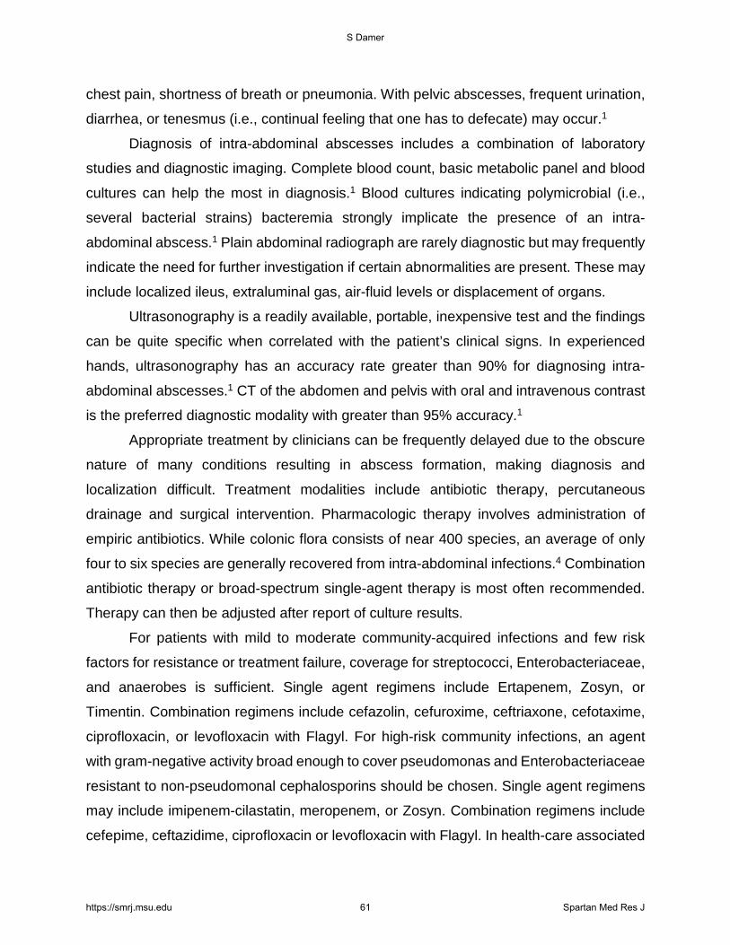
Spartan Med Res Jhttpssmrjmsuedu
S Damer
chest pain shortness of breath or pneumonia With pelvic abscesses frequent urination
diarrhea or tenesmus (ie continual feeling that one has to defecate) may occur1
Diagnosis of intra-abdominal abscesses includes a combination of laboratory
studies and diagnostic imaging Complete blood count basic metabolic panel and blood
cultures can help the most in diagnosis1 Blood cultures indicating polymicrobial (ie
several bacterial strains) bacteremia strongly implicate the presence of an intra-
abdominal abscess1 Plain abdominal radiograph are rarely diagnostic but may frequently
indicate the need for further investigation if certain abnormalities are present These may
include localized ileus extraluminal gas air-fluid levels or displacement of organs
Ultrasonography is a readily available portable inexpensive test and the findings
can be quite specific when correlated with the patientrsquos clinical signs In experienced
hands ultrasonography has an accuracy rate greater than 90 for diagnosing intra-
abdominal abscesses1 CT of the abdomen and pelvis with oral and intravenous contrast
is the preferred diagnostic modality with greater than 95 accuracy1
Appropriate treatment by clinicians can be frequently delayed due to the obscure
nature of many conditions resulting in abscess formation making diagnosis and
localization difficult Treatment modalities include antibiotic therapy percutaneous
drainage and surgical intervention Pharmacologic therapy involves administration of
empiric antibiotics While colonic flora consists of near 400 species an average of only
four to six species are generally recovered from intra-abdominal infections4 Combination
antibiotic therapy or broad-spectrum single-agent therapy is most often recommended
Therapy can then be adjusted after report of culture results
For patients with mild to moderate community-acquired infections and few risk
factors for resistance or treatment failure coverage for streptococci Enterobacteriaceae
and anaerobes is sufficient Single agent regimens include Ertapenem Zosyn or
Timentin Combination regimens include cefazolin cefuroxime ceftriaxone cefotaxime
ciprofloxacin or levofloxacin with Flagyl For high-risk community infections an agent
with gram-negative activity broad enough to cover pseudomonas and Enterobacteriaceae
resistant to non-pseudomonal cephalosporins should be chosen Single agent regimens
may include imipenem-cilastatin meropenem or Zosyn Combination regimens include
cefepime ceftazidime ciprofloxacin or levofloxacin with Flagyl In health-care associated
61
Vol 2 No2 Winter 2017
Pelvic Abscess with Presentation as Inability to Ambulate
infections with known colonization of methicillin-resistant Staphalococcus aureus
(MRSA) vancomycin should be added In patients who are known to be colonized by
highly-resistant organisms to include vancomycin-resistant enterococci (VRE) agents
such as linezolid and daptomycin should be used4
Physicians should often consider an infectious disease consult Percutaneous CT
guided catheter drainage is the standard treatment of most intra-abdominal abscesses2
Surgical drainage becomes mandatory when residual fluid cannot be evacuated with
catheter irrigation manipulation or additional drain placement
Intra-abdominal abscesses have a mortality rate of 10 to 405 Outcome depends
on the patientrsquos primary illness and general medical condition rather than on the specific
nature and location of the abscess Risk factors include multiple surgical procedures age
older than 50 years delay in initial intervention greater than 24 hours
immunocompromised conditions poor nutritional status multiple organ failure and
complex recurrent or persistent abscesses4 Multiple organ failure is a primary cause of
death4
Case Report A Caucasian male in his early 60s presented to the McLaren Macomb Emergency
Department (ED) with a complaint of bilateral hip pain and inability to ambulate The pain
had been present for the past four-to-five days and was progressive in nature The patient
admitted to being fairly active and normally able to ambulate without the assistance of a
cane or walker up until symptoms began He described his pain as 10 out of 10 constant
pain in the anterior bilateral hip joints with mild radiation into his lumbar back and groin
with no radiation into his legs His pain increased with ambulation but improved with rest
He attempted to take three doses of aspirin at home without any relief in his symptoms
and began using a walker to help him ambulate
The patient had an extensive medical history This history included sigmoid
diverticulitis with sigmoid colectomy in 2012 deep vein thrombosis duodenal ulcer
benign prostatic hypertrophy and hypertension He also suffered from chronic obstructive
pulmonary disease chronic thrombocytopenia peripheral vascular disease
atherosclerotic coronary artery disease and paroxysmal atrial fibrillation He had been
62
Spartan Med Res Jhttpssmrjmsuedu
S Damer
diagnosed with end stage renal disease and was on hemodialysis He subsequently had
multiple hemodialysis catheter infections with bacteremia (MRSA) obstructive uropathy
with chronic hydronephrosis and several peritoneal and pelvic abdominal abscesses He
denied having noticed any recent changes in his ostomy output
The patientrsquos past surgical history included inferior vena cava filter placement
cholecystectomy sigmoid colectomy with end colostomy numerous cystoscopies
bilateral ureteral stents peroneal abscess incision and drainage
esophagogastroduodenoscopy colonoscopy several IR drainages of pelvic abscesses
exploratory laparotomy with enterolysis temporary catheter placement and numerous
arteriovenous fistula creations
The patient took amiodarone Prilosec Imdur and aspirin He had no allergies to
any medications He denied use of nicotine alcohol or any illicit drugs He lived with and
took care of his elderly mother His father had died from a cerebral vascular accident and
his mother had a history of coronary artery disease
Physical exam revealed a well-nourished well-hydrated male who was in no acute
distress He had been brought from home via ambulance as ambulation was too painful
His vital signs demonstrated hypotension with a blood pressure of 7151 Patient admits
that this was his typical baseline His pain was worse on the right than the left He had
increased tenderness to log roll axial compression and passive flexion of the right hip
when compared to the left
He had intact two-point discrimination and light touch in the L2-S1 nerve
distributions bilaterally He had intact +2 out of 4 dorsalis pedis and posterior tibial pulses
found with Doppler ultrasound He elicited 5 out of 5 muscle strength in his bilateral lower
extremities There was no tenderness to palpation over the greater trochanteric region of
either hip He had a negative straight leg raise bilaterally The patient had severe brawny
edema and dryness to his lower extremities
Laboratory studies demonstrated a normocytic anemia chronic thrombocytopenia
hyponatremia and chronic kidney disease His sedimentation rate and c-reactive protein
levels were elevated When the patient was sent over for x-ray imaging left hip right hip
and pelvis x-rays demonstrated no abnormalities Lumbosacral x-ray demonstrated mild
scoliosis with diffuse moderate degenerative change (Image 1) The patient was offered
63
Vol 2 No2 Winter 2017
Pelvic Abscess with Presentation as Inability to Ambulate
pain medication but refused any other treatment He was admitted to the hospital due to
his intractable bilateral hip pain and inability to ambulate Orthopedics Nephrology and
Physical TherapyOccupational Therapy consults were placed
Orthopedics was the first service to evaluate the patient on hospital Day 2 and
ordered a CT of the abdomen and pelvis with contrast This study found that superior to
the bladder there was a 49 x 38 cm fluid collection with peripheral enhancement
concerning for abscess (Image 2) Internal Medicine then evaluated the patient and
reviewed the results of the CT scan Blood cultures were obtained and the General
Surgery service was consulted A repeat CT with oral contrast was ordered by General
Surgery on hospital Day 3 This test showed that the adjacent loops of the intestines along
the anterior and superior margin of the fluid collection were partially opacified without
obvious extravasation of oral contrast in the region suggesting fistula (ie
communication with the bowel) (Image 3)
The blood cultures grew out E coli x 2 after 24 hours The patient was placed on
Zosyn and vancomycin On hospital Day 5 Interventional Radiology performed CT
guidance drainage of the pelvic abscess with catheter placement Culture results would
later grow E coli The Urology service was also consulted as the patient had history of
hydronephrosis due to obstructive uropathy with bilateral stent placement This service
was asked to evaluate the need for stent replacement and integrity of the bladder with its
close approximation to the abscess
On hospital Day 6 the patient went to the operating room with Urology for a
bilateral retrograde urogram The study demonstrated findings consistent with
communication of the urinary bladder with the abscess cavity previously drained The
stents were exchanged a urinary catheter was placed and a urine sample was obtained
for which culture results came back positive for Ecoli The patient remained on Zosyn
As the patient continued to experience significant bilateral hip pain an MRI of the
patientrsquos thoraciclumbar spine and bilateral hips was ordered on Day 7 A thoracic spine
MRI demonstrated a small central disc protrusion at the Thoracic 4-T5 level No spinal
canal stenosis or foraminal encroachment The MRI of the lumbar spine and bilateral hips
was unable to be completed since the patient could not tolerate the pain long enough to
have the study performed
64
Spartan Med Res Jhttpssmrjmsuedu
S Damer
The pelvic drain was removed between hospital Days 9 and 11 although progress
notes are unclear as to the specific day On hospital Day 15 the patient was discharged
to a subacute rehabilitation unit on a two-week course of oral Augmentin He had worked
with Physical Therapy throughout his hospital stay The patient slowly regained some
ability to ambulate but it is unknown whether or not this was from resolving his pelvic
abscess or due to rehabilitation with Physical Therapy He was to follow up with the
urologist in two-to-three weeks for urinary catheter removal
DISCUSSION This case describes a patient who suffered from the inability to ambulate who was
eventually diagnosed with an intra-abdominal abscess This patient had presented to the
emergency department with bilateral hip pain which would be an unusual presenting
symptom for his diagnosed pathology As earlier described most common complaints
associated with an intra-abdominal abscess include abdominal paintenderness
distention nausea anorexia fever fast heart rate or high white blood cell count The
patient met none of these criteria Throughout the patientrsquos hospital stay he never had a
complaint related to his gastrointestinal system On Day 3 of his hospitalization he spiked
a white blood cell count but remained afebrile throughout his course The question arises
as to whether the patientrsquos complaint has any relation to the intra-abdominal abscess
found on CT imaging
After an in-depth review of the patientrsquos medical record it was revealed that he
had an extensive medical history The patient has suffered from diverticulitis with
perforation and abscess formation He had several drainages of the abscess completed
by an interventional radiologist prior to and after his sigmoid colectomy with end
ileostomy that was performed in September 2012
Throughout his multiple hospitalizations he had recurrence of an intraperitoneal
abscess that was located superior to the bladder and adjacent to the left iliopsoas muscle
Several studies showed altered attenuation of the iliopsoas muscle and evidence that the
abscess was contiguous with the muscle CT imaging conducted in December 2012
indicated that the abscess involved the left iliopsoas bursa It is this
65
Vol 2 No2 Winter 2017
Pelvic Abscess with Presentation as Inability to Ambulate
involvementextension for which the patientrsquos presenting complaint could possibly have
relation to his found pathology however no direct data concerning this existed
The psoas muscle arises from the transverse processes and lateral aspects of the
vertebral bodies between the 12th thoracic and 5th lumbar vertebrae It courses downward
passing deep to the inguinal ligament and anterior to the hip joint capsule to form a tendon
that inserts into the lesser trochanter of the femur The iliacus muscle joins the psoas to
insert via the same tendon These two muscles are the main hip flexors The tendon is
separated from the hip capsule by the iliopsoas bursa This bursa is the largest bursa in
the body and exists to help reduce rubbing between the iliopsoas muscle and the femur
This bursa communicates with the hip joint space in up to 15 of persons which may
facilitate spread of infection between these sites6
The iliopsoas muscle can be involved in several pathologic conditions to include
iliopsoas tendinitisbursitis iliopsoas syndrome and psoas abscesses Although
uncommon injuries iliopsoas tendinitisbursitis occurs when the tendon and bursa
becomes inflamed They are overuse injuries that result from overloading the hips with
repetitive movements7 People who participate in activities such as golf hockey
cheerleading gymnastics and resistance training are most susceptible to injury Iliopsoas
syndrome frequently begins as a bilateral muscle spasm which eventually becomes
prominent on one side
Psoas abscesses are a collection of pus in the iliopsoas muscle compartment
which may arise from contiguous spread from adjacent structures or by hematogenous
route from a distant site6 They are divided into primary and secondary abscesses
Primary abscess occurs as a result of hematogenous or lymphatic seeding Risk factors
include diabetes IV drug abuse HIV renal failure or other forms of immunosuppression
Secondary abscesses occur as a result of a direct spread of infection to the psoas muscle
from adjacent structures The structures include vertebrae hip arthroplasty GI tract
aorta and genitourinary tract
Symptoms of iliopsoas tendinitisbursitis and psoas syndromes include pain
tenderness swelling heat or redness and loss of normal mobility Signs of psoas abscess
include back or flank pain fever inguinal mass limp anorexia and weight loss Pain can
66
Spartan Med Res Jhttpssmrjmsuedu
S Damer
be present in up to 91 of cases with localization to the back flank or lower abdomen
and possible radiation to the hip andor posterior thigh6
Originally described in 1881 the classic clinical presentation of a psoas abscess
included back pain limp and fever8 Newer case studies have demonstrated that these
symptoms may only be present in 30 of cases8 Many patients present with nonspecific
complaints such as malaise low grade fever abdominalflank discomfort a flexed and
externally rotated hip and pain on movement of the hip Pain is due to irritation of muscle
belly and referred pain from nerve roots L2-L4 Due to the vague and nonspecific
presenting symptoms of a psoas abscess they are commonly misdiagnosed although
data concerning the misdiagnosis rate is sparse8 One case studied reported a psoas
abscess that was previously misdiagnosed as a deep vein thrombosis9
After review of the presenting symptoms of iliopsoas bursitistendonitis syndrome
and psoas abscess the clinical conclusion can be made that this patient may have been
suffering from referred pain to the hip from his pelvic abscess that abutted the left
iliopsoas muscle This is a speculation since there are no direct reports available
concerning a direct correlation between this manrsquos pelvic abscesses and hip pain
However several discrepancies arose during his workup The patient had suffered
from right hip pain yet the patientrsquos abscess directly invaded the left iliopsoas muscle He
also had more pain when asked to perform hip flexion and preferred to lay flat on his back
without axial loading Research has found a correlation with patients suffering from psoas
abscesses and that they prefer to lay with their hips flexed6 In addition the patient had
presented several times in the past for recurrent pelvic abscess and failed to complain of
hip pain during those admissions Finally the patient may have had pathology of his
lumbar spine or hips that could have been identified if the patient had been able to tolerate
the MRI imaging procedure
In support of the theory of the patientrsquos suffering from referred pain the patient did
experience pain upon movement of the hip and had improvement in his symptoms after
IR drainage and antibiotic administration Hip pain especially in Crohnrsquos patients should
prompt consideration of a psoas abscess as the incidence has been estimated to be
between 04 and 436 Psoas abscess has also been described in the setting of
appendicitis colorectal cancer ulcerative colitis and following abdominal surgery so
67
Vol 2 No2 Winter 2017
Pelvic Abscess with Presentation as Inability to Ambulate
evidence exists with other GI pathology While a question was made during the patientrsquos
hospital stay as to whether his symptomatology was somehow related to his pathology
versus incidental the author does not believe the theory of referred pain is far-fetched
CONCLUSIONS This report presents a case of a Caucasian male in his early 60s who presented
to the emergency department with complaint of bilateral hip pain and inability to ambulate
He was admitted to the hospital and found to have a recurrent pelvic abscess which
extended to his left iliopsoas muscle The patient failed to demonstrate the typical
symptoms of a pelvic abscess and therefore his pathology was not revealed initially in the
emergency department It was not until the patient had been hospitalized for a day and
his medical record had been thoroughly reviewed when his diagnosis was made Even
then his presenting complaints could not be fully explained Additional imaging studies
failed to demonstrate any intrinsic pathology to the hips themselves
This case demonstrated an atypical presentation of a pelvic abscess but questions
remained as to whether the patientrsquos symptoms were due to referred pain to the hips
Conditions such as iliopsoas tendinitisbursitis and psoas syndromes present with pain
tenderness swelling heat or redness and loss of normal mobility while signs of a psoas
abscess include back or flank pain fever inguinal mass limp anorexia and weight loss
These clinical findings are more consistent with what the patient in the case study had
presented to providers Further investigations are required to determine the percentage
of patients with pelvic abscesses who initially present with a component of hip pain
The authors report no external funding source for this study
The authors declare no conflict of interest
Submitted for publication June 2017 Accepted for publication December 2017
68
Spartan Med Res Jhttpssmrjmsuedu
Rl
S Damer
ACKNOWLEDGEMENTS Julie Lata DO revised this case report critically for important intellectual content
and gave final pre-publication approval of this final article draft
REFERENCES 1 Saber AA Abdominal Abscess Medscape 2016 Jun 21 2017 Oct 1 Available from httpemedicinemedscapecomarticle1979032-overviewa5
2 Ansari P Intra-abdominal Abscesses Merck Manual 2017 Jan 2017 Oct 1 Available from httpswwwmerckmanualscomprofessionalgastrointestinal-disordersacute-abdomen-and-surgical-gastroenterologyintra-abdominal-abscessesv891088
3 De Koster K Referred Pain Physiopedia 2017 Oct 1 Available from httpswwwphysio-pediacomReferred_Pain
4 Barshak M Antimicrobial Approach to Intra-abdominal Infections in Adults Up to Date 2016 Mar 14 2017 Oct 1 Available from httpwwwuptodatecomcontentsantimicrobial-approach-to-intra-abdominal-infections-in-adults
5 Daley B Peritonitis and Abdominal Sepsis Medscape 2017 Jan 11 2017 Oct 1 Available from httpemedicinemedscapecomarticle180234-overview
6 Spelman D Psoas abscess Up to Date 2016 May 9 2017 Oct 1 Available from httpswwwuptodatecomcontentspsoas-abscess
7 Tufo A Psoas syndrome A frequently missed diagnosis J Amer Osteo Assoc 2012112(1)522-528
8 Shields D Robinson P Crowley TP Iliopsoas abscess ndash A review and update on the literature Intl J Surg 201210 (1)466-469
9 Kong V Oosthuizen G Mthethwa M Reddy K Clarke D Delayed presentation of a psoas abscess previously misdiagnosed as deep vein thrombosis A potentially devastating tumor Intl J Case Rep Images 20134(2)111-114
69
Vol 2 No2 Winter 2017
Pelvic Abscess with Presentation as Inability to Ambulate
TABLES AND FIGURES
Image 1 Pelvis X-ray
70
Spartan Med Res Jhttpssmrjmsuedu
S Damer
Image 2 Fluid Collection - 49 x 38 cm
71
Vol 2 No2 Winter 2017
Pelvic Abscess with Presentation as Inability to Ambulate
Image 3 Pelvic Abscess without Signs of Communication with Bowel
72

Vol 2 No2 Winter 2017
Pelvic Abscess with Presentation as Inability to Ambulate
infections with known colonization of methicillin-resistant Staphalococcus aureus
(MRSA) vancomycin should be added In patients who are known to be colonized by
highly-resistant organisms to include vancomycin-resistant enterococci (VRE) agents
such as linezolid and daptomycin should be used4
Physicians should often consider an infectious disease consult Percutaneous CT
guided catheter drainage is the standard treatment of most intra-abdominal abscesses2
Surgical drainage becomes mandatory when residual fluid cannot be evacuated with
catheter irrigation manipulation or additional drain placement
Intra-abdominal abscesses have a mortality rate of 10 to 405 Outcome depends
on the patientrsquos primary illness and general medical condition rather than on the specific
nature and location of the abscess Risk factors include multiple surgical procedures age
older than 50 years delay in initial intervention greater than 24 hours
immunocompromised conditions poor nutritional status multiple organ failure and
complex recurrent or persistent abscesses4 Multiple organ failure is a primary cause of
death4
Case Report A Caucasian male in his early 60s presented to the McLaren Macomb Emergency
Department (ED) with a complaint of bilateral hip pain and inability to ambulate The pain
had been present for the past four-to-five days and was progressive in nature The patient
admitted to being fairly active and normally able to ambulate without the assistance of a
cane or walker up until symptoms began He described his pain as 10 out of 10 constant
pain in the anterior bilateral hip joints with mild radiation into his lumbar back and groin
with no radiation into his legs His pain increased with ambulation but improved with rest
He attempted to take three doses of aspirin at home without any relief in his symptoms
and began using a walker to help him ambulate
The patient had an extensive medical history This history included sigmoid
diverticulitis with sigmoid colectomy in 2012 deep vein thrombosis duodenal ulcer
benign prostatic hypertrophy and hypertension He also suffered from chronic obstructive
pulmonary disease chronic thrombocytopenia peripheral vascular disease
atherosclerotic coronary artery disease and paroxysmal atrial fibrillation He had been
62
Spartan Med Res Jhttpssmrjmsuedu
S Damer
diagnosed with end stage renal disease and was on hemodialysis He subsequently had
multiple hemodialysis catheter infections with bacteremia (MRSA) obstructive uropathy
with chronic hydronephrosis and several peritoneal and pelvic abdominal abscesses He
denied having noticed any recent changes in his ostomy output
The patientrsquos past surgical history included inferior vena cava filter placement
cholecystectomy sigmoid colectomy with end colostomy numerous cystoscopies
bilateral ureteral stents peroneal abscess incision and drainage
esophagogastroduodenoscopy colonoscopy several IR drainages of pelvic abscesses
exploratory laparotomy with enterolysis temporary catheter placement and numerous
arteriovenous fistula creations
The patient took amiodarone Prilosec Imdur and aspirin He had no allergies to
any medications He denied use of nicotine alcohol or any illicit drugs He lived with and
took care of his elderly mother His father had died from a cerebral vascular accident and
his mother had a history of coronary artery disease
Physical exam revealed a well-nourished well-hydrated male who was in no acute
distress He had been brought from home via ambulance as ambulation was too painful
His vital signs demonstrated hypotension with a blood pressure of 7151 Patient admits
that this was his typical baseline His pain was worse on the right than the left He had
increased tenderness to log roll axial compression and passive flexion of the right hip
when compared to the left
He had intact two-point discrimination and light touch in the L2-S1 nerve
distributions bilaterally He had intact +2 out of 4 dorsalis pedis and posterior tibial pulses
found with Doppler ultrasound He elicited 5 out of 5 muscle strength in his bilateral lower
extremities There was no tenderness to palpation over the greater trochanteric region of
either hip He had a negative straight leg raise bilaterally The patient had severe brawny
edema and dryness to his lower extremities
Laboratory studies demonstrated a normocytic anemia chronic thrombocytopenia
hyponatremia and chronic kidney disease His sedimentation rate and c-reactive protein
levels were elevated When the patient was sent over for x-ray imaging left hip right hip
and pelvis x-rays demonstrated no abnormalities Lumbosacral x-ray demonstrated mild
scoliosis with diffuse moderate degenerative change (Image 1) The patient was offered
63
Vol 2 No2 Winter 2017
Pelvic Abscess with Presentation as Inability to Ambulate
pain medication but refused any other treatment He was admitted to the hospital due to
his intractable bilateral hip pain and inability to ambulate Orthopedics Nephrology and
Physical TherapyOccupational Therapy consults were placed
Orthopedics was the first service to evaluate the patient on hospital Day 2 and
ordered a CT of the abdomen and pelvis with contrast This study found that superior to
the bladder there was a 49 x 38 cm fluid collection with peripheral enhancement
concerning for abscess (Image 2) Internal Medicine then evaluated the patient and
reviewed the results of the CT scan Blood cultures were obtained and the General
Surgery service was consulted A repeat CT with oral contrast was ordered by General
Surgery on hospital Day 3 This test showed that the adjacent loops of the intestines along
the anterior and superior margin of the fluid collection were partially opacified without
obvious extravasation of oral contrast in the region suggesting fistula (ie
communication with the bowel) (Image 3)
The blood cultures grew out E coli x 2 after 24 hours The patient was placed on
Zosyn and vancomycin On hospital Day 5 Interventional Radiology performed CT
guidance drainage of the pelvic abscess with catheter placement Culture results would
later grow E coli The Urology service was also consulted as the patient had history of
hydronephrosis due to obstructive uropathy with bilateral stent placement This service
was asked to evaluate the need for stent replacement and integrity of the bladder with its
close approximation to the abscess
On hospital Day 6 the patient went to the operating room with Urology for a
bilateral retrograde urogram The study demonstrated findings consistent with
communication of the urinary bladder with the abscess cavity previously drained The
stents were exchanged a urinary catheter was placed and a urine sample was obtained
for which culture results came back positive for Ecoli The patient remained on Zosyn
As the patient continued to experience significant bilateral hip pain an MRI of the
patientrsquos thoraciclumbar spine and bilateral hips was ordered on Day 7 A thoracic spine
MRI demonstrated a small central disc protrusion at the Thoracic 4-T5 level No spinal
canal stenosis or foraminal encroachment The MRI of the lumbar spine and bilateral hips
was unable to be completed since the patient could not tolerate the pain long enough to
have the study performed
64
Spartan Med Res Jhttpssmrjmsuedu
S Damer
The pelvic drain was removed between hospital Days 9 and 11 although progress
notes are unclear as to the specific day On hospital Day 15 the patient was discharged
to a subacute rehabilitation unit on a two-week course of oral Augmentin He had worked
with Physical Therapy throughout his hospital stay The patient slowly regained some
ability to ambulate but it is unknown whether or not this was from resolving his pelvic
abscess or due to rehabilitation with Physical Therapy He was to follow up with the
urologist in two-to-three weeks for urinary catheter removal
DISCUSSION This case describes a patient who suffered from the inability to ambulate who was
eventually diagnosed with an intra-abdominal abscess This patient had presented to the
emergency department with bilateral hip pain which would be an unusual presenting
symptom for his diagnosed pathology As earlier described most common complaints
associated with an intra-abdominal abscess include abdominal paintenderness
distention nausea anorexia fever fast heart rate or high white blood cell count The
patient met none of these criteria Throughout the patientrsquos hospital stay he never had a
complaint related to his gastrointestinal system On Day 3 of his hospitalization he spiked
a white blood cell count but remained afebrile throughout his course The question arises
as to whether the patientrsquos complaint has any relation to the intra-abdominal abscess
found on CT imaging
After an in-depth review of the patientrsquos medical record it was revealed that he
had an extensive medical history The patient has suffered from diverticulitis with
perforation and abscess formation He had several drainages of the abscess completed
by an interventional radiologist prior to and after his sigmoid colectomy with end
ileostomy that was performed in September 2012
Throughout his multiple hospitalizations he had recurrence of an intraperitoneal
abscess that was located superior to the bladder and adjacent to the left iliopsoas muscle
Several studies showed altered attenuation of the iliopsoas muscle and evidence that the
abscess was contiguous with the muscle CT imaging conducted in December 2012
indicated that the abscess involved the left iliopsoas bursa It is this
65
Vol 2 No2 Winter 2017
Pelvic Abscess with Presentation as Inability to Ambulate
involvementextension for which the patientrsquos presenting complaint could possibly have
relation to his found pathology however no direct data concerning this existed
The psoas muscle arises from the transverse processes and lateral aspects of the
vertebral bodies between the 12th thoracic and 5th lumbar vertebrae It courses downward
passing deep to the inguinal ligament and anterior to the hip joint capsule to form a tendon
that inserts into the lesser trochanter of the femur The iliacus muscle joins the psoas to
insert via the same tendon These two muscles are the main hip flexors The tendon is
separated from the hip capsule by the iliopsoas bursa This bursa is the largest bursa in
the body and exists to help reduce rubbing between the iliopsoas muscle and the femur
This bursa communicates with the hip joint space in up to 15 of persons which may
facilitate spread of infection between these sites6
The iliopsoas muscle can be involved in several pathologic conditions to include
iliopsoas tendinitisbursitis iliopsoas syndrome and psoas abscesses Although
uncommon injuries iliopsoas tendinitisbursitis occurs when the tendon and bursa
becomes inflamed They are overuse injuries that result from overloading the hips with
repetitive movements7 People who participate in activities such as golf hockey
cheerleading gymnastics and resistance training are most susceptible to injury Iliopsoas
syndrome frequently begins as a bilateral muscle spasm which eventually becomes
prominent on one side
Psoas abscesses are a collection of pus in the iliopsoas muscle compartment
which may arise from contiguous spread from adjacent structures or by hematogenous
route from a distant site6 They are divided into primary and secondary abscesses
Primary abscess occurs as a result of hematogenous or lymphatic seeding Risk factors
include diabetes IV drug abuse HIV renal failure or other forms of immunosuppression
Secondary abscesses occur as a result of a direct spread of infection to the psoas muscle
from adjacent structures The structures include vertebrae hip arthroplasty GI tract
aorta and genitourinary tract
Symptoms of iliopsoas tendinitisbursitis and psoas syndromes include pain
tenderness swelling heat or redness and loss of normal mobility Signs of psoas abscess
include back or flank pain fever inguinal mass limp anorexia and weight loss Pain can
66
Spartan Med Res Jhttpssmrjmsuedu
S Damer
be present in up to 91 of cases with localization to the back flank or lower abdomen
and possible radiation to the hip andor posterior thigh6
Originally described in 1881 the classic clinical presentation of a psoas abscess
included back pain limp and fever8 Newer case studies have demonstrated that these
symptoms may only be present in 30 of cases8 Many patients present with nonspecific
complaints such as malaise low grade fever abdominalflank discomfort a flexed and
externally rotated hip and pain on movement of the hip Pain is due to irritation of muscle
belly and referred pain from nerve roots L2-L4 Due to the vague and nonspecific
presenting symptoms of a psoas abscess they are commonly misdiagnosed although
data concerning the misdiagnosis rate is sparse8 One case studied reported a psoas
abscess that was previously misdiagnosed as a deep vein thrombosis9
After review of the presenting symptoms of iliopsoas bursitistendonitis syndrome
and psoas abscess the clinical conclusion can be made that this patient may have been
suffering from referred pain to the hip from his pelvic abscess that abutted the left
iliopsoas muscle This is a speculation since there are no direct reports available
concerning a direct correlation between this manrsquos pelvic abscesses and hip pain
However several discrepancies arose during his workup The patient had suffered
from right hip pain yet the patientrsquos abscess directly invaded the left iliopsoas muscle He
also had more pain when asked to perform hip flexion and preferred to lay flat on his back
without axial loading Research has found a correlation with patients suffering from psoas
abscesses and that they prefer to lay with their hips flexed6 In addition the patient had
presented several times in the past for recurrent pelvic abscess and failed to complain of
hip pain during those admissions Finally the patient may have had pathology of his
lumbar spine or hips that could have been identified if the patient had been able to tolerate
the MRI imaging procedure
In support of the theory of the patientrsquos suffering from referred pain the patient did
experience pain upon movement of the hip and had improvement in his symptoms after
IR drainage and antibiotic administration Hip pain especially in Crohnrsquos patients should
prompt consideration of a psoas abscess as the incidence has been estimated to be
between 04 and 436 Psoas abscess has also been described in the setting of
appendicitis colorectal cancer ulcerative colitis and following abdominal surgery so
67
Vol 2 No2 Winter 2017
Pelvic Abscess with Presentation as Inability to Ambulate
evidence exists with other GI pathology While a question was made during the patientrsquos
hospital stay as to whether his symptomatology was somehow related to his pathology
versus incidental the author does not believe the theory of referred pain is far-fetched
CONCLUSIONS This report presents a case of a Caucasian male in his early 60s who presented
to the emergency department with complaint of bilateral hip pain and inability to ambulate
He was admitted to the hospital and found to have a recurrent pelvic abscess which
extended to his left iliopsoas muscle The patient failed to demonstrate the typical
symptoms of a pelvic abscess and therefore his pathology was not revealed initially in the
emergency department It was not until the patient had been hospitalized for a day and
his medical record had been thoroughly reviewed when his diagnosis was made Even
then his presenting complaints could not be fully explained Additional imaging studies
failed to demonstrate any intrinsic pathology to the hips themselves
This case demonstrated an atypical presentation of a pelvic abscess but questions
remained as to whether the patientrsquos symptoms were due to referred pain to the hips
Conditions such as iliopsoas tendinitisbursitis and psoas syndromes present with pain
tenderness swelling heat or redness and loss of normal mobility while signs of a psoas
abscess include back or flank pain fever inguinal mass limp anorexia and weight loss
These clinical findings are more consistent with what the patient in the case study had
presented to providers Further investigations are required to determine the percentage
of patients with pelvic abscesses who initially present with a component of hip pain
The authors report no external funding source for this study
The authors declare no conflict of interest
Submitted for publication June 2017 Accepted for publication December 2017
68
Spartan Med Res Jhttpssmrjmsuedu
Rl
S Damer
ACKNOWLEDGEMENTS Julie Lata DO revised this case report critically for important intellectual content
and gave final pre-publication approval of this final article draft
REFERENCES 1 Saber AA Abdominal Abscess Medscape 2016 Jun 21 2017 Oct 1 Available from httpemedicinemedscapecomarticle1979032-overviewa5
2 Ansari P Intra-abdominal Abscesses Merck Manual 2017 Jan 2017 Oct 1 Available from httpswwwmerckmanualscomprofessionalgastrointestinal-disordersacute-abdomen-and-surgical-gastroenterologyintra-abdominal-abscessesv891088
3 De Koster K Referred Pain Physiopedia 2017 Oct 1 Available from httpswwwphysio-pediacomReferred_Pain
4 Barshak M Antimicrobial Approach to Intra-abdominal Infections in Adults Up to Date 2016 Mar 14 2017 Oct 1 Available from httpwwwuptodatecomcontentsantimicrobial-approach-to-intra-abdominal-infections-in-adults
5 Daley B Peritonitis and Abdominal Sepsis Medscape 2017 Jan 11 2017 Oct 1 Available from httpemedicinemedscapecomarticle180234-overview
6 Spelman D Psoas abscess Up to Date 2016 May 9 2017 Oct 1 Available from httpswwwuptodatecomcontentspsoas-abscess
7 Tufo A Psoas syndrome A frequently missed diagnosis J Amer Osteo Assoc 2012112(1)522-528
8 Shields D Robinson P Crowley TP Iliopsoas abscess ndash A review and update on the literature Intl J Surg 201210 (1)466-469
9 Kong V Oosthuizen G Mthethwa M Reddy K Clarke D Delayed presentation of a psoas abscess previously misdiagnosed as deep vein thrombosis A potentially devastating tumor Intl J Case Rep Images 20134(2)111-114
69
Vol 2 No2 Winter 2017
Pelvic Abscess with Presentation as Inability to Ambulate
TABLES AND FIGURES
Image 1 Pelvis X-ray
70
Spartan Med Res Jhttpssmrjmsuedu
S Damer
Image 2 Fluid Collection - 49 x 38 cm
71
Vol 2 No2 Winter 2017
Pelvic Abscess with Presentation as Inability to Ambulate
Image 3 Pelvic Abscess without Signs of Communication with Bowel
72

Spartan Med Res Jhttpssmrjmsuedu
S Damer
diagnosed with end stage renal disease and was on hemodialysis He subsequently had
multiple hemodialysis catheter infections with bacteremia (MRSA) obstructive uropathy
with chronic hydronephrosis and several peritoneal and pelvic abdominal abscesses He
denied having noticed any recent changes in his ostomy output
The patientrsquos past surgical history included inferior vena cava filter placement
cholecystectomy sigmoid colectomy with end colostomy numerous cystoscopies
bilateral ureteral stents peroneal abscess incision and drainage
esophagogastroduodenoscopy colonoscopy several IR drainages of pelvic abscesses
exploratory laparotomy with enterolysis temporary catheter placement and numerous
arteriovenous fistula creations
The patient took amiodarone Prilosec Imdur and aspirin He had no allergies to
any medications He denied use of nicotine alcohol or any illicit drugs He lived with and
took care of his elderly mother His father had died from a cerebral vascular accident and
his mother had a history of coronary artery disease
Physical exam revealed a well-nourished well-hydrated male who was in no acute
distress He had been brought from home via ambulance as ambulation was too painful
His vital signs demonstrated hypotension with a blood pressure of 7151 Patient admits
that this was his typical baseline His pain was worse on the right than the left He had
increased tenderness to log roll axial compression and passive flexion of the right hip
when compared to the left
He had intact two-point discrimination and light touch in the L2-S1 nerve
distributions bilaterally He had intact +2 out of 4 dorsalis pedis and posterior tibial pulses
found with Doppler ultrasound He elicited 5 out of 5 muscle strength in his bilateral lower
extremities There was no tenderness to palpation over the greater trochanteric region of
either hip He had a negative straight leg raise bilaterally The patient had severe brawny
edema and dryness to his lower extremities
Laboratory studies demonstrated a normocytic anemia chronic thrombocytopenia
hyponatremia and chronic kidney disease His sedimentation rate and c-reactive protein
levels were elevated When the patient was sent over for x-ray imaging left hip right hip
and pelvis x-rays demonstrated no abnormalities Lumbosacral x-ray demonstrated mild
scoliosis with diffuse moderate degenerative change (Image 1) The patient was offered
63
Vol 2 No2 Winter 2017
Pelvic Abscess with Presentation as Inability to Ambulate
pain medication but refused any other treatment He was admitted to the hospital due to
his intractable bilateral hip pain and inability to ambulate Orthopedics Nephrology and
Physical TherapyOccupational Therapy consults were placed
Orthopedics was the first service to evaluate the patient on hospital Day 2 and
ordered a CT of the abdomen and pelvis with contrast This study found that superior to
the bladder there was a 49 x 38 cm fluid collection with peripheral enhancement
concerning for abscess (Image 2) Internal Medicine then evaluated the patient and
reviewed the results of the CT scan Blood cultures were obtained and the General
Surgery service was consulted A repeat CT with oral contrast was ordered by General
Surgery on hospital Day 3 This test showed that the adjacent loops of the intestines along
the anterior and superior margin of the fluid collection were partially opacified without
obvious extravasation of oral contrast in the region suggesting fistula (ie
communication with the bowel) (Image 3)
The blood cultures grew out E coli x 2 after 24 hours The patient was placed on
Zosyn and vancomycin On hospital Day 5 Interventional Radiology performed CT
guidance drainage of the pelvic abscess with catheter placement Culture results would
later grow E coli The Urology service was also consulted as the patient had history of
hydronephrosis due to obstructive uropathy with bilateral stent placement This service
was asked to evaluate the need for stent replacement and integrity of the bladder with its
close approximation to the abscess
On hospital Day 6 the patient went to the operating room with Urology for a
bilateral retrograde urogram The study demonstrated findings consistent with
communication of the urinary bladder with the abscess cavity previously drained The
stents were exchanged a urinary catheter was placed and a urine sample was obtained
for which culture results came back positive for Ecoli The patient remained on Zosyn
As the patient continued to experience significant bilateral hip pain an MRI of the
patientrsquos thoraciclumbar spine and bilateral hips was ordered on Day 7 A thoracic spine
MRI demonstrated a small central disc protrusion at the Thoracic 4-T5 level No spinal
canal stenosis or foraminal encroachment The MRI of the lumbar spine and bilateral hips
was unable to be completed since the patient could not tolerate the pain long enough to
have the study performed
64
Spartan Med Res Jhttpssmrjmsuedu
S Damer
The pelvic drain was removed between hospital Days 9 and 11 although progress
notes are unclear as to the specific day On hospital Day 15 the patient was discharged
to a subacute rehabilitation unit on a two-week course of oral Augmentin He had worked
with Physical Therapy throughout his hospital stay The patient slowly regained some
ability to ambulate but it is unknown whether or not this was from resolving his pelvic
abscess or due to rehabilitation with Physical Therapy He was to follow up with the
urologist in two-to-three weeks for urinary catheter removal
DISCUSSION This case describes a patient who suffered from the inability to ambulate who was
eventually diagnosed with an intra-abdominal abscess This patient had presented to the
emergency department with bilateral hip pain which would be an unusual presenting
symptom for his diagnosed pathology As earlier described most common complaints
associated with an intra-abdominal abscess include abdominal paintenderness
distention nausea anorexia fever fast heart rate or high white blood cell count The
patient met none of these criteria Throughout the patientrsquos hospital stay he never had a
complaint related to his gastrointestinal system On Day 3 of his hospitalization he spiked
a white blood cell count but remained afebrile throughout his course The question arises
as to whether the patientrsquos complaint has any relation to the intra-abdominal abscess
found on CT imaging
After an in-depth review of the patientrsquos medical record it was revealed that he
had an extensive medical history The patient has suffered from diverticulitis with
perforation and abscess formation He had several drainages of the abscess completed
by an interventional radiologist prior to and after his sigmoid colectomy with end
ileostomy that was performed in September 2012
Throughout his multiple hospitalizations he had recurrence of an intraperitoneal
abscess that was located superior to the bladder and adjacent to the left iliopsoas muscle
Several studies showed altered attenuation of the iliopsoas muscle and evidence that the
abscess was contiguous with the muscle CT imaging conducted in December 2012
indicated that the abscess involved the left iliopsoas bursa It is this
65
Vol 2 No2 Winter 2017
Pelvic Abscess with Presentation as Inability to Ambulate
involvementextension for which the patientrsquos presenting complaint could possibly have
relation to his found pathology however no direct data concerning this existed
The psoas muscle arises from the transverse processes and lateral aspects of the
vertebral bodies between the 12th thoracic and 5th lumbar vertebrae It courses downward
passing deep to the inguinal ligament and anterior to the hip joint capsule to form a tendon
that inserts into the lesser trochanter of the femur The iliacus muscle joins the psoas to
insert via the same tendon These two muscles are the main hip flexors The tendon is
separated from the hip capsule by the iliopsoas bursa This bursa is the largest bursa in
the body and exists to help reduce rubbing between the iliopsoas muscle and the femur
This bursa communicates with the hip joint space in up to 15 of persons which may
facilitate spread of infection between these sites6
The iliopsoas muscle can be involved in several pathologic conditions to include
iliopsoas tendinitisbursitis iliopsoas syndrome and psoas abscesses Although
uncommon injuries iliopsoas tendinitisbursitis occurs when the tendon and bursa
becomes inflamed They are overuse injuries that result from overloading the hips with
repetitive movements7 People who participate in activities such as golf hockey
cheerleading gymnastics and resistance training are most susceptible to injury Iliopsoas
syndrome frequently begins as a bilateral muscle spasm which eventually becomes
prominent on one side
Psoas abscesses are a collection of pus in the iliopsoas muscle compartment
which may arise from contiguous spread from adjacent structures or by hematogenous
route from a distant site6 They are divided into primary and secondary abscesses
Primary abscess occurs as a result of hematogenous or lymphatic seeding Risk factors
include diabetes IV drug abuse HIV renal failure or other forms of immunosuppression
Secondary abscesses occur as a result of a direct spread of infection to the psoas muscle
from adjacent structures The structures include vertebrae hip arthroplasty GI tract
aorta and genitourinary tract
Symptoms of iliopsoas tendinitisbursitis and psoas syndromes include pain
tenderness swelling heat or redness and loss of normal mobility Signs of psoas abscess
include back or flank pain fever inguinal mass limp anorexia and weight loss Pain can
66
Spartan Med Res Jhttpssmrjmsuedu
S Damer
be present in up to 91 of cases with localization to the back flank or lower abdomen
and possible radiation to the hip andor posterior thigh6
Originally described in 1881 the classic clinical presentation of a psoas abscess
included back pain limp and fever8 Newer case studies have demonstrated that these
symptoms may only be present in 30 of cases8 Many patients present with nonspecific
complaints such as malaise low grade fever abdominalflank discomfort a flexed and
externally rotated hip and pain on movement of the hip Pain is due to irritation of muscle
belly and referred pain from nerve roots L2-L4 Due to the vague and nonspecific
presenting symptoms of a psoas abscess they are commonly misdiagnosed although
data concerning the misdiagnosis rate is sparse8 One case studied reported a psoas
abscess that was previously misdiagnosed as a deep vein thrombosis9
After review of the presenting symptoms of iliopsoas bursitistendonitis syndrome
and psoas abscess the clinical conclusion can be made that this patient may have been
suffering from referred pain to the hip from his pelvic abscess that abutted the left
iliopsoas muscle This is a speculation since there are no direct reports available
concerning a direct correlation between this manrsquos pelvic abscesses and hip pain
However several discrepancies arose during his workup The patient had suffered
from right hip pain yet the patientrsquos abscess directly invaded the left iliopsoas muscle He
also had more pain when asked to perform hip flexion and preferred to lay flat on his back
without axial loading Research has found a correlation with patients suffering from psoas
abscesses and that they prefer to lay with their hips flexed6 In addition the patient had
presented several times in the past for recurrent pelvic abscess and failed to complain of
hip pain during those admissions Finally the patient may have had pathology of his
lumbar spine or hips that could have been identified if the patient had been able to tolerate
the MRI imaging procedure
In support of the theory of the patientrsquos suffering from referred pain the patient did
experience pain upon movement of the hip and had improvement in his symptoms after
IR drainage and antibiotic administration Hip pain especially in Crohnrsquos patients should
prompt consideration of a psoas abscess as the incidence has been estimated to be
between 04 and 436 Psoas abscess has also been described in the setting of
appendicitis colorectal cancer ulcerative colitis and following abdominal surgery so
67
Vol 2 No2 Winter 2017
Pelvic Abscess with Presentation as Inability to Ambulate
evidence exists with other GI pathology While a question was made during the patientrsquos
hospital stay as to whether his symptomatology was somehow related to his pathology
versus incidental the author does not believe the theory of referred pain is far-fetched
CONCLUSIONS This report presents a case of a Caucasian male in his early 60s who presented
to the emergency department with complaint of bilateral hip pain and inability to ambulate
He was admitted to the hospital and found to have a recurrent pelvic abscess which
extended to his left iliopsoas muscle The patient failed to demonstrate the typical
symptoms of a pelvic abscess and therefore his pathology was not revealed initially in the
emergency department It was not until the patient had been hospitalized for a day and
his medical record had been thoroughly reviewed when his diagnosis was made Even
then his presenting complaints could not be fully explained Additional imaging studies
failed to demonstrate any intrinsic pathology to the hips themselves
This case demonstrated an atypical presentation of a pelvic abscess but questions
remained as to whether the patientrsquos symptoms were due to referred pain to the hips
Conditions such as iliopsoas tendinitisbursitis and psoas syndromes present with pain
tenderness swelling heat or redness and loss of normal mobility while signs of a psoas
abscess include back or flank pain fever inguinal mass limp anorexia and weight loss
These clinical findings are more consistent with what the patient in the case study had
presented to providers Further investigations are required to determine the percentage
of patients with pelvic abscesses who initially present with a component of hip pain
The authors report no external funding source for this study
The authors declare no conflict of interest
Submitted for publication June 2017 Accepted for publication December 2017
68
Spartan Med Res Jhttpssmrjmsuedu
Rl
S Damer
ACKNOWLEDGEMENTS Julie Lata DO revised this case report critically for important intellectual content
and gave final pre-publication approval of this final article draft
REFERENCES 1 Saber AA Abdominal Abscess Medscape 2016 Jun 21 2017 Oct 1 Available from httpemedicinemedscapecomarticle1979032-overviewa5
2 Ansari P Intra-abdominal Abscesses Merck Manual 2017 Jan 2017 Oct 1 Available from httpswwwmerckmanualscomprofessionalgastrointestinal-disordersacute-abdomen-and-surgical-gastroenterologyintra-abdominal-abscessesv891088
3 De Koster K Referred Pain Physiopedia 2017 Oct 1 Available from httpswwwphysio-pediacomReferred_Pain
4 Barshak M Antimicrobial Approach to Intra-abdominal Infections in Adults Up to Date 2016 Mar 14 2017 Oct 1 Available from httpwwwuptodatecomcontentsantimicrobial-approach-to-intra-abdominal-infections-in-adults
5 Daley B Peritonitis and Abdominal Sepsis Medscape 2017 Jan 11 2017 Oct 1 Available from httpemedicinemedscapecomarticle180234-overview
6 Spelman D Psoas abscess Up to Date 2016 May 9 2017 Oct 1 Available from httpswwwuptodatecomcontentspsoas-abscess
7 Tufo A Psoas syndrome A frequently missed diagnosis J Amer Osteo Assoc 2012112(1)522-528
8 Shields D Robinson P Crowley TP Iliopsoas abscess ndash A review and update on the literature Intl J Surg 201210 (1)466-469
9 Kong V Oosthuizen G Mthethwa M Reddy K Clarke D Delayed presentation of a psoas abscess previously misdiagnosed as deep vein thrombosis A potentially devastating tumor Intl J Case Rep Images 20134(2)111-114
69
Vol 2 No2 Winter 2017
Pelvic Abscess with Presentation as Inability to Ambulate
TABLES AND FIGURES
Image 1 Pelvis X-ray
70
Spartan Med Res Jhttpssmrjmsuedu
S Damer
Image 2 Fluid Collection - 49 x 38 cm
71
Vol 2 No2 Winter 2017
Pelvic Abscess with Presentation as Inability to Ambulate
Image 3 Pelvic Abscess without Signs of Communication with Bowel
72
Vol 2 No2 Winter 2017
Pelvic Abscess with Presentation as Inability to Ambulate
pain medication but refused any other treatment He was admitted to the hospital due to
his intractable bilateral hip pain and inability to ambulate Orthopedics Nephrology and
Physical TherapyOccupational Therapy consults were placed
Orthopedics was the first service to evaluate the patient on hospital Day 2 and
ordered a CT of the abdomen and pelvis with contrast This study found that superior to
the bladder there was a 49 x 38 cm fluid collection with peripheral enhancement
concerning for abscess (Image 2) Internal Medicine then evaluated the patient and
reviewed the results of the CT scan Blood cultures were obtained and the General
Surgery service was consulted A repeat CT with oral contrast was ordered by General
Surgery on hospital Day 3 This test showed that the adjacent loops of the intestines along
the anterior and superior margin of the fluid collection were partially opacified without
obvious extravasation of oral contrast in the region suggesting fistula (ie
communication with the bowel) (Image 3)
The blood cultures grew out E coli x 2 after 24 hours The patient was placed on
Zosyn and vancomycin On hospital Day 5 Interventional Radiology performed CT
guidance drainage of the pelvic abscess with catheter placement Culture results would
later grow E coli The Urology service was also consulted as the patient had history of
hydronephrosis due to obstructive uropathy with bilateral stent placement This service
was asked to evaluate the need for stent replacement and integrity of the bladder with its
close approximation to the abscess
On hospital Day 6 the patient went to the operating room with Urology for a
bilateral retrograde urogram The study demonstrated findings consistent with
communication of the urinary bladder with the abscess cavity previously drained The
stents were exchanged a urinary catheter was placed and a urine sample was obtained
for which culture results came back positive for Ecoli The patient remained on Zosyn
As the patient continued to experience significant bilateral hip pain an MRI of the
patientrsquos thoraciclumbar spine and bilateral hips was ordered on Day 7 A thoracic spine
MRI demonstrated a small central disc protrusion at the Thoracic 4-T5 level No spinal
canal stenosis or foraminal encroachment The MRI of the lumbar spine and bilateral hips
was unable to be completed since the patient could not tolerate the pain long enough to
have the study performed
64
Spartan Med Res Jhttpssmrjmsuedu
S Damer
The pelvic drain was removed between hospital Days 9 and 11 although progress
notes are unclear as to the specific day On hospital Day 15 the patient was discharged
to a subacute rehabilitation unit on a two-week course of oral Augmentin He had worked
with Physical Therapy throughout his hospital stay The patient slowly regained some
ability to ambulate but it is unknown whether or not this was from resolving his pelvic
abscess or due to rehabilitation with Physical Therapy He was to follow up with the
urologist in two-to-three weeks for urinary catheter removal
DISCUSSION This case describes a patient who suffered from the inability to ambulate who was
eventually diagnosed with an intra-abdominal abscess This patient had presented to the
emergency department with bilateral hip pain which would be an unusual presenting
symptom for his diagnosed pathology As earlier described most common complaints
associated with an intra-abdominal abscess include abdominal paintenderness
distention nausea anorexia fever fast heart rate or high white blood cell count The
patient met none of these criteria Throughout the patientrsquos hospital stay he never had a
complaint related to his gastrointestinal system On Day 3 of his hospitalization he spiked
a white blood cell count but remained afebrile throughout his course The question arises
as to whether the patientrsquos complaint has any relation to the intra-abdominal abscess
found on CT imaging
After an in-depth review of the patientrsquos medical record it was revealed that he
had an extensive medical history The patient has suffered from diverticulitis with
perforation and abscess formation He had several drainages of the abscess completed
by an interventional radiologist prior to and after his sigmoid colectomy with end
ileostomy that was performed in September 2012
Throughout his multiple hospitalizations he had recurrence of an intraperitoneal
abscess that was located superior to the bladder and adjacent to the left iliopsoas muscle
Several studies showed altered attenuation of the iliopsoas muscle and evidence that the
abscess was contiguous with the muscle CT imaging conducted in December 2012
indicated that the abscess involved the left iliopsoas bursa It is this
65
Vol 2 No2 Winter 2017
Pelvic Abscess with Presentation as Inability to Ambulate
involvementextension for which the patientrsquos presenting complaint could possibly have
relation to his found pathology however no direct data concerning this existed
The psoas muscle arises from the transverse processes and lateral aspects of the
vertebral bodies between the 12th thoracic and 5th lumbar vertebrae It courses downward
passing deep to the inguinal ligament and anterior to the hip joint capsule to form a tendon
that inserts into the lesser trochanter of the femur The iliacus muscle joins the psoas to
insert via the same tendon These two muscles are the main hip flexors The tendon is
separated from the hip capsule by the iliopsoas bursa This bursa is the largest bursa in
the body and exists to help reduce rubbing between the iliopsoas muscle and the femur
This bursa communicates with the hip joint space in up to 15 of persons which may
facilitate spread of infection between these sites6
The iliopsoas muscle can be involved in several pathologic conditions to include
iliopsoas tendinitisbursitis iliopsoas syndrome and psoas abscesses Although
uncommon injuries iliopsoas tendinitisbursitis occurs when the tendon and bursa
becomes inflamed They are overuse injuries that result from overloading the hips with
repetitive movements7 People who participate in activities such as golf hockey
cheerleading gymnastics and resistance training are most susceptible to injury Iliopsoas
syndrome frequently begins as a bilateral muscle spasm which eventually becomes
prominent on one side
Psoas abscesses are a collection of pus in the iliopsoas muscle compartment
which may arise from contiguous spread from adjacent structures or by hematogenous
route from a distant site6 They are divided into primary and secondary abscesses
Primary abscess occurs as a result of hematogenous or lymphatic seeding Risk factors
include diabetes IV drug abuse HIV renal failure or other forms of immunosuppression
Secondary abscesses occur as a result of a direct spread of infection to the psoas muscle
from adjacent structures The structures include vertebrae hip arthroplasty GI tract
aorta and genitourinary tract
Symptoms of iliopsoas tendinitisbursitis and psoas syndromes include pain
tenderness swelling heat or redness and loss of normal mobility Signs of psoas abscess
include back or flank pain fever inguinal mass limp anorexia and weight loss Pain can
66
Spartan Med Res Jhttpssmrjmsuedu
S Damer
be present in up to 91 of cases with localization to the back flank or lower abdomen
and possible radiation to the hip andor posterior thigh6
Originally described in 1881 the classic clinical presentation of a psoas abscess
included back pain limp and fever8 Newer case studies have demonstrated that these
symptoms may only be present in 30 of cases8 Many patients present with nonspecific
complaints such as malaise low grade fever abdominalflank discomfort a flexed and
externally rotated hip and pain on movement of the hip Pain is due to irritation of muscle
belly and referred pain from nerve roots L2-L4 Due to the vague and nonspecific
presenting symptoms of a psoas abscess they are commonly misdiagnosed although
data concerning the misdiagnosis rate is sparse8 One case studied reported a psoas
abscess that was previously misdiagnosed as a deep vein thrombosis9
After review of the presenting symptoms of iliopsoas bursitistendonitis syndrome
and psoas abscess the clinical conclusion can be made that this patient may have been
suffering from referred pain to the hip from his pelvic abscess that abutted the left
iliopsoas muscle This is a speculation since there are no direct reports available
concerning a direct correlation between this manrsquos pelvic abscesses and hip pain
However several discrepancies arose during his workup The patient had suffered
from right hip pain yet the patientrsquos abscess directly invaded the left iliopsoas muscle He
also had more pain when asked to perform hip flexion and preferred to lay flat on his back
without axial loading Research has found a correlation with patients suffering from psoas
abscesses and that they prefer to lay with their hips flexed6 In addition the patient had
presented several times in the past for recurrent pelvic abscess and failed to complain of
hip pain during those admissions Finally the patient may have had pathology of his
lumbar spine or hips that could have been identified if the patient had been able to tolerate
the MRI imaging procedure
In support of the theory of the patientrsquos suffering from referred pain the patient did
experience pain upon movement of the hip and had improvement in his symptoms after
IR drainage and antibiotic administration Hip pain especially in Crohnrsquos patients should
prompt consideration of a psoas abscess as the incidence has been estimated to be
between 04 and 436 Psoas abscess has also been described in the setting of
appendicitis colorectal cancer ulcerative colitis and following abdominal surgery so
67
Vol 2 No2 Winter 2017
Pelvic Abscess with Presentation as Inability to Ambulate
evidence exists with other GI pathology While a question was made during the patientrsquos
hospital stay as to whether his symptomatology was somehow related to his pathology
versus incidental the author does not believe the theory of referred pain is far-fetched
CONCLUSIONS This report presents a case of a Caucasian male in his early 60s who presented
to the emergency department with complaint of bilateral hip pain and inability to ambulate
He was admitted to the hospital and found to have a recurrent pelvic abscess which
extended to his left iliopsoas muscle The patient failed to demonstrate the typical
symptoms of a pelvic abscess and therefore his pathology was not revealed initially in the
emergency department It was not until the patient had been hospitalized for a day and
his medical record had been thoroughly reviewed when his diagnosis was made Even
then his presenting complaints could not be fully explained Additional imaging studies
failed to demonstrate any intrinsic pathology to the hips themselves
This case demonstrated an atypical presentation of a pelvic abscess but questions
remained as to whether the patientrsquos symptoms were due to referred pain to the hips
Conditions such as iliopsoas tendinitisbursitis and psoas syndromes present with pain
tenderness swelling heat or redness and loss of normal mobility while signs of a psoas
abscess include back or flank pain fever inguinal mass limp anorexia and weight loss
These clinical findings are more consistent with what the patient in the case study had
presented to providers Further investigations are required to determine the percentage
of patients with pelvic abscesses who initially present with a component of hip pain
The authors report no external funding source for this study
The authors declare no conflict of interest
Submitted for publication June 2017 Accepted for publication December 2017
68
Spartan Med Res Jhttpssmrjmsuedu
Rl
S Damer
ACKNOWLEDGEMENTS Julie Lata DO revised this case report critically for important intellectual content
and gave final pre-publication approval of this final article draft
REFERENCES 1 Saber AA Abdominal Abscess Medscape 2016 Jun 21 2017 Oct 1 Available from httpemedicinemedscapecomarticle1979032-overviewa5
2 Ansari P Intra-abdominal Abscesses Merck Manual 2017 Jan 2017 Oct 1 Available from httpswwwmerckmanualscomprofessionalgastrointestinal-disordersacute-abdomen-and-surgical-gastroenterologyintra-abdominal-abscessesv891088
3 De Koster K Referred Pain Physiopedia 2017 Oct 1 Available from httpswwwphysio-pediacomReferred_Pain
4 Barshak M Antimicrobial Approach to Intra-abdominal Infections in Adults Up to Date 2016 Mar 14 2017 Oct 1 Available from httpwwwuptodatecomcontentsantimicrobial-approach-to-intra-abdominal-infections-in-adults
5 Daley B Peritonitis and Abdominal Sepsis Medscape 2017 Jan 11 2017 Oct 1 Available from httpemedicinemedscapecomarticle180234-overview
6 Spelman D Psoas abscess Up to Date 2016 May 9 2017 Oct 1 Available from httpswwwuptodatecomcontentspsoas-abscess
7 Tufo A Psoas syndrome A frequently missed diagnosis J Amer Osteo Assoc 2012112(1)522-528
8 Shields D Robinson P Crowley TP Iliopsoas abscess ndash A review and update on the literature Intl J Surg 201210 (1)466-469
9 Kong V Oosthuizen G Mthethwa M Reddy K Clarke D Delayed presentation of a psoas abscess previously misdiagnosed as deep vein thrombosis A potentially devastating tumor Intl J Case Rep Images 20134(2)111-114
69
Vol 2 No2 Winter 2017
Pelvic Abscess with Presentation as Inability to Ambulate
TABLES AND FIGURES
Image 1 Pelvis X-ray
70
Spartan Med Res Jhttpssmrjmsuedu
S Damer
Image 2 Fluid Collection - 49 x 38 cm
71
Vol 2 No2 Winter 2017
Pelvic Abscess with Presentation as Inability to Ambulate
Image 3 Pelvic Abscess without Signs of Communication with Bowel
72
Spartan Med Res Jhttpssmrjmsuedu
S Damer
The pelvic drain was removed between hospital Days 9 and 11 although progress
notes are unclear as to the specific day On hospital Day 15 the patient was discharged
to a subacute rehabilitation unit on a two-week course of oral Augmentin He had worked
with Physical Therapy throughout his hospital stay The patient slowly regained some
ability to ambulate but it is unknown whether or not this was from resolving his pelvic
abscess or due to rehabilitation with Physical Therapy He was to follow up with the
urologist in two-to-three weeks for urinary catheter removal
DISCUSSION This case describes a patient who suffered from the inability to ambulate who was
eventually diagnosed with an intra-abdominal abscess This patient had presented to the
emergency department with bilateral hip pain which would be an unusual presenting
symptom for his diagnosed pathology As earlier described most common complaints
associated with an intra-abdominal abscess include abdominal paintenderness
distention nausea anorexia fever fast heart rate or high white blood cell count The
patient met none of these criteria Throughout the patientrsquos hospital stay he never had a
complaint related to his gastrointestinal system On Day 3 of his hospitalization he spiked
a white blood cell count but remained afebrile throughout his course The question arises
as to whether the patientrsquos complaint has any relation to the intra-abdominal abscess
found on CT imaging
After an in-depth review of the patientrsquos medical record it was revealed that he
had an extensive medical history The patient has suffered from diverticulitis with
perforation and abscess formation He had several drainages of the abscess completed
by an interventional radiologist prior to and after his sigmoid colectomy with end
ileostomy that was performed in September 2012
Throughout his multiple hospitalizations he had recurrence of an intraperitoneal
abscess that was located superior to the bladder and adjacent to the left iliopsoas muscle
Several studies showed altered attenuation of the iliopsoas muscle and evidence that the
abscess was contiguous with the muscle CT imaging conducted in December 2012
indicated that the abscess involved the left iliopsoas bursa It is this
65
Vol 2 No2 Winter 2017
Pelvic Abscess with Presentation as Inability to Ambulate
involvementextension for which the patientrsquos presenting complaint could possibly have
relation to his found pathology however no direct data concerning this existed
The psoas muscle arises from the transverse processes and lateral aspects of the
vertebral bodies between the 12th thoracic and 5th lumbar vertebrae It courses downward
passing deep to the inguinal ligament and anterior to the hip joint capsule to form a tendon
that inserts into the lesser trochanter of the femur The iliacus muscle joins the psoas to
insert via the same tendon These two muscles are the main hip flexors The tendon is
separated from the hip capsule by the iliopsoas bursa This bursa is the largest bursa in
the body and exists to help reduce rubbing between the iliopsoas muscle and the femur
This bursa communicates with the hip joint space in up to 15 of persons which may
facilitate spread of infection between these sites6
The iliopsoas muscle can be involved in several pathologic conditions to include
iliopsoas tendinitisbursitis iliopsoas syndrome and psoas abscesses Although
uncommon injuries iliopsoas tendinitisbursitis occurs when the tendon and bursa
becomes inflamed They are overuse injuries that result from overloading the hips with
repetitive movements7 People who participate in activities such as golf hockey
cheerleading gymnastics and resistance training are most susceptible to injury Iliopsoas
syndrome frequently begins as a bilateral muscle spasm which eventually becomes
prominent on one side
Psoas abscesses are a collection of pus in the iliopsoas muscle compartment
which may arise from contiguous spread from adjacent structures or by hematogenous
route from a distant site6 They are divided into primary and secondary abscesses
Primary abscess occurs as a result of hematogenous or lymphatic seeding Risk factors
include diabetes IV drug abuse HIV renal failure or other forms of immunosuppression
Secondary abscesses occur as a result of a direct spread of infection to the psoas muscle
from adjacent structures The structures include vertebrae hip arthroplasty GI tract
aorta and genitourinary tract
Symptoms of iliopsoas tendinitisbursitis and psoas syndromes include pain
tenderness swelling heat or redness and loss of normal mobility Signs of psoas abscess
include back or flank pain fever inguinal mass limp anorexia and weight loss Pain can
66
Spartan Med Res Jhttpssmrjmsuedu
S Damer
be present in up to 91 of cases with localization to the back flank or lower abdomen
and possible radiation to the hip andor posterior thigh6
Originally described in 1881 the classic clinical presentation of a psoas abscess
included back pain limp and fever8 Newer case studies have demonstrated that these
symptoms may only be present in 30 of cases8 Many patients present with nonspecific
complaints such as malaise low grade fever abdominalflank discomfort a flexed and
externally rotated hip and pain on movement of the hip Pain is due to irritation of muscle
belly and referred pain from nerve roots L2-L4 Due to the vague and nonspecific
presenting symptoms of a psoas abscess they are commonly misdiagnosed although
data concerning the misdiagnosis rate is sparse8 One case studied reported a psoas
abscess that was previously misdiagnosed as a deep vein thrombosis9
After review of the presenting symptoms of iliopsoas bursitistendonitis syndrome
and psoas abscess the clinical conclusion can be made that this patient may have been
suffering from referred pain to the hip from his pelvic abscess that abutted the left
iliopsoas muscle This is a speculation since there are no direct reports available
concerning a direct correlation between this manrsquos pelvic abscesses and hip pain
However several discrepancies arose during his workup The patient had suffered
from right hip pain yet the patientrsquos abscess directly invaded the left iliopsoas muscle He
also had more pain when asked to perform hip flexion and preferred to lay flat on his back
without axial loading Research has found a correlation with patients suffering from psoas
abscesses and that they prefer to lay with their hips flexed6 In addition the patient had
presented several times in the past for recurrent pelvic abscess and failed to complain of
hip pain during those admissions Finally the patient may have had pathology of his
lumbar spine or hips that could have been identified if the patient had been able to tolerate
the MRI imaging procedure
In support of the theory of the patientrsquos suffering from referred pain the patient did
experience pain upon movement of the hip and had improvement in his symptoms after
IR drainage and antibiotic administration Hip pain especially in Crohnrsquos patients should
prompt consideration of a psoas abscess as the incidence has been estimated to be
between 04 and 436 Psoas abscess has also been described in the setting of
appendicitis colorectal cancer ulcerative colitis and following abdominal surgery so
67
Vol 2 No2 Winter 2017
Pelvic Abscess with Presentation as Inability to Ambulate
evidence exists with other GI pathology While a question was made during the patientrsquos
hospital stay as to whether his symptomatology was somehow related to his pathology
versus incidental the author does not believe the theory of referred pain is far-fetched
CONCLUSIONS This report presents a case of a Caucasian male in his early 60s who presented
to the emergency department with complaint of bilateral hip pain and inability to ambulate
He was admitted to the hospital and found to have a recurrent pelvic abscess which
extended to his left iliopsoas muscle The patient failed to demonstrate the typical
symptoms of a pelvic abscess and therefore his pathology was not revealed initially in the
emergency department It was not until the patient had been hospitalized for a day and
his medical record had been thoroughly reviewed when his diagnosis was made Even
then his presenting complaints could not be fully explained Additional imaging studies
failed to demonstrate any intrinsic pathology to the hips themselves
This case demonstrated an atypical presentation of a pelvic abscess but questions
remained as to whether the patientrsquos symptoms were due to referred pain to the hips
Conditions such as iliopsoas tendinitisbursitis and psoas syndromes present with pain
tenderness swelling heat or redness and loss of normal mobility while signs of a psoas
abscess include back or flank pain fever inguinal mass limp anorexia and weight loss
These clinical findings are more consistent with what the patient in the case study had
presented to providers Further investigations are required to determine the percentage
of patients with pelvic abscesses who initially present with a component of hip pain
The authors report no external funding source for this study
The authors declare no conflict of interest
Submitted for publication June 2017 Accepted for publication December 2017
68
Spartan Med Res Jhttpssmrjmsuedu
Rl
S Damer
ACKNOWLEDGEMENTS Julie Lata DO revised this case report critically for important intellectual content
and gave final pre-publication approval of this final article draft
REFERENCES 1 Saber AA Abdominal Abscess Medscape 2016 Jun 21 2017 Oct 1 Available from httpemedicinemedscapecomarticle1979032-overviewa5
2 Ansari P Intra-abdominal Abscesses Merck Manual 2017 Jan 2017 Oct 1 Available from httpswwwmerckmanualscomprofessionalgastrointestinal-disordersacute-abdomen-and-surgical-gastroenterologyintra-abdominal-abscessesv891088
3 De Koster K Referred Pain Physiopedia 2017 Oct 1 Available from httpswwwphysio-pediacomReferred_Pain
4 Barshak M Antimicrobial Approach to Intra-abdominal Infections in Adults Up to Date 2016 Mar 14 2017 Oct 1 Available from httpwwwuptodatecomcontentsantimicrobial-approach-to-intra-abdominal-infections-in-adults
5 Daley B Peritonitis and Abdominal Sepsis Medscape 2017 Jan 11 2017 Oct 1 Available from httpemedicinemedscapecomarticle180234-overview
6 Spelman D Psoas abscess Up to Date 2016 May 9 2017 Oct 1 Available from httpswwwuptodatecomcontentspsoas-abscess
7 Tufo A Psoas syndrome A frequently missed diagnosis J Amer Osteo Assoc 2012112(1)522-528
8 Shields D Robinson P Crowley TP Iliopsoas abscess ndash A review and update on the literature Intl J Surg 201210 (1)466-469
9 Kong V Oosthuizen G Mthethwa M Reddy K Clarke D Delayed presentation of a psoas abscess previously misdiagnosed as deep vein thrombosis A potentially devastating tumor Intl J Case Rep Images 20134(2)111-114
69
Vol 2 No2 Winter 2017
Pelvic Abscess with Presentation as Inability to Ambulate
TABLES AND FIGURES
Image 1 Pelvis X-ray
70
Spartan Med Res Jhttpssmrjmsuedu
S Damer
Image 2 Fluid Collection - 49 x 38 cm
71
Vol 2 No2 Winter 2017
Pelvic Abscess with Presentation as Inability to Ambulate
Image 3 Pelvic Abscess without Signs of Communication with Bowel
72

Vol 2 No2 Winter 2017
Pelvic Abscess with Presentation as Inability to Ambulate
involvementextension for which the patientrsquos presenting complaint could possibly have
relation to his found pathology however no direct data concerning this existed
The psoas muscle arises from the transverse processes and lateral aspects of the
vertebral bodies between the 12th thoracic and 5th lumbar vertebrae It courses downward
passing deep to the inguinal ligament and anterior to the hip joint capsule to form a tendon
that inserts into the lesser trochanter of the femur The iliacus muscle joins the psoas to
insert via the same tendon These two muscles are the main hip flexors The tendon is
separated from the hip capsule by the iliopsoas bursa This bursa is the largest bursa in
the body and exists to help reduce rubbing between the iliopsoas muscle and the femur
This bursa communicates with the hip joint space in up to 15 of persons which may
facilitate spread of infection between these sites6
The iliopsoas muscle can be involved in several pathologic conditions to include
iliopsoas tendinitisbursitis iliopsoas syndrome and psoas abscesses Although
uncommon injuries iliopsoas tendinitisbursitis occurs when the tendon and bursa
becomes inflamed They are overuse injuries that result from overloading the hips with
repetitive movements7 People who participate in activities such as golf hockey
cheerleading gymnastics and resistance training are most susceptible to injury Iliopsoas
syndrome frequently begins as a bilateral muscle spasm which eventually becomes
prominent on one side
Psoas abscesses are a collection of pus in the iliopsoas muscle compartment
which may arise from contiguous spread from adjacent structures or by hematogenous
route from a distant site6 They are divided into primary and secondary abscesses
Primary abscess occurs as a result of hematogenous or lymphatic seeding Risk factors
include diabetes IV drug abuse HIV renal failure or other forms of immunosuppression
Secondary abscesses occur as a result of a direct spread of infection to the psoas muscle
from adjacent structures The structures include vertebrae hip arthroplasty GI tract
aorta and genitourinary tract
Symptoms of iliopsoas tendinitisbursitis and psoas syndromes include pain
tenderness swelling heat or redness and loss of normal mobility Signs of psoas abscess
include back or flank pain fever inguinal mass limp anorexia and weight loss Pain can
66
Spartan Med Res Jhttpssmrjmsuedu
S Damer
be present in up to 91 of cases with localization to the back flank or lower abdomen
and possible radiation to the hip andor posterior thigh6
Originally described in 1881 the classic clinical presentation of a psoas abscess
included back pain limp and fever8 Newer case studies have demonstrated that these
symptoms may only be present in 30 of cases8 Many patients present with nonspecific
complaints such as malaise low grade fever abdominalflank discomfort a flexed and
externally rotated hip and pain on movement of the hip Pain is due to irritation of muscle
belly and referred pain from nerve roots L2-L4 Due to the vague and nonspecific
presenting symptoms of a psoas abscess they are commonly misdiagnosed although
data concerning the misdiagnosis rate is sparse8 One case studied reported a psoas
abscess that was previously misdiagnosed as a deep vein thrombosis9
After review of the presenting symptoms of iliopsoas bursitistendonitis syndrome
and psoas abscess the clinical conclusion can be made that this patient may have been
suffering from referred pain to the hip from his pelvic abscess that abutted the left
iliopsoas muscle This is a speculation since there are no direct reports available
concerning a direct correlation between this manrsquos pelvic abscesses and hip pain
However several discrepancies arose during his workup The patient had suffered
from right hip pain yet the patientrsquos abscess directly invaded the left iliopsoas muscle He
also had more pain when asked to perform hip flexion and preferred to lay flat on his back
without axial loading Research has found a correlation with patients suffering from psoas
abscesses and that they prefer to lay with their hips flexed6 In addition the patient had
presented several times in the past for recurrent pelvic abscess and failed to complain of
hip pain during those admissions Finally the patient may have had pathology of his
lumbar spine or hips that could have been identified if the patient had been able to tolerate
the MRI imaging procedure
In support of the theory of the patientrsquos suffering from referred pain the patient did
experience pain upon movement of the hip and had improvement in his symptoms after
IR drainage and antibiotic administration Hip pain especially in Crohnrsquos patients should
prompt consideration of a psoas abscess as the incidence has been estimated to be
between 04 and 436 Psoas abscess has also been described in the setting of
appendicitis colorectal cancer ulcerative colitis and following abdominal surgery so
67
Vol 2 No2 Winter 2017
Pelvic Abscess with Presentation as Inability to Ambulate
evidence exists with other GI pathology While a question was made during the patientrsquos
hospital stay as to whether his symptomatology was somehow related to his pathology
versus incidental the author does not believe the theory of referred pain is far-fetched
CONCLUSIONS This report presents a case of a Caucasian male in his early 60s who presented
to the emergency department with complaint of bilateral hip pain and inability to ambulate
He was admitted to the hospital and found to have a recurrent pelvic abscess which
extended to his left iliopsoas muscle The patient failed to demonstrate the typical
symptoms of a pelvic abscess and therefore his pathology was not revealed initially in the
emergency department It was not until the patient had been hospitalized for a day and
his medical record had been thoroughly reviewed when his diagnosis was made Even
then his presenting complaints could not be fully explained Additional imaging studies
failed to demonstrate any intrinsic pathology to the hips themselves
This case demonstrated an atypical presentation of a pelvic abscess but questions
remained as to whether the patientrsquos symptoms were due to referred pain to the hips
Conditions such as iliopsoas tendinitisbursitis and psoas syndromes present with pain
tenderness swelling heat or redness and loss of normal mobility while signs of a psoas
abscess include back or flank pain fever inguinal mass limp anorexia and weight loss
These clinical findings are more consistent with what the patient in the case study had
presented to providers Further investigations are required to determine the percentage
of patients with pelvic abscesses who initially present with a component of hip pain
The authors report no external funding source for this study
The authors declare no conflict of interest
Submitted for publication June 2017 Accepted for publication December 2017
68
Spartan Med Res Jhttpssmrjmsuedu
Rl
S Damer
ACKNOWLEDGEMENTS Julie Lata DO revised this case report critically for important intellectual content
and gave final pre-publication approval of this final article draft
REFERENCES 1 Saber AA Abdominal Abscess Medscape 2016 Jun 21 2017 Oct 1 Available from httpemedicinemedscapecomarticle1979032-overviewa5
2 Ansari P Intra-abdominal Abscesses Merck Manual 2017 Jan 2017 Oct 1 Available from httpswwwmerckmanualscomprofessionalgastrointestinal-disordersacute-abdomen-and-surgical-gastroenterologyintra-abdominal-abscessesv891088
3 De Koster K Referred Pain Physiopedia 2017 Oct 1 Available from httpswwwphysio-pediacomReferred_Pain
4 Barshak M Antimicrobial Approach to Intra-abdominal Infections in Adults Up to Date 2016 Mar 14 2017 Oct 1 Available from httpwwwuptodatecomcontentsantimicrobial-approach-to-intra-abdominal-infections-in-adults
5 Daley B Peritonitis and Abdominal Sepsis Medscape 2017 Jan 11 2017 Oct 1 Available from httpemedicinemedscapecomarticle180234-overview
6 Spelman D Psoas abscess Up to Date 2016 May 9 2017 Oct 1 Available from httpswwwuptodatecomcontentspsoas-abscess
7 Tufo A Psoas syndrome A frequently missed diagnosis J Amer Osteo Assoc 2012112(1)522-528
8 Shields D Robinson P Crowley TP Iliopsoas abscess ndash A review and update on the literature Intl J Surg 201210 (1)466-469
9 Kong V Oosthuizen G Mthethwa M Reddy K Clarke D Delayed presentation of a psoas abscess previously misdiagnosed as deep vein thrombosis A potentially devastating tumor Intl J Case Rep Images 20134(2)111-114
69
Vol 2 No2 Winter 2017
Pelvic Abscess with Presentation as Inability to Ambulate
TABLES AND FIGURES
Image 1 Pelvis X-ray
70
Spartan Med Res Jhttpssmrjmsuedu
S Damer
Image 2 Fluid Collection - 49 x 38 cm
71
Vol 2 No2 Winter 2017
Pelvic Abscess with Presentation as Inability to Ambulate
Image 3 Pelvic Abscess without Signs of Communication with Bowel
72

Spartan Med Res Jhttpssmrjmsuedu
S Damer
be present in up to 91 of cases with localization to the back flank or lower abdomen
and possible radiation to the hip andor posterior thigh6
Originally described in 1881 the classic clinical presentation of a psoas abscess
included back pain limp and fever8 Newer case studies have demonstrated that these
symptoms may only be present in 30 of cases8 Many patients present with nonspecific
complaints such as malaise low grade fever abdominalflank discomfort a flexed and
externally rotated hip and pain on movement of the hip Pain is due to irritation of muscle
belly and referred pain from nerve roots L2-L4 Due to the vague and nonspecific
presenting symptoms of a psoas abscess they are commonly misdiagnosed although
data concerning the misdiagnosis rate is sparse8 One case studied reported a psoas
abscess that was previously misdiagnosed as a deep vein thrombosis9
After review of the presenting symptoms of iliopsoas bursitistendonitis syndrome
and psoas abscess the clinical conclusion can be made that this patient may have been
suffering from referred pain to the hip from his pelvic abscess that abutted the left
iliopsoas muscle This is a speculation since there are no direct reports available
concerning a direct correlation between this manrsquos pelvic abscesses and hip pain
However several discrepancies arose during his workup The patient had suffered
from right hip pain yet the patientrsquos abscess directly invaded the left iliopsoas muscle He
also had more pain when asked to perform hip flexion and preferred to lay flat on his back
without axial loading Research has found a correlation with patients suffering from psoas
abscesses and that they prefer to lay with their hips flexed6 In addition the patient had
presented several times in the past for recurrent pelvic abscess and failed to complain of
hip pain during those admissions Finally the patient may have had pathology of his
lumbar spine or hips that could have been identified if the patient had been able to tolerate
the MRI imaging procedure
In support of the theory of the patientrsquos suffering from referred pain the patient did
experience pain upon movement of the hip and had improvement in his symptoms after
IR drainage and antibiotic administration Hip pain especially in Crohnrsquos patients should
prompt consideration of a psoas abscess as the incidence has been estimated to be
between 04 and 436 Psoas abscess has also been described in the setting of
appendicitis colorectal cancer ulcerative colitis and following abdominal surgery so
67
Vol 2 No2 Winter 2017
Pelvic Abscess with Presentation as Inability to Ambulate
evidence exists with other GI pathology While a question was made during the patientrsquos
hospital stay as to whether his symptomatology was somehow related to his pathology
versus incidental the author does not believe the theory of referred pain is far-fetched
CONCLUSIONS This report presents a case of a Caucasian male in his early 60s who presented
to the emergency department with complaint of bilateral hip pain and inability to ambulate
He was admitted to the hospital and found to have a recurrent pelvic abscess which
extended to his left iliopsoas muscle The patient failed to demonstrate the typical
symptoms of a pelvic abscess and therefore his pathology was not revealed initially in the
emergency department It was not until the patient had been hospitalized for a day and
his medical record had been thoroughly reviewed when his diagnosis was made Even
then his presenting complaints could not be fully explained Additional imaging studies
failed to demonstrate any intrinsic pathology to the hips themselves
This case demonstrated an atypical presentation of a pelvic abscess but questions
remained as to whether the patientrsquos symptoms were due to referred pain to the hips
Conditions such as iliopsoas tendinitisbursitis and psoas syndromes present with pain
tenderness swelling heat or redness and loss of normal mobility while signs of a psoas
abscess include back or flank pain fever inguinal mass limp anorexia and weight loss
These clinical findings are more consistent with what the patient in the case study had
presented to providers Further investigations are required to determine the percentage
of patients with pelvic abscesses who initially present with a component of hip pain
The authors report no external funding source for this study
The authors declare no conflict of interest
Submitted for publication June 2017 Accepted for publication December 2017
68
Spartan Med Res Jhttpssmrjmsuedu
Rl
S Damer
ACKNOWLEDGEMENTS Julie Lata DO revised this case report critically for important intellectual content
and gave final pre-publication approval of this final article draft
REFERENCES 1 Saber AA Abdominal Abscess Medscape 2016 Jun 21 2017 Oct 1 Available from httpemedicinemedscapecomarticle1979032-overviewa5
2 Ansari P Intra-abdominal Abscesses Merck Manual 2017 Jan 2017 Oct 1 Available from httpswwwmerckmanualscomprofessionalgastrointestinal-disordersacute-abdomen-and-surgical-gastroenterologyintra-abdominal-abscessesv891088
3 De Koster K Referred Pain Physiopedia 2017 Oct 1 Available from httpswwwphysio-pediacomReferred_Pain
4 Barshak M Antimicrobial Approach to Intra-abdominal Infections in Adults Up to Date 2016 Mar 14 2017 Oct 1 Available from httpwwwuptodatecomcontentsantimicrobial-approach-to-intra-abdominal-infections-in-adults
5 Daley B Peritonitis and Abdominal Sepsis Medscape 2017 Jan 11 2017 Oct 1 Available from httpemedicinemedscapecomarticle180234-overview
6 Spelman D Psoas abscess Up to Date 2016 May 9 2017 Oct 1 Available from httpswwwuptodatecomcontentspsoas-abscess
7 Tufo A Psoas syndrome A frequently missed diagnosis J Amer Osteo Assoc 2012112(1)522-528
8 Shields D Robinson P Crowley TP Iliopsoas abscess ndash A review and update on the literature Intl J Surg 201210 (1)466-469
9 Kong V Oosthuizen G Mthethwa M Reddy K Clarke D Delayed presentation of a psoas abscess previously misdiagnosed as deep vein thrombosis A potentially devastating tumor Intl J Case Rep Images 20134(2)111-114
69
Vol 2 No2 Winter 2017
Pelvic Abscess with Presentation as Inability to Ambulate
TABLES AND FIGURES
Image 1 Pelvis X-ray
70
Spartan Med Res Jhttpssmrjmsuedu
S Damer
Image 2 Fluid Collection - 49 x 38 cm
71
Vol 2 No2 Winter 2017
Pelvic Abscess with Presentation as Inability to Ambulate
Image 3 Pelvic Abscess without Signs of Communication with Bowel
72

Vol 2 No2 Winter 2017
Pelvic Abscess with Presentation as Inability to Ambulate
evidence exists with other GI pathology While a question was made during the patientrsquos
hospital stay as to whether his symptomatology was somehow related to his pathology
versus incidental the author does not believe the theory of referred pain is far-fetched
CONCLUSIONS This report presents a case of a Caucasian male in his early 60s who presented
to the emergency department with complaint of bilateral hip pain and inability to ambulate
He was admitted to the hospital and found to have a recurrent pelvic abscess which
extended to his left iliopsoas muscle The patient failed to demonstrate the typical
symptoms of a pelvic abscess and therefore his pathology was not revealed initially in the
emergency department It was not until the patient had been hospitalized for a day and
his medical record had been thoroughly reviewed when his diagnosis was made Even
then his presenting complaints could not be fully explained Additional imaging studies
failed to demonstrate any intrinsic pathology to the hips themselves
This case demonstrated an atypical presentation of a pelvic abscess but questions
remained as to whether the patientrsquos symptoms were due to referred pain to the hips
Conditions such as iliopsoas tendinitisbursitis and psoas syndromes present with pain
tenderness swelling heat or redness and loss of normal mobility while signs of a psoas
abscess include back or flank pain fever inguinal mass limp anorexia and weight loss
These clinical findings are more consistent with what the patient in the case study had
presented to providers Further investigations are required to determine the percentage
of patients with pelvic abscesses who initially present with a component of hip pain
The authors report no external funding source for this study
The authors declare no conflict of interest
Submitted for publication June 2017 Accepted for publication December 2017
68
Spartan Med Res Jhttpssmrjmsuedu
Rl
S Damer
ACKNOWLEDGEMENTS Julie Lata DO revised this case report critically for important intellectual content
and gave final pre-publication approval of this final article draft
REFERENCES 1 Saber AA Abdominal Abscess Medscape 2016 Jun 21 2017 Oct 1 Available from httpemedicinemedscapecomarticle1979032-overviewa5
2 Ansari P Intra-abdominal Abscesses Merck Manual 2017 Jan 2017 Oct 1 Available from httpswwwmerckmanualscomprofessionalgastrointestinal-disordersacute-abdomen-and-surgical-gastroenterologyintra-abdominal-abscessesv891088
3 De Koster K Referred Pain Physiopedia 2017 Oct 1 Available from httpswwwphysio-pediacomReferred_Pain
4 Barshak M Antimicrobial Approach to Intra-abdominal Infections in Adults Up to Date 2016 Mar 14 2017 Oct 1 Available from httpwwwuptodatecomcontentsantimicrobial-approach-to-intra-abdominal-infections-in-adults
5 Daley B Peritonitis and Abdominal Sepsis Medscape 2017 Jan 11 2017 Oct 1 Available from httpemedicinemedscapecomarticle180234-overview
6 Spelman D Psoas abscess Up to Date 2016 May 9 2017 Oct 1 Available from httpswwwuptodatecomcontentspsoas-abscess
7 Tufo A Psoas syndrome A frequently missed diagnosis J Amer Osteo Assoc 2012112(1)522-528
8 Shields D Robinson P Crowley TP Iliopsoas abscess ndash A review and update on the literature Intl J Surg 201210 (1)466-469
9 Kong V Oosthuizen G Mthethwa M Reddy K Clarke D Delayed presentation of a psoas abscess previously misdiagnosed as deep vein thrombosis A potentially devastating tumor Intl J Case Rep Images 20134(2)111-114
69
Vol 2 No2 Winter 2017
Pelvic Abscess with Presentation as Inability to Ambulate
TABLES AND FIGURES
Image 1 Pelvis X-ray
70
Spartan Med Res Jhttpssmrjmsuedu
S Damer
Image 2 Fluid Collection - 49 x 38 cm
71
Vol 2 No2 Winter 2017
Pelvic Abscess with Presentation as Inability to Ambulate
Image 3 Pelvic Abscess without Signs of Communication with Bowel
72

Spartan Med Res Jhttpssmrjmsuedu
Rl
S Damer
ACKNOWLEDGEMENTS Julie Lata DO revised this case report critically for important intellectual content
and gave final pre-publication approval of this final article draft
REFERENCES 1 Saber AA Abdominal Abscess Medscape 2016 Jun 21 2017 Oct 1 Available from httpemedicinemedscapecomarticle1979032-overviewa5
2 Ansari P Intra-abdominal Abscesses Merck Manual 2017 Jan 2017 Oct 1 Available from httpswwwmerckmanualscomprofessionalgastrointestinal-disordersacute-abdomen-and-surgical-gastroenterologyintra-abdominal-abscessesv891088
3 De Koster K Referred Pain Physiopedia 2017 Oct 1 Available from httpswwwphysio-pediacomReferred_Pain
4 Barshak M Antimicrobial Approach to Intra-abdominal Infections in Adults Up to Date 2016 Mar 14 2017 Oct 1 Available from httpwwwuptodatecomcontentsantimicrobial-approach-to-intra-abdominal-infections-in-adults
5 Daley B Peritonitis and Abdominal Sepsis Medscape 2017 Jan 11 2017 Oct 1 Available from httpemedicinemedscapecomarticle180234-overview
6 Spelman D Psoas abscess Up to Date 2016 May 9 2017 Oct 1 Available from httpswwwuptodatecomcontentspsoas-abscess
7 Tufo A Psoas syndrome A frequently missed diagnosis J Amer Osteo Assoc 2012112(1)522-528
8 Shields D Robinson P Crowley TP Iliopsoas abscess ndash A review and update on the literature Intl J Surg 201210 (1)466-469
9 Kong V Oosthuizen G Mthethwa M Reddy K Clarke D Delayed presentation of a psoas abscess previously misdiagnosed as deep vein thrombosis A potentially devastating tumor Intl J Case Rep Images 20134(2)111-114
69
Vol 2 No2 Winter 2017
Pelvic Abscess with Presentation as Inability to Ambulate
TABLES AND FIGURES
Image 1 Pelvis X-ray
70
Spartan Med Res Jhttpssmrjmsuedu
S Damer
Image 2 Fluid Collection - 49 x 38 cm
71
Vol 2 No2 Winter 2017
Pelvic Abscess with Presentation as Inability to Ambulate
Image 3 Pelvic Abscess without Signs of Communication with Bowel
72

Vol 2 No2 Winter 2017
Pelvic Abscess with Presentation as Inability to Ambulate
TABLES AND FIGURES
Image 1 Pelvis X-ray
70
Spartan Med Res Jhttpssmrjmsuedu
S Damer
Image 2 Fluid Collection - 49 x 38 cm
71
Vol 2 No2 Winter 2017
Pelvic Abscess with Presentation as Inability to Ambulate
Image 3 Pelvic Abscess without Signs of Communication with Bowel
72
Spartan Med Res Jhttpssmrjmsuedu
S Damer
Image 2 Fluid Collection - 49 x 38 cm
71
Vol 2 No2 Winter 2017
Pelvic Abscess with Presentation as Inability to Ambulate
Image 3 Pelvic Abscess without Signs of Communication with Bowel
72
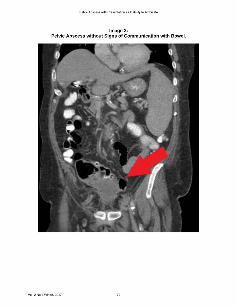
Vol 2 No2 Winter 2017
Pelvic Abscess with Presentation as Inability to Ambulate
Image 3 Pelvic Abscess without Signs of Communication with Bowel
72


![Journal of Medical Diagnostic - Longdom · Tubo-Ovarian Abscess (TOA) is a common condition in women . in genital activity [1]. But its association with pregnancy is exceptional [2-3]](https://img.pdfslide.net/doc/110x75/60fe6664a11b686ae964b303/journal-of-medical-diagnostic-longdom-tubo-ovarian-abscess-toa-is-a-common-condition.jpg)