Embed Size (px)
Citation preview
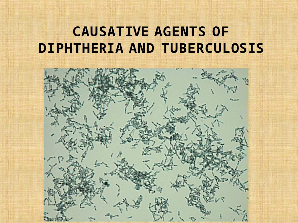
CAUSATIVE AGENTS OF DIPHTHERIA AND TUBERCULOSIS
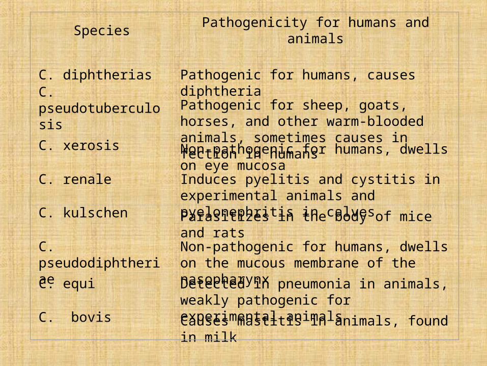
Species Pathogenicity for humans and animals
C. diphtherias Pathogenic for humans, causes diphtheria
C. pseudotuberculosis Pathogenic for sheep, goats, horses, and other warm-blooded animals, sometimes causes in fection in humans
C. xerosis Non-pathogenic for humans, dwells on eye mucosa
C. renale Induces pyelitis and cystitis in experimental animals and pyelonephritis in calves
C. kulschen Parasitizes in the body of mice and rats
C. pseudodiphtheriae Non-pathogenic for humans, dwells on the mucous membrane of the nasopharynx
C. equi Detected in pneumonia in animals, weakly pathogenic for experimental animals
C. bovis Causes mastitis in animals, found in milk
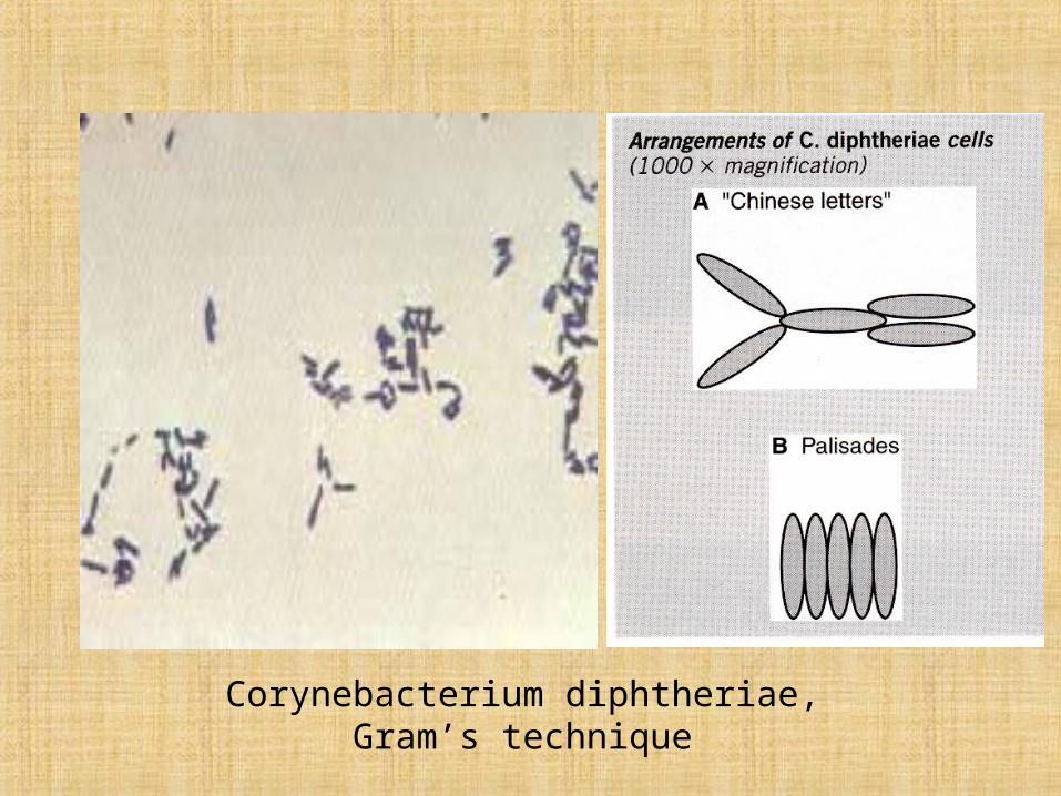
Corynebacterium diphtheriae, Gram’s technique
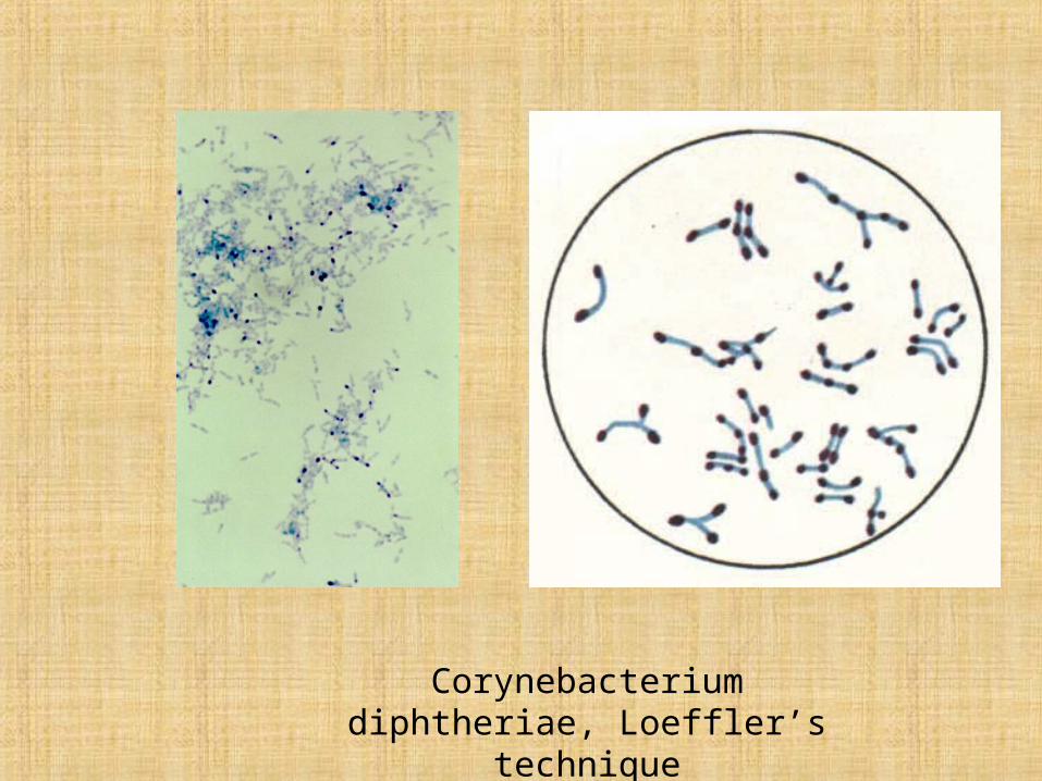
Corynebacterium diphtheriae, Loeffler’s technique
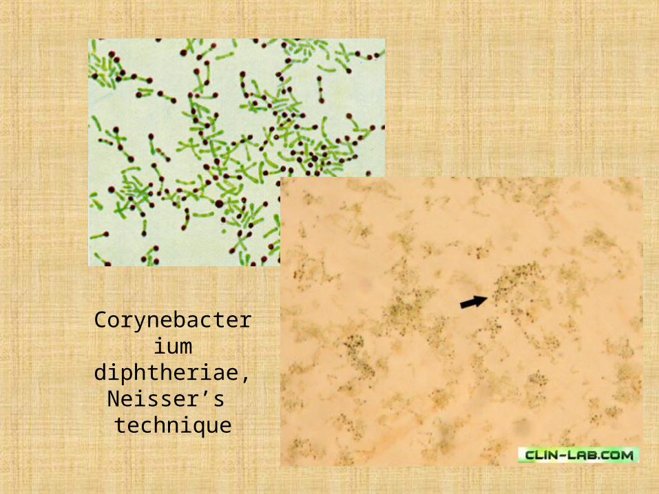
Corynebacterium diphtheriae, Neisser’s technique
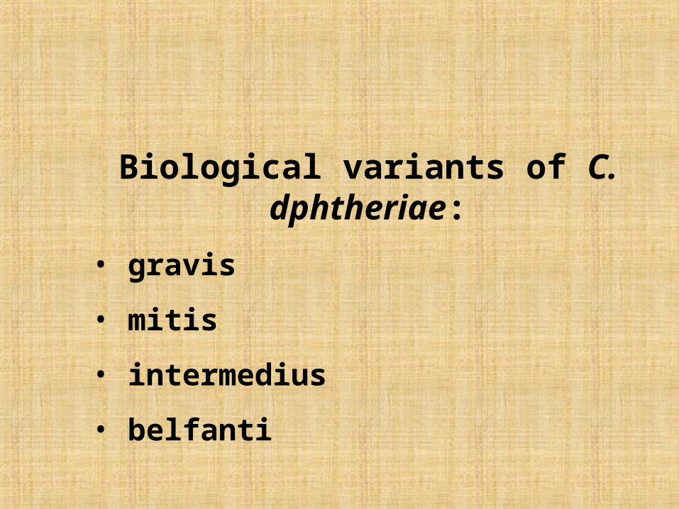
Biological variants of С. dphtheriae:
• gravis
• mitis
• intermedius
• belfanti
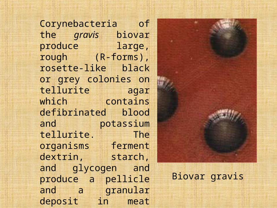
Biovar gravis
Corynebacteria of the gravis biovar produce large, rough (R-forms), rosette-like black or grey colonies on tellurite agar which contains defibrinated blood and potassium tellurite. The organisms ferment dextrin, starch, and glycogen and produce a pellicle and a granular deposit in meat broth. They are usually highly toxic with very marked invasive properties.
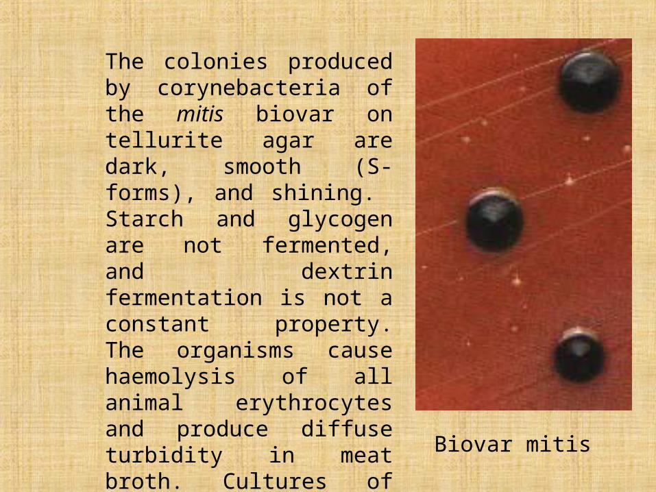
Biovar mitis
The colonies produced by corynebacteria of the mitis biovar on tellurite agar are dark, smooth (S-forms), and shining. Starch and glycogen are not fermented, and dextrin fermentation is not a constant property. The organisms cause haemolysis of all animal erythrocytes and produce diffuse turbidity in meat broth. Cultures of this biovar are usually less toxic and invasive than those of the gravis biovar.
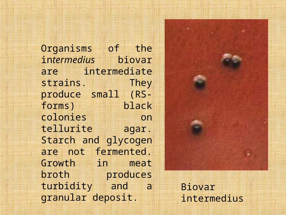
Biovar intermedius
Organisms of the intermedius biovar are intermediate strains. They produce small (RS-forms) black colonies on tellurite agar. Starch and glycogen are not fermented. Growth in meat broth produces turbidity and a granular deposit.
Fermentative properties. All three biovars of C. diphtheriae do not coagulate milk, do not break down urea, produce no indole, and slowly produce hydrogen sulphide. They reduce nitrates to nitrites. Potassium tellurite is also reduced, and for this reason C. diphtheriae colonies grown on tellurite agar turn black or grey. Glucose and levulose are fermented whereas galactose, maltose, starch, dextrin, and glycerin fermentation is variable. Exposure to factors in the external environment renders the organisms incapable of carbohydrate fermentation.
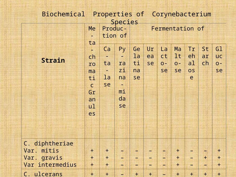
Strain
Me-ta-chromatic Granule
s
Produc-tion of
Fermentation of
Ca-ta-
lase
Py-razina-midase
Gelatinase
Urease
Lacto-se
Malto-se
Trehalose
Starch
Gluco-se
C. diphtheriaeVar. mitisVar. gravisVar intermedius
+++
+++
–––
–––
–––
–––
+++
–––
–+–
+++
C. ulcerans + + – + + – + + + +
C. pseudotuberculosis + + – – + – + – – +
C. pseudodiphtheriticum + + + – + – – – – –
C. xerosis + + + – – – – – – +
Biochemical Properties of Corynebacterium Species
Antigenic structure. Eleven serovars of C. diphtheriae have been deter-mined on the basis of the agglutination reaction. They all produce toxins which do not differ from each other and are neutralized completely by the standard diphtheria antitoxin. A number of authors have confirmed the presence of type-specific thermolabile surface protein antigens (K-antigens) and group-specific thermostable
somatic polysaccharide antigens (O-antigens) in the diphtheria corynebacteria
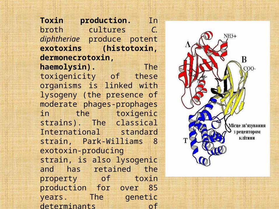
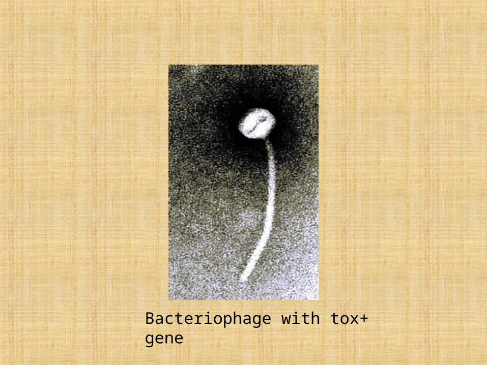
Toxin production. In broth cultures C. diphtheriae produce potent exotoxins (histotoxin, dermonecrotoxin, haemolysin). The toxigenicity of these organisms is linked with lysogeny (the presence of moderate phages-prophages in the toxigenic strains). The classical International standard strain, Park-Williams 8 exotoxin-producing strain, is also lysogenic and has retained the property of toxin production for over 85 years. The genetic determinants of toxigenicity (tox+ genes) are located in the genome of the prophage, which is integrated with the C. diphtheriae nucleoid.
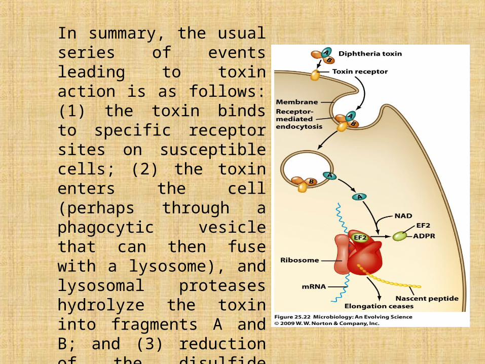
In summary, the usual series of events leading to toxin action is as follows: (1) the toxin binds to specific receptor sites on susceptible cells; (2) the toxin enters the cell (perhaps through a phagocytic vesicle that can then fuse with a lysosome), and lysosomal proteases hydrolyze the toxin into fragments A and B; and (3) reduction of the disulfide bridges (perhaps by glutathione) releases fragment A from fragment B; and (4) fragment A can then enzymatically inactivate EF-2.
The diphtheria toxin is unstable, and is destroyed easily by exposure to heat, light, and oxygen of the air, but is relatively resistant to super-sonic vibrations. The toxin is transformed into the toxoid by mixture with 0.3-0.4 per cent formalin and maintenance at 38-40° C for a period of 3 or 4 weeks. The toxoid is more resistant to physical and chemical factors than the toxin.
Bacteriophage with tox+ gene
Pathogenicity for animals. Animals do not naturally acquire diphtheria. Although, virulent diphtheria organisms were found to be pre-sent in horses, cows, and dogs, the epidemiological significance of animals in diphtheria is negligible.
Among the laboratory animals, guinea pigs and rabbits are most susceptible to the disease. Inoculation of these animals with a culture or toxin gives rise to typical manifestations of a toxinfection and the appearance of inflammation, oedema, and necrosis at the site of inoculation. The internal organs become congested, particularly the adrenals in which haemorrhages occur.
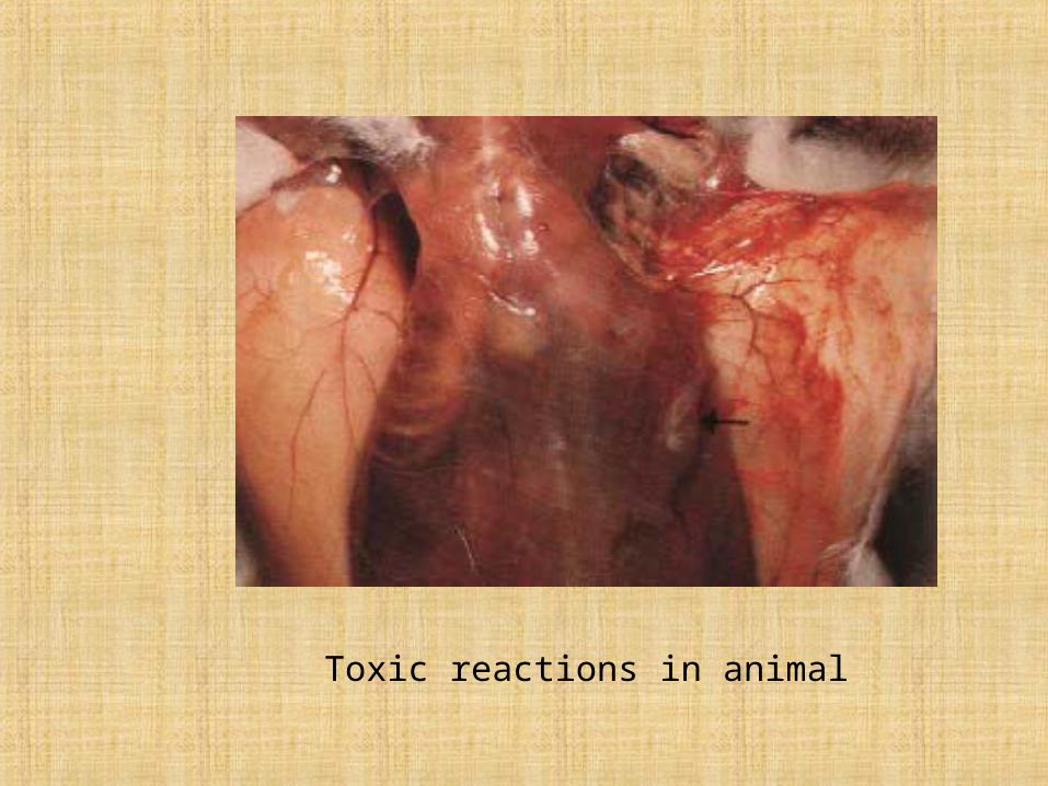
Toxic reactions in animal
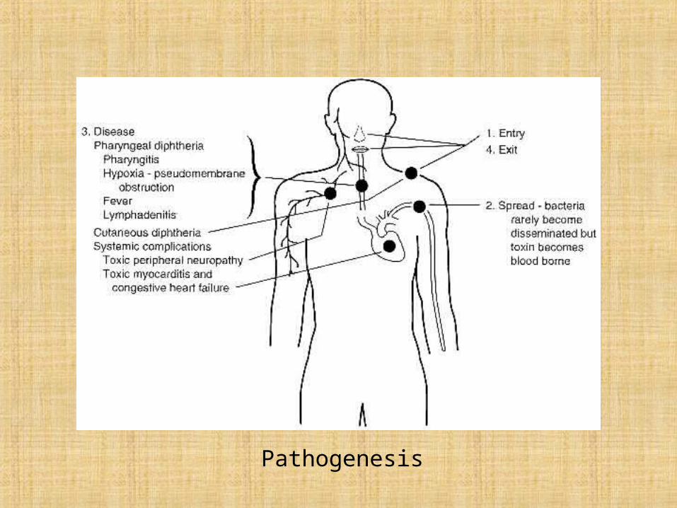
Pathogenesis
Pathogenesis and disease in man. Patients suffering from the disease and carriers are the sources of infection in diphtheria. The disease is transmitted by an air-droplet route, and sometimes with dust particles. Transmission by various objects (toys, dishes, books, towels, handkerchiefs, etc.) and foodstuffs (milk, cold dishes, etc.) contaminated with C. diphtheriae is also possible.
Histotoxin plays the principal role in the pathogenesis of diphtheria. It blocks protein synthesis in the cells of mammals and inactivates transferase, the enzyme responsible for the formation of the polypeptide chain.
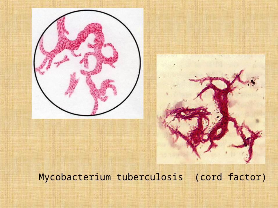
C. diphtheriae penetrate into the blood and tissues of sick humans and infected animals. The diffusion factor due to which these organisms are capable of invasion is formed of a complex of K-antigen and lipids of the wall of bacterial cells. The lipids contain corynemicolic and corynemicolenic acids, the cord factor (trehalose dimicolate), and mannose and inositol phosphatides. The cord factor causes the death of mice, destroys mitochondria, and disturbs the processes of respiration and phosphorylation. The necrotic factor, alpha-glutaric acid, and haemolysin are considered to be factors of invasiveness.
Clinical studies and experiments on animals have provided evidence of the influence of pathogenic staphylococci and streptococci, on the development of diphtheria, the infection becoming more severe in the presence of these organisms. Hypersensitivity to C. diphtheriae and to the products of their metabolism is of definite significance in the pathogenesis of diphtheria.
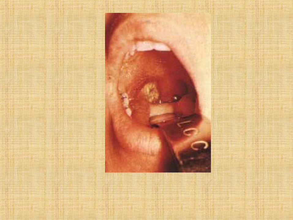
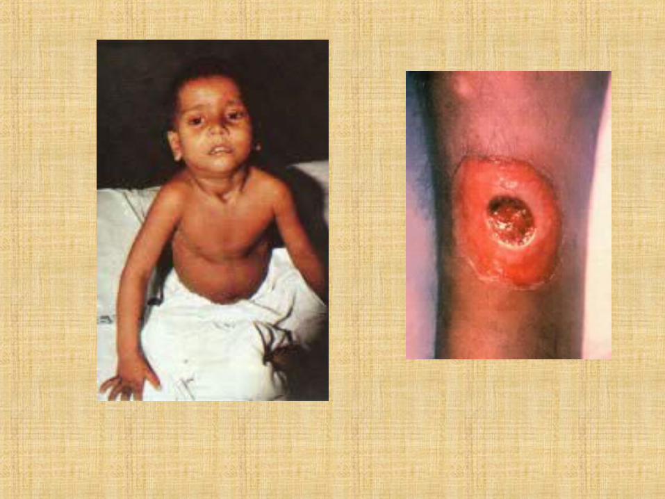
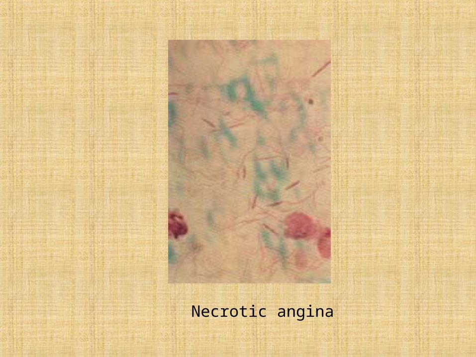
In man, membranes containing a large number of C. diphtheriae and other bacteria are formed at the site of entry of the causative agent (pharynx, nose, trachea, eye conjunctiva, skin, vulva, vagina, and wounds). The toxin produces diphtheria! inflammation and necrosis in the mucous membranes or skin. On being absorbed, the toxin affects the nerve cells, cardiac muscle, and parenchymatous organs and causes severe toxaemia.
Deep changes take place in the cardiac muscle, vessels, adrenals, and in the central and peripheral nervous systems.
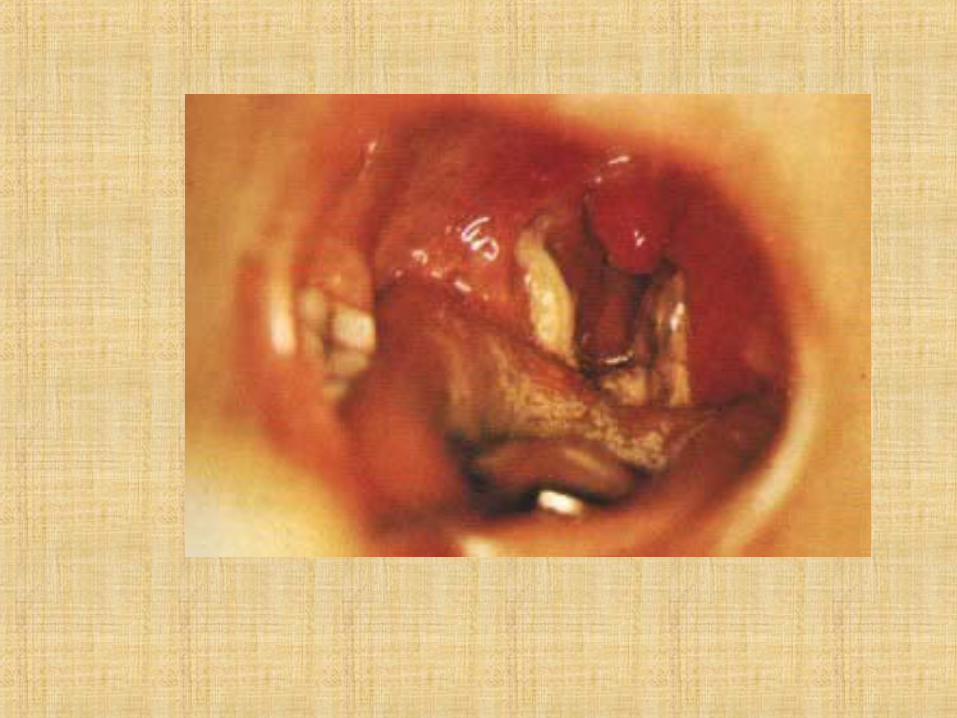
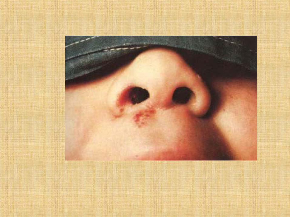
According to the site of the lesion, faucial diphtheria and diphtheritic croup occur most frequently, and nasal diphtheria somewhat less frequently. The incidence of diphtheria of the eyes, ears, genital organs, and skin is relatively rare. Faucial diphtheria constitutes more than 90 per cent of all the diphtherial cases, and nasal diphtheria takes the second place.
Immunity following diphtheria depends mainly on the antitoxin con-tent m the blood However, a definite role of the antibacterial component, associated with phagocytosis and the presence of opsonins, agglutinins, precipitins, and complement-fixing substances cannot be ruled out. Therefore, immunity produced by diphtheria is anti-infectious (anti-toxic and antibacterial) in character.
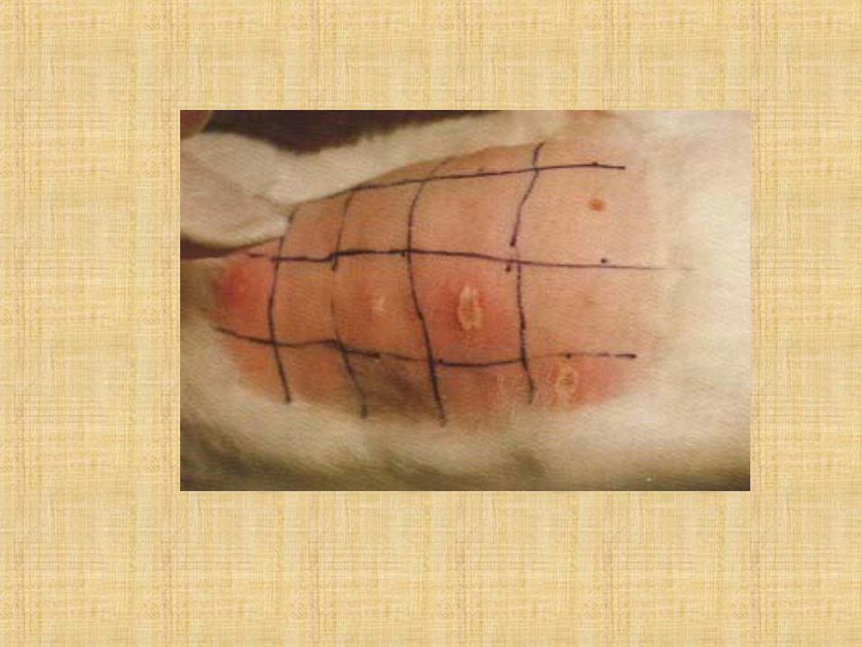
Schick test. This test is used for detecting the presence of antitoxin in children's blood. The toxin is injected intracutaneously into the forearm in a 0 2 ml volume which is equivalent to 1/40 DLM for guinea pigs. A positive reaction, which indicates susceptibility to the disease, is manifested by an erythematous swelling measuring 2 cm in diameter which appears at the site of injection in 24-48 hours. The Schick test is positive when the blood contains either no antitoxin or not more than0.005 units per millilitre of blood serum. A negative Schick reaction indicates, to a certain degree, insusceptibility to diphtheria.
In view of the fact that the diphtheria exotoxin produces a state of sensitization and causes the development of severe reaction in many children, it is advisable to restrict the application of the Schick test and conduct it with great care.
Children from 1 to 4 years old are most susceptible to diphtheria. A relative increase of the incidence of the disease among individuals 15years of age and older has been noted in recent years.
Diphtheria leaves a less stable immunity than do other children's diseases (measles, whooping cough). Diphtheria reinfection occurs in 6-7 per cent of the cases.
Laboratory diagnosis. Discharges from the pharynx, nose, and, some-times, from the vulva, eyes, and skin are collected with a sterile cotton-wool swab for examination.
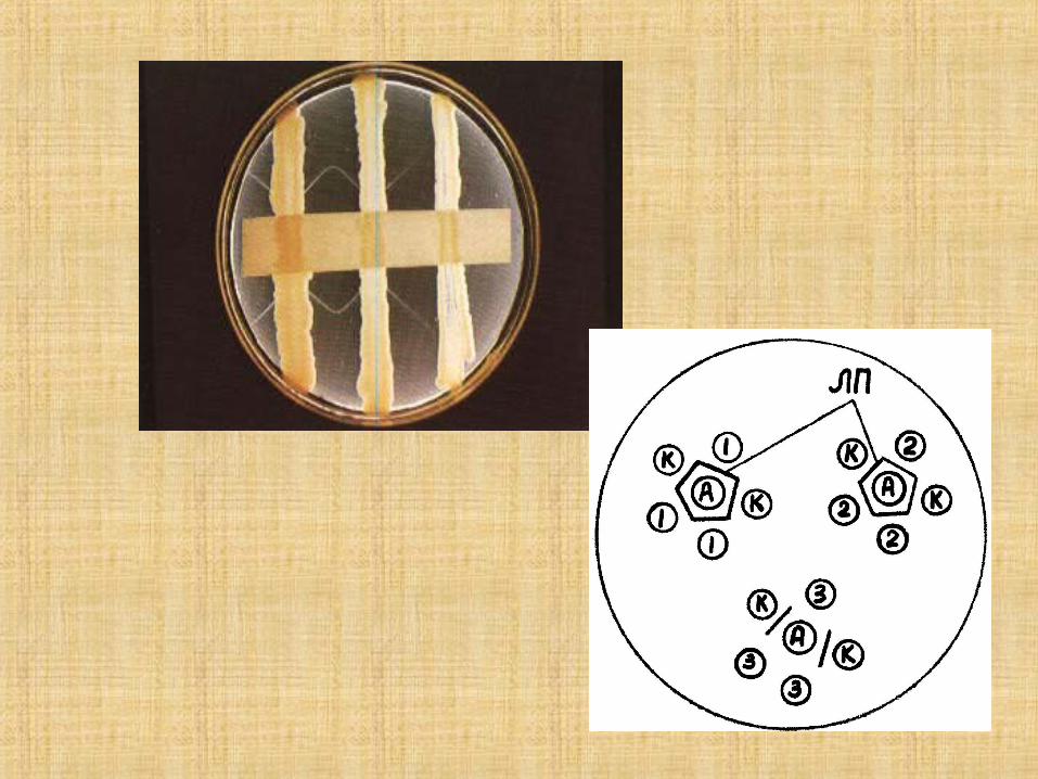
The toxigenic and non-toxigenic strains of diphtheria corynebacteria are differentiated either by subcutaneous or intracutaneous infection of guinea pigs, or by the agar precipitation method, the latter being relatively simple and may be carried out in any laboratory. It is based on the ability of the diphtheria toxin to react with the antitoxin and produce a precipitate resembling arrow-tendrils.
Necrotic angina
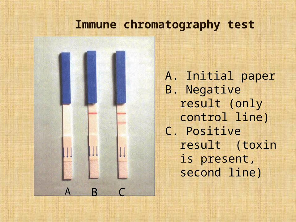
Immune chromatography test
А. Initial paperВ. Negative result (only
control line)С. Positive result (toxin
is present, second line)
А В С
Treatment. According to the physician's prescriptions, patients are given antitoxin in doses ranging from 5000 to 15000 units in mildly severe cases, and from 30 000 to 50 000 units in severe cases of the disease. Penicillin, streptomycin, tetracycline, erythromycin, sulphonamides, and cardiac drugs are also employed. Diphtheria toxoid is recommended in definite doses for improving the immunobiological state of the body, i.e for stimulating antitoxin production.
Prophylaxis. General control measures comprise early diagnosis, prompt hospitalization, thorough disinfection of premises and objects, recognition of carriers, and systematic health education.
Specific prophylaxis is afforded by active immunization. A number of preparations are used: the pertussis-diphtheria vaccine, purified adsorbed toxoid, pertussis-diphtheria-tetanus vaccine All preparations are used according to instructions and directions.
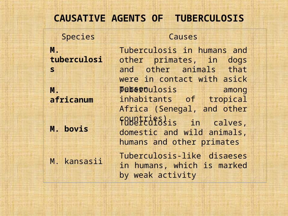
Species Causes
M. tuberculosisTuberculosis in humans and other primates, in dogs and other animals that were in contact with asick person
M. africanumTuberculosis among inhabitants of tropical Africa (Senegal, and other countries)
M. bovisTuberculosis in calves, domestic and wild animals, humans and other primates
M. kansasiiTuberculosis-like disaeses in humans, which is marked by weak activity
CAUSATIVE AGENTS OF TUBERCULOSIS
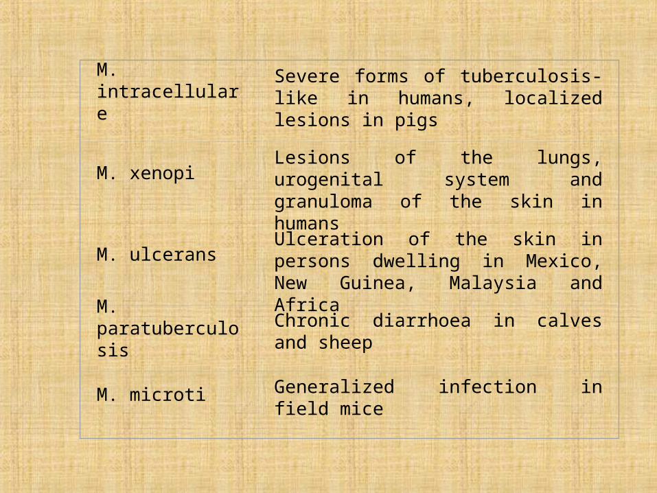
M. intracellulare Severe forms of tuberculosis-like in humans, localized lesions in pigs
M. xenopiLesions of the lungs, urogenital system and granuloma of the skin in humans
M. ulceransUlceration of the skin in persons dwelling in Mexico, New Guinea, Malaysia and Africa
M. paratuberculosis Chronic diarrhoea in calves and sheep
M. microti Generalized infection in field mice
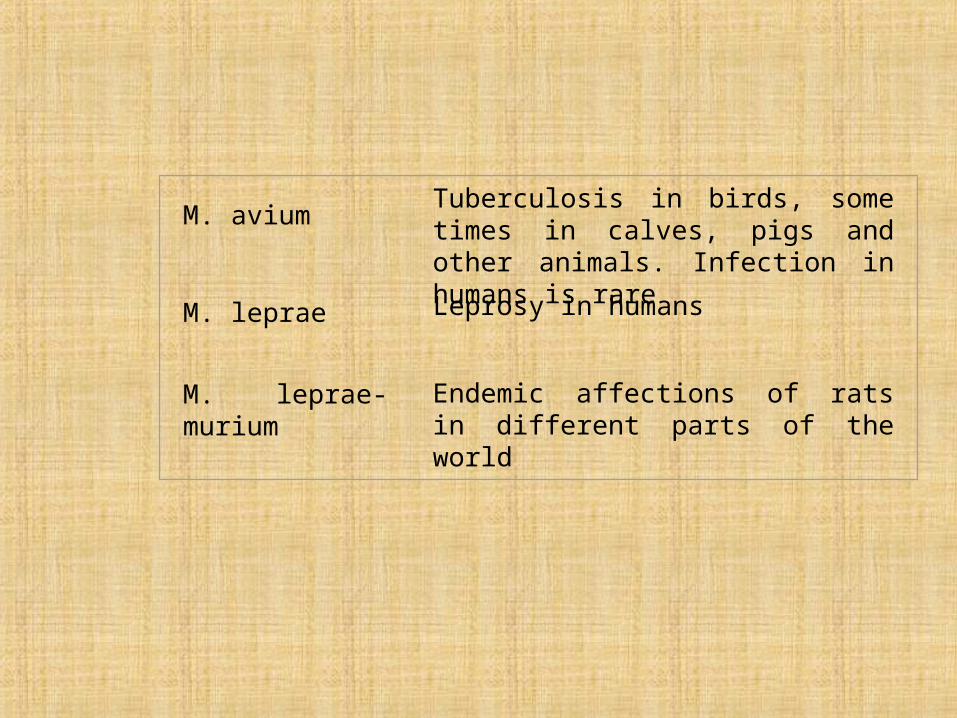
M. aviumTuberculosis in birds, some times in calves, pigs and other animals. Infection in humans is rare
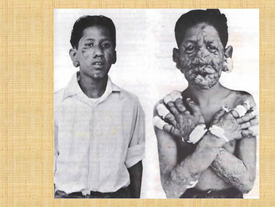
M. leprae Leprosy in humans
M. leprae-muriumEndemic affections of rats in different parts of the world
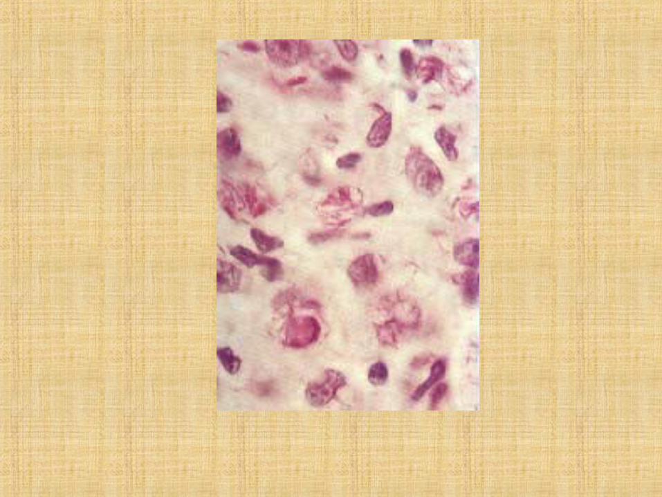
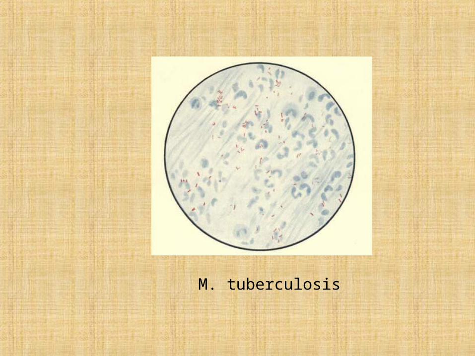
Morphology. M. tuberculosis is a slender, straight or slightly curved rod, 1-4 mcm in length and 0.3-0.6 mcm in breadth. It may have small terminal swellings. The organisms are non-motile, Gram-positive, pleomorphous, and do not form spores or capsules. They stain poorly by the ordinary methods but are stained well by the Ziehl-Neelsen method.
Rod-like, thread-like, branching, granular, coccoid, and filterable forms are encountered.
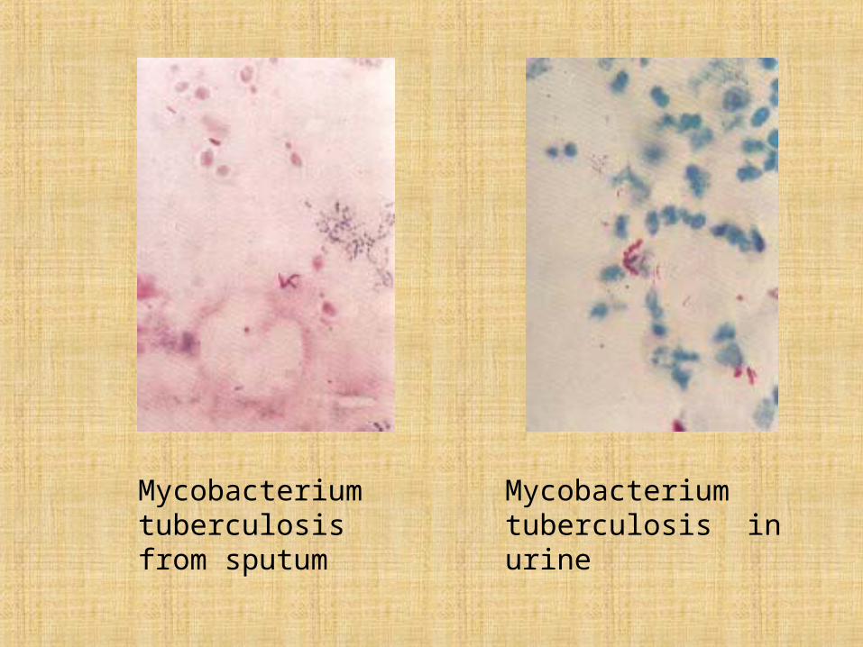
Mycobacterium tuberculosis from sputum
Mycobacterium tuberculosis in urine
Electron microscopy has revealed the presence of granules and vacuoles located terminally in the cells of mycobacteria. The cytoplasm of young cultures is homogeneous, while that of old cultures is granular. M tuberculosis is acid-fast due to the fact that it contains mycolic acid and lipids
The lipids of M. tuberculosis consist of three fractions: (1) phosphatide which is soluble in ether; (2) fat which is soluble in ether and acetone. (3) wax which is soluble in chloroform and ether.
Nonacid-fast granular forms, which readily stain violet by Gram's method and known as Much's granules, and acid-fast Slenger's fragments of M. tuberculosis also occur. The G +C content in DNA ranges from 62 to 70 per cent.
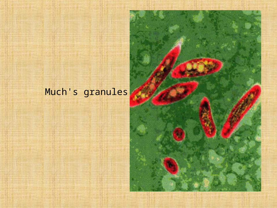
Much's granules
Chemical composition. The fact that as much as 40% of the dry weight of mycobacteria may consist of lipid undoubtedly accounts for many of their unusual growth and staining characteristics A comprehensive discussion of mycobacterial lipids is beyond the scope of this text, but one class of lipids, the mycosides, is unique to acid-fast organisms and is involved in some manner with the pathogenicity of the mycobacteria
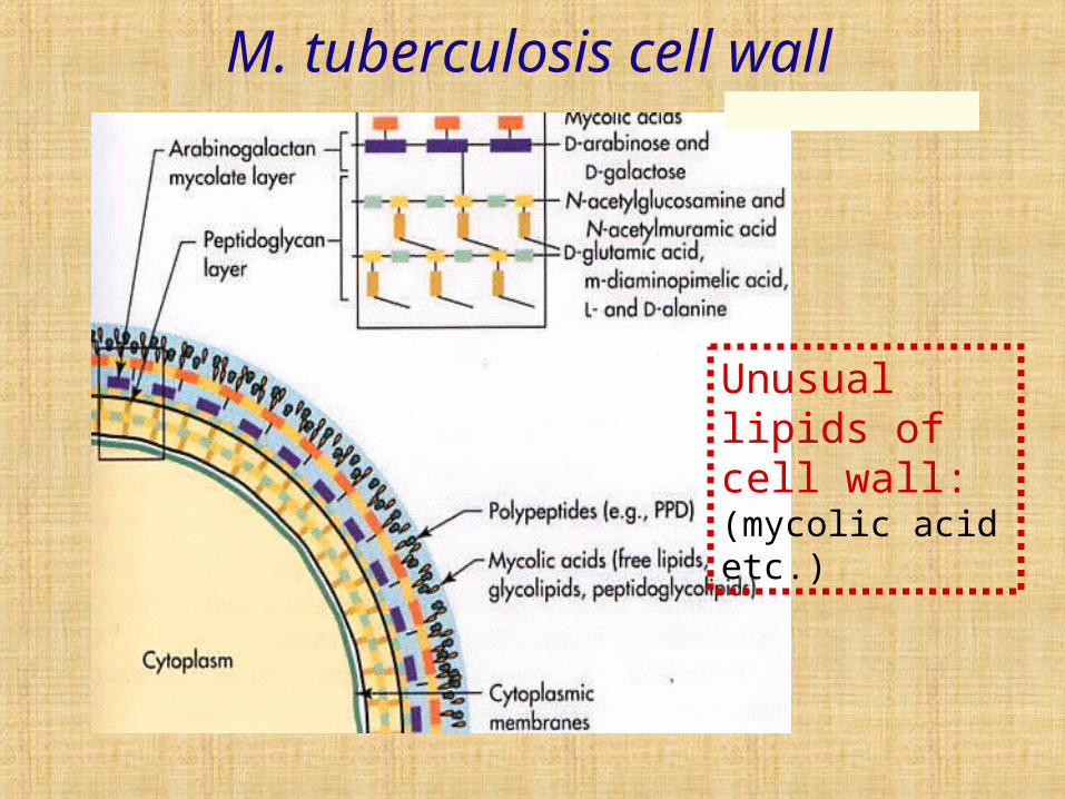
M. tuberculosis cell wall
Unusual lipids of cell wall: (mycolic acid etc.)
Mycobacterium tuberculosis (cord factor)
Cultivation. The organisms grow on selective media, e. g. coagulated serum, glycerin agar, glycerin potato, glycerin broth and egg media (Petroffs, Petragnani's, Dorset's, Loewenstein's, Lubenau's, Vinogradov's, etc.) They may be cultured on Soton's synthetic medium which contains asparagine, glycerin, iron citrate, potassium phosphate, and other substances.
Certain levels of vitamins (biotin, nicotinic acid, riboflavin. etc.) are necessary for the growth of M. tuberculosis. Scarcely visible growth appears 8-10 days after inoculation on glycerin (2-3 per cent) agar, but in 2-3 weeks a dry cream-coloured pellicle is produced. The best and quickest (on the sixth-eighth day) growth is obtained on Petroffs egg medium which consists of egg yolk, meat extract, agar, glycerin, and gentian violet.
On glycerin (4-5 per cent) meat-peptone broth the organisms produce a thin delicate film in 10-15 days, which thickens gradually, becomes brittle, wrinkled, and yellow; the broth remains clear. M. tuberculosis can be successfully cultivated by Pryce's microculture method or Shkolnikova's deep method in citrated rabbit or sheep blood. Growth becomes visible in 3-6 days. Synthetic and semisynthetic media are employed for cultivating M. tuberculosis in special laboratories. The organisms are aerobic, and their optimal growth temperature is 37 C. They do not grow below 24 and above 42 C. The reaction of the medium is almost neutral (pH 6.4-7.0), but growth is possible at pH ranging from 6.0 to 8.0. M. tuberculosis dissociate from typical R-forms to the atypical S-forms. Some strains produce a yellow pigment in old cultures.
M. tuberculosis
Colonies of M. tuberculosis
Growth properties of Mycobacteria
M. tuberculosisM. kansasii
(photochromogen)M. gordonae
(scotochromogen)
Fermentative properties. The organisms have been found to contain proteolytic enzymes which break down proteins in alkaline and acid medium. They also contain dehydrogenases which ferment ammo acids, alcohols, glycerin, and numerous carbohydrates. M. tuberculosis is cap-able of causing reduction (they reduce salts of telluric acid, potassium tellurite, and break down olive and castor oils, etc.). The organisms produce lecithinase, glycerophosphatase, and urease which ferment lecithin, phosphatides, and urea.
Toxin production. M. tuberculosis does not produce an exotoxin. It contains toxic substances which are liberated when the cell decomposes
In 1890 R. Koch isolated from the tubercle bacillus a substance known as tuberculin. There are several tuberculin preparations. The Old Koch's tuberculin is a 5-6-week-old glycerin broth culture sterilized for 30 minutes by a continuous current of steam (100 C), evaporated at 70 C to one tenth of the initial volume, and filtered through a porcelain filter.
The New Koch's tuberculin consists of desiccated M. tuberculosis which are triturated in 50 per cent glycerin to a homogeneous mass. A tuberculin has been derived from the bovine variety of M. tuberculosis, which contains protein substances, fatty acids, lipids, neutral fats, and crystalline alcohol. There is also a tuberculin free of waste sub-stances and designated PPD (purified protein derivative) or PT (purificatum tuberculinum).
Tuberculin is toxic for guinea pigs which are affected with tuberculosis (injection of 0.1 ml of the standard preparation is fatal for 50 percent of experimental animals). Small doses of tuberculin produce no changes in healthy guinea pigs.
The chemical composition of the toxic substances contained in M.tuberculosis has not yet been ascertained. It is known that the toxin of the tubercle mycobacteria is composed of proteins (albumins and nucleoproteins). Phosphatides have been isolated from the virulent types of the organism and are capable of producing characteristic lesions in rabbits. Phthioic acid is the most active.
Extremely toxic substances have been extracted from M. tuberculosis after boiling in vaseline oil. They are fatal to guinea pigs in doses of one-thousandth of a milligram
Virulent mycobacteria differ from the non-virulent organisms in that they contain a great number of lipopolysaccharide components. The lipid fraction (cord factor) responsible for adhesion of mycobacteria and their growth in cords and strands is marked by high toxicity. The cord factor of M. tuberculosis destroys the mitochondria of the cells of the infected body and causes disorders in respiration and phosphorylation.
Antigenic structure. On the basis of agglutination and complement-fixation reaction a number of types of mycobacteria have been distinguished: mammalian (human, bovine, and rodent), avian, poikilotherm, and saprophytic. The human type does not differ serologically from the bovine or murine types. Mycobacterial antigens produce agglutinis, opsonins, precipitins, and complement-fixing antibodies in low titres. Tuberculin is considered to be a peculiar antigen (hapten).
A high molecular tuberculin may be considered to be a full-value antigen capable of stimulating the production of corresponding antibodies.
M. tuberculosis and tuberculin possess allergenic properties and produce local, focal, and generalized reactions in the body infected with tuberculosis.
According to data supplied by a number of investigators, the M tuberculosis antigen contains proteins, lipids, and particularly large amounts of phosphatides and lipopolysaccharides. Experiments on animals have proved that the lipopolysaccharide-protein complexes protect the body from infection with M. tuberculosis. Tuberculin is widely used for allergic tests, which are employed for determining infection with M. tuberculosis.
Resistance. Tubercle bacilli are more resistant to external effects as compared to other non-sporeforming bacteria as a result of their high lipid content (25-40 per cent).
The organisms survive in the flowing water for over a year, in soil and manure up to 6 months, on the pages of books over a period of3 months, in dried sputum for 2 months, in distilled water for several weeks, and in gastric juice for 6 hours. They are easily rendered harm-less at temperatures ranging from 100 to 120°C. The organisms are sensitive to exposure to sunlight.
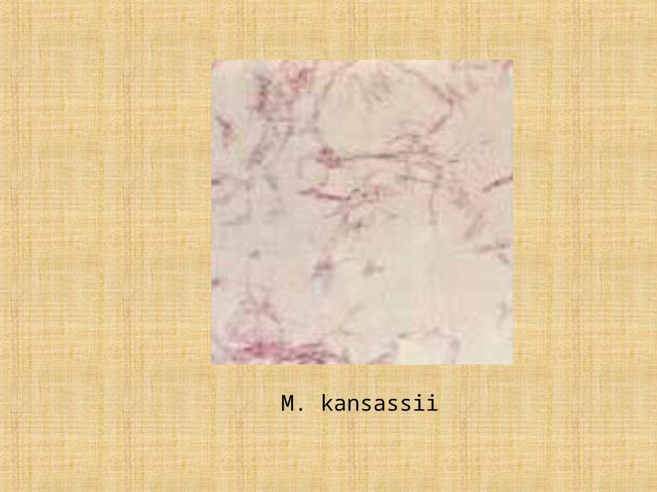
M. kansassii
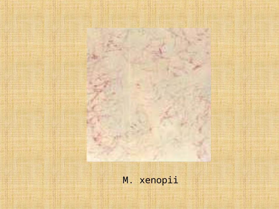
M. xenopii
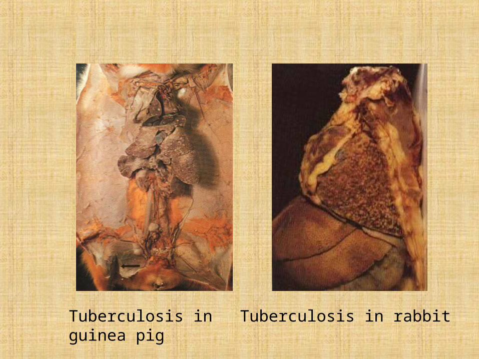
Tuberculosis in guinea pig
Tuberculosis in rabbit
Pathogenesis and disease in man. It has been shown that tuberculosis in man is caused by several types of mycobacteria — the human type(M. tuberculosis), the bovine type (M. bovis), etc. The share of atypical mycobacteria which cause a variety of clinical forms of tuberculosis among humans has recently grown to 50 per cent.
Infection with tuberculosis takes place through the respiratory tract by the droplets and dust, and, sometimes, per os through contaminated foodstuffs, and through the skin and mucous membranes. Intrauterine infection via the placenta may also occur.

One drop = 3 bacteriaDuring 5 min = 3000 drops =
9000 microbes!
http://catalog.cmsp.com/datav3/it060009.htm
With air-borne infection, the primary infectious centre develops in the lungs, but if infection takes place through the alimentary tract, the primary focus is in the mesenteric lymgh nodes. When body resistance is low and conditions of work and life are unfavourable, the organisms may leave the site of primary localization and spread throughout the body, causing a generalized infection. At present, there is a point of view which maintains that localization of the infectious focus in the lungs is preceded by a lympho-haematogenic dispersion of M. tuberculosis throughout the body. The duration of the incubation period in tuberculosis is comparatively long, from several weeks to 40 years and more.
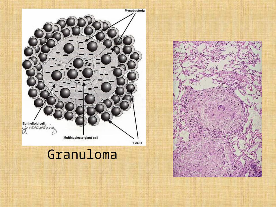
Granuloma

Tuberculosis forms
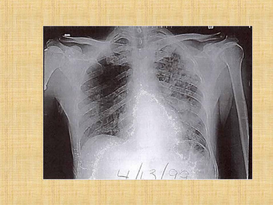
Pulmonary tuberculosis Tuberculous meningitidis Tuberculosis of the kidney Genital tuberculosis Tuberculosis of bones and joints Ocular tuberculosis Lymphatic tuberculosis Cutaneous tuberculosis Tuberculosis of the larynx Intestine tuberculosis, tuberculous peritonitis
http://www-medlib.med.utah.edu/WebPath/TUTORIAL/MTB/MTB002.html
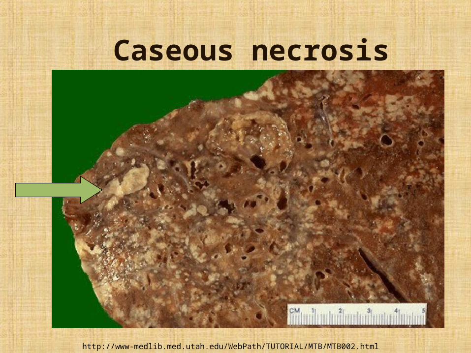
Caseous necrosis
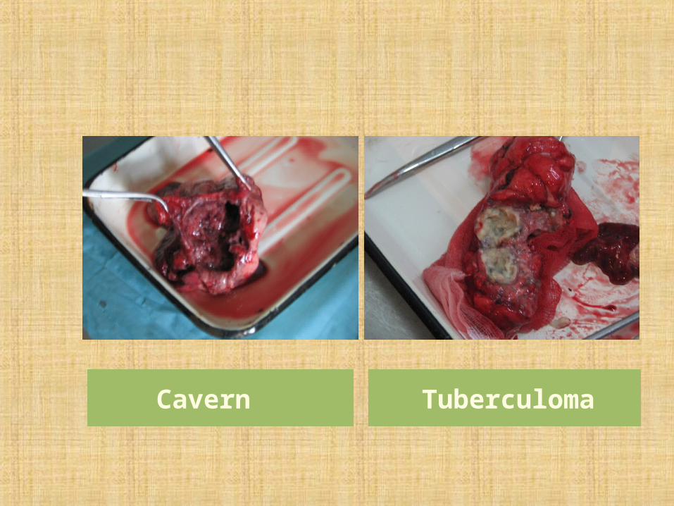
Cavern Tuberculoma
Immunity. Man is naturally resistant to tuberculosis, this property being hereditary. On the basis of the allergic reaction. X-ray examination, and patho-anatomical changes it has been shown that in a great number of cases infection does not result in disease. There are approximately 80 per cent of adults over 20 years of age among infected persons and no more than 10 per cent of them become ill, and only 5 per cent immediately after infection.
There is a characteristic immunity produced by tuberculosis. Inoculation of M. tuberculosis into healthy guinea pigs causes no visible changes during the first days after infection. But a compact tubercle which undergoes ulceration is formed in 10-14 days. The lymph nodes become enlarged and hard, a generalized process develops, and the animal dies.
When tuberculous animals are inoculated with M. tuberculosis, an ulcer is formed at the site of injection. This ulcer shortly heals and no involvement of the lymph nodes or generalization of infection takes place. These facts were established by Koch and advanced the knowledge on a number of problems concerning pathogenesis and immunity in tuberculosis. Particular importance was attributed to non-sterile (infectious)immunity which has been widely reproduced artificially (by BCG vaccination). It is understood that immunity to tuberculosis is usually non-sterile. However, as in brucellosis, the phase of non-sterile immunity in tuberculosis is followed by the phase of sterile immunity.
Interference of M. tuberculosis with BCG strains and other non-virulent mycobacteria which are capable of blocking tissue and organ cells sensitive to virulent tuberculous mycobacteria plays a definite role in the complex of defence mechanisms of the body.
The genetic factor (which has been studied in detail in twins) plays an obvious role in immunity in tuberculosis. The concordance in affection with the disease is 67 per cent among monozygotic twins, 25.6 per cent among dizygotic twins, 25.6 per cent among brothers and sisters, and7 per cent in husband and wife.
A new component which affects M. tuberculosis has been found to be present in human blood. Individuals devoid of this component are more susceptible to tuberculosis.
Among the defence factors phages should be mentioned. They affect both virulent and avirulent M. tuberculosis strains. The discovery of phages is of certain practical importance. They may be used in diagnosis and, probably, in the treatment of tuberculosis.
Many tissues are capable of producing enzymes which break down mycobacteria. Such properties are characteristic of enzymes of the nuclease group.
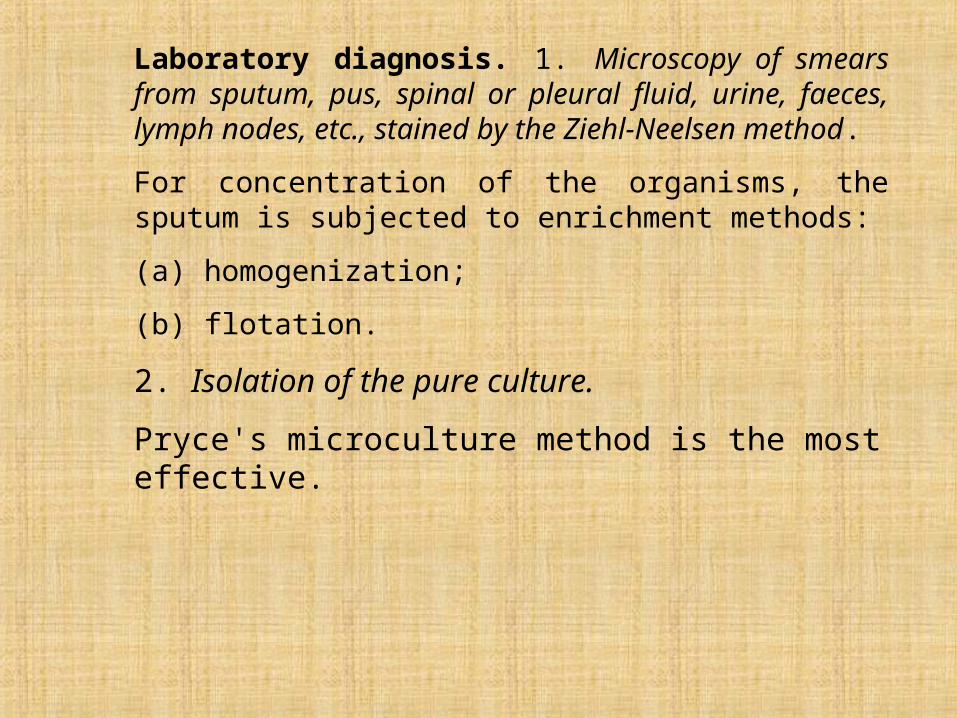
Laboratory diagnosis. 1. Microscopy of smears from sputum, pus, spinal or pleural fluid, urine, faeces, lymph nodes, etc., stained by the Ziehl-Neelsen method.
For concentration of the organisms, the sputum is subjected to enrichment methods:
(a) homogenization;
(b) flotation.
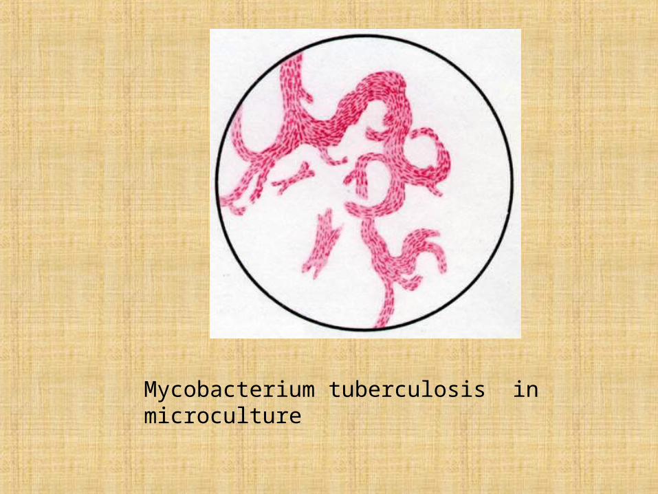
2. Isolation of the pure culture.
Pryce's microculture method is the most effective.
M. tuberculosis
Mycobacterium tuberculosis in microculture
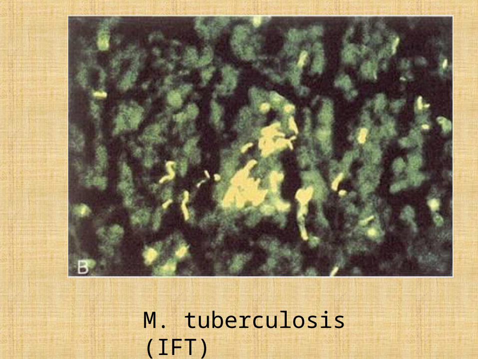
M. tuberculosis (IFT)
The virulent and non-virulent M. tuberculosis strains are differentiated by their growth on butyrate albumin agar (Middlebrook-Dubos test). The virulent strains grow in the form of plaits, and the non-virulent strains form irregular clusters. The above authors suggested the differentiation of the virulent and non-virulent strains by staining the smears with neutral red which has an affinity for virulent mycobacteria and stains them purple-pink (non-virulent strains are stained yellow).
3. Biological method. Inoculation of guinea pigs produces an infiltrate at the site of injection of the material, lymph node enlargement, and generalized tuberculosis. The animals die 1-1.5 months after inoculation. Post-mortem examination reveals the presence of numerous tubercles in the internal organs. Specimens are obtained from lymph nodes by puncture 5-10 days after inoculation and examined for the presence of tubercle bacilli. The tuberculin test is carried out 3-4 weeks after infection. The atypical strains and L-forms are non-pathogenic for guinea pigs
4. Complement-fixation reaction (positive in 80 per cent of cases with chronic pulmonary tuberculosis, in 20-25 per cent of patients with skin tuberculosis, and in 5-10 per cent of healthy people).
5. Indirect haemagglutination reaction (Middlebrook-Dubos test).Sheep erythrocytes, on which polysaccharides of M. tuberculosis or tuberculin are adsorbed, are agglutinated in serum of tuberculosis patients.
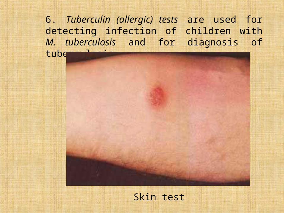
6. Tuberculin (allergic) tests are used for detecting infection of children with M. tuberculosis and for diagnosis of tuberculosis.
Skin test
Treatment is accomplished with antibacterial preparations. They include derivatives of isonicotinic acid hydrazide (tubazide, phthivazide, etc.), streptomycin, and PAS — preparations of the first series. Preparations of the second series (cycloserine, kanamycin, biomycin, etc) are used to enhance the therapeutic effect. The isolated M. tuberculosis are tested for sensitivity to drugs which are added to fluid or solid media indifferent concentrations.
The control of tuberculosis in a population requires the location and treatment of infected persons who spread tubercle bacilli by way of pulmonary secretions. However, even though there are annually over 26,000 new cases and 3000 deaths reported in the United States, tuberculosis is usually a slow, chronic disease, and it is exceedingly difficult to find infected persons until they have experienced months or years of active infection. For early detection, therefore, one must rely on the tuberculin skin test, and a positive reaction is interpreted as denoting an infected person, whether or not the disease is quiescent or active.

For this reason, control relies heavily on preventive therapy, and the Tuberculosis Advisory Committee to the Centres for Disease Control has recommended that the following persons be considered potential candidates for active disease (in the order listed) and that they be treated with daily oral INH for 1 year:
1. Household members and other close associates of persons with recently diagnosed tuberculosis.
2. Positive tuberculin reactors with findings on a chest roentgenogram consistent with nonprogressive tuberculosis, even in the absence of bacteriologic findings.
3. All persons who have converted from a tuberculinnegative to a tuberculin-positive response within the last 2 years.
4. Positive tuberculin reactors undergoing prolonged therapy with adrenocorticoids, receiving immunosuppressive therapy, having leukaemia or Hodgkin's disease, having diabetes mellitus, having silicosis, or who have had a gastrectomy.
5. All persons younger than 35 years of age who are positive tuberculin skin reactors. INH therapy is not recommended for positive tuberculin reactors 35 years of age or older, because prolonged treatment with INH causes occasional progressive liver disease; although the risk is low for persons younger than 35 years of age, the incidence rate increases to 1.2% of persons between 35 and 49, and to 2.3% for those older than 50 years.
Some individuals older than 55 years of age may not respond to tuberculin even though they were at one time tuberculin positive. Such persons, however, may experience a booster effect from the initial testing and become tuberculin positive to a subsequent test given a year or more later, indicating a conversion resulting from an infection with M tuberculosis. Such an interpretation can be avoided if negative reactors are given a repeat test 1 week or 10 days after the first test. Positive reactions to the second test would then be attributed to a booster effect rather than to a new infection.
Prophylaxis is insured by early diagnosis, timely detection of patients with atypical forms of the disease, routine check up of patients and recovered patients, disinfection of milk and meat derived from sick animals, and other measures.
Active immunization of human beings is of great importance in the control of tuberculosis. It lowers significantly the incidence of the disease and the death rate, gives protection against the development of severe cases, and lowers the body sensitivity to the effect of tubercle mycobacteria and to the products of their disintegration. Active immunity makes the body capable of fixing and rendering harmless the causative agent, stimulates biochemical activity of tissues and intensifies the production of antibacterial substances. Immunization produces a certain type of infectious immunity.
Intracutaneous immunization and revaccination have been carried out in the world 1962. For these requirements a special dry BCG vaccine is produced. It is given in a single injection to newborn infants. Revaccination is carried out at the age of 7, 12, 17, 23, and 27-30 years.
Postvaccinal immunity is produced within 3 or 4 weeks and remains for 1-1.5 to 15 years.
To prevent tuberculosis among carriers or those who had recovered from the disease, preventive chemotherapy with Isoniazid (isonicotinicacid hydrazide) is applied.
Living conditions play an important part in the incidence of tuberculosis. Deterioration of the conditions increases the incidence of the disease and death rate (wars, famine, unemployment, economical crises, and other disasters).
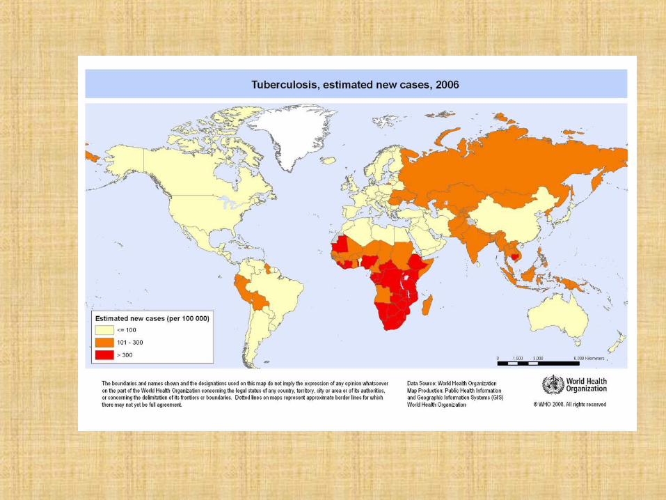
According to WHO, a total of more than 15 million tuberculosis patients have been recorded in the world. The incidence of tuberculosis is very high in Latin America, India, and Africa.
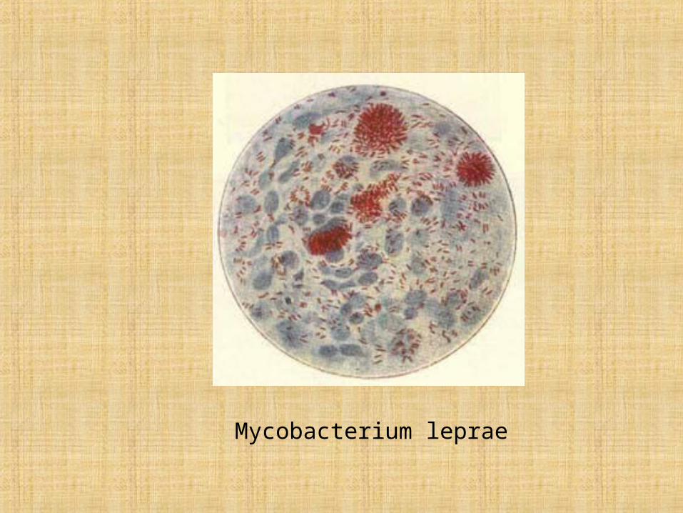
Mycobacterium leprae
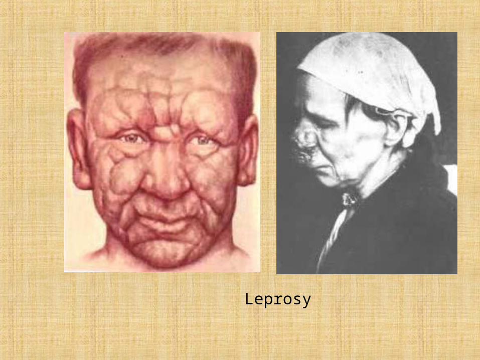
Leprosy