Embed Size (px)
Citation preview
T h e n e w e ngl a nd j o u r na l o f m e dic i n e
n engl j med 383;10 nejm.org September 3, 2020958
Review Article
From the Center for Interstitial Lung Dis-eases and Sarcoidosis, Department of Respiratory Medicine, Erasmus MC–Uni-versity Medical Center Rotterdam, Rot-terdam, the Netherlands (M.W.); and the Department of Respiratory Medicine, Na-tional Coordinating Reference Center for Rare Pulmonary Diseases, Louis Pradel Hospital, and Claude Bernard University — both in Lyon, France (V.C.). Address reprint requests to Dr. Wijsenbeek at the Center for Interstitial Lung Diseases and Sarcoidosis, Department of Respiratory Medicine, Erasmus MC, University Medi-cal Center Rotterdam, 3015 GD Rotterdam, the Netherlands, or at m . wijsenbeek-lourens @ erasmusmc . nl.
N Engl J Med 2020;383:958-68.DOI: 10.1056/NEJMra2005230Copyright © 2020 Massachusetts Medical Society.
Diffuse parenchymal lung diseases encompass a large number of conditions, with a wide range of causes, clinical manifestations, and imaging and pathological features, as well as variable outcomes. Despite
the intrinsic heterogeneity of this group of diseases, in most of them, the pulmo-nary alveolar walls are infiltrated by various combinations of inflammatory cells, fibrosis, and proliferation of certain cells that make up the normal alveolar wall. Since these pathologic abnormalities predominate in the lung interstitium, the disorders are termed interstitial lung diseases (ILDs).
Idiopathic pulmonary fibrosis (IPF) is the archetypal and most common fibrotic ILD. IPF is characterized by an imaging and pathological pattern of usual intersti-tial pneumonia (UIP) without an identifiable cause or association with a disease known to be associated with pulmonary fibrosis. It occurs more commonly in men than in women (sex ratio, 7:3) and is more common in people older than 60 years of age than in younger people.1,2 IPF is a chronic and irreversible disease, usually progressing to respiratory failure and death (median interval between diagnosis and death, 3 years).3 In contrast to IPF, other ILDs are generally characterized by a younger mean age at presentation (20 to 60 years) and a more balanced sex ratio. The variable underlying pathological features of other ILDs, with fibrosis gener-ally less prominent than inflammatory infiltration, also translate into more hetero-geneous and often less severe outcomes, as compared with IPF. However, a num-ber of these other ILDs are also characterized by progressive fibrosis.4 As in any other organ, fibrosis in the lungs can be a manifestation of several clinical enti-ties, and if the fibrosis is progressive, it will ultimately result in organ failure,5 causing respiratory symptoms, limited exercise capacity, an impaired quality of life, and an increased risk of death.6
ILDs are typically assigned to many disease categories for classification and management purposes, roughly on the basis of a known underlying disease (e.g., pulmonary fibrosis associated with rheumatoid arthritis), an inciting agent (e.g., pneumoconiosis), or the absence of a known cause (e.g., IPF).4,7 In this review, we address pulmonary fibrosis in various contexts and disease entities, emphasizing the commonalities in pathophysiological features, clinical manifestations, and diag-nostic features, as well as the similarly progressive nature of many of these diseases.
Epidemiol o gy
Although each of the individual fibrosing ILDs is rare, collectively they affect a considerable number of patients, representing a substantial burden of disease. The overall prevalence of ILD is estimated to be up to 76.0 cases per 100,000 people in Europe and 74.3 cases per 100,000 in the United States. Sarcoidosis, connective-tissue disease (CTD)–associated ILDs, and IPF are the most common fibrotic ILDs, with an estimated prevalence of 30.2, 12.1, and 8.2 cases per 100,000, respec-tively8 (Table 1). Among all patients with fibrotic ILDs other than IPF, 13 to 40%
Jeffrey M. Drazen, M.D., Editor
Spectrum of Fibrotic Lung DiseasesMarlies Wijsenbeek, M.D., and Vincent Cottin, M.D.
The New England Journal of Medicine Downloaded from nejm.org by KEVIN ROSTEING on November 9, 2020. For personal use only. No other uses without permission.
Copyright © 2020 Massachusetts Medical Society. All rights reserved.
n engl j med 383;10 nejm.org September 3, 2020 959
Fibrotic Lung Diseases
have a progressive fibrosing phenotype,18 repre-senting up to 20 patients per 100,000 people in Europe and up to 28 patients per 100,000 in the United States (Fig. S1 in the Supplementary Ap-pendix, available with the full text of this article at NEJM.org).19
Pulmonary fibrosis occurs throughout the world, with geographic variation.19 The prevalence of IPF, estimated to be 8 to 60 cases per 100,000 population,8,20 is higher in North America and Europe than in the rest of the world, whereas the prevalence of sarcoidosis is higher in northern Europe and among Black persons and is lower in Japan.21
Pathoph ysiol o gy
The formation of fibrosis is an essential response of the body against pathogens and in normal wound healing.22 In pulmonary fibrosis, various and often disease-specific triggers set off exag-gerated cascades of inflammatory and fibrotic responses, leading to downstream fibrotic tissue remodeling and extracellular-matrix deposition,23 which in turn perpetuate fibrosis formation (Fig. 1). Much is still unknown about the patho-physiology of specific disease entities and the factors that differentiate normal wound repair from progression to fibrosis. Although triggers, susceptibility, and initial inflammatory respons-es vary among diseases, the current assumption is that in later phases, common mechanisms play a role.23
A variety of genetic studies have identified both common and rare variants that are associ-ated with enhanced susceptibility to pulmonary fibrosis, with remarkable similarities between familial IPF and other fibrotic ILDs.24 For exam-ple, a frequent polymorphism in the promoter of MUC5B, which is involved in airway clearance and bacterial host defense, is associated with increased risks of IPF, rheumatoid arthritis with ILD25 (RA–ILD), and chronic hypersensitivity pneumonitis (CHP)26 but not systemic sclerosis with ILD (SSc–ILD), sarcoidosis, or antisynthe-tase syndrome. Telomere shortening and telomere-related gene mutations (TERT, TERC, RTEL1, and PARN) are found in IPF, RA–ILD, and CHP.24,26 Some rare genetic variants, such as telomere-related gene mutations, are clearly associated with progressive disease.24
Besides shared genetic risk factors, different ILDs have heterogeneous, overlapping initial
pathways7 (Fig. 1). In IPF, an as yet undefined insult to alveolar epithelial-cell integrity may ini-tiate disease through the interaction between epithelial cells and myofibroblasts.5 Granuloma-tous inflammation in response to a putative, persistent, unknown trigger progresses to fibro-sis in only a small percentage of patients with sarcoidosis.13 In SSc–ILD, a combination of in-flammation, endothelial dysfunction, and vascu-lopathy leads to pulmonary fibrosis in a major-ity of patients, driving the prognosis.23 Studies investigating specific conditions suggest that various inflammatory responses may lead to a profibrotic environment and cytokine milieu (in-cluding, especially, transforming growth factor β, connective-tissue growth factor, platelet-derived growth factor, and WNT and hedgehog signal-ing). Shared downstream pathways may activate and sustain a complex interplay leading to fibro-blast activation and differentiation into myofi-broblasts, which further orchestrate fibrogene-sis.23 Once established, structural tissue changes and the profibrotic milieu form a feed-forward loop, leading to self-perpetuating fibrosis.
Dise a se En ti ties w i th Pul mona r y Fibrosis
ILDs can be divided into five broad clinical cate-gories: ILDs related to distinct primary diseases (e.g., sarcoidosis, Langerhans-cell granulomato-sis, eosinophilic pneumonia, lymphangioleiomyo-matosis, and pulmonary alveolar proteinosis); ILDs related to environmental exposures, includ-ing pneumoconiosis due to inhalation of inor-ganic substances and hypersensitivity pneumo-nitis mostly related to inhalation of organic particles (e.g., domestic or occupational expo-sure to mold or birds or other exposures); ILDs induced by drugs, illicit drugs, or irradiation; ILDs associated with CTDs, including RA–ILD and SSc–ILD, idiopathic inflammatory myopathy, and primary Sjögren’s disease; and idiopathic interstitial pneumonias,27 which include IPF, idio-pathic nonspecific interstitial pneumonia, and other, less common entities.
Pulmonary fibrosis can occur in the context of many of these ILDs (Table 1 and Table S1). A separation can be made between pulmonary fi-brosis in the context of underlying systemic dis-eases, such as CTDs and sarcoidosis, and condi-tions that are restricted to the lung, such as CHP, drug-induced pulmonary fibrosis, idiopathic non-
The New England Journal of Medicine Downloaded from nejm.org by KEVIN ROSTEING on November 9, 2020. For personal use only. No other uses without permission.
Copyright © 2020 Massachusetts Medical Society. All rights reserved.
n engl j med 383;10 nejm.org September 3, 2020960
T h e n e w e ngl a nd j o u r na l o f m e dic i n eTa
ble
1. C
linic
al C
hara
cter
istic
s of
Sel
ecte
d B
road
Cat
egor
ies
of P
ulm
onar
y Fi
bros
is.*
Con
ditio
nM
ain
Clin
ical
Fe
atur
esFi
ndin
gs o
n C
hest
Im
agin
gO
ther
Fea
ture
s
Not
Cha
ract
eris
tic o
f IPF
Man
agem
ent
Prog
nosi
s
Ris
k Fa
ctor
s fo
r Pr
ogre
ssiv
e Fi
bros
is o
r D
eath
Rel
ativ
e Pr
eval
ence
†
Prog
ress
ive
Fibr
osin
g Ph
enot
ype
% o
f pat
ient
s
IPF3
Vel
cro-
like
crac
kles
; fin
ger
club
bing
(3
0–50
% o
f pa-
tie
nts)
; mal
e:
fem
ale
ratio
, 3:1
; ag
e >5
0 yr
Def
inite
or
prob
a-bl
e U
IP p
atte
rn,
inde
term
inat
e pa
tter
n fo
r U
IP
(and
bio
psy
findi
ngs
or c
lin-
ical
cou
rse
sug-
gest
ive
of IP
F)
NA
Ant
ifibr
otic
ther
apy
(pir
feni
done
, ni
nted
anib
)
Med
ian
surv
ival
, 3–
4 yr
; pot
en-
tial f
or s
low
ing
prog
ress
ion
Old
er a
ge, m
ale
sex,
hon
ey-
com
bing
or
UIP
pat
tern
on
CT,
FV
C <
70%
1290
–100
SSc–
ILD
9,10
Ray
naud
’s p
heno
m-
enon
, ski
n th
ick-
enin
g, fi
nger
tip
lesi
ons,
tela
ngi-
ecta
sia,
gas
tro-
esop
hage
al r
eflu
x,
vasc
ulop
athy
Mor
e co
mm
on
fibro
tic N
SIP
than
UIP
pat
-te
rn
Youn
ger
age,
mor
e w
omen
than
men
af-
fect
ed, m
ultis
yste
mic
in
volv
emen
t, au
to-
imm
une
sero
logi
c fin
ding
s (a
nti–
Scl-7
0,
antic
entr
omer
e,
and
anti–
RN
A p
oly-
mer
ase
III a
ntib
od-
ies)
, abn
orm
al n
ail-
fold
cap
illar
osco
py
Imm
unos
uppr
essi
ve
ther
apy:
myc
ophe
no-
late
; alte
rnat
ivel
y, IV
cy
clop
hosp
ham
ide,
az
athi
opri
ne, r
itux-
imab
, toc
ilizu
mab
Ant
ifibr
otic
ther
apy
(nin
teda
nib)
Stem
-cel
l tra
nspl
anta
tion
or lu
ng tr
ansp
lant
a-tio
n in
sel
ect p
atie
nts
10-Y
r m
orta
lity,
40
%; 3
5% o
f SS
c-re
late
d de
aths
due
to
ILD
; pos
sibl
e st
abili
zatio
n w
ith tr
eatm
ent
Diff
use
cuta
neou
s SS
c, <
7 yr
sin
ce
diag
nosi
s, m
ale
sex,
Bla
ck r
ace,
an
ti–Sc
l-70
antib
odie
s,
dise
ase
exte
nt
on C
T >2
0%,
redu
ced
FVC
an
d D
Lco
940
Rhe
umat
oid
ar
thri
tis–
ILD
11, 1
2
Mor
ning
stif
fnes
s,
sym
met
ric
arth
ri-
tis, s
ynov
itis,
join
t er
osio
ns, r
heum
a-to
id n
odul
es
Pred
omin
ance
of
UIP
pat
tern
ov
er N
SIP
or
inde
term
inat
e pa
tter
n, m
ulti-
com
part
men
t in
volv
emen
t (a
ssoc
iatio
n of
ai
rway
s or
ple
u-ra
l inv
olve
men
t)
Aut
oim
mun
e se
rolo
gic
feat
ures
(A
CPA
s, b
ut
rheu
mat
oid
fact
or
less
spe
cific
)
Lack
of e
vide
nce
for
imm
unos
uppr
essi
ve
ther
apy;
ritu
xim
ab,
abat
acep
t, or
myc
o-ph
enol
ate
occa
sion
-al
ly u
sed;
ant
ifibr
otic
th
erap
y (n
inte
dani
b)
used
in c
ases
of
prog
ress
ive
fibro
sis;
pi
rfen
idon
e is
und
er
inve
stig
atio
n‡
Med
ian
surv
ival
, 3
yr (
UIP
pat
tern
) or
long
er (
othe
r pa
tter
ns);
effe
ct
of tr
eatm
ent o
n lu
ng d
isea
se
unkn
own
Old
er a
ge, m
ale
sex,
dis
ease
ex
tent
on
CT
>20%
, hon
-ey
com
bing
or
UIP
pat
tern
on
CT,
FV
C <
70%
832
Sarc
oido
sis,
fib
rotic
(s
tage
IV)13
Mul
tisys
tem
dis
ease
in
any
org
an,
espe
cial
ly s
kin,
ey
e, h
eart
, liv
er,
and
lym
ph n
odes
; pu
lmon
ary
in-
volv
emen
t in
90%
of
cas
es; w
ide
rang
e of
clin
ical
ph
enot
ypes
Upp
er-lo
be,
peri
bron
cho-
vasc
ular
, and
ly
mph
atic
dis
-tr
ibut
ion;
den
se
peri
hila
r fib
rotic
or
cav
itate
d m
asse
s; b
ron-
chia
l dis
tort
ion,
re
ticul
ar o
paci
-tie
s, a
nd tr
ac-
tion
bron
chie
c-ta
sis;
UIP
-like
pa
tter
n ra
re
Youn
ger
age;
Fem
ale:
m
ale
ratio
, 1:1
; mul
ti-or
gan
invo
lvem
ent;
abse
nce
of b
ibas
ilar
crac
kles
and
clu
b-bi
ng; n
onca
seat
ing
epith
elio
id-c
ell g
ran-
ulom
as w
ith g
iant
ce
lls o
n pa
thol
ogic
al
eval
uatio
n
Mon
itori
ng a
lone
or
trea
tmen
t with
gl
ucoc
ortic
oids
; m
etho
trex
ate
or
azat
hiop
rine
as
glu-
coco
rtic
oid-
spar
ing
agen
t or
seco
nd-li
ne
ther
apy;
infli
xim
ab
or a
dalim
umab
as
thir
d-lin
e th
erap
y;
lack
of e
vide
nce
for
leflu
nom
ide
and
hydr
oxyc
hlor
oqui
ne
for
lung
dis
ease
; be
nefit
of a
ntifi
brot
ic
ther
apy
uncl
ear
10-Y
r m
orta
lity,
ab
out 1
0%;
75%
of s
arco
id-
osis
-rel
ated
de
aths
due
to
lung
dis
ease
; ge
nera
lly r
e-sp
onsi
ve to
im
mun
omod
u-la
tion
Bla
ck r
ace,
dis
ease
ex
tent
on
CT
>20%
, pul
mo-
nary
hyp
erte
n-si
on, f
emal
e se
x
4513
The New England Journal of Medicine Downloaded from nejm.org by KEVIN ROSTEING on November 9, 2020. For personal use only. No other uses without permission.
Copyright © 2020 Massachusetts Medical Society. All rights reserved.
n engl j med 383;10 nejm.org September 3, 2020 961
Fibrotic Lung Diseases
Con
ditio
nM
ain
Clin
ical
Fe
atur
esFi
ndin
gs o
n C
hest
Im
agin
gO
ther
Fea
ture
s
Not
Cha
ract
eris
tic o
f IPF
Man
agem
ent
Prog
nosi
s
Ris
k Fa
ctor
s fo
r Pr
ogre
ssiv
e Fi
bros
is o
r D
eath
Rel
ativ
e Pr
eval
ence
†
Prog
ress
ive
Fibr
osin
g Ph
enot
ype
% o
f pat
ient
s
Chr
onic
fib
rotic
hy
pers
ensi
-tiv
ity p
neu-
mon
itis14
, 15
Prol
onge
d ex
posu
re
to in
hale
d pa
r-tic
les,
pre
dom
i-na
ntly
org
anic
an
tigen
s; o
nset
of
sym
ptom
s ov
er a
pe
riod
of 6
mo
or
mor
e§
Ret
icul
atio
n an
d ho
neyc
ombi
ng,
with
per
ibro
n-ch
ovas
cula
r,
uppe
r- a
nd
mid
dle-
zone
di
stri
butio
n;
grou
nd-g
lass
at-
tenu
atio
n w
ith
mos
aici
sm a
nd
air
trap
ping
Offe
ndin
g in
hale
d an
ti-ge
n no
t alw
ays
iden
-tif
ied;
rec
urre
nt e
pi-
sode
s of
sym
ptom
s;
BA
L ly
mph
ocyt
osis
(>
20%
of c
ases
);
posi
tive
prec
ipiti
ns;
biop
sy, i
f per
form
ed
show
ing
airw
ay-
cent
ric
lym
phoc
ytic
in
filtr
atio
n, lo
ose
gran
ulom
as, a
nd g
i-an
t cel
ls
Expo
sure
avo
idan
ce;
limite
d ev
iden
ce fo
r gl
ucoc
ortic
oids
and
im
mun
osup
pres
sive
th
erap
y (m
ycop
he-
nola
te o
r az
athi
o-pr
ine)
; ant
ifibr
otic
th
erap
y (n
inte
dani
b)
for
prog
ress
ive
fibro
-si
s; lu
ng tr
ansp
lant
a-tio
n in
rar
e ca
ses
5-Yr
sur
viva
l, 50
–80
%; p
oten
tial
for
impr
ove-
men
t or
stab
i-liz
atio
n w
ith
trea
tmen
t
Pers
iste
nt e
xpo-
sure
to o
ffend
-in
g an
tigen
, ho
neyc
ombi
ng
or U
IP p
atte
rn
on C
T
321
Unc
lass
ifiab
le
fibro
tic
ILD
16, 1
7
Dem
ogra
phic
fea-
ture
s va
ry; m
e-di
an a
ge, 6
0–65
yr
; non
spec
ific
sym
ptom
s w
ith
dysp
nea
and
coug
h; n
o fir
st-
choi
ce d
iagn
osis
; of
ten
subt
le a
uto-
imm
une
feat
ures
Non
spec
ific
fea-
ture
s ge
nera
lly
not m
eetin
g cr
i-te
ria
for
mai
n pa
tter
ns
Maj
or d
iscr
epan
cy
amon
g cl
inic
al, i
m-
agin
g, a
nd h
isto
logi
c fe
atur
es; n
ondi
ag-
nost
ic C
T fin
ding
s an
d no
bio
psy
perf
orm
ed o
r bi
opsy
re
sults
non
cont
ribu
-to
ry
Lim
ited
evid
ence
for
gluc
ocor
ticoi
ds;
imm
unos
uppr
es-
sive
ther
apy
ofte
n fir
st-li
ne; a
ntifi
brot
ic
ther
apy
(pir
feni
done
or
nin
teda
nib)
in
prog
ress
ive
fibro
sis
5-Yr
sur
viva
l, 45
–70%
; var
i-ab
le d
isea
se
cour
se
Hon
eyco
mbi
ng
on im
agin
g,
prog
ress
ive
decl
ine
in lu
ng
func
tion
853
* M
ore
com
preh
ensi
ve in
form
atio
n is
pro
vide
d in
Tab
le S
1. A
CPA
s de
note
s an
ti–ci
trul
linat
ed p
rote
in a
ntib
odie
s, B
AL
bron
choa
lveo
lar
lava
ge, D
L co
diff
usin
g ca
paci
ty o
f the
lung
for
car-
bon
mon
oxid
e, F
VC
forc
ed v
ital c
apac
ity, I
LD in
ters
titia
l lun
g di
seas
e, I
PF id
iopa
thic
pul
mon
ary
fibro
sis,
IV
intr
aven
ous,
NA
not
app
licab
le, N
SIP
nons
peci
fic in
ters
titia
l pne
umon
ia, S
Sc
syst
emic
scl
eros
is, a
nd U
IP u
sual
inte
rstit
ial p
neum
onia
.†
Rel
ativ
e pr
eval
ence
is t
he e
stim
ated
pre
vale
nce
amon
g al
l pat
ient
s w
ith I
LD.
‡ T
he t
rial
is o
ngoi
ng (
Clin
ical
Tria
ls.g
ov n
umbe
r, N
CT0
2999
178)
.§
A li
st o
f inh
aled
org
anic
ant
igen
s th
at c
an c
ause
fibr
otic
hyp
erse
nsiti
vity
pne
umon
itis
is a
vaila
ble
at w
ww
. hpl
ung .
com
.
The New England Journal of Medicine Downloaded from nejm.org by KEVIN ROSTEING on November 9, 2020. For personal use only. No other uses without permission.
Copyright © 2020 Massachusetts Medical Society. All rights reserved.
n engl j med 383;10 nejm.org September 3, 2020962
T h e n e w e ngl a nd j o u r na l o f m e dic i n e
Alveolus
Early Phase (Underlying Disease–Specific)
Late Phase (Shared Self-Perpetuating Fibrosis)
Exaggeratedimmune response
Edema
Type 1 alveolarepithelial cell
Lymphocytes andmacrophages
Epithelial-cellinjury
Repairedtype 2 alveolarepithelial cells
Type 2 alveolarepithelial cell
Pericyte
Subpleuralhoneycombing
MyofibroblastsFibroblastic
focus
Collagen
Fibroblasts
Pleura
Granuloma
Respiratory bronchiole
Normalinterstitium
Exaggeratedimmune response
injury
Late Phase (Shared Self-Perpetuating Fibrosis)
Exaggeratedimmune response
Exaggerated
Alveolus
Exaggeratedimmune response
Exaggeratedimmune response
Early Phase (Underlying Disease–Specific)
• Tobacco smoking• Occupational exposure• Air pollution• Microaspiration• Viral infection
Environmental risk factorsepithelial cell
Residentfibroblast
Pleura
Extracellularmatrix production
Lungremodeling
Myofibroblasts
Tissue stiffnessand hypoxia
Fibrosis
Microvascularendothelial
injury
Chronicinflammation
Fibroblastic focus
Subpleural honeycombing
NSIP
Granuloma
Endothelial-celldifferentiation
CapillaryCapillary
Fibrocyte
Monocyte
Epithelial-celldifferentiation
EdemaEdema
Granuloma
FibrosisFibrosis
Autoimmunity
Exaggerated
Activatedhelper T cell
Persistentantigen
Normallung tissuelung tissue
Partial or completeresolution
Genetic predisposition
Aging
The New England Journal of Medicine Downloaded from nejm.org by KEVIN ROSTEING on November 9, 2020. For personal use only. No other uses without permission.
Copyright © 2020 Massachusetts Medical Society. All rights reserved.
n engl j med 383;10 nejm.org September 3, 2020 963
Fibrotic Lung Diseases
specific interstitial pneumonia, and IPF.3,27 There is also overlap between groups (e.g., drug-induced pulmonary fibrosis in CTD and a genetic predis-position in various ILDs). Owing to the epidemi-
ology and burden of fibrosis within each diag-nostic category, clinicians most often see patients with CTD–ILD, IPF, CHP, sarcoidosis, or unclassifiable fibrotic ILD.
Currently, there is a specific interest in the potential development of fibrosis after coronavi-rus disease 2019 (Covid-19). Although infection with severe acute respiratory syndrome corona-virus 2 (SARS-CoV-2) causes a range of pulmo-nary symptoms, male sex, older age, obesity, and coexisting conditions appear to be risk factors for the development of SARS.28 Pulmonary fibro-sis is a known complication of acute respiratory distress syndrome (ARDS), and there are simi-larities in the fibroproliferative response and risk factors between lung fibrosis in the context of ARDS and lung fibrosis in the context of other diseases.29 Nevertheless, analysis of long-term follow-up data after ARDS29 or infection with another strain of SARS-CoV in 200330 showed fibrotic changes that remained mostly stable over time and had little clinical relevance.29 The long-term effect and the disease course of pul-monary fibrosis caused by Covid-19 are cur-rently under investigation in prospective studies.
Di agnos tic A pproach
Other than disease-specific symptoms, cough, progressive exertional dyspnea, and exercise limitation are the main presenting symptoms. The diagnosis is often delayed by several months or even years. A thorough history, including en-vironmental exposures, medication use, and ex-trapulmonary signs, should be taken.2 On chest auscultation, fine crackles (also called Velcro rales or crepitations) are indicative of fibrosis,31 although squeaks may be heard in patients with hypersensitivity pneumonitis. Premature graying of hair and hematologic abnormalities may be a sign of telomeropathy-related fibrosis. In CTDs, pulmonary fibrosis may develop either after the underlying condition is diagnosed or before the extrapulmonary manifestations are observed.32 Hands, joints, and skin should be thoroughly examined.32 Serologic testing is recommended, including for antinuclear antibodies and anti–citrullinated peptide antibodies.2 If there is a clinical suspicion of an autoimmune condition, consultation with a rheumatologist and more extensive serologic testing are recommended.
High-resolution computed tomographic (CT)
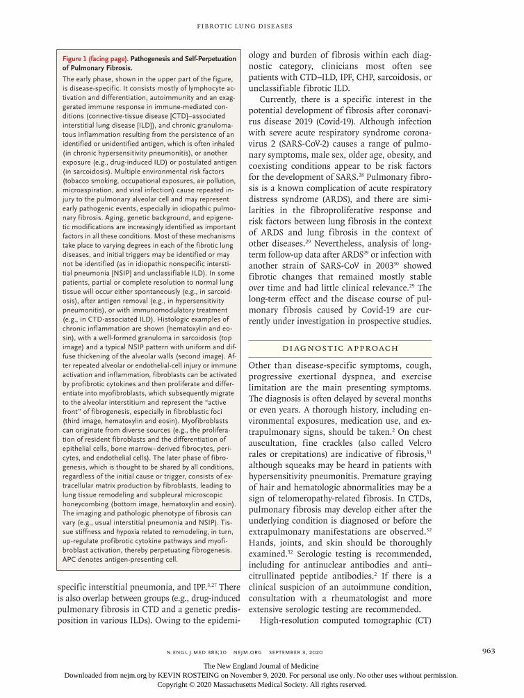
Figure 1 (facing page). Pathogenesis and Self-Perpetuation of Pulmonary Fibrosis.
The early phase, shown in the upper part of the figure, is disease-specific. It consists mostly of lymphocyte ac-tivation and differentiation, autoimmunity and an exag-gerated immune response in immune-mediated con-ditions (connective-tissue disease [CTD]–associated interstitial lung disease [ILD]), and chronic granuloma-tous inflammation resulting from the persistence of an identified or unidentified antigen, which is often inhaled (in chronic hypersensitivity pneumonitis), or another exposure (e.g., drug-induced ILD) or postulated antigen (in sarcoidosis). Multiple environmental risk factors (tobacco smoking, occupational exposures, air pollution, microaspiration, and viral infection) cause repeated in-jury to the pulmonary alveolar cell and may represent early pathogenic events, especially in idiopathic pulmo-nary fibrosis. Aging, genetic background, and epigene-tic modifications are increasingly identified as important factors in all these conditions. Most of these mechanisms take place to varying degrees in each of the fibrotic lung diseases, and initial triggers may be identified or may not be identified (as in idiopathic nonspecific intersti-tial pneumonia [NSIP] and unclassifiable ILD). In some patients, partial or complete resolution to normal lung tissue will occur either spontaneously (e.g., in sarcoid-osis), after antigen removal (e.g., in hypersensitivity pneumonitis), or with immunomodulatory treatment (e.g., in CTD-associated ILD). Histologic examples of chronic inflammation are shown (hematoxylin and eo-sin), with a well-formed granuloma in sarcoidosis (top image) and a typical NSIP pattern with uniform and dif-fuse thickening of the alveolar walls (second image). Af-ter repeated alveolar or endothelial-cell injury or immune activation and inflammation, fibroblasts can be activated by profibrotic cytokines and then proliferate and differ-entiate into myofibroblasts, which subsequently migrate to the alveolar interstitium and represent the “active front” of fibrogenesis, especially in fibroblastic foci (third image, hematoxylin and eosin). Myofibroblasts can originate from diverse sources (e.g., the prolifera-tion of resident fibroblasts and the differentiation of epithelial cells, bone marrow–derived fibrocytes, peri-cytes, and endothelial cells). The later phase of fibro-genesis, which is thought to be shared by all conditions, regardless of the initial cause or trigger, consists of ex-tracellular matrix production by fibroblasts, leading to lung tissue remodeling and subpleural microscopic honeycombing (bottom image, hematoxylin and eosin). The imaging and pathologic phenotype of fibrosis can vary (e.g., usual interstitial pneumonia and NSIP). Tis-sue stiffness and hypoxia related to remodeling, in turn, up-regulate profibrotic cytokine pathways and myofi-broblast activation, thereby perpetuating fibrogenesis. APC denotes antigen-presenting cell.
The New England Journal of Medicine Downloaded from nejm.org by KEVIN ROSTEING on November 9, 2020. For personal use only. No other uses without permission.
Copyright © 2020 Massachusetts Medical Society. All rights reserved.
n engl j med 383;10 nejm.org September 3, 2020964
T h e n e w e ngl a nd j o u r na l o f m e dic i n e
scanning of the chest establishes the diagnosis of pulmonary fibrosis by revealing reticulation, architectural distortion, and lung volume loss and may identify patterns suggestive of specific causes (Table 1, Table S1, and Fig. S2).2,3 The UIP pattern is the hallmark of pulmonary fibrosis, observed frequently in IPF, in RA–ILD, and in advanced disease irrespective of the underlying condition.3,33 In contrast, the most common pat-tern in SSc–ILD is that of nonspecific interstitial pneumonia, which consists of mixed reticulation and ground-glass attenuation to a varying ex-tent, often with traction bronchiectasis, central axial distribution, and sparing of the subpleural area. Expiratory imaging may be useful, espe-cially in CHP.34 Pulmonary-function testing as-sesses the level of disease impairment and is the most frequently used measure for monitoring the course of disease and response to therapy. In patients with pulmonary fibrosis, testing typi-cally shows a restrictive lung-function pattern (decreased forced vital capacity [FVC], normal or increased ratio of forced expiratory volume in 1 second to FVC, decreased total lung capacity, and low residual volume), together with a de-creased diffusing capacity of the lung for carbon monoxide. However, normal lung function does not rule out the presence of pulmonary fibrosis.
If the combination of clinical findings and imaging is not diagnostic, more invasive diag-nostic procedures may be needed. Bronchoalveo-lar lavage contributes to the diagnosis of hyper-sensitivity pneumonitis and sarcoidosis. Bronchial mucosa and lymph-node biopsies are performed when sarcoidosis is suspected. It is recommend-ed that all collected information be synthesized by a multidisciplinary team experienced in ILD (Fig. 2), which may either establish a diagnosis or discuss the indication for further diagnostic procedures such as thoracoscopic lung biopsy or transbronchial cryobiopsy. Weighing diagnostic yield and therapeutic consequences against po-tential risks associated with each procedure is crucial for discussion among the members of the multidisciplinary team and with the patient.35 Consideration of the course of the disease in a given patient is important in guiding diagnosis and management and may reduce the need for invasive diagnostic procedures. Although a first-choice diagnosis can be made with sufficient confidence in the majority of cases,36 a subgroup of ILD cases remains unclassifiable even after thorough assessment.16
Pro gr essi v e Pul mona r y Fibrosis
The natural course of untreated IPF is character-ized by progression to respiratory failure in vir-tually every patient with a secure diagnosis.3 In contrast, more than half of all patients with a diagnosis of pulmonary fibrosis other than IPF have stable, chronic disease or improvement with immunomodulatory therapy.6 Despite treatment that is considered appropriate, however, a pro-portion of patients will have progressive pulmo-nary fibrosis associated with worsening respi-ratory symptoms, a decline in lung function, a decreased quality of life, and a risk of early death, independent of the classification of the ILD.6,37 Outcomes may be similar to those of IPF, especially in patients with a UIP pattern, such as those with RA–ILD and some patients with CHP (Fig. S3).
The risk of progressive disease and the prog-nosis depend on the underlying entity (Table 1 and Table S1). However, the longitudinal disease course4,27 varies and needs to be identified indi-vidually, since it has implications for management decisions and occasionally may lead to a reconsid-eration of the diagnosis. No serum biomarker has been validated for monitoring disease pro-gression or assessing the respective components of inflammation and fibrosis in pathogenesis. Scores, especially those based on sex, age, FVC, and diffusing capacity of the lung for carbon monoxide, have been developed to assess the prognosis.38 In case series, predictors of disease progression, despite immunomodulatory therapy, include demographic characteristics (e.g., per-sons of African descent with SSc–ILD or sarcoid-osis), more extensive disease on CT imaging, greater impairment in lung function, presence of honeycombing33 and a UIP pattern on CT, and persistence of the agent causing the disease (Table 1).
There is no standard definition of disease progression in patients with pulmonary fibrosis. Because a decline in FVC is predictive of death in patients with IPF,39 it has been used as an end point in pivotal studies of antifibrotic drugs.40 In a clinical trial evaluating the efficacy of anti-fibrotic therapy in patients with progressive fi-brosing ILD,41 patients were required to meet at least one of the following criteria for disease progression within the 24 months before screen-ing: a relative decline in the FVC of 10% or more of the predicted value, a composite of a relative
The New England Journal of Medicine Downloaded from nejm.org by KEVIN ROSTEING on November 9, 2020. For personal use only. No other uses without permission.
Copyright © 2020 Massachusetts Medical Society. All rights reserved.
n engl j med 383;10 nejm.org September 3, 2020 965
Fibrotic Lung Diseases
decline in the FVC of 5 to 10% of the predicted value and worsening symptoms or an increase in disease extent on chest CT, or worsening symp-toms and an increase in disease extent on chest CT. Other criteria have also been used.37,42-44 In clinical practice, no threshold or rate of decline has been formally accepted; however, assessment of the progression of fibrosis is usually based on serial lung-function tests performed at 3-to-6-
month intervals. Since small variations in FVC may be confounded by measurement errors, multi-modal assessment of disease progression also includes worsening of symptoms and exercise capacity, increased fibrosis on imaging, decreased diffusing capacity of the lung for carbon monox-ide, need for oxygen supplementation, and clinical events predicting early death (acute exacerbation of fibrosis or nonelective hospitalization) (Fig. 3).43
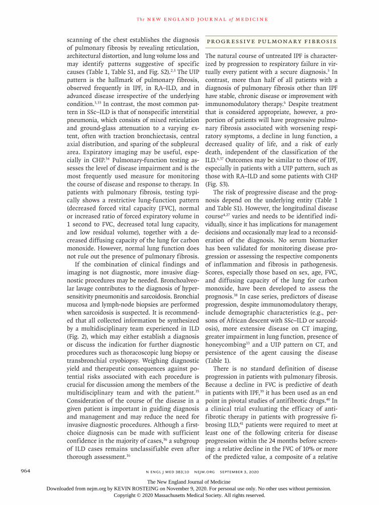
Figure 2. Algorithm for the Diagnosis of Pulmonary Fibrosis.
A multidisciplinary team can play a central role in the diagnosis and management of ILD. Key results of the diagnostic approach are dis-cussed by members of the team, which includes clinicians, radiologists, pathologists, and other health care providers. The participants consider all data available and propose a first-choice diagnosis, assess the need for biopsy and confidence in the diagnosis, and consider possible alternative diagnoses, the potential for disease progression, and the prognosis. On the basis of this discussion, a management decision is made. However, not everyone in the world has equal access to a multidisciplinary team, and even when a multidisciplinary team is available, resources and time may not permit a team discussion of all cases. If a case cannot be addressed by a multidisciplinary team, it is important for the treating clinician to realize that many factors (outermost purple circles) need to be considered before diag-nostic or therapeutic decisions can be made. The high-resolution CT (HRCT) scans at the upper right show a usual interstitial pneumo-nia pattern (top) and an NSIP pattern (bottom). The flow-volume loop shows a typical restrictive pattern (a decrease in forced vital capacity [FVC]) that is often seen in pulmonary fibrosis. The black line represents a predicted normal flow-volume loop for a patient of similar age, sex, height, and race, and the gray zone around it represents the 95% confidence interval. The FVC is shown on the horizontal axis as the volume from the origin to the intersection of the loop with that axis.
Flow-volumeloop in ILD
Exhaled Volume (liters)
Flow
(lite
rs/s
ec)
Biopsyresults
Clinicaldata
HRCT ofthe chest
Broncho-alveolarlavage
Biologic featuresAutoimmunity
Multi-disciplinarydiscussion
Possible needfor biopsy
First-choicediagnosis
Alternativediagnoses
Diagnosticconfidence
PrognosisPotential
for diseaseprogression Lung function
15
10
5
21 43 65 870
−5
−10
−15
The New England Journal of Medicine Downloaded from nejm.org by KEVIN ROSTEING on November 9, 2020. For personal use only. No other uses without permission.
Copyright © 2020 Massachusetts Medical Society. All rights reserved.
n engl j med 383;10 nejm.org September 3, 2020966
T h e n e w e ngl a nd j o u r na l o f m e dic i n e
M a nagemen t
For most patients, a diagnosis of pulmonary fi-brosis is a life-altering verdict. The uncertainty about prognosis in combination with an increas-ing symptom burden has a major effect on the
quality of life of patients and their family mem-bers. According to the underlying condition, treatment can be aimed at ameliorating the dis-ease or slowing down disease progression while improving or maintaining quality of life45 (Fig. 3).
Educating patients and sharing decisions are
Figure 3. Algorithm for the Management of Pulmonary Fibrosis.
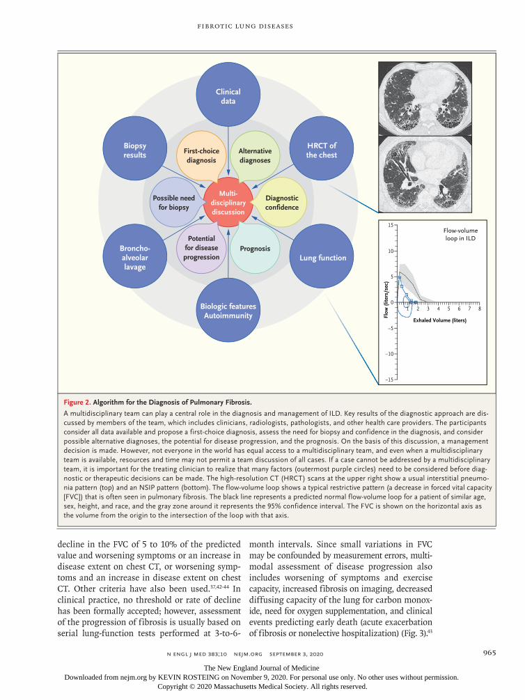
Once a diagnosis of fibrotic ILD has been made, first-line therapy consists of treatment of the underlying disorder, which is often immu-nomodulatory therapy. Depending on the underlying condition, antifibrotic therapy is considered in cases of disease progression despite appropriate first-line therapy. The sequence of medications used may depend on the individual patient and the disease entity. Nonphar-macologic treatment should be considered throughout the disease course. Monitoring for disease progression is based mainly on serial pulmonary-function tests (with progression characterized by a consistent decline in FVC), which are often combined with one or more of the following: measurement of the diffusing capacity of the lung for carbon monoxide, assessment of symptoms and exercise capacity, CT findings (with an increase in the extent of fibrosis indicating progression), measurement of oxygen saturation during exercise, and supplemental oxygen requirements. Acute exacerbations of pulmonary fibrosis also represent progressive disease. In patients with idio-pathic pulmonary fibrosis (IPF), antifibrotic agents should be offered at diagnosis. If there is disease progression, the diagnosis and treat-ment options should be reviewed before adding the treatments shown in purple. ABA denotes abatacept, ADA adalimumab, AZA azathi-oprine, CPM cyclophosphamide, IFX infliximab, MMF mycophenolate mofetil, MTX methotrexate, RA–ILD rheumatoid arthritis and ILD, RTX rituximab, SSc–ILD systemic sclerosis and ILD, and TCL tocilizumab.
Chronic hypersensitivitypneumonitis
Antigeneviction
SarcoidosisIdiopathic
NSIPIPF
Unclassifiable ILD
Management
Monitoring of Disease Course
Disease progression Stable disease Ameliorization
SSc–ILD
Consider immunomodulation treatment or observation
RA–ILD
Glucocorticoids
MMF, CPM, TCL(AZA, RTX)
Glucocorticoids
RTX, ABA,MMF
Consider antifibrotic agents (nintedanib)
Nonpharmacologic treatmentSupplemental oxygen, psychosocial support, smoking cessation, rehabilitation, symptom palliation, end-of-life care
MMF(AZA)
Glucocorticoids
MTX(AZA, IFX, ADA)
Glucocorticoids
MMF, AZA, or otherimmunosuppressants
Antifibrotic agents(pirfenidone or nintedanib)
Glucocorticoids
Regular Follow-upReconsider Management
The New England Journal of Medicine Downloaded from nejm.org by KEVIN ROSTEING on November 9, 2020. For personal use only. No other uses without permission.
Copyright © 2020 Massachusetts Medical Society. All rights reserved.
n engl j med 383;10 nejm.org September 3, 2020 967
Fibrotic Lung Diseases
important, especially since there are many off-label treatment options with potentially serious side effects. Preventing exposures and events that may drive further disease progression is essen-tial. Avoidance of the offending antigen in pa-tients with CHP and cessation of tobacco smok-ing are priorities. Pneumococcal and influenza vaccinations are recommended. On the basis of expert opinion, supplemental oxygen is indicated in patients with resting hypoxemia (partial pres-sure of arterial oxygen [Pao2] of <55 mm Hg, oxygen saturation as measured by pulse oximetry of <89%, or Pao2 of <60 mm Hg and cor pulmo-nale or polycythemia).46 Pulmonary rehabilitation45 and use of ambulatory oxygen in patients with isolated exertional hypoxemia47 improve the qual-ity of life, reduce breathlessness, and increase walking ability. Identification and accurate treat-ment of coexisting conditions are essential. Lung transplantation is an option in select patients, although extrapulmonary disease or severe coex-isting conditions may disqualify some patients, especially those with CTDs, from consideration as candidates for transplantation.48 For many patients, the focus is on palliative care.49
Decisions about pharmacologic treatment are guided by the underlying diagnosis and by the disease course. For patients with IPF, treatment with antifibrotic drugs (pirfenidone or nintedanib) is recommended.50 In most cases of fibrosing ILD other than IPF, immunomodulation with the use of glucocorticoids, immunosuppressive ther-apy, or both is indicated and is generally used as first-line therapy if there is a suspicion of in-flammation-driven disease.18,37,51 Except for SSc–ILD and sarcoidosis, however, the evidence in sup-port of this approach is very weak.51 In patients with a UIP pattern, there is theoretical concern that immunosuppression may not be beneficial or might even be harmful, as was previously shown in IPF.52
Nintedanib has been approved by the Food and Drug Administration (FDA) and the Euro-pean Medicines Agency (EMA) for patients with SSc–ILD and for patients with chronic fibrosing ILDs with a progressive phenotype. This agent is not associated with an improvement in function but reduces the decline in FVC by about half,41 supporting the notion that progressive pulmo-nary fibrosis may be amenable to antifibrotic therapy regardless of the underlying specific disease. Pirfenidone reduces disease progression in patients with progressive, unclassifiable, fi-brotic ILD.44 In considering pharmacologic treat-ment, the benefit of long-term preservation of lung function should be balanced against the risk of side effects. Many questions remain, however, about appropriate timing and sequence of these treatments.
Fu t ur e Dir ec tions
Pulmonary fibrosis is a pathologic process that stems from multiple underlying causes. Moni-toring disease progression has become a priority in guiding treatment decisions. We hope that, in the coming years, different biomarkers and novel techniques such as molecular classifiers53 will provide more insights into assessing and moni-toring fibrosis-driven as compared with inflam-mation-driven disease activity, resulting in more individualized targeted treatments, since it is clear that a “one size fits all” approach does not apply to the broad spectrum of fibrosing dis-eases. Current research efforts may lead to ear-lier diagnosis and interventions to prevent, halt, and potentially reverse the development of life-limiting lung fibrosis.
Disclosure forms provided by the authors are available with the full text of this article at NEJM.org.
We thank Jan von der Thüsen, M.D., Ph.D., for the histologic images.
References1. Katzenstein AL, Myers JL. Idiopathic pulmonary fibrosis: clinical relevance of pathologic classification. Am J Respir Crit Care Med 1998; 157: 1301-15.2. Raghu G, Remy-Jardin M, Myers JL, et al. Diagnosis of idiopathic pulmonary fibrosis: an official ATS/ERS/JRS/ALAT clinical practice guideline. Am J Respir Crit Care Med 2018; 198(5): e44-e68.3. Lederer DJ, Martinez FJ. Idiopathic pulmonary fibrosis. N Engl J Med 2018; 378: 1811-23.4. Wells AU, Brown KK, Flaherty KR,
Kolb M, Thannickal VJ. What’s in a name? That which we call IPF, by any other name would act the same. Eur Respir J 2018; 51: 1800692.5. Rockey DC, Bell PD, Hill JA. Fibrosis — a common pathway to organ injury and failure. N Engl J Med 2015; 372: 1138-49.6. Cottin V, Wollin L, Fischer A, Qua-resma M, Stowasser S, Harari S. Fibrosing interstitial lung diseases: knowns and un-knowns. Eur Respir Rev 2019; 28: 180100.7. Wolters PJ, Blackwell TS, Eickelberg O, et al. Time for a change: is idiopathic pul-
monary fibrosis still idiopathic and only fibrotic? Lancet Respir Med 2018; 6: 154-60.8. Duchemann B, Annesi-Maesano I, Jacobe de Naurois C, et al. Prevalence and incidence of interstitial lung diseases in a multi-ethnic county of Greater Paris. Eur Respir J 2017; 50: 1602419.9. Perelas A, Silver RM, Arrossi AV, Highland KB. Systemic sclerosis-associ-ated interstitial lung disease. Lancet Respir Med 2020; 8: 304-20.10. van den Hoogen F, Khanna D, Fran-sen J, et al. 2013 Classification criteria for
The New England Journal of Medicine Downloaded from nejm.org by KEVIN ROSTEING on November 9, 2020. For personal use only. No other uses without permission.
Copyright © 2020 Massachusetts Medical Society. All rights reserved.
n engl j med 383;10 nejm.org September 3, 2020968
Fibrotic Lung Diseases
systemic sclerosis: an American College of Rheumatology/European League against Rheumatism collaborative initiative. Arthri-tis Rheum 2013; 65: 2737-47.11. Aletaha D, Neogi T, Silman AJ, et al. 2010 Rheumatoid arthritis classification criteria: an American College of Rheuma-tology/European League against Rheuma-tism collaborative initiative. Ann Rheum Dis 2010; 69: 1580-8.12. Shaw M, Collins BF, Ho LA, Raghu G. Rheumatoid arthritis-associated lung dis-ease. Eur Respir Rev 2015; 24: 1-16.13. Grunewald J, Grutters JC, Arkema EV, Saketkoo LA, Moller DR, Müller-Quern-heim J. Sarcoidosis. Nat Rev Dis Primers 2019; 5: 45.14. Raghu G, Remy-Jardin M, Ryerson CJ, et al. Diagnosis of hypersensitivity pneu-monitis in adults. An official ATS/JRS/ALAT Clinical Practice Guideline. Am J Respir Crit Care Med 2020; 202(3): e36-e69.15. Vasakova M, Morell F, Walsh S, Leslie K, Raghu G. Hypersensitivity pneumonitis: perspectives in diagnosis and manage-ment. Am J Respir Crit Care Med 2017; 196: 680-9.16. Ryerson CJ, Urbania TH, Richeldi L, et al. Prevalence and prognosis of unclas-sifiable interstitial lung disease. Eur Res-pir J 2013; 42: 750-7.17. Hyldgaard C, Bendstrup E, Wells AU, Hilberg O. Unclassifiable interstitial lung diseases: clinical characteristics and sur-vival. Respirology 2017; 22: 494-500.18. Wijsenbeek M, Kreuter M, Olson A, et al. Progressive fibrosing interstitial lung diseases: current practice in diagnosis and management. Curr Med Res Opin 2019; 35: 2015-24.19. Olson AL, Gifford AH, Inase N, Fernández Pérez ER, Suda T. The epidemi-ology of idiopathic pulmonary fibrosis and interstitial lung diseases at risk of a progressive-fibrosing phenotype. Eur Respir Rev 2018; 27: 180077.20. Raghu G, Chen S-Y, Hou Q, Yeh W-S, Collard HR. Incidence and prevalence of idiopathic pulmonary fibrosis in US adults 18-64 years old. Eur Respir J 2016; 48: 179-86.21. Valeyre D, Prasse A, Nunes H, Uzun-han Y, Brillet PY, Müller-Quernheim J. Sarcoidosis. Lancet 2014; 383: 1155-67.22. Thannickal VJ, Zhou Y, Gaggar A, Duncan SR. Fibrosis: ultimate and proxi-mate causes. J Clin Invest 2014; 124: 4673-7.23. Distler JHW, Györfi A-H, Ramanujam M, Whitfield ML, Königshoff M, Lafyatis R. Shared and distinct mechanisms of fibro-sis. Nat Rev Rheumatol 2019; 15: 705-30.24. Adegunsoye A, Vij R, Noth I. Integrat-ing genomics into management of fi-brotic interstitial lung disease. Chest 2019; 155: 1026-40.25. Juge P-A, Lee JS, Ebstein E, et al. MUC5B promoter variant and rheumatoid arthritis with interstitial lung disease. N Engl J Med 2018; 379: 2209-19.26. Ley B, Torgerson DG, Oldham JM, et al. Rare protein-altering telomere-related gene
variants in patients with chronic hyper-sensitivity pneumonitis. Am J Respir Crit Care Med 2019; 200: 1154-63.27. Travis WD, Costabel U, Hansell DM, et al. An official American Thoracic Society/European Respiratory Society statement: update of the international multidisci-plinary classification of the idiopathic interstitial pneumonias. Am J Respir Crit Care Med 2013; 188: 733-48.28. Figliozzi S, Masci PG, Ahmadi N, et al. Predictors of adverse prognosis in Covid-19: a systematic review and meta-analysis. Eur J Clin Invest 2020 July 29 (Epub ahead of print).29. Burnham EL, Janssen WJ, Riches DW, Moss M, Downey GP. The fibroprolifera-tive response in acute respiratory distress syndrome: mechanisms and clinical sig-nificance. Eur Respir J 2014; 43: 276-85.30. Chang YC, Yu CJ, Chang SC, et al. Pul-monary sequelae in convalescent patients after severe acute respiratory syndrome: evaluation with thin-section CT. Radiology 2005; 236: 1067-75.31. Sgalla G, Walsh SLF, Sverzellati N, et al. “Velcro-type” crackles predict specific ra-diologic features of fibrotic interstitial lung disease. BMC Pulm Med 2018; 18: 103.32. Mathai SC, Danoff SK. Management of interstitial lung disease associated with con-nective tissue disease. BMJ 2016; 352: h6819.33. Adegunsoye A, Oldham JM, Bellam SK, et al. Computed tomography honeycomb-ing identifies a progressive fibrotic pheno-type with increased mortality across di-verse interstitial lung diseases. Ann Am Thorac Soc 2019; 16: 580-8.34. Barnett J, Molyneaux PL, Rawal B, et al. Variable utility of mosaic attenuation to distinguish fibrotic hypersensitivity pneu-monitis from idiopathic pulmonary fibro-sis. Eur Respir J 2019; 54: 54.35. Kolb M, Raghu G, Wells A. Prognostic impact of typical and probable usual in-terstitial pneumonia pattern in idiopathic pulmonary fibrosis: is the debate about biopsy a Star Wars saga? Eur Respir J 2020; 55: 2000590.36. Walsh SLF, Lederer DJ, Ryerson CJ, et al. Diagnostic likelihood thresholds that define a working diagnosis of idiopathic pulmonary fibrosis. Am J Respir Crit Care Med 2019; 200: 1146-53.37. George PM, Spagnolo P, Kreuter M, et al. Progressive fibrosing interstitial lung disease; consensus recommendations, clin-ical uncertainties and research priorities. Lancet Respir Med (in press).38. Ryerson CJ, Vittinghoff E, Ley B, et al. Predicting survival across chronic inter-stitial lung disease: the ILD-GAP model. Chest 2014; 145: 723-8.39. Paterniti MO, Bi Y, Rekić D, Wang Y, Karimi-Shah BA, Chowdhury BA. Acute exacerbation and decline in forced vital capacity are associated with increased mor-tality in idiopathic pulmonary fibrosis. Ann Am Thorac Soc 2017; 14: 1395-402.40. Karimi-Shah BA, Chowdhury BA. Forced vital capacity in idiopathic pulmonary fibro-
sis — FDA review of pirfenidone and nin-tedanib. N Engl J Med 2015; 372: 1189-91.41. Flaherty KR, Wells AU, Cottin V, et al. Nintedanib in progressive fibrosing inter-stitial lung diseases. N Engl J Med 2019; 381: 1718-27.42. Behr J, Neuser P, Prasse A, et al. Explor-ing efficacy and safety of oral pirfenidone for progressive, non-IPF lung fibrosis (RELIEF) — a randomized, double-blind, placebo-controlled, parallel group, multi-center, phase II trial. BMC Pulm Med 2017; 17: 122.43. Cottin V. Treatment of progressive fi-brosing interstitial lung diseases: a mile-stone in the management of interstitial lung diseases. Eur Respir Rev 2019; 28: 190109.44. Maher TM, Corte TJ, Fischer A, et al. Pirfenidone in patients with unclassifi-able progressive fibrosing interstitial lung disease: a double-blind, randomised, pla-cebo-controlled, phase 2 trial. Lancet Respir Med 2020; 8: 147-57.45. Wijsenbeek MS, Holland AE, Swigris JJ, Renzoni EA. Comprehensive support-ive care for patients with fibrosing inter-stitial lung disease. Am J Respir Crit Care Med 2019; 200: 152-9.46. Lim RK, Humphreys C, Morisset J, Hol-land AE, Johannson KA, O2 Delphi Collabo-rators. Oxygen in patients with fibrotic in-terstitial lung disease: an international Delphi survey. Eur Respir J 2019; 54: 54.47. Visca D, Mori L, Tsipouri V, et al. Effect of ambulatory oxygen on quality of life for patients with fibrotic lung disease (AmbOx): a prospective, open-label, mixed-method, crossover randomised controlled trial. Lancet Respir Med 2018; 6: 759-70.48. Weill D, Benden C, Corris PA, et al. A consensus document for the selection of lung transplant candidates: 2014 — an update from the Pulmonary Transplanta-tion Council of the International Society for Heart and Lung Transplantation. J Heart Lung Transplant 2015; 34: 1-15.49. Kreuter M, Bendstrup E, Russell AM, et al. Palliative care in interstitial lung disease: living well. Lancet Respir Med 2017; 5: 968-80.50. Raghu G, Rochwerg B, Zhang Y, et al. An official ATS/ERS/JRS/ALAT clinical prac-tice guideline: treatment of idiopathic pulmonary fibrosis — an update of the 2011 clinical practice guideline. Am J Respir Crit Care Med 2015; 192(2): e3-e19.51. Maher TM, Wuyts W. Management of fibrosing interstitial lung diseases. Adv Ther 2019; 36: 1518-31.52. The Idiopathic Pulmonary Fibrosis Clin-ical Research Network. Prednisone, azathio-prine, and N-acetylcysteine for pulmonary fibrosis. N Engl J Med 2012; 366: 1968-77.53. Raghu G, Flaherty KR, Lederer DJ, et al. Use of a molecular classifier to identify usual interstitial pneumonia in convention-al transbronchial lung biopsy samples: a prospective validation study. Lancet Respir Med 2019; 7: 487-96.Copyright © 2020 Massachusetts Medical Society.
The New England Journal of Medicine Downloaded from nejm.org by KEVIN ROSTEING on November 9, 2020. For personal use only. No other uses without permission.
Copyright © 2020 Massachusetts Medical Society. All rights reserved.