Embed Size (px)
Citation preview

fmicb-07-00501 April 12, 2016 Time: 17:33 # 1
REVIEWpublished: 14 April 2016
doi: 10.3389/fmicb.2016.00501
Edited by:Agostinho Carvalho,
University of Minho, Portugal
Reviewed by:Igor C. Almeida,
University of Texas at El Paso, USALauren Ashley Cowart,
Medical University of South Carolina,USA
*Correspondence:Maurizio Del Poeta
Specialty section:This article was submitted toFungi and Their Interactions,
a section of the journalFrontiers in Microbiology
Received: 27 December 2015Accepted: 28 March 2016
Published: 14 April 2016
Citation:Singh A and Del Poeta M (2016)Sphingolipidomics: An Important
Mechanistic Tool for Studying FungalPathogens. Front. Microbiol. 7:501.
doi: 10.3389/fmicb.2016.00501
Sphingolipidomics: An ImportantMechanistic Tool for Studying FungalPathogensAshutosh Singh1,2 and Maurizio Del Poeta1,2*
1 Department of Molecular Genetics and Microbiology, Stony Brook University, Stony Brook, NY, USA, 2 VeteransAdministration Medical Center, Northport, NY, USA
Sphingolipids form of a unique and complex group of bioactive lipids in fungi.Structurally, sphingolipids of fungi are quite diverse with unique differences in thesphingoid backbone, amide linked fatty acyl chain and the polar head group. Two of themost studied and conserved sphingolipid classes in fungi are the glucosyl- or galactosyl-ceramides and the phosphorylinositol containing phytoceramides. Comprehensivestructural characterization and quantification of these lipids is largely based onadvanced analytical mass spectrometry based lipidomic methods. While separation ofcomplex lipid mixtures is achieved through high performance liquid chromatography,the soft – electrospray ionization tandem mass spectrometry allows a high sensitivityand selectivity of detection. Herein, we present an overview of lipid extraction,chromatographic separation and mass spectrometry employed in qualitative andquantitative sphingolipidomics in fungi.
Keywords: sphingolipids, high performance liquid chromatography, electrospray ionization tandem massspectrometry, fungi, fungal infections, ceramide
INTRODUCTION
As a group, sphingolipids are essential components of all eukaryotic cell membranes (Dickson andLester, 2002). In fungi, sphingolipids play an important role in a variety of biological processeslike cell division (Epstein et al., 2012), heat stress response (Jenkins et al., 1997), acid/alkalinetolerance (Luberto et al., 2001; Rittershaus et al., 2006), hyphae formation (Oura and Kajiwara,2010), domain formation (Marquês et al., 2015) spore germination (Levery et al., 2002), endocytosis(Zanolari et al., 2000), signal transduction (Obeid et al., 2002), apoptosis (Cheng et al., 2003),pathogenesis and virulence (Luberto et al., 2001; Rittershaus et al., 2006). Endowed with uniquechemical structure and synthesized by fungal specific enzymes, these sphingolipids are ideal drug
Abbreviations: CE, collision energy; CID, collision-induced dissociation; ESI, electrospray ionization; FAB, fastatom bombardment; FT-ICR, Fourier-transform ion cyclotron resonance; GC, gas chromatography; GC-EI-MS, gaschromatography-electron impact-mass spectrometry; Gcs1, glucosylceramide synthase; HILIC, hydrophilic-interactionliquid chromatography; HPLC, high-performance liquid chromatography; IPC, inositolphosphoryl ceramide; LCB, long-chain base; MALDI, matrix-assisted laser desorption/ionization; MIPC, mannosyl-inositol phosphorylceramide; MS/MS,tandem mass spectrometry; MSn, multiple-stage mass spectrometry; (M(IP)2C, mannosyldiinositolphosphoryl ceramide;MRM, multiple-reaction monitoring; NL, neutral loss; NMR, nuclear magnetic resonance spectroscopy; PREIS, precursor-ion scanning; QqTOF MS, hybrid quadrupole time-of–flight mass spectrometer; RF, radiofrequency; SRM, single-reactionmonitoring; Smt1, sphingolipid C9 methyltransferase; Sld8, 18-desaturase; SPE, solid-phase extraction; TLC, thin-layerchromatography; TMS, trimethylsilyl; TOF, time-of-flight; UDP, uridine 5-diphosphate.
Frontiers in Microbiology | www.frontiersin.org 1 April 2016 | Volume 7 | Article 501

fmicb-07-00501 April 12, 2016 Time: 17:33 # 2
Singh and Del Poeta Lipid Profiling of Pathogenic Fungi
targets (Mor et al., 2015). In this context, it is important tocharacterize sphingolipids in greater detail.
Characterization of various fungal sphingolipids requires theunderstanding of three key components (Figure 1). These are: thepathways of biosynthesis and degradation; structures of varioussphingolipids being synthesized; understanding the ways forefficient extraction of sphingolipids from the cell and the precisemethods for their analysis. A tremendous amount of literaturein the field of fungal lipid metabolism allows us to categoricallyunderstand these components. In the sections below, we havedescribed a literature based review of these key componentsof sphingolipid characterization, with an emphasis on massspectrometry based structural and functional characterization ofsphingolipids from pathogenic fungi.
In general, the structure of sphingolipids comprise a LCBbackbone amide- linked to a fatty acid at C2 position and esterlinked to a polar head group at C1 position (Del Poeta et al.,2014). There is a large diversity in sphingoid bases of mammals(Kendall et al., 2015), plants (Luttgeharm et al., 2015), and fungi(Del Poeta et al., 2014). In fungal cells, de novo sphingolipidbiosynthesis begins with condensation of L-serine and palmitoyl-CoA to form 3-ketodihydrosphingosine which is then reducedto dihydrosphingosine (d18:0 backbone; sphinganine; Figure 2A;Buede et al., 1991; Nagiec et al., 1994). This step leads to theformation of an 18 C containing LCB. Dihydrosphingosine isthen amide linked to a fatty acid (usually α-hydroxylated, 18 or16 C containing) by ceramide synthases to form dihydroceramide(Lahiri and Futerman, 2005). Further, a 14-desaturation of thesphingoid backbone of dihydroceramide forms ceramide (d18:1backbone; 4-sphingenine; Ternes et al., 2011; Rodriguez-Cuencaet al., 2015). Next, a 18-desaturation of the sphingoid backboneof 14-ceramide leads to formation of 14, 18-ceramide (d18:2backbone; 4,8-sphingadienine; Sperling et al., 2000; Figure 2A).
A sphingolipid C9-methyl transferase catalyzes the additionof a methyl group at C9 position of the 4,8-sphingadieninebase of 14, 18-ceramide to form 9-methyl-14, 18-ceramide(d19:2 backbone; 9-methyl-4,8-sphingadienine; Ternes et al.,2006; Singh et al., 2012; Figure 2A). Finally, a Gcs1 catalyzesthe transfer of glucose moiety from the UDP-glucose onto theC1 hydroxyl group of the ceramide forming glucosylceramide(Rittershaus et al., 2006). Although all the enzymatic stepsare not well characterized, some have been studied in fungilike Cryptococcus neoformans (Rittershaus et al., 2006; Singhet al., 2012), Aspergillus nidulans (Li et al., 2006), A. fumigatus(Levery et al., 2002; Kotz et al., 2010), Fusarium graminearum(Rittenour et al., 2011) and Candida albicans (Cheon et al.,2012). Notably, glucosylceramide biosynthesis is absent inSaccharomyces cerevisiae and C. glabrata (Leipelt et al., 2001).
In plants, 8-sphingenine (d18:1) and 4,8-sphingenine (d18:2)represent the major sphingoid bases, however, presence of the cis-and trans- isomeric forms result in nine different C18 sphingoidbases (Ohnishi et al., 1983). This results in a complex mixture ofglucosylceramide pool in plants (Imai et al., 2012). In mammals,the major sphingoid base is 4-sphingenine (d18:1) which is linkedto 16 C fatty acid in the glucosylceramide structure (Leipelt et al.,2001). In contrast, the fungal glucosylceramide structure is ratherunique. The sphingoid backbone is composed of 9-methyl-4,8-sphingadienine which is amide linked to α-hydroxylated C18:0fatty acid (Figure 2B; Del Poeta et al., 2014). Certain fungalspecies contain galactose instead of glucose in this cerebrosidestructure (Warnecke and Heinz, 2003). The α-hydroxylatedC16:0 and α-hydroxylated C18:1 are the two other major fattyacyls reported in the fungal cerebroside structure (Barreto-Bergter et al., 2004). The structures of different cerebrosidesproduced by fungi have been extensively reviewed by Barreto-Bergter et al. (2011).
FIGURE 1 | Key components of sphingolipid characterization in fungi.
Frontiers in Microbiology | www.frontiersin.org 2 April 2016 | Volume 7 | Article 501

fmicb-07-00501 April 12, 2016 Time: 17:33 # 3
Singh and Del Poeta Lipid Profiling of Pathogenic Fungi
FIGURE 2 | Sphingolipid biosynthesis in fungi. (A) Scheme of fungal sphingolipid biosynthetic pathway. Enzymatic steps with reverse reaction shown in bracketsare as follows: (1) Serine C-palmitoyl transferase; (2) 3-ketosphinganine reductase; (3) Dihydrosphingosine kinase [Dihydrosphingosine-1P-phosphatase]; (4)Long-chain base (LCB) phosphate lyase; (5) Dihydroceramide synthase; (6) Dihydroceramide C4 desaturase; (7) Ceramidase [Ceramide synthase]; (8) Sphingosinekinase [Sphingosine-1P-phosphatase]; (9) Ceramide C8 desaturase; (10) Sphingolipid C9 methyltransferase (Smt1); (11) Hexosyl ceramide synthase [Hexosylceramidase]; (12) Dihydrosphingosine C4-hydroxylase (13) Phytosphingosine kinase [Phytosphingosine -1P-phosphatase]; (14) Ceramide synthase [Ceramidase];(15) Sphingolipid α-hydroxylase; (16) IPC synthase; (17) IPC mannosyl transferase; (18) Inositolphosphotransferase; (19) Inositol phosphosphingolipid phospholipaseC. Major sphingoid backbone in fungi is composed of 18 C-atoms (shown in blue) and the fatty acyls attached to various sphingolipids comprise of 16–26 C-atoms
(Continued)
Frontiers in Microbiology | www.frontiersin.org 3 April 2016 | Volume 7 | Article 501
fmicb-07-00501 April 12, 2016 Time: 17:33 # 4
Singh and Del Poeta Lipid Profiling of Pathogenic Fungi
FIGURE 2 | Continued
(shown in red). Putative conversion steps of dihydroceramide to phytoceramide and to IPC are represented by dashed arrows. (B) Structure of fungal glucosylceramide.Long chain sphingoid backbone amide linked to a fatty acyl and linked by β-glycosidic bond to a polar head group (glucose) at C1 position. Unique features of fungalglucosylceramide are: 14 and 18 double bonds in the sphingoid backbone (shown in green), 9-methylation in the sphingoid backbone (shown in yellow), α-hydroxylfatty acyl (shown in orange) and hydroxyl groups in the hexose moiety (shown in blue). (C) Structure of fungal IPC. The distinguished features of IPC’s in fungi are:C3-hydroxylation of sphingoid backbone and α-hydroxyl fatty acyl (both shown in orange), phosphate linked inositol group (shown in grey). ‘R’ represents 16–24carbons in the fatty acyl chain.
Fungi also produce phytoceramide (Heung et al., 2006).Dihydrosphingosine C4-hydroxylase catalyzes the addition ofa hydroxyl group at C4 position of dihydrosphingosinebackbone to produce phytosphingosine (t18:0 backbone; 4-hydroxysphinganine; Li et al., 2007; Figure 2A). Ceramidesynthases then transfer a non-hydroxylated fatty acid (18, 24or 26 C containing) to amide group at C2 position to formphytoceramide (Cheon et al., 2012). The fatty acyl moietyof the phytoceramides is α-hydroxylated by a sphingolipidα-hydroxylase to form αOH-phytoceramides (Haak et al.,1997). Both phytoceramides and αOH-phytoceramides arefurther converted to complex phosphosphingolipids such as IPC(transfer of phosphorylinositol group to phytoceramide), MIPC,(transfer of mannosyl group to IPC) and M(IP)2C, (transferof a second phosphorylinositol group to MIPC; Guan andWenk, 2008). Although IPC derivatives are not reported inmammals, certain plants and kinetoplastid protozoa do produceIPC derivatives (Heung et al., 2006).
The t18:0 phytosphingosine backbone, C2 hygroxylated fattyacyls and phosphorylinositol containing polar head group are theunique feature of IPC derivatives (Figure 2C). IPC structuresand pathway of synthesis have been characterized in severalfungi like S. cereviseae (Ejsing et al., 2009; Voynova et al., 2014),C. neoformans (Luberto et al., 2001), A. fumigatus (Kuroda et al.,1999; Heidler and Radding, 2000), A. nidulans (Bennion et al.,2003), Histoplasma capsulatum (Guimarães et al., 2014), andC. albicans (Singh et al., 2010; Cheon et al., 2012).
The re-cycling of fungal sphingolipids involves the productionof sphingosine, sphingosine-1-phosphate, dihydrosphingosine-1-phosphate, and phytosphingosine-1-phosphate (Figure 2A).The ultimate degradation steps of sphingolipids are constitutedby the catabolism of dihydrosphingosine-1-phosphate andsphingosine-1-phosphate into palmitaldehyde and phosphorylethanolamine (Kim et al., 2000; Serra and Saba, 2010) and thephytosphingosine-1-phosphate into 2-hydroxy-palmitaldehydeand phosphoryl ethanolamine (Kondo et al., 2014), by the long-chain basse-1-phosphate lyases (Figure 2A).
The ability of fungal cells to produce sphingolipids withdifferent sphingoid backbone structures adds to the complexityof the lipid mixtures (Guimarães et al., 2014). In additionto the backbone structure, the degree of unsaturation,fatty acyl chain lengths, methylation and hydroxylationmodifications, all make it quite difficult to analyze these lipidsusing classical techniques like in vivo labeling (Chigornoet al., 2006), TLC (Urban et al., 1980) and GC (Cacas et al.,2012). Therefore, advanced analytical methods for theanalyses of these lipids are employed to characterize theirstructures.
In last two decades, several mass spectrometry techniques havebeen used to identify and characterize the total sphingolipid poolor the “sphingolipidome.” A wide variety of mass spectrometrybased methods are available in literature that allows accurateanalyses of these complex sphingolipid mixtures (Merrill et al.,2005, 2009; Haynes et al., 2009; Wenk, 2010; Köfeler et al., 2012).Below we describe methods of extraction, chromatographicseparation and the mass spectrometry based strategies to analyzesphingolipids.
LIPID EXTRACTION
Extraction of lipids is the most crucial step for lipid analysisby both classical and high throughput techniques. Currentlyseveral modified adaptations of Folch method (Folch et al.,1957) and Bligh and Dyer method (Bligh and Dyer, 1959) arebeing employed to extract lipids from fungal cells (Prasad andGhannoum, 1996; Schneiter and Daum, 2006; Ejsing et al.,2009; Haynes et al., 2009). In our laboratory and others, weuse the method described by Mandala et al. (1995) for lipidextraction. This method has shown a good extraction efficiencyand reproducibility for lipid analyses. The scheme of lipidextraction from fungal cells is shown in Figure 3. Fungal cellsare extracted in ethanol: dH2O:diethylether:pyridine:NH4OH(15:15:5:1:0.018; v/v) at 60◦C for 1 h as described previously(Hanson and Lester, 1980). Lipid extract is then subjected to asolution of methanol:chloroform (2:1; v/v) followed by additionof 1/3rd volume chloroform and 1/3rd volume dH2O and thelower organic phase reserved (Bligh and Dyer, 1959). Organicphase is dried in SpeedVac, flushed with N2 and stored in−20◦C. At this stage, these lipid extracts can be used for theestimation of inorganic phosphate (Pi) content, dry lipid weightor for the isolation and purification specific lipid classes (Merrillet al., 2009; Rana et al., 2015). Both Pi content and dry lipidweight have been used to normalize the lipid amounts. Severalgroups have employed SPE techniques to further purify specificsphingolipid groups (Bodennec et al., 2000; Barreto-Bergteret al., 2004). Extensive lipid purification by SPE is cumbersomeand time consuming, and is usually not required for routinelipid analysis; however, is preferred for a thorough structuralcharacterization of complex glycosphingolipids (Barreto-Bergteret al., 2004, 2011).
Glycerolipids are the major lipid contaminants that are co-extracted with sphingolipids (Bligh and Dyer, 1959). A mildalkaline methanolysis (Clarke and Dawson, 1981), usually for60 min at room temperature is sufficient to hydrolyze theester linkages of fatty acyls of glycerolipids. This allows a
Frontiers in Microbiology | www.frontiersin.org 4 April 2016 | Volume 7 | Article 501

fmicb-07-00501 April 12, 2016 Time: 17:33 # 5
Singh and Del Poeta Lipid Profiling of Pathogenic Fungi
FIGURE 3 | Procedure of sphingolipid extraction. Prior to extraction,fungal cell extracts or lyophilized media are spiked with suitable internalstandards. First extraction is performed using Mandala extraction buffer(ethanol: dH2O: diethyl ether: pyridine: 4.2N NH4OH; 15: 15: 5: 1: 0.018; v/v).Subsequently, a second Bligh and Dyer extraction is performed usingmethanol and chloroform. Samples at this stage can be used to determine thePi content, lipid dry weight or to purify specific sphingolipid components usingsolid phase extraction. Finally, samples are base hydrolyzed using mild alkalinebase hydrolysis (0.6 M KOH in methanol) to remove the glycerol backbonecontaining lipid contaminants. For certain fungal masses it is required to firstcrush the sample using glass beads in the lipid extraction buffer itself or tomake the sample into powder using pestle and mortar in liquid N2 prior toextraction.
clean extraction of alkaline-stable components, which are highlyenriched in sphingolipids.
For accurate quantification of lipids by mass spectrometry,internal standards must be added prior to lipid extraction(Rana et al., 2015). Although, absolute quantification of eachlipid species by mass spectrometry requires the use of isotope-labeled standard for that species (Ecker and Liebisch, 2014);however, presently this is not possible due to high synthesiscosts and the large number of lipids being analyzed. Additionof one internal standard per lipid class being analyzed is widelyaccepted (LIPID MAPS Consortium). This is primarily becausethe ionization of lipid species is largely dependent upon thespecific head group rather than the attached fatty acyls (Köfeler
et al., 2012). The C17-sphingoid backbone lipids (Avanti PolarLipids Inc., Alabaster, AL, USA; Matreya Inc., Pleasant Gap, PA,USA) are routinely used as internal standards as these closelyresemble C18 lipids in physicochemical properties and ionizationefficiencies (Rana et al., 2015). Quantification of sphingolipidspecies with different chain lengths can be achieved using thecalibration curves of closest chain length standards (Rana et al.,2015).
METHODS OF LIPID ANALYSIS
In Vivo Labeling, Thin-layerChromatography and GasChromatography Mass SpectrometryFor several decades, in vivo labeling has been used to characterizethe sphingolipid metabolic pathways in fungi (Chigorno et al.,2006). The technique involves the uptake of a radiolabeledprecursor by cells. The radiolabeled precursor then getsincorporated into the complex lipid structures. Heavy labeled[14C]-palmitate and [14C]-serine are the most commonly usedlabeled precursors to follow sphingolipid synthesis (Chigornoet al., 2006). However, these labels get incorporated intoother untargeted lipids and give high background signals.[3H]-dihydrosphingosine provides a more sphingolipid specificlabeling (Karashima et al., 2013). [3H]-inositol has been usedto focus on IPC derivatives (Haak et al., 1997) and [3H]-or [14C]- glucose or galactose have been used to focus onglycosphingolipids (Sasaki, 1981). Unfortunately, the very longhalf-life of these radiolabeled precursors presents a serious risk tohealth if exposed and is a challenge during disposal (Ecker andLiebisch, 2014).
Lipid extracts from labeled or unlabeled cells can be resolvedusing the TLC technique (Figure 4A). It is a simple, costeffective, fast and sensitive method to qualitatively characterizelipids. Identification of broad lipid classes by TLC is simple andachieved by comparing their mobility to the standard (Urbanet al., 1980). The most commonly used solvent systems to resolvesphingolipids on a TLC include: chloroform:methanol:dH2O(65:25:4; v/v), chloroform:methanol:4.2N NH4OH (9:7:2 and40:10:1; v/v) and chloroform:methanol:acetic acid (90:2:8; v/vMandala et al., 1995; Bodennec et al., 2000; Barreto-Bergteret al., 2004). Unlabeled sphingolipids are visualized usingiodine (Palumbo and Zullo, 1987) or orcinol/resorcinol staining(Tadano and Ishizuka, 1982) and the labeled sphingolipid arevisualized by autoradiography on an X-ray film (Kondo et al.,2014).
Structural composition of the purified sphingolipids can bedetermined using GC-EI-MS (Cacas et al., 2012). Only thevolatile components that can be carried by the carrier gas (He)can be analyzed by GCMS. Most sphingolipid structures arenon-volatile. For this reason, sphingolipids are hydrolyzed torelease the fatty acid, sugar, and LCB components, which arethen derivatized into methyl esters or TMS derivatives andanalyzed by GC-EI-MS (Figure 4A; Christie, 1989; Matsubaraand Hayashi, 1991; Johnson and Brown, 1992; Merkle and
Frontiers in Microbiology | www.frontiersin.org 5 April 2016 | Volume 7 | Article 501

fmicb-07-00501 April 12, 2016 Time: 17:33 # 6
Singh and Del Poeta Lipid Profiling of Pathogenic Fungi
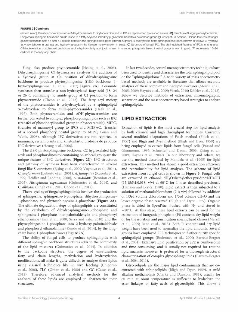
FIGURE 4 | Strategy for mass spectrometry based identification of sphinglipids. (A) Alternate strategies to confirm the sphingolipid structure. Qualitativeanalysis of sphingolipids can be performed using commercially available standards by High performance thin-layer chromatography (HP-TLC) or TLC. Sphingolipidcomponents like sphingoid backbone, sugar and fatty can be analyzed using Gas chromatography mass spectrometry (GCMS). For sphingoid base and sugaranalysis, samples are prepared using following steps: acid methanolysis (1 N HCl in methanol), N-acetylation (pyridine and acetic anhydride) andper-O-trimethylsilylation [TriSil reagent; 1-(Trimethylsilyl)imidazole – Pyridine mixture]. Sphingoid bases and sugars are analyzed as N-acetyl-per-O-trimethylsilylderivatives and monosaccharide methyl glycosides, respectively. For fatty acyl analysis, samples are prepared using acid methanolysis, re-extraction in n-hexane andper-O-trimethylsilylated (TriSil) to form fatty acid methyl esters (FAMEs). (B) A scheme of HPLC-ESI-MS/MS. Lipids are resolved by HPLC and thereafter analyzed bytandem mass spectrometry (MS/MS). ‘Q1’ and ‘Q3’ quadruples are mass analyzers and the quadrupole ‘q’ operates in radio frequency leading to collision-induceddissociation (CID). Electrospray ionization (ESI) or Atmospheric pressure chemical ionization (APCI, not shown) is used as ionization methods. (C) Scheme for Q1-MSor full scan. The molecular ions or m/z ratio are recorded in Q1. (D) Scheme for product ion scanning. Only a select ion with specific m/z is recorded in Q1 andundergoes CID. Resultant ions are recorded in Q3. ‘M’ represents the molecular ion; ‘X’ represents a fragment loss. (E) Scheme for parent ion scanning. A full scanof ions recorded in Q1 and following CID, only a select fragment ion is recorded in Q3. (F) Scheme for single-reaction monitoring (SRM). A preselected ion isrecorded in Q1, a select parent ion undergoes CID and a select fragment ion is recorded in Q3. (G) Scheme for multiple-reaction monitoring (MRM). Multiple SRM’scan be set up simultaneously depending upon the scanning efficiency of the instrument. (H) Scheme for neutral loss (NL) scanning. Ions are recorded in both Q1 andQ3 with a set mass difference between the two.
Frontiers in Microbiology | www.frontiersin.org 6 April 2016 | Volume 7 | Article 501

fmicb-07-00501 April 12, 2016 Time: 17:33 # 7
Singh and Del Poeta Lipid Profiling of Pathogenic Fungi
Poppe, 1994). GC-EI-MS is not a preferred technique toanalyze sphingolipids because fragmentation obtained in EI-MS is not very consistent as high energy electrons are usedfor fragmentation, strong ionization may completely destroythe molecular ion and extensive derivatization may generatecomplicated spectral patterns with poor resolution (Cacas et al.,2012).
High-performance LiquidChromatography (HPLC)Various sphingolipid classes can readily be separated by HPLCon both the normal phase C18 and reverse phase C8 columns.Normal phase HPLC utilizes the head group properties toobtain separation (for example, ceramides and hexosylceramides;Merrill et al., 2005). However, the reverse phase HPLC usingC8 column is capable of resolving sphingolipid classes basedon their hydrophobicity of carbon backbone and degree ofunsaturation (for example, sphingosine and dihydrosphingosine;Rana et al., 2015). During most of the routine sphingolipidanalysis the reverse phase HPLC when coupled with massspectrometry is a powerful tool for simultaneous separation anddetection of sphingolipid species. The main goal of HPLC isto chromatographically separate the sphingolipid species thatcannot be resolved based on m/z (mass-to-charge) by the massspectrometers, like isomeric and isobaric species, and to improvethe sensitivity of detection. Several different buffer systems havebeen described as mobile phases (Merrill et al., 2005). Thebinary buffer system most frequently used as the mobile phasein sphingolipid analysis is: 2 mM ammonium formate + 0.2%formic acid in H2O (Buffer 1) and 1 mM ammoniumformate + 0.2% formic acid in methanol (Buffer 2). A gradientelution of analytes using buffers 1 and 2 allows completeseparation of sphingolipid species. Additional separation onHPLC may be achieved by lowering the flow rate of themobile phase and changing the gradient conditions (Rana et al.,2015).
Fungal lipid extracts may contain a mixture of glucosyl-and galactosyl- ceramides (Barreto-Bergter et al., 2004). Dueto a remarkable similarity in their backbone structures theselipid species are difficult to separate. These lipid species canbe chromatographically separated using HILIC on silica basedcolumns using isocratic elution (Zama et al., 2009). Theelution buffer contains acetonitrile:methanol:acetic acid (97:2:1;v/v)+ 5 mM ammonium acetate (Merrill et al., 2005).
Advanced Mass SpectrometryA wide variety of mass spectrometry platforms are now availablethat can be used to analyze the molecular composition ofcomplex lipid mixtures (Köfeler et al., 2012). Mass spectrometryis a structure-based analysis of biological molecules (Wenk,2010), and has been extensively used to characterize varioussphingolipid species (Sugawara et al., 2010; Brügger, 2014; Liet al., 2014; Ogiso et al., 2014).
Several different ionization methods have been used insphingolipid analysis, for example, the ESI (Han and Gross,1994), MALDI (Jones et al., 2014), and FAB (Ahn et al., 2009).
These ionization techniques are coupled with mass analyzers likeMS/MS (Shaner et al., 2009), MSn (Ito et al., 2013), TOF (Jia et al.,2015), FT-ICR (Guo et al., 2012), and QqTOF MS (Ejsing et al.,2006). The advantages and disadvantages of these techniqueshave been described in detail previously (Köfeler et al., 2012).
For sphingolipidomic purposes, ESI-MS/MS approach is mostextensively employed. Coupled with reverse phase HPLC, ESI-MS/MS provides a simple, sensitive, and structure specificquantification of sphingolipids. Although the setup of otherlipidomic platforms may be different but the basic concepts ofmass spectrometry remain the same.
The primary requisition of any analytes to be analyzed bymass spectrometry depends on the fact that whether they can bereadily ionized in gaseous phase without in source fragmentation(Han and Gross, 1994). ESI is a soft-ionization technique thatallows the formation of intact positively or negatively chargedions that are then transmitted to the mass analyzers. In a typicalESI source, analytes in solvents are infused into the ion sourcevia a narrow capillary needle. A high voltage, positive or negativedepending upon the lipids being targeted, is applied between thetip of the needle and the inlet of mass analyzer. This results in theformation of a charged droplet which gets desolvated under highvoltage, vacuum and a drying N2 gas, leading to the productionof charged molecular ions in gaseous phase (Brügger, 2014).Sphingolipids are readily ionized in ESI source to form [M+H]+molecular ions for long chain sphingoid bases, ceramides, andglycosphingolipids, or to from [M−H]− molecular ions for IPCderivatives (Shaner et al., 2009).
A scheme for HPLC-ESI-MS/MS is summarized in Figure 4B.Different scanning approaches have developed around this triplequadrupole set up (Han and Gross, 1994; Merrill et al., 2005;Bielawski et al., 2006; Guan and Wenk, 2006; Brügger, 2014; Ranaet al., 2015), and these are:
1) Full scan (Figure 4C): In a full scan, the m/z of molecularions generated in ESI are recorded in the first quadrupoleor the first mass analyzer. Full scan approach is widelyused in non-targeted lipidomics. However, no structuralinformation can be deduced from these scanning.
2) Product ion scanning (Figure 4D): In this mode, the firstmass analyzer allows a molecular ion of specific m/z to passto the collision cell. Based on the applied CE the molecularion is fragmented into product ions using an inert collisiongas (N2 or Ar) via RF; this process is referred as CID. Them/z of these fragments is recorded in the third quadrupoleor the second mass analyzer. For sphingolipids, the production analysis provides valuable structural information aboutthe sphingoid backbone, polar head groups and the fattyacyl attached. For example, in positive ion mode, at highCE, glucosylceramide is fragmented into a characteristicion representing the loss of double dehydration productof sphingoid backbone (Figure 4D, Table 1). Similarions characteristic to specific sphingolipid structures areidentified and further used for lipid detection. Optimizationof CE is important to obtain correct fragmentation of themolecular ions into desired daughter ions. CE required forachieving CID of different sphingolipid molecular species
Frontiers in Microbiology | www.frontiersin.org 7 April 2016 | Volume 7 | Article 501

fmicb-07-00501 April 12, 2016 Time: 17:33 # 8
Singh and Del Poeta Lipid Profiling of Pathogenic Fungi
TABLE 1 | Sphingoid backbones and polar head groups of fungal sphingolipids.
STRUCTURE Name Precursor ion
Sphinganine (d 18:0);Dihydrosphingosine;D-erythro-2-Amino-1,3-octadecanediol
m/z 284.3 [M + H−H2o]+
m/z 266.3 [M + H−2H2o]+
4-Sphingenine(d18:l);Sphingosine; (2S,3R,4E)-2-Amino-4-octadecene-1,3-diol
m/z 282.3 [M + H−H2o]+
m/z 264.3[M + H−2H20]+
8-Sphingenine (d18:1);(2S,3R,4E)-2-Ammo-8-octadecene-1,3-diol
m/z 264.3 [M + H−2H20]+
4R-Hydroxysphinganine (t18:0);Phytosphingosine
m/z 282.3 [M + H−2H20]+
m/z 264.3 [M + H−3H20]+
4R-Hydroxy-eicosasphinganine (t20:0) m/z 310.3 [M + H−2H20]+
4,8-Sphingadiene (d18:2);sphinga-4E,8E-diene
m/z 262.3 [M + H−2H20]+
9-Methyl-4,8-sphingadiene (dl9:2);9-methyl-sphinga-4E,8E-dienine
m/z 276.3 [M + H−2H20]+
4-Amino-9-methyl-4,8-nonadecadiene-1,3-diol (d20:2) m/z 290.3 [M + H−2H2O]+
4-Hydroxy-9-methyl 1-4,8-sphingadiene (d19:2OH) m/z 292.3 [M + H−2H2O]+
m/z 274.3 [M + H−3H2O]+
4-Amino-9-methyl-8-nonadecene-1,3,4-triol(d20:1) m/z 308.3 [M + H−2H2O]+
Inositol phosphate m/z 259.1 [M − H]−
m/z 241.1 [M − H−H2O]−
Mannosyl inositol phosphate m/z 421.1 [M − H−H2O]−
Structure, common name and characteristic fragment positive and negative precursor ions of complex sphingolipid structures of fungi.
may vary depending upon hydroxylations, unsaturations,and carbon chain length.
3) Parent- or precursor-ion scanning (PREIS; Figure 4E): Here,the mass analyzer records molecular ions based on their
m/z, these ions undergo CID, and only a select daughterion is passed into the third quadrupole. This mode is veryuseful in the analysis of sphingolipid species which posses acommon backbone or headgroup fragment.
Frontiers in Microbiology | www.frontiersin.org 8 April 2016 | Volume 7 | Article 501

fmicb-07-00501 April 12, 2016 Time: 17:33 # 9
Singh and Del Poeta Lipid Profiling of Pathogenic Fungi
4) Single-reaction monitoring (SRM; Figure 4F): Only a selectparent ion is recorded in the first quadrupole and undergoesCID and m/z of a select daughter ion is recorded in the thirdquadrupole. SRM’s are highly selective and very sensitivemethods.
5) Multiple-reaction monitoring (MRM; Figure 4G): SeveralSRM reactions can be simultaneously recorded by themass spectrometer, however, this largely depends uponthe scanning speed of the instrument. MRM scanningrepresents a targeted lipidomics approach and is quitesuccessfully used for the analysis of sphingolipids.
6) Neutral loss (NL) scanning (Figure 4H): Both first and thirdquadrupole records the m/z, with a constant mass offsetbetween them. For example, a NL of 18 amu is set, whichrepresents a loss of water molecule from the molecular ionand is characteristic of sphingolipid structures (Rana et al.,2015).
A large amount of literature containing the fragmentationpattern of different sphingolipid classes is now available. Mostof these fragmentation data (product ions) were acquired usingdirect infusion ESI-MS/MS approaches. This, however, requiresextensive offline purification of sphingolipid of interest, usuallyusing SPE (Barreto-Bergter et al., 2004). Fortunately a largeamount of literature containing the fragmentation patterns ofvarious fungal sphingolipids is available and sphingolipid class-specific fragment and precursor ions are known for most classes(Guan and Wenk, 2006). In targeted lipidomics, the uniqueprecursor and parent ions are selected for each sphingolipidspecies and a MRM based method is employed for analyses(Bielawski et al., 2006).
ANALYSIS OF FUNGAL SPHINGOLIPIDS
By using a precursor m/z and product m/z ion pairs of molecularspecies of interest, an accurate detection and quantification canbe done. Although the complete sphingolipidome for most fungiis yet to be determined, many sphingolipid specific backbonesand polar head groups have been characterized (summarized inTable 1). Targeted methods using PREIS and MRM scanningmethods have been developed using these fragments to trace thesphingolipid metabolism of many fungi (Bielawski et al., 2006;Guan and Wenk, 2006; Ejsing et al., 2009; Sugawara et al., 2010;Brügger, 2014; Li et al., 2014; Ogiso et al., 2014).
For example, dihydrosphingosine (d18:0) and sphingosine(d18:1) can be identified using precursor ions of m/z 302 and300, and product ions of m/z 266 and 264, respectively, in thepositive ion mode. Ions of m/z 266 and 264 are a result ofloss of two water molecules from the sphingoid backbone. Lossof one water molecule from the backbone results ions of m/z284 and 282. However, the lipid extracts may contain a mixtureof d18:1 backbone with a difference in the position of doublebonds, like 4-Sphingenine (major species) and 8-Sphingenine.In this situation, a prior chromatographic separation of thesespecies is important and can be achieved by optimizing themobile phase. The d18:0 backbone fragment ion m/z 266
is used to analyze dihydroceramide species while the d18:1backbone fragment ion m/z 264 is used to analyze ceramidespecies. The phytosphingosine backbone (t18:0) is abundantin fungal lipid extract, and is constituent of phytoceramidesand IPC derivatives. A precursor ion m/z 282 is characteristicto t18:0 backbone and results from the loss of two watermolecules. Similarly, a rather less abundant t20:0 (4R-Hydroxy-eicosasphinganine) backbone is also detectable in phytoceramideand IPC structures using the positive ion precursor m/z 310.The presence of phosphorylinositol group/s in IPC, MIPC, andM(IP)2C structure often results in poor ionization efficienciesin positive ion mode. However, these lipids are readily ionizedusing negative ion mode and can be detected as [M–H]−. Theprecursor ion with m/z 241 which represents a dehydrationproduct of the ion m/z 259 (the phosphorylinositol group) andcan be used to analyze IPC and M(IP)2C species. Similarly, theprecursor ion m/z 421 is used to analyze MIPC species (Table 1;Guan and Wenk, 2006; Ejsing et al., 2009; Sugawara et al.,2010).
The mass spectrometric analyses of purified lipids, especiallycerebrosides, from different fungal sources have revealed severalother complex backbone structures (Barreto-Bergter et al., 2011).Among these 9-methyl-4,8-sphingadiene (d19:2) is predominantin most fungal species like Cryptococcus, Aspergillus, Candida,and several others (Del Poeta et al., 2014). The d19:2 backbonehas a characteristic fragment of m/z 276 that results from the lossof two water molecules. The corresponding hexosylceramide canbe identified in positive ion mode using [M + H]+ as the parention. The [M + H]+ ions 756, 754, and 728 represent three mostabundant hexosylceramides in fungi. The 2-hydroxy fatty acylgroup in these structures is C18:0, C18:1, and C16:0. It is importantto mention here that often the ions for these lipid species presentthemselves as Na+ or K+ adducts in positive ion or Cl− adductin the negative ion mode. Analysis of such samples requiresoptimization of the CE and accounting for the altered massduring the analysis. Interestingly, the differential fragmentationpattern of Cl− adduct of hexosylceramide in negative ESI-MS/MSmode can be used to identify the nature of the hexose moiety(glucose or galactose). This achievable by monitoring the peakintensity ratios of two characteristic product ions: m/z 179 and89. The ion intensity patterns of 179 < 89 and 179 > 89 representgalactose and glucose moiety in the hexosylceramide structure,respectively (Han and Cheng, 2005).
The 4,8-Sphingadiene (d18:2) backbone containingsphingolipid species are also common in fungi. These canbe analyzed using a precursor ion m/z 262. Although,the cis- or trans- isomers of d19:2 and d18:2 are notreported in fungi, however, an HPLC-ESI-MS/MS basedmethod for analysis of these was recently described usingplant lipid extracts (Imai et al., 2012). Several uniquesphingoid backbones in cerebroside structures have alsobeen identified in fungi like 4-Hydroxy-9-methyl-4,8-sphingadiene (d19:2OH) in Scedosporium apiospermum andPseudallescheria boydii (Pinto et al., 2002; Rollin-Pinheiro et al.,2014), 4-Amino-9-methyl-8-nonadecene-1,3,4-triol (d20:1)and 4-Amino-9-methyl-4,8-nonadecadiene-1,3-diol (d20:2) inCladosporium resinae (Barreto-Bergter et al., 2011).
Frontiers in Microbiology | www.frontiersin.org 9 April 2016 | Volume 7 | Article 501

fmicb-07-00501 April 12, 2016 Time: 17:33 # 10
Singh and Del Poeta Lipid Profiling of Pathogenic Fungi
Our ability to accurately quantify different sphingolipidstructures has been efficiently used to establish the sphingolipidbiosynthetic pathways in fungi. Considering that differences inthe biosynthesis of various sphingolipid structures can havedrastic physiological affect on fungi, it becomes necessary tounderstand the enzymatic steps involved therein. This has beensuccessfully achieved for several fungal species by comparing the
sphingolipid profiles of the wild type and various sphingolipidpathway mutants (Kuroda et al., 1999; Heidler and Radding,2000; Luberto et al., 2001; Bennion et al., 2003; Ejsing et al., 2009;Cheon et al., 2012; Guimarães et al., 2014; Voynova et al., 2014).In a similar scenario, our lab worked out the glucosylceramidebiosynthetic pathway of C. neofomans (shown in Figure 5A;Rittershaus et al., 2006; Singh et al., 2012). Lipid analysis of wild
FIGURE 5 | Analysis of ceramides and glucosylceramides of Cryptococcus neoformans using MRM approach. (A) Partial pathway and structures ofglucosylceramide biosynthetic pathway of C. neoformans. 14-Cer [I] is converted to 14, 18-Cer [II] by 18-desaturase (Sld8). 14–8-Cer is then methylated at C9position of sphingoid backbone to from 14–18,9Me-Cer [III] by Smt1. A Gcs1 transfers the glucose group to sphingoid backbone of ceramide structures at C1position forming a β1-3-glycosidic bond. This results in formation of 14-GlcCer (IV), 14, 18-GlcCer (V) and 14–18,9Me-GlcCer (VI) structures. The amide-linkedfatty acyl in these structures is α-hydroxylated, a key feature. The respective SRM transitions used for the identification and quantification of these structures areshown as (Parent ion→Daughter ion). (B) Analysis of ceramide and glucosylcamides in sphingolipid pathway mutants of C. neoformans. The wild type strain CnWTshows a major pool of 14–18,9Me-GlcCer (VI), where all other structures are present in negligible amounts. The Cn1smt1 strain does not produce14–18,9Me-GlcCer (VI) structure and accumulates 14, 18-Cer [II] which is converted to 14, 18-GlcCer [IV] by Gcs1. The Cn1gcs1 shows an accumulation of14–18,9Me-Cer (III). The pathway trends are represented by gray arrows in the wild type and mutant strains.
Frontiers in Microbiology | www.frontiersin.org 10 April 2016 | Volume 7 | Article 501

fmicb-07-00501 April 12, 2016 Time: 17:33 # 11
Singh and Del Poeta Lipid Profiling of Pathogenic Fungi
type C. neofomans strain (CnWT) showed that 1′-β-D-glucosyl-2′-N-hydroxyoctadecanoyl-9-methyl-4,8-sphingadiene (or 14–18,9Me-GlcCer, detected using the SRM transition of m/z 756to 276; loss of double dehydrated sphingoid base) was themost abundant cerebroside structure (Figure 5B, panel 1). Thecorresponding ceramide species 2′-N-hydroxyoctadecanoyl-9-methyl-4,8-sphingadiene (or 14–18,9Me-Cer, detected usingthe SRM transition of m/z 594 to 576; water loss) was alsodetectable. The deletion of SMT1 (Cn1smt1) abolishes theSmt1 activity, leading to an accumulation of un-methylatedceramide and glucosylceramide structures (Figure 5B, panel2). These are 2′-N-hydroxyoctadecanoyl-4,8-sphingadiene (or14–18-Cer, detected using the SRM transition of m/z 580 to562; water loss) and 1′-β-D-glucosyl-2′-N-hydroxyoctadecanoyl-4,8-sphingadiene (or 14–18-GlcCer, detected using the SRMtransition of m/z 742 to 262; loss of double dehydrated sphingoidbase), respectively. The 14–18,9Me-Cer and 14–18,9Me-GlcCer structures are absent in Cn1smt1 mutant. These dataconfirmed that methylation of ceramide by Smt1 is a pre-requisiteto the formation of methylated glucosylceramide. A deletionof glucosylceramide synthase GCS1 (Cn1smt1) leads to anaccumulation of 14–18,9Me-Cer; however, all glucosylceramidestructures are completely absent, suggesting that Gcs1 is the keyGcs1 in C. neofomans. A similar approach can also be used tostudy the function of Sld8, (uncharacterized in C. neoformans),where 2′-N-hydroxyoctadecanoyl-4-sphingenine (14-Cer) canbe detected using the SRM transition of m/z 582 to 564(water loss) and 1′-β-D-glucosyl-2′-N-hydroxyoctadecanoyl-4-sphingenine (14- GlcCer) can be detected using the SRMtransition of m/z 744 to 264 (loss of double dehydratedsphingoid base). Thus, combined with genetic approaches, massspectrometry becomes extremely useful in understanding thelipid metabolism.
Although, HPLC-ESI-MS/MS presents as a powerful toolfor qualitative and quantitative analysis of sphingolipids infungi as well as other biological systems. It is importantto note that ESI-MS/MS only gives limited informationabout the structure, especially if we are looking at an
uncharacterized structure. For example, it is not possible to assignpositions of double bonds, hydroxyl groups, and methylationbased on limited fragmentation information. Extensive MSn
is necessary to provide some insights into the possiblearrangement of the structure (Serb et al., 2009). However,such studies require more sophisticated instrumentation withhigh mass resolution, high sensitivity and sub part per-millionaccuracy are required. Confirmation of the exact structureis only possible using NMR spectroscopy of the purifiedlipid species (Sarkar et al., 2015). A routine sphingolipidomeprofiling of fungal lipids provides limited information regardingthe rate at which these structures are synthesized or theactivity of the enzymes that synthesize them. More detailedinformation of the metabolic flux of sphingolipid species may beachievable by using the labeling approaches like stable isotope(Ecker and Liebisch) or isotopomer analysis (Hellerstein andNeese, 1999). Regardless of its limitations HPLC-ESI-MS/MSremains a method of choice for quantitative sphingolipidomeprofiling.
AUTHOR CONTRIBUTIONS
All authors listed, have made substantial, direct and intellectualcontribution to the work, and approved it for publication.
FUNDING
This work was supported by NIH Grants AI56168, AI71142,AI116420, AI100631, and T32AI007539 and by a Merit ReviewGrant I01BX002624 from the Veterans Affairs Program inBiomedical Laboratory Research and Development to MD.
ACKNOWLEDGMENT
We thank the valuable inputs from Arielle Bryan.
REFERENCESAhn, Y. M., Lee, W. W., Jung, J. H., Lee, S. G., and Hong, J. (2009). Structural
determination of glucosylceramides isolated from marine sponge by fast atombombardment collision-induced dissociation linked scan at constant B/E.J. Mass Spectrom. 44, 1698–708. doi: 10.1002/jms.1678
Barreto-Bergter, E., Pinto, M. R., and Rodrigues, M. L. (2004). Structure andbiological functions of fungal cerebrosides. An. Acad. Bras. Cienc. 76, 67–84.doi: 10.1590/S0001-37652004000100007
Barreto-Bergter, E., Sassaki, G. L., and de Souza, L. M. (2011). Structuralanalysis of fungal cerebrosides. Front. Microbiol. 2:239. doi: 10.3389/fmicb.2011.00239
Bennion, B., Park, C., Fuller, M., Lindsey, R., Momany, M., Jennemann, R.,et al. (2003). Glycosphingolipids of the model fungus Aspergillus nidulans:characterization of GIPCs with oligo-alpha-mannose-type glycans. J. Lipid Res.44, 2073–2088. doi: 10.1194/jlr.M300184-JLR200
Bielawski, J., Szulc, Z. M., Hannun, Y. A., and Bielawska, A. (2006).Simultaneous quantitative analysis of bioactive sphingolipids by high-performance liquid chromatography-tandem mass spectrometry. Methods 39,82–91. doi: 10.1016/j.ymeth.2006.05.004
Bligh, E. G., and Dyer, W. J. (1959). A rapid method of total lipid extraction andpurification. Can. J. Biochem. Physiol. 37, 911–917. doi: 10.1139/o59-099
Bodennec, J., Koul, O., Aguado, I., Brichon, G., Zwingelstein, G., andPortoukalian, J. (2000). A procedure for fractionation of sphingolipid classes bysolid-phase extraction on aminopropyl cartridges. J. Lipid Res. 41, 1524–1531.
Brügger, B. (2014). Lipidomics: analysis of the lipid composition of cellsand subcellular organelles by electrospray ionization mass spectrometry.Annu. Rev. Biochem. 83, 79–98. doi: 10.1146/annurev-biochem-060713-035324
Buede, R., Rinker-Schaffer, C., Pinto, W. J., Lester, R. L., and Dickson, R. C.(1991). Cloning and characterization of LCB1, a Saccharomyces gene requiredfor biosynthesis of the long-chain base component of sphingolipids. J. Bacteriol.173, 4325–4332.
Cacas, J. L., Melser, S., Domergue, F., Joubès, J., Bourdenx, B., Schmitter, J. M., et al.(2012). Rapid nanoscale quantitative analysis of plant sphingolipid long-chainbases by GC-MS. Anal. Bioanal. Chem. 403, 2745–2755. doi: 10.1007/s00216-012-6060-1
Cheng, J., Park, T. S., Chio, L. C., Fischl, A. S., and Ye, X. S. (2003). Induction ofapoptosis by sphingoid long-chain bases in Aspergillus nidulans. Mol. Cell. Biol.23, 163–177. doi: 10.1128/MCB.23.1.163-177.2003
Frontiers in Microbiology | www.frontiersin.org 11 April 2016 | Volume 7 | Article 501

fmicb-07-00501 April 12, 2016 Time: 17:33 # 12
Singh and Del Poeta Lipid Profiling of Pathogenic Fungi
Cheon, S. A., Bal, J., Song, Y., Hwang, H. M., Kim, A. R., Kang, W. K., et al.(2012). Distinct roles of two ceramide synthases, CaLag1p and CaLac1p, inthe morphogenesis of Candida albicans. Mol. Microbiol. 83, 728–745. doi:10.1111/j.1365-2958.2011.07961.x
Chigorno, V., Sciannamblo, M., Mikulak, J., Prinetti, A., and Sonnino, S.(2006). Efflux of sphingolipids metabolically labeled with [1-3H]sphingosine,L-[3- 3H]serine and [9,10- 3H]palmitic acid from normal cells in culture.Glycoconj. J. 23, 159–165. doi: 10.1007/s10719-006-7921-7
Christie, W. W. (1989). Gas Chromatography and Lipids: A Practical Guide. Ayr:The Oily Press.
Clarke, N. G., and Dawson, R. M. (1981). Alkaline O leads to N-transacylation.A new method for the quantitative deacylation of phospholipids. Biochem. J.195, 301–306. doi: 10.1042/bj1950301
Del Poeta, M., Nimrichter, L., Rodrigues, M. L., and Luberto, C. (2014). Synthesisand biological properties of fungal glucosylceramide. PLoS Pathog. 10:e1003832.doi: 10.1371/journal.ppat.1003832
Dickson, R. C., and Lester, R. L. (2002). Sphingolipid functions inSaccharomyces cerevisiae. Biochim. Biophys. Acta 1583, 13–25. doi:10.1016/S1388-1981(02)00210-X
Ecker, J., and Liebisch, G. (2014). Application of stable isotopes toinvestigate the metabolism of fatty acids, glycerophospholipid andsphingolipid species. Prog. Lipid Res. 54, 14–31. doi: 10.1016/j.plipres.2014.01.002
Ejsing, C. S., Moehring, T., Bahr, U., Duchoslav, E., Karas, M., Simons, K., et al.(2006). Collision-induced dissociation pathways of yeast sphingolipids andtheir molecular profiling in total lipid extracts: a study by quadrupole TOF andlinear ion trap-orbitrap mass spectrometry. J. Mass Spectrom. 41, 372–389. doi:10.1002/jms.997
Ejsing, C. S., Sampaio, J. L., Surendranath, V., Duchoslav, E., Ekroos, K., Klemm,R. W., et al. (2009). Global analysis of the yeast lipidome by quantitativeshotgun mass spectrometry. Proc. Natl. Acad. Sci. U.S.A. 106, 2136–2141. doi:10.1073/pnas.0811700106
Epstein, S., Castillon, G. A., Qin, Y., and Riezman, H. (2012). An essential functionof sphingolipids in yeast cell division. Mol. Microbiol. 84, 1018–1032. doi:10.1111/j.1365-2958.2012.08087.x
Folch, J., Lees, M., and Sloane Stanley, G. H. (1957). A simple method for theisolation and purification of total lipides from animal tissues. J. Biol. Chem. 226,497–509.
Guan, X., and Wenk, M. R. (2008). Biochemistry of inositol lipids. Front. Biosci. 13,3239–3251. doi: 10.2741/2923
Guan, X. L., and Wenk, M. R. (2006). Mass spectrometry-based profiling ofphospholipids and sphingolipids in extracts from Saccharomyces cerevisiae.Yeast 23, 465–477. doi: 10.1002/yea.1362
Guimarães, L. L., Toledo, M. S., Ferreira, F. A., Straus, A. H., and Takahashi, H. K.(2014). Structural diversity and biological significance of glycosphingolipids inpathogenic and opportunistic fungi. Front. Cell. Infect. Microbiol. 4:138. doi:10.3389/fcimb.2014.00138
Guo, Y., Wang, X., Qiu, L., Qin, X., Liu, H., Wang, Y., et al. (2012). Probing gender-specific lipid metabolites and diagnostic biomarkers for lung cancer usingFourier transform ion cyclotron resonance mass spectrometry. Clin. Chim. Acta414, 135–141. doi: 10.1016/j.cca.2012.08.010
Haak, D., Gable, K., Beeler, T., and Dunn, T. (1997). Hydroxylation ofSaccharomyces cerevisiae ceramides requires Sur2p and Scs7p. J. Biol. Chem.272, 29704–29710. doi: 10.1074/jbc.272.47.29704
Han, X., and Cheng, H. (2005). Characterization and direct quantitation ofcerebroside molecular species from lipid extracts by shotgun lipidomics. J. LipidRes. 46, 163–175. doi: 10.1194/jlr.D400022-JLR200
Han, X., and Gross, R. W. (1994). Electrospray ionization mass spectroscopicanalysis of human erythrocyte plasma membrane phospholipids. Proc.Natl. Acad. Sci. U.S.A. 91, 10635–10639. doi: 10.1073/pnas.91.22.10635
Hanson, B. A., and Lester, R. L. (1980). The extraction of inositol-containingphospholipids and phosphatidylcholine from Saccharomyces cerevisiae andNeurospora crassa. J. Lipid Res. 21, 309–315.
Haynes, C. A., Allegood, J. C., Park, H., and Sullards, M. C. (2009).Sphingolipidomics: methods for the comprehensive analysis of sphingolipids.J. Chromatogr. B Analyt. Technol. Biomed. Life Sci. 877, 2696–2708. doi:10.1016/j.jchromb.2008.12.057
Heidler, S. A., and Radding, J. A. (2000). Inositol phosphoryl transferasesfrom human pathogenic fungi. Biochim. Biophys. Acta 1500, 147–152. doi:10.1016/S0925-4439(99)00097-6
Hellerstein, M. K., and Neese, R. A. (1999). Mass isotopomer distribution analysisat eight years: theoretical, analytic, and experimental considerations. Am. J.Physiol. 276(6 Pt 1), E1146–E1170.
Heung, L. J., Luberto, C., and Del Poeta, M. (2006). Role of sphingolipids inmicrobial pathogenesis. Infect. Immun. 74, 28–39. doi: 10.1128/IAI.74.1.28-39.2006
Imai, H., Hattori, H., and Watanabe, M. (2012). An improved method for analysisof glucosylceramide species having cis-8 and trans-8 isomers of sphingoid basesby LC-MS/MS. Lipids 47, 1221–1229. doi: 10.1007/s11745-012-3725-7
Ito, E., Waki, H., Miseki, K., Shimada, T., Sato, T. A., Kakehi, K., et al.(2013). Structural characterization of neutral glycosphingolipids usinghigh-performance liquid chromatography-electrospray ionization massspectrometry with a repeated high-speed polarity and MSn switching system.Glycoconj. J. 30, 881–888. doi: 10.1007/s10719-013-9492-8
Jenkins, G. M., Richards, A., Wahl, T., Mao, C., Obeid, L., and Hannun, Y. (1997).Involvement of yeast sphingolipids in the heat stress response of Saccharomycescerevisiae. J. Biol. Chem. 272, 32566–32572. doi: 10.1074/jbc.272.51.32566
Jia, Z., Li, S., Cong, P., Wang, Y., Sugawara, T., Xue, C., et al. (2015). Highthroughput analysis of cerebrosides from the sea cucumber Pearsonothriagraeffei by liquid chromatography-quadrupole-time-of-flight massspectrometry. J. Oleo Sci. 64, 51–60. doi: 10.5650/jos.ess14136
Johnson, S. B., and Brown, R. E. (1992). Simplified derivatization for determiningsphingolipid fatty acyl composition by gas chromatography-mass spectrometry.J. Chromatogr. 605, 281–286. doi: 10.1016/0021-9673(92)85248-R
Jones, E. E., Dworski, S., Canals, D., Casas, J., Fabrias, G., Schoenling, D.,et al. (2014). On-tissue localization of ceramides and other sphingolipidsby MALDI mass spectrometry imaging. Anal. Chem. 86, 8303–8311. doi:10.1021/ac501937d
Karashima, T., Kajiwara, K., and Funato, K. (2013). Metabolic labeling ofyeast sphingolipids with radioactive D-erythro-[4,5-3H]dihydrosphingosine.Bio Protoc. 3:e862.
Kendall, A. C., Pilkington, S. M., Massey, K. A., Sassano, G., Rhodes, L. E., andNicolaou, A. (2015). Distribution of bioactive lipid mediators in human skin.J. Invest. Dermatol. 135, 1510–1520. doi: 10.1038/jid.2015.41
Kim, S., Fyrst, H., and Saba, J. (2000). Accumulation of phosphorylated sphingoidlong chain bases results in cell growth inhibition in Saccharomyces cerevisiae.Genetics 156, 1519–1529.
Köfeler, H. C., Fauland, A., Rechberger, G. N., and Trötzmüller, M. (2012).Mass spectrometry based lipidomics: an overview of technological platforms.Metabolites 2, 19–38. doi: 10.3390/metabo2010019
Kondo, N., Ohno, Y., Yamagata, M., Obara, T., Seki, N., Kitamura, T., et al.(2014). Identification of the phytosphingosine metabolic pathway leading toodd-numbered fatty acids. Nat. Commun. 5:5338. doi: 10.1038/ncomms6338
Kotz, A., Wagener, J., Engel, J., Routier, F., Echtenacher, B., Pich, A., et al.(2010). The mitA gene of Aspergillus fumigatus is required for mannosylationof inositol-phosphorylceramide, but is dispensable for pathogenicity. FungalGenet. Biol. 47, 169–178. doi: 10.1016/j.fgb.2009.10.001
Kuroda, M., Hashida-Okado, T., Yasumoto, R., Gomi, K., Kato, I., andTakesako, K. (1999). An aureobasidin A resistance gene isolated fromAspergillus is a homolog of yeast AUR1, a gene responsible for inositolphosphorylceramide (IPC) synthase activity. Mol. Gen. Genet. 261, 290–296.doi: 10.1007/s004380050969
Lahiri, S., and Futerman, A. H. (2005). LASS5 is a bona fide dihydroceramidesynthase that selectively utilizes palmitoyl-CoA as acyl donor. J. Biol. Chem. 280,33735–33738. doi: 10.1074/jbc.M506485200
Leipelt, M., Warnecke, D., Zahringer, U., Ott, C., Muller, F., Hube, B., et al. (2001).Glucosylceramide synthases, a gene family responsible for the biosynthesis ofglucosphingolipids in animals, plants, and fungi. J. Biol. Chem. 276, 33621–33629. doi: 10.1074/jbc.m104952200
Levery, S. B., Momany, M., Lindsey, R., Toledo, M. S., Shayman, J. A.,Fuller, M., et al. (2002). Disruption of the glucosylceramide biosyntheticpathway in Aspergillus nidulans and Aspergillus fumigatus by inhibitors ofUDP-Glc:ceramide glucosyltransferase strongly affects spore germination, cellcycle, and hyphal growth. FEBS Lett. 525, 59–64. doi: 10.1016/S0014-5793(02)03067-3
Frontiers in Microbiology | www.frontiersin.org 12 April 2016 | Volume 7 | Article 501

fmicb-07-00501 April 12, 2016 Time: 17:33 # 13
Singh and Del Poeta Lipid Profiling of Pathogenic Fungi
Li, M., Yang, L., Bai, Y., and Liu, H. (2014). Analytical methods in lipidomics andtheir applications. Anal. Chem. 86, 161–75. doi: 10.1021/ac403554h
Li, S., Bao, D., Yuen, G., Harris, S. D., and Calvo, A. M. (2007). basA regulates cellwall organization and asexual/sexual sporulation ratio in Aspergillus nidulans.Genetics 176, 243–253. doi: 10.1534/genetics.106.068239
Li, S., Du, L., Yuen, G., and Harris, S. D. (2006). Distinct ceramide synthasesregulate polarized growth in the filamentous fungus Aspergillus nidulans. Mol.Biol. Cell 17, 1218–1227. doi: 10.1091/mbc.E05-06-0533
Luberto, C., Toffaletti, D. L., Wills, E. A., Tucker, S. C., Casadevall, A.,Perfect, J. R., et al. (2001). Roles for inositol-phosphoryl ceramide synthase1 (IPC1) in pathogenesis of C. neoformans. Genes Dev. 15, 201–212. doi:10.1101/gad.856001
Luttgeharm, K. D., Chen, M., Mehra, A., Cahoon, R. E., Markham, J. E., andCahoon, E. B. (2015). Overexpression of Arabidopsis ceramide synthasesdifferentially affects growth, sphingolipid metabolism, programmed celldeath, and mycotoxin resistance. Plant Physiol. 169, 1108–1117. doi:10.1104/pp.15.00987
Mandala, S. M., Thornton, R. A., Frommer, B. R., Curotto, J. E., Rozdilsky, W.,Kurtz, M. B., et al. (1995). The discovery of australifungin, a novel inhibitorof sphinganine N-acyltransferase from Sporormiella australis. Producingorganism, fermentation, isolation, and biological activity. J. Antibiot. (Tokyo)48, 349–356. doi: 10.7164/antibiotics.48.349
Marquês, J. T., Cordeiro, A. M., Viana, A. S., Herrmann, A., Marinho, H. S.,and de Almeida, R. F. (2015). Formation and properties of membrane-ordereddomains by phytoceramide: role of sphingoid base hydroxylation. Langmuir 31,9410–9421. doi: 10.1021/acs.langmuir.5b02550
Matsubara, T., and Hayashi, A. (1991). Fragmentation pathways of O-trimethylsilylethers of dihydroxy long-chain bases analysed by linked-scan massspectrometry. J. Chromatogr. 562, 119–124. doi: 10.1016/0378-4347(91)80570-3
Merkle, R. K., and Poppe, I. (1994). Carbohydrate composition analysis ofglycoconjugates by gas-liquid chromatography/mass spectrometry. MethodsEnzymol. 230, 1–15. doi: 10.1016/0076-6879(94)30003-8
Merrill, A. H. Jr., Stokes, T. H., Momin, A., Park, H., Portz, B. J., Kelly, S.,et al. (2009). Sphingolipidomics: a valuable tool for understanding the roles ofsphingolipids in biology and disease. J. Lipid Res. 50(Suppl.), S97–S102. doi:10.1194/jlr.R800073-JLR200
Merrill, A. H. Jr., Sullards, M. C., Allegood, J. C., Kelly, S., and Wang, E. (2005).Sphingolipidomics: high-throughput, structure-specific, and quantitativeanalysis of sphingolipids by liquid chromatography tandem mass spectrometry.Methods 36, 207–224. doi: 10.1016/j.ymeth.2005.01.009
Mor, V., Rella, A., Farnoud, A. M., Singh, A., Munshi, M., Bryan, A., et al. (2015).Identification of a new class of antifungals targeting the synthesis of fungalsphingolipids. MBio 6:e00647. doi: 10.1128/mBio.00647-15
Nagiec, M. M., Baltisberger, J. A., Wells, G. B., Lester, R. L., and Dickson,R. C. (1994). The LCB2 gene of Saccharomyces and the related LCB1gene encode subunits of serine palmitoyltransferase, the initial enzyme insphingolipid synthesis. Proc. Natl. Acad. Sci. U.S.A. 91, 7899–7902. doi:10.1073/pnas.91.17.7899
Obeid, L. M., Okamoto, Y., and Mao, C. (2002). Yeast sphingolipids: metabolismand biology. Biochim. Biophys. Acta 1585, 163–171. doi: 10.1016/S1388-1981(02)00337-2
Ogiso, H., Taniguchi, M., Araya, S., Aoki, S., Wardhani, L. O., Yamashita, Y.,et al. (2014). Comparative analysis of biological sphingolipids withglycerophospholipids and diacylglycerol by LC-MS/MS. Metabolites 4,98–114. doi: 10.3390/metabo4010098
Ohnishi, M., Ito, S., and Fujino, Y. (1983). Characterization of sphingolipidsin spinach leaves. Biochim. Biophys. Acta 752, 416–422. doi: 10.1016/0005-2760(83)90271-0
Oura, T., and Kajiwara, S. (2010). Candida albicans sphingolipid C9-methyltransferase is involved in hyphal elongation. Microbiology 156(Pt4), 1234–1243. doi: 10.1099/mic.0.033985-0
Palumbo, G., and Zullo, F. (1987). The use of iodine staining for the quantitativeanalysis of lipids separated by thin layer chromatography. Lipids 22, 201–205.doi: 10.1007/BF02537303
Pinto, M. R., Rodrigues, M. L., Travassos, L. R., Haido, R. M., Wait, R., and Barreto-Bergter, E. (2002). Characterization of glucosylceramides in Pseudallescheriaboydii and their involvement in fungal differentiation. Glycobiology 12, 251–260. doi: 10.1093/glycob/12.4.251
Prasad, R., and Ghannoum, M. A. (1996). Lipids of Pathogenic Fungi. Boca Raton,FL: CRC Press.
Rana, N. A., Singh, A., Del Poeta, M., and Hannun, Y. A. (2015). “Qualitative andquantitative measurements of sphingolipids by mass spectrometry,” in BioactiveSphingolipids in Cancer Biology and Therapy, eds Y. A. Hannun, C. Luberto, C.Mao, and L. M. Obeid (Cham: Springer International Publishing), 313–338.
Rittenour, W. R., Chen, M., Cahoon, E. B., and Harris, S. D. (2011).Control of glucosylceramide production and morphogenesis by the Bar1ceramide synthase in Fusarium graminearum. PLoS ONE 6:e19385. doi:10.1371/journal.pone.0019385
Rittershaus, P. C., Kechichian, T. B., Allegood, J., Merrill, A. H. J., Hennig, M.,Luberto, C., et al. (2006). Glucosylceramide is an essential regulator ofpathogenicity of Cryptococcus neoformans. J. Clin. Invest. 116, 1651–1659. doi:10.1172/jci27890
Rodriguez-Cuenca, S., Barbarroja, N., and Vidal-Puig, A. (2015). Dihydroceramidedesaturase 1, the gatekeeper of ceramide induced lipotoxicity. Biochim. Biophys.Acta. 1851, 40–50. doi: 10.1016/j.bbalip.2014.09.021
Rollin-Pinheiro, R., Liporagi-Lopes, L. C., de Meirelles, J. V., Souza, L. M., andBarreto-Bergter, E. (2014). Characterization of Scedosporium apiospermumglucosylceramides and their involvement in fungal development andmacrophage functions. PLoS ONE 9:e98149. doi: 10.1371/journal.pone.0098149
Sarkar, A., Drouillard, S., Rivet, A., and Perez, S. (2015). Databases ofconformations and NMR structures of glycan determinants. Glycobiology 25,1480–1490. doi: 10.1093/glycob/cwv054
Sasaki, T. (1981). Extensive radioactive labeling of glycolipids in cultured hamsterfibroblasts by incubation of whole cells with UDP-[14C]glucose and UDP-[14C]galactose. Biochim. Biophys. Acta 666, 418–425. doi: 10.1016/0005-2760(81)90301-5
Schneiter, R., and Daum, G. (2006). Extraction of yeast lipids. Methods Mol. Biol.313, 41–45.
Serb, A., Schiopu, C., Flangea, C., Sisu, E., and Zamfir, A. D. (2009). Top-downglycolipidomics: fragmentation analysis of ganglioside oligosaccharide coreand ceramide moiety by chip-nanoelectrospray collision-induced dissociationMS2-MS6. J. Mass Spectrom. 44, 1434–1442. doi: 10.1002/jms.1625
Serra, M., and Saba, J. D. (2010). Sphingosine 1-phosphate lyase, a key regulatorof sphingosine 1-phosphate signaling and function. Adv. Enzyme Regul. 50,349–362. doi: 10.1016/j.advenzreg.2009.10.024
Shaner, R. L., Allegood, J. C., Park, H., Wang, E., Kelly, S., Haynes, C. A.,et al. (2009). Quantitative analysis of sphingolipids for lipidomics using triplequadrupole and quadrupole linear ion trap mass spectrometers. J. Lipid Res. 50,1692–1707. doi: 10.1194/jlr.D800051-JLR200
Singh, A., Prasad, T., Kapoor, K., Mandal, A., Roth, M., Welti, R., et al.(2010). Phospholipidome of Candida: each species of Candida hasdistinctive phospholipid molecular species. OMICS 14, 665–677. doi:10.1089/omi.2010.0041
Singh, A., Wang, H., Silva, L. C., Na, C., Prieto, M., Futerman, A. H., et al.(2012). Methylation of glycosylated sphingolipid modulates membrane lipidtopography and pathogenicity of Cryptococcus neoformans. Cell. Microbiol. 14,500–516. doi: 10.1111/j.1462-5822.2011.01735.x
Sperling, P., Lee, M., Girke, T., Zähringer, U., Stymne, S., and Heinz, E. (2000).A bifunctional delta-fatty acyl acetylenase/desaturase from the moss Ceratodonpurpureus. A new member of the cytochrome b5 superfamily. Eur. J. Biochem.267, 3801–3811. doi: 10.1046/j.1432-1327.2000.01418.x
Sugawara, T., Aida, K., Duan, J., and Hirata, T. (2010). Analysis ofglucosylceramides from various sources by liquid chromatography-ion trap mass spectrometry. J. Oleo Sci. 59, 387–394. doi: 10.5650/jos.59.387
Tadano, K., and Ishizuka, I. (1982). Isolation and characterization of the sulfatedgangliotriaosylceramide from rat kidney. J. Biol. Chem. 257, 1482–1490.
Ternes, P., Sperling, P., Albrecht, S., Franke, S., Cregg, J. M., Warnecke, D.,et al. (2006). Identification of fungal sphingolipid C9-methyltransferasesby phylogenetic profiling. J. Biol. Chem. 281, 5582–5592. doi:10.1074/jbc.M512864200
Ternes, P., Wobbe, T., Schwarz, M., Albrecht, S., Feussner, K., Riezman, I.,et al. (2011). Two pathways of sphingolipid biosynthesis are separated in theyeast Pichia pastoris. J. Biol. Chem. 86, 11401–1114. doi: 10.1074/jbc.M110.193094
Frontiers in Microbiology | www.frontiersin.org 13 April 2016 | Volume 7 | Article 501

fmicb-07-00501 April 12, 2016 Time: 17:33 # 14
Singh and Del Poeta Lipid Profiling of Pathogenic Fungi
Urban, P. F., Harth, S., Freysz, L., and Dreyfus, H. (1980). Brain and retinalganglioside composition from different species determined by TLC andHPTLC. Adv. Exp. Med. Biol. 125, 149–157. doi: 10.1007/978-1-4684-7844-0-14
Voynova, N. S., Mallela, S. K., Vazquez, H. M., Cerantola, V., Sonderegger, M.,Knudsen, J., et al. (2014). Characterization of yeast mutants lacking alkalineceramidases YPC1 and YDC1. FEMS Yeast Res. 14, 776–788. doi: 10.1111/1567-1364.12169
Warnecke, D., and Heinz, E. (2003). Recently discovered functions ofglucosylceramides in plants and fungi. Cell. Mol. Life Sci. 60, 919–941.
Wenk, M. R. (2010). Lipidomics: new tools and applications. Cell 143, 888–895.doi: 10.1016/j.cell.2010.11.033
Zama, K., Hayashi, Y., Ito, S., Hirabayashi, Y., Inoue, T., Ohno, K., et al. (2009).Simultaneous quantification of glucosylceramide and galactosylceramideby normal-phase HPLC using O-phtalaldehyde derivatives preparedwith sphingolipid ceramide N-deacylase. Glycobiology 19, 767–775. doi:10.1093/glycob/cwp047
Zanolari, B., Friant, S., Funato, K., Sutterlin, C., Stevenson, B. J., andRiezman, H. (2000). Sphingoid base synthesis requirement for endocytosis inSaccharomyces cerevisiae. EMBO J. 19, 2824–2833. doi: 10.1093/emboj/19.12.2824
Conflict of Interest Statement: The authors declare that the research wasconducted in the absence of any commercial or financial relationships that couldbe construed as a potential conflict of interest.
Copyright © 2016 Singh and Del Poeta. This is an open-access article distributedunder the terms of the Creative Commons Attribution License (CC BY). Theuse, distribution or reproduction in other forums is permitted, provided theoriginal author(s) or licensor are credited and that the original publication inthis journal is cited, in accordance with accepted academic practice. No use,distribution or reproduction is permitted which does not comply with theseterms.
Frontiers in Microbiology | www.frontiersin.org 14 April 2016 | Volume 7 | Article 501