Embed Size (px)
Citation preview
DMD #5249
1
Hepatic, extrahepatic, microsomal, and mitochondrial activation
of the N-hydroxylated prodrugs
benzamidoxime, guanoxabenz, and Ro 48-3656
Bernd Clement, Sabine Mau, Stephanie Deters, Antje Havemeyer
Institute of Pharmacy, Christian-Albrechts-University of Kiel, Kiel, Germany
(B.C., S.M., S.D., A.H.)
DMD Fast Forward. Published on August 23, 2005 as doi:10.1124/dmd.105.005249
Copyright 2005 by the American Society for Pharmacology and Experimental Therapeutics.
This article has not been copyedited and formatted. The final version may differ from this version.DMD Fast Forward. Published on August 23, 2005 as DOI: 10.1124/dmd.105.005249
at ASPE
T Journals on Septem
ber 9, 2018dm
d.aspetjournals.orgD
ownloaded from
DMD #5249
2
Running title: Activation of N-hydroxylated prodrugs
Corresponding author: Prof. Dr. Bernd Clement, Christian-Albrechts-Universität,
Pharmazeutisches Institut, Gutenbergstraße 76, D-24118 Kiel, Germany,
phone: 0431-8801126, fax: 0431-8801352; email: [email protected]
Number of text pages: 25
Number of tables: 6
Number of figures: 6
Number of references: 42
Number of words in the abstract: 234
Number of words in the Introduction: 654
Number of words in the discussion: 1418
Non-standard Abbreviations:
p-HMB p-hydroxymercuribenzoic acid
OMV outer membrane vesicles
P450 Cytochrome P450
CR rotenone-insensitive cytochrome c reductase
SuccCytc succinate cytochrome c reductase
BAORed benzamidoxime reductase
This article has not been copyedited and formatted. The final version may differ from this version.DMD Fast Forward. Published on August 23, 2005 as DOI: 10.1124/dmd.105.005249
at ASPE
T Journals on Septem
ber 9, 2018dm
d.aspetjournals.orgD
ownloaded from
DMD #5249
3
Abstract
In previous studies it was shown that liver microsomes from rabbit, rat, pig, and
human are involved in the reduction of N-hydroxylated amidines, guanidines, and
amidinohydrazones of various drugs and model compounds (Clement, 2002). One
responsible enzyme system, the microsomal benzamidoxime reductase, consisting of
cytochrome b5, its reductase, and a P450 isoenzyme, was isolated from pig liver
microsomes (Clement et al., 1997) . Further investigations followed to establish
whether such enzyme systems are also present in microsomes of other organs such
as brain, lung, and intestine. In addition the mitochondrial reduction in human and
porcine liver and also kidney preparations were studied. The reductase activities
were measured by following the reduction of benzamidoxime to benzamidine, of
guanoxabenz to guanabenz, and of Ro 48-3656 to Ro 44-3888. Interestingly,
preparations of all tested organs were capable of reducing the three compounds. The
highest specific rates were found in kidney followed by liver, brain, lung, and
intestine, and usually the mitochondrial reduction rates were superior. From the
determined characteristics, similarities between the enzyme systems in the different
organs and organelles were detected. Furthermore, properties of the benzamidoxime
reductase located in the outer membrane of pig liver mitochondria were studied. In
summary, these results demonstrate that in addition to the microsomal reduction,
mitochondria are involved to a great extent in the activation of amidoxime prodrugs.
The importance of extrahepatic metabolism in the reduction of N-hydroxylated
prodrugs is demonstrated.
This article has not been copyedited and formatted. The final version may differ from this version.DMD Fast Forward. Published on August 23, 2005 as DOI: 10.1124/dmd.105.005249
at ASPE
T Journals on Septem
ber 9, 2018dm
d.aspetjournals.orgD
ownloaded from

DMD #5249
4
Numerous drugs and drug candidates contain strongly basic functional groups, such
as guanidines, amidinohydrazones, and amidines. Because of their strong basicity
they are protonated under physiological conditions and usually not absorbed in the
gastrointestinal tract. In our laboratory a prodrug principle was developed in particular
for amidines by N-hydroxylating this functional group to the corresponding
amidoximes, substances with a less basic functional group. Consequently, they are
not protonated under physiological conditions and can easily be absorbed as free
bases. By creating these prodrugs of the active principle the oral bioavailability of
amidines can be increased. Thus amidoximes and similar functional groups can be
used as prodrugs for amidines and related groups.
The prodrug principle was developed for pentamidine (Clement, 1993) and was then
applied to other amidines such as trypanocidal compounds (Zhou et al., 2004),
glycoprotein IIIa/IIb receptor antagonist, such as Sibrafiban (Weller et al., 1996) and
the thrombin inhibitor ximelagatran which was recently approved and is the first orally
available direct thrombin inhibitor on the market (Gustafsson et al., 2001)
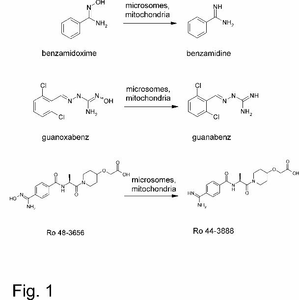
Benzamidoxime is a model compound for such amidoxime prodrugs which are known
to be reduced by liver microsomes from different species as well as by the purified
enzyme system from pig liver (Clement et al., 1997). Therefore the reduction of this
substrate and its HPLC analysis can be taken as a reliable activity assay (Fig. 1).
Sibrafiban is a non-marketed oral platelet aggregation inhibitor and a double-prodrug.
After oral administration the amidoxime ethylester sibrafiban is adsorbed and
hydrolyzed into Ro 48-5656 (amidoxime and free acid) and by reduction of the
N-hydroxylated structure Ro 48-3656 into the active metabolite Ro 44-3888 (Timm et
al., 1997) (Fig.1).
The third substrate under investigation was the centrally acting α2-adrenoreceptor
agonist guanoxabenz (Benzérial). The reduction of guanoxabenz to its
This article has not been copyedited and formatted. The final version may differ from this version.DMD Fast Forward. Published on August 23, 2005 as DOI: 10.1124/dmd.105.005249
at ASPE
T Journals on Septem
ber 9, 2018dm
d.aspetjournals.orgD
ownloaded from
DMD #5249
5
amidinohydrazone guanabenz (Wytensin, Hipten, Rexitene) has been described
in previous studies with pig, rabbit, and human liver microsomes (Clement et al.,
1996). This compound was chosen as a model compound for
N-hydroxyamidinohydrazones as a prodrug of an amidinohydrazone. Furthermore,
guanoxabenz has been shown to be mutagenic (Clement et al., 1996). Thus the
reduction is of high toxicological interest as a detoxification reaction.
Previous investigations demonstrated that microsomal enzymes from rabbit, rat, pig,
and human liver are involved in the reduction of N-hydroxylated xenobiotics
(Clement, 2002).
In addition, one enzyme system capable of reducing N-hydroxylated derivatives of
strongly basic functional groups has been identified so far in pig liver microsomes
consisting of cytochrome b5, NADH cytochrome b5 reductase, and a P450 isoenzyme
of the subfamily 2D (Clement et al., 1997).
The microsomal enzyme system showed similarities with the microsomal
hydroxylamine reductase, a porcine liver enzyme with insensibility to oxygen and
highest activity in acid medium (Kadlubar and Ziegler, 1974), and the mitochondrial
reduction of benzamidoxime showed common characteristics to the mitochondrial
hydroxylamine reductase, a cyanide and mercury-sensitive NADH-dependent
enzyme system (Bernheim and Hochstein, 1968).
The aim of this study was to investigate in which organs the N-reduction to the active
amidine forms can take place and consequently can take effect. In addition, the
presence of these reducing systems in the gastrointestinal tract can decrease the
bioavailability, because the amidoximes might already be reduced to their amidines
before absorption. On the other hand, the absence of such enzyme systems might be
correlated with organ typical genotoxic properties of several N-hydroxylated
This article has not been copyedited and formatted. The final version may differ from this version.DMD Fast Forward. Published on August 23, 2005 as DOI: 10.1124/dmd.105.005249
at ASPE
T Journals on Septem
ber 9, 2018dm
d.aspetjournals.orgD
ownloaded from
DMD #5249
6
structures. Thus this reductase may play a vital role protecting humans and other
mammals against accumulation of noxious metabolites.
In this study the results of the investigations with liver, kidney, brain, lung, and
intestine microsomes using the reduction of benzamidoxime to benzamidine
(Clement et al., 1997), of guanoxabenz to guanabenz (Clement et al., 1996), and the
sibrafiban-metabolite Ro 48-3656 to the active form of sibrafiban Ro 44-3888 are
summarized.
This report demonstrates that porcine and human microsomes and mitochondria of
all tested organs are able to reduce N-hydroxylated structures.
Furthermore, characteristics and intramitochondrial localisation of the mitochondrial
benzamidoxime reductase were investigated.
This article has not been copyedited and formatted. The final version may differ from this version.DMD Fast Forward. Published on August 23, 2005 as DOI: 10.1124/dmd.105.005249
at ASPE
T Journals on Septem
ber 9, 2018dm
d.aspetjournals.orgD
ownloaded from
DMD #5249
7
Materials and Methods
Benzamidoxime (N-hydroxy-benzenecarboximidamide) was synthesized from
benzonitrile, and hydroxylamine as described (Krüger, 1885). Guanabenz acetate
( 2-[(2,6-dichlorophenyl)methylene]-hydrazinecarboximidamide. Acetate salt ) was
kindly supplied by Wyeth-Pharma GmbH (Münster, Germany), guanoxabenz-HCl
( 2-[(2,6-dichlorophenyl)methylene]-N-hydroxy-hydrazinecarboximidamide. Hydro-
chloride salt.) by Laboratoires Houdé (Paris, France), and Ro 48-3656 ( [[1-[(2S)-2-
[[4-[(hydroxyamino)iminomethyl]benzoyl]amino]-1-oxopropyl]-4-piperidinyl]oxy-acetic
acid), and Ro 44-3888 ( [[1-[(2S)-2-[[4-(aminoiminomethyl)benzoyl]amino]-1-
oxopropyl]-4-piperidinyl]oxy]-acetic acid) by Hoffmann La Roche (Basel, Switzerland).
All other chemicals were commercially available. Hydroxylamine-HCl, dipotassium
hydrogen phosphate, phosphoric acid, tetramethylammonium chloride, cytochrome c
(from horse heart), mannitol, 1-octanesulfonic acid (sodium salt), Percoll®,
p-hydroxymercuribenzoic acid (sodium salt), rotenone, succinic acid (disodium salt),
MOPS, and benzamidine were purchased from Sigma-Aldrich (Taufkirchen,
Germany). Methanol from J.T. Baker (Derventer, Holland), Hepes from Biochrom AG
(Berlin, Germany). Acetonitrile was obtained from Promochem (Wesel, Germany),
Triton X-100 from Serva Feinbiochemica (Heidelberg, Germany), acetic acid, BSA,
DTT, EDTA (disodium salt), EGTA, potassium cyanide, potassium dihydrogen
phosphate, NADH (disodium salt), NADPH (tetrasodium salt), sucrose, Tris-HCl,
potassium hexacyanoferrate (III), and all other chemicals from Merck KGaA
(Darmstadt, Germany) unless otherwise stated. Acetonitrile and methanol were of
HPLC grade, while other chemicals and solvents were of analytical grade.
This article has not been copyedited and formatted. The final version may differ from this version.DMD Fast Forward. Published on August 23, 2005 as DOI: 10.1124/dmd.105.005249
at ASPE
T Journals on Septem
ber 9, 2018dm
d.aspetjournals.orgD
ownloaded from
DMD #5249
8
Preparation of subcellular fractions:
Preparation of microsomes. Microsomes were prepared by differential centrifugation.
All steps were performed in standard phosphate buffer pH 7.4 (4.8 mM KH2PO4,
15.2 mM K2HPO4, 0.25 M sucrose, 1mM EDTA). Centrifugation steps and storage
were carried out in standard phosphate buffer without EDTA.
Briefly the tissues were excised, washed, homogenized with a homogenizer
(developed from University of Austin, Texas, USA), and centrifuged at 9000g for
30 minutes and then twice at 100000g for 60 minutes. The resulting pellet was
suspended in phosphate buffer with a motorized teflon pestle glass tube
homogenizer, adjusted to a pH of 7.4 and frozen at –80°C. All organs except
intestine and lung were pooled. All operations were performed at 0-4°C.
Preparation of mitochondria. Pig liver mitochondria: Pig livers (slaughterhouse) were
placed in 10 mM Tris-HCl pH 7.4, containing 0.25 M sucrose, 1 mM EDTA, 1 mM
DTT, and perfused with the same buffer. All operations were performed at 0-4°C. The
livers were homogenized. The homogenate was centrifuged at 750g for 20 minutes.
The supernatant was collected and the pellet was resuspended and centrifuged
again. The supernatants were combined and crude mitochondria were sedimented by
centrifugation at 7710g for 20 minutes. The 7710g sediment was washed four times
by careful resuspending in buffer and following centrifugation. In order to minimize
microsomal contamination, the centrifugal force was gradually diminished at each
centrifugation step: the suspended mitochondria were centrifuged at the first
sedimentation step at 7350g for 20 minutes, and then at 6450g for 20 minutes, 5580g
for 20 minutes, and finally at 5050g for 20 minutes.
The final pellet was resuspended in buffer and stored at –80°C.
In other cases and for isolation of the outer mitochondrial membrane pig liver
mitochondria were obtained by differential centrifugation and isotonic Percoll gradient
This article has not been copyedited and formatted. The final version may differ from this version.DMD Fast Forward. Published on August 23, 2005 as DOI: 10.1124/dmd.105.005249
at ASPE
T Journals on Septem
ber 9, 2018dm
d.aspetjournals.orgD
ownloaded from
DMD #5249
9
(Hovius et al., 1990). The livers were excised, perfused, and homogenized in
isolation buffer (0.25 M sucrose, 1 mM EDTA, 10 mM KH2PO4, 1 mM DTT,
0.1% (w/v) BSA, pH 7.4). The homogenate was centrifuged at 600g for 15 minutes.
The pellet was discarded, and this centrifugation step was repeated four times. The
crude mitochondrial fraction was then sedimented from the supernatant by
centrifugation at 10300g for 20 minutes. The pellet was resuspended in isolation
buffer, the suspension was layered on top of Percoll-buffer (30% (v/v) Percoll in
225 mM mannitol, 1 mM EGTA, 25 mM Hepes, 0.1% (w/v) BSA, pH 7.4), and
centrifuged at 95000g for 40 minutes. The mitochondrial fraction was collected and
washed twice by centrifugation at 6300g for 20 minutes. The final mitochondrial pellet
was resuspended in isolation buffer and stored at –80°C. All operations were
performed at 0-4°C.
Pig kidney mitochondria: Kidney cortex was prepared as described above for the
preparation of pig liver mitochondria with slight modifications.
Human liver and kidney mitochondria: Human liver and kidney mitochondria were
prepared from pooled liver / kidney samples of cancer patients. Only morphologically
intact tissue was used. The separation was performed as described above for the
preparation of pig liver mitochondria with slight modifications. Prior consent of the
local medical ethics committee and from the donors were obtained for these studies.
Isolation of mitochondrial outer membrane vesicles (OMV-fraction) from pig liver: The
OMV was purified using the swell disruption method followed by two steps of sucrose
density gradient centrifugation (de Kroon et al., 1997). Purified mitochondria were
sedimented by centrifugation at 10000g for 20 minutes und resuspended in hypotonic
buffer (2.5 M KH2PO4, 2.5 mM K2HPO4, 5 mM EDTA, pH 7.2). After 20 minutes
stirring on ice the suspension was homogenized and the swell disruption
homogenate was loaded on a discontinuous sucrose gradient in tubes (70 ml) of the
This article has not been copyedited and formatted. The final version may differ from this version.DMD Fast Forward. Published on August 23, 2005 as DOI: 10.1124/dmd.105.005249
at ASPE
T Journals on Septem
ber 9, 2018dm
d.aspetjournals.orgD
ownloaded from
DMD #5249
10
following layers (all containing basic buffer: 10 mM MOPS, 2.5 mM EDTA, pH 7.2):
15 ml 1.1 M sucrose, 25 ml 0.25 M sucrose.
After centrifugation at 141000g for 70 minutes the OMV band was collected at the
interface between the 1.1 M and 0.25 M sucrose layers and suspended in 1.1 M
sucrose buffer. 40 ml of this suspension was loaded on the bottom of a second
discontinuous gradient, consisting of the following layers (all containing basic buffer):
10 ml 1.065 M sucrose and enough basic buffer to fill the tube. After centrifugation at
141000g for 16 hours the OMV-fraction was collected, suspended in basic buffer and
washed twice by centrifugation at 235000g for 70 minutes. The final OMV pellet was
resuspended in storage buffer (220 mM mannitol, 70 mM sucrose, 2 mM Hepes,
pH 7.4), and stored at –80°C. All operations were performed at 0-4°C.
Enzyme purification. Cytochrome b5 was purified from pig liver microsomes
according to a published method (Taniguchi et al., 1984; Clement et al., 1997).
Protein determination. Protein was assayed using bicinchonic acid (Smith et al.,
1985), according to the manufactor’s directions (BCA protein assay kit, Pierce,
Rockford, USA)
Enzyme assays. Cytochrome b5 was estimated from difference spectra between the
oxidized and the NADH reduced preparation (Estabrook and Werringloer, 1978).
Cytochrome P450 was determined by carbon monxide difference spectra (Omura
and Sato, 1964). In the case of mitochondria it was necessary to preincubate the
enzyme preparation with 0.05% Triton X-100. NADH cytochrome b5 reductase was
determined by a modification of the ferricyanide reduction assay (Mihara and Sato,
1978). Rotenone-insensitive NADH cytochrome c reductase (marker of mitochondrial
outer membrane) and succinate cytochrome c reductase (marker of mitochondrial
inner membrane) were determined according to the method of Sottocasa et al.,
(1967).
This article has not been copyedited and formatted. The final version may differ from this version.DMD Fast Forward. Published on August 23, 2005 as DOI: 10.1124/dmd.105.005249
at ASPE
T Journals on Septem
ber 9, 2018dm
d.aspetjournals.orgD
ownloaded from
DMD #5249
11
NADPH cytochrome c reductase (microsomal marker) was measured
spectrophotometrically (Yasukochi and Masters, 1976).
Mitochondria were examined for microsomal impurities by assaying NADPH
cytochrome c reductase, microsomes were examined for mitochondrial impurities by
assaying succinate cytochrome c reductase.
Calculation of apparent kinetic parameters. To determine reduction kinetics, activities
were measured at a minimum of 0.5 mM substrate concentrations with two
replications at each concentration level. Apparent kinetic parameters Km and Vmax
were estimated using nonlinear regression analysis (Sigma Plot 5.0; SPSS Science,
Chicago, IL).
Incubation with benzamidoxime. Incubations were carried out under aerobic
conditions at 37°C in a shaking water bath. Incubation mixtures contained 6-600 µg
protein of various origins, 0.8-1.5 mM benzamidoxime and 0.4-1.0 mM NADH in a
total volume of 300 µl (microsomal preparations) or 150 µl (mitochondrial
preparations) potassium phosphate buffer pH 5.5 (brain microsomes), pH 6.3 (human
liver microsomes), or pH 7.0 (inestine microsomes). For all other preparations pH 6.0
was used. After a preincubation time of 3 minutes at 37°C the reaction was initiated
by the addition of NADH and terminated after 15 – 40 minutes by adding aliquots of
methanol. The precipitated proteins were sedimented by centrifugation and the
supernatant was analysed by HPLC.
Incubation with guanoxabenz: Incubations were performed aerobically at 37°C in a
shaking water bath with an incubation mixture consisting of 56-600 µg protein of
various origin, 1.0-3.0 mM guanoxabenz and 1.0-1.5 mM NADH in a total volume of
300 µl (microsomal preparations) or 150 µl (mitochondrial preparations) 100 mM
potassium phosphate buffer pH 6.0 (all mitochondrial preparations, lung
microsomes), pH 6.3 ( pig kidney microsomes, pig and human liver microsomes), pH
This article has not been copyedited and formatted. The final version may differ from this version.DMD Fast Forward. Published on August 23, 2005 as DOI: 10.1124/dmd.105.005249
at ASPE
T Journals on Septem
ber 9, 2018dm
d.aspetjournals.orgD
ownloaded from
DMD #5249
12
6.5 (brain microsomes, human kidney microsomes), or 7.0 (intestine microsomes).
After a preincubation time of 3-8 minutes at 37°C the reaction was started by the
addition of NADH and terminated after 20-30 minutes by adding aliquots of methanol.
The precipitated proteins were sedimented by centrifugation and the supernatant was
analysed by HPLC.
Incubation with Ro 48-3656. Incubations were carried out under aerobic conditions at
37°C in a shaking water bath. The usual incubation mixture contained 56-1000 µg
protein of various origins, 1.0-2.0 mM Ro 48-3656 and 0.8-1.0 mM NADH in a total
volume of 300 µl (microsomal preparations) or 150 µl (mitochondrial preparations)
100 mM potassium phosphate buffer pH 6.0 (mitochondrial preparations) or 6.3
(microsomal preparations). After 3 minutes of preincubation at 37°C the reaction was
started by the addition of NADH. The samples were incubated for 20-30 minutes and
stopped by adding aliquots of methanol. The precipitated proteins were sedimented
by centrifugation and the supernatant was analysed by HPLC.
Inhibition studies. Incubations with benzamidoxime were performed as described
above with slight modifications. After preincubation (5-30 minutes) of protein with
inhibitor and NADH the reaction was started by the addition of benzamidoxime.
Studies were performed with 0-100 µM p-HMB, 0-2000 µM potassium cyanide and 0-
1M hydroxylamine-hydrochloride.
HPLC analysis. For HPLC analysis, the following system was used: WatersTM 600 S
controller with pump 616, 486 TAD UV detector, autosampler 717 plus and EZ
Chrom chromatographie software Version 6.7 or EZ Chrom Client chromatography
software Version 2.8.3. (Scientific Software Inc.; San Ramon, CA). Solvents used in
the analysis were filtered through a 0.45 µm Sartolon membrane filter (Sartorius AG,
Göttingen, Germany) and degassed with helium or by sonication.
This article has not been copyedited and formatted. The final version may differ from this version.DMD Fast Forward. Published on August 23, 2005 as DOI: 10.1124/dmd.105.005249
at ASPE
T Journals on Septem
ber 9, 2018dm
d.aspetjournals.orgD
ownloaded from
DMD #5249
13
HPLC Method for the reduction of benzamidoxime to benzamidine. The separation
was carried out isocratically by 10 mM 1-octylsulfonate sodium salt and 17%
acetonitrile (v/v) (pH not adjusted) by a LiChroCART 250-4 HPLC-Cartridge with
LiChrospher 60 RP-select B (5 µm) and a LiChroCART 4-4 guard column (Merck
KgaA, Darmstadt, Germany). The mobile phase was passed through the column at a
rate of 1.0 ml/min. The effluent was monitored at 229 nm. For the determination of
the recovery rate, incubation mixtures with defined concentrations of synthetic
reference substance (5-200 µM) were incubated and worked up under the same
conditions as the experimental samples but without adding cofactor. The standard
curves were linear over this range with correlation coefficients of 0.999 (n = 48). The
signals (peak areas) obtained were compared with those of the same amount of
benzamidine dissolved in the mobile phase. The recovery rate from pig liver
mitochondria amounted to 105% (r = 0.9993). Similar values were obtained from the
other enzymes sources. The retention times were 7.9 ± 0.1 min (benzamidoxime)
and 26.7 ± 0.1 min (benzamidine).
The formation of the reductive metabolite benzamidine was also already identified by
comparison of its HPLC, TLC and mass spectral characteristics with the data of the
reference compound (Clement et al., 1988).
HPLC Method for the reduction of guanoxabenz to guanabenz.: The separation was
carried out isocratically by 30% (v/v) methanol, 0.5% (v/v) acetic acid and 69.5% (v/v)
water (pH 4.0 with conc. NH3) by a LiChroCART 125-4 HPLC-Cartridge with
LiChrospher 60 RP-select B (5 µm) and a LiChroCART 4-4 guard column (Merck
KGaA, Darmstadt, Germany). The mobile phase was passed through the column at a
rate of 1.0 ml/min. The effluent was monitored at 272 nm. For the determination of
the recovery rate, incubation mixtures with defined concentrations of synthetic
This article has not been copyedited and formatted. The final version may differ from this version.DMD Fast Forward. Published on August 23, 2005 as DOI: 10.1124/dmd.105.005249
at ASPE
T Journals on Septem
ber 9, 2018dm
d.aspetjournals.orgD
ownloaded from
DMD #5249
14
reference substance (1–100 µM) were incubated and worked up under the same
conditions as the experimental samples but without adding cofactor. The standard
curves were linear over this range with correlation coefficients of 0.9986 (n = 40). The
signals (peak areas) obtained were compared with those of the same amount of
guanoxabenz dissolved in the mobile phase. The recovery rate from pig liver
microsomes amounted to 107% (r = 0.9993). Similar values were obtained from the
other enzymes sources. The retention times were 14.2 ± 0.4 min (guanoxabenz) and
19.4 ± 0.5 min (guanabenz).
The formation of guanabenz as the reductive metabolite of guanoxabenz was already
confirmed by LC-MS analysis (Clement et al., 1996).
HPLC Method for the reduction of Ro 48-3656 to Ro 44-3888: The separation was
carried out isocratically by 96% (v/v) 100 mM phosphate buffer with 10 mM
tetramethylammonium chloride (pH 4.5 with conc. H3PO4) and 4% (v/v) acetonitrile
by a LiChroCART 250-4 HPLC-Cartidge with LiChrospher 60 RP-select B (5 µm)
and a LiChroCART 4-4 guard column (Merck KGaA, Darmstadt, Germany). The
mobile phase was passed through the column at a rate of 1.0 ml/min. The effluent
was monitored at 240 nm. For the determination of the recovery rate, incubation
mixtures with defined concentrations of reference substance (10 – 400 µM) were
incubated and performed under the same conditions as the experimental samples
but without adding cofactor. The standard curves were linear over this range with
correlation coefficients of 0.9986 (n = 44). The signals (peak areas) obtained were
compared with those of the same amount of Ro 44-3888 dissolved in the mobile
phase. The recovery rate from pig kidney microsomes amounted to 104%
(r = 0.9941). Similar values were obtained from the other enzymes sources. The
retention times were 6.5 ± 1.1 min (Ro 44-3888) and 11.4 ± 2.0 min (Ro 48-3656).
The retention times for the metabolite agreed with those of the reference substance.
This article has not been copyedited and formatted. The final version may differ from this version.DMD Fast Forward. Published on August 23, 2005 as DOI: 10.1124/dmd.105.005249
at ASPE
T Journals on Septem
ber 9, 2018dm
d.aspetjournals.orgD
ownloaded from
DMD #5249
15
The active metabolite Ro 44-3888 was also already determined utilising HPLC-
column switching combined with turbo ion spray single quadrupole mass
spectrometry (Timm et al., 1997).
This article has not been copyedited and formatted. The final version may differ from this version.DMD Fast Forward. Published on August 23, 2005 as DOI: 10.1124/dmd.105.005249
at ASPE
T Journals on Septem
ber 9, 2018dm
d.aspetjournals.orgD
ownloaded from
DMD #5249
16
Results
Characterisation of subcellular fractions.
The comparison of the activities of the two marker enzymes NADPH cytochrome c
reductase (microsomal impurities) and succinate cytochrome c reductase
(mitochondrial impurities) indicates that the microsomal preparations are usually less
contaminated than the mitochondrial preparations. Based on the specific acitivity of
succinate cytochrome c reductase in mitochondria the corresponding microsome
fraction preparations usually contain < 10% mitochondria, while based on the specific
acitivity of enzymes NADPH cytochrome c reductase in microsomes the
corresponding mitochondria preparations contain usually about 20% microsomal
impurities.
Concentrations and activities of cytochrome b5, NADH cytochrome b5 reductase, and
cytochrome P450 in investigated subcellular fractions are listed in Table 1 and 2.
NADH cytochrome b5 reductase activity measured in the OMV-fraction was
(21.1 ± 0.25) U/mg (n=3), cytochrome b5 content measured in the OMV-fraction was
(0.06 ± 0.01) nmol/mg (n=3). Low contents of cytochrome P450 were detectable in
the OMV-fraction but could not be quantified.
Microsomal and mitochondrial reduction of N-hydroxylated structures.
Reduction of benzamidoxime to benzamidine: The reduction of the model substrate
benzamidoxime to benzamidine was detected at physiological pH in all microsomal
and mitochondrial preparations (Table 3 ). The rate of the mitochondrial
benzamidoxime reduction was higher than in microsomes and it could be shown that
the specific rate of reduction depended on the origin of the organelles. The specific
This article has not been copyedited and formatted. The final version may differ from this version.DMD Fast Forward. Published on August 23, 2005 as DOI: 10.1124/dmd.105.005249
at ASPE
T Journals on Septem
ber 9, 2018dm
d.aspetjournals.orgD
ownloaded from
DMD #5249
17
rates were highest in kidney, followed by liver, brain, lung, and intestine except for
human kidney mitochondria (Table 3).
The preferred cosubstrate of the reduction is NADH and the pH optimum is usually at
weak acid pH (data not shown).
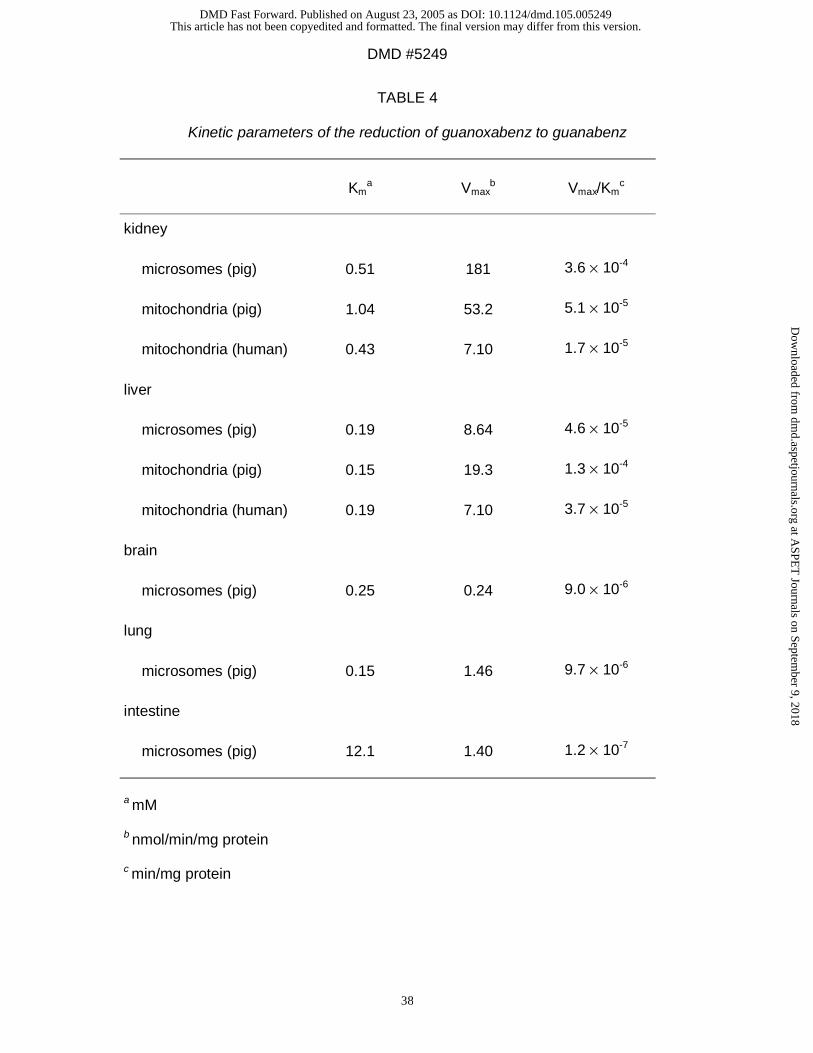
Reduction of guanoxabenz to guanabenz: Guanoxabenz which is known to be
reduced by liver microsomes (Clement et al., 1996) was also transformed by all
preparations (Table 4). Replacing NADH by NADPH decreased the reduction rates
(data not shown). These results demonstrate again that NADH was the preferred
cosubstrate and that the reduction rate was higher in mitochondria than in
microsomes. The reduction in microsomes was again more pronounced in kidney
than in liver, followed by lung, intestine, and brain (Table 4).
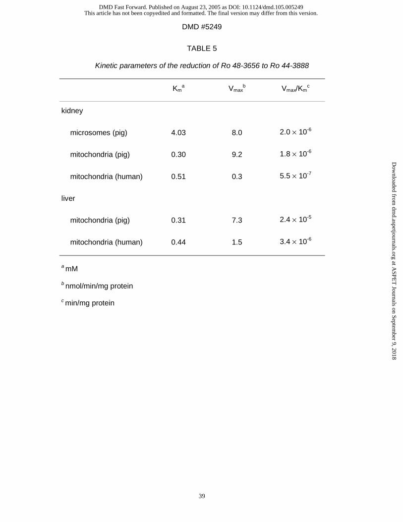
Reduction of Ro 48-3656 to Ro 44-3888: Ro 48-3656 was reduced to its metabolite
Ro 44-3888 (Table 5).
In mitochondria of pig organs NADH was the preferred cosubstrate (data not shown).
In contrast to the other two drugs the kidney and liver rates were very similar ( pig
preparations) or the reduction exhibits greater activity in liver mitochondria ( human
preparations ) (Table 5).
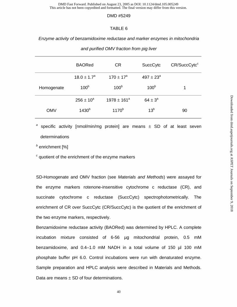
Characterisation of the mitochondrial benzamidoxime reductase.
An outer membrane enriched fraction (OMV-fraction) obtained from pig liver
mitochondria was analysed for marker enzymes of the outer and the inner
mitochondrial membrane, rotenone-insensitive cytochrome c reductase, and
succinate cytochrome c reductase, respectively, as summarized in Table 6. The
quotient of the enrichment of the two enzyme markers indicates that a high degree of
purification of the OMV fraction was obtained. The distribution of the benzamidoxime
reductase was close to that of the outer membrane marker, showing an 11-fold
This article has not been copyedited and formatted. The final version may differ from this version.DMD Fast Forward. Published on August 23, 2005 as DOI: 10.1124/dmd.105.005249
at ASPE
T Journals on Septem
ber 9, 2018dm
d.aspetjournals.orgD
ownloaded from
DMD #5249
18
enrichment of rotenone-insensitive cytochrome c reductase, and 14-fold enrichment
of benzamidoxime reductase in the OMV-fraction over the starting swell-disruption
homogenate (see Materials and Methods) and indicates its outer membrane
localisation.
As shown in Table 3 the OMV-fraction is a very potent preparation for reducing the
model substrate benzamidoxime. The preferred cosubstrate of the reduction was
NADH and the pH optimum was pH 6.0. The reduction also occured at pH 7.4 (data
not shown). The catalytic efficiency was increased about a hundredfold in
comparison to the unfractioned pig liver mitochondria (Table 6).
The NADH-dependent benzamidoxime reductase was completely inhibited by the
NADH b5-reductase inhibitor p-HMB (Shimada et al., 1998) at concentrations of
80 µM in mitochondria and 20 µM in the OMV-fraction. A 50% inhibition of
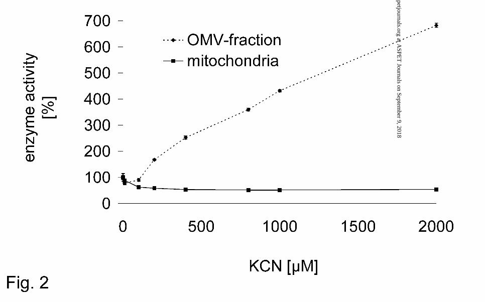
mitochondrial benzamidoxime reduction was observed at concentrations of > 400 µM
potassium cyanide (Fig. 2).
However, the benzamidoxime reductase activity of the OMV-fraction was insensitive
and in contrast even enhanced by cyanide (Fig. 2).
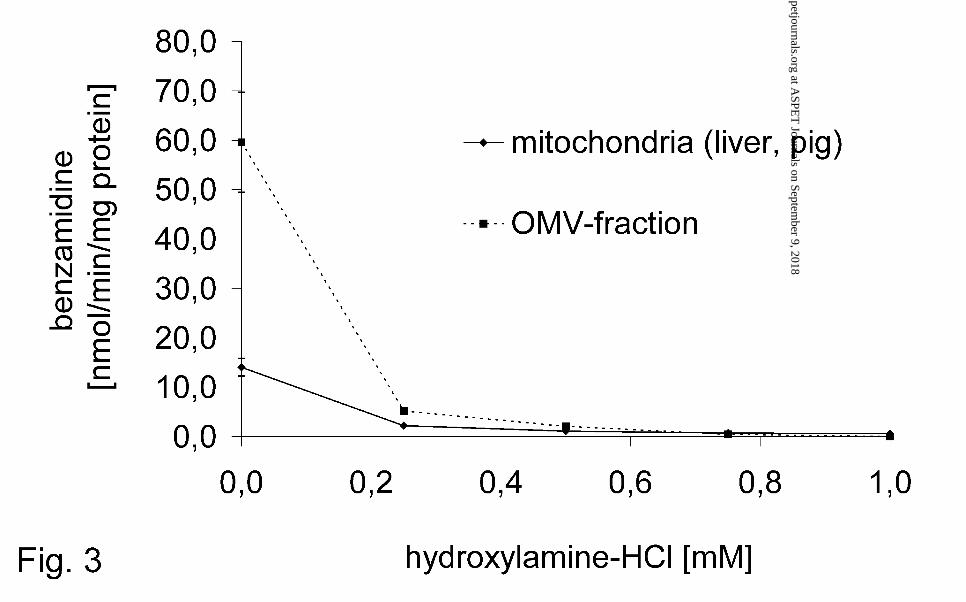
25 µM hydroxylamine-HCl almost completely inhibited the benzamidoxime reductase
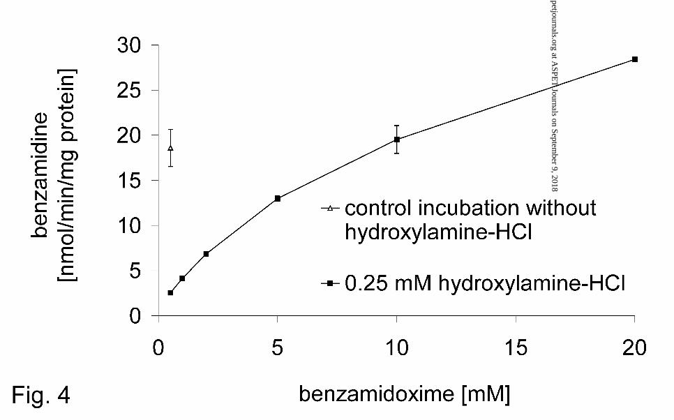
activity in mitochondria and the OMV-fraction (Fig. 3). However, increasing
concentrations of benzamidoxime in the incubation mixture reversed the inhibitory
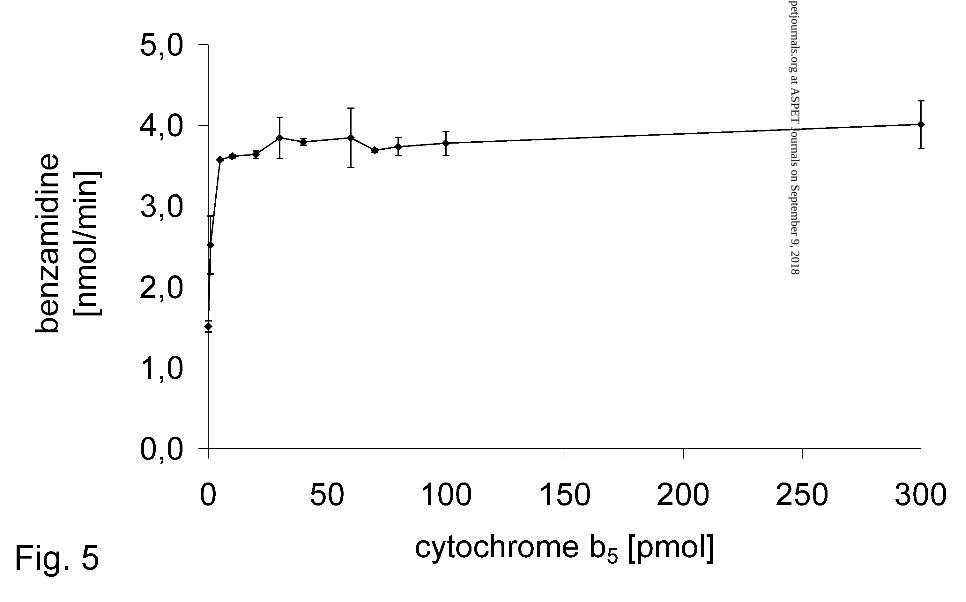
effect of hydroxylamine-HCl (Fig. 4). The rate of the benzamidoxime reduction in the
OMV-fraction could be doubled by adding microsomal purified cytochrome b5 (Fig. 5).
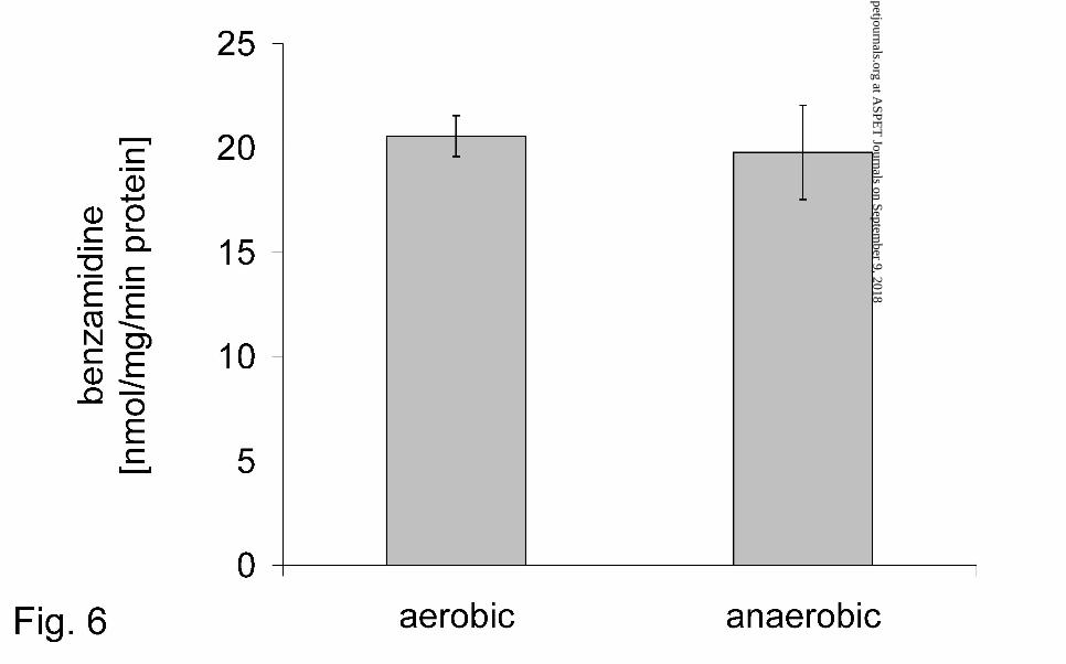
The mitochondrial reduction of benzamidoxime was oxygen-insensitive as well as the
microsomal hydroxylamine reductase (Kadlubar and Ziegler, 1974) (Fig. 6).
This article has not been copyedited and formatted. The final version may differ from this version.DMD Fast Forward. Published on August 23, 2005 as DOI: 10.1124/dmd.105.005249
at ASPE
T Journals on Septem
ber 9, 2018dm
d.aspetjournals.orgD
ownloaded from
DMD #5249
19
Discussion
Extrahepatic metabolism for the activation of N-hydroxylated prodrugs.
The reduction of benzamidoxime was described using liver microsomes as enzyme
source (Clement et al., 1997; Andersson et al., 2005) and the reduction of
guanoxabenz was also investigated with liver microsomes (Clement et al., 1996) and
in addition by cytosolic fractions (Dambrova et al., 1998).
The objective of these investigations was to demonstrate the reduction of several
amidoximes to amidines by microsomes and also by mitochondria of different organs
(Fig. 1). Although the liver is usually the main organ for drug metabolism the
conversion rates in porcine kidney preparations were usually higher than in liver
preparations and mitochondrial reduction rates were higher than microsomal ones
(Table 3, 4, 5). The only exception of mitochondrial superiority was the conversion of
guanoxabenz (Table 4): in this case the microsomal kidney preparations showed
reduction rates which were three times higher than those of the mitochondrial
preparations of liver and kidney.
The specific microsomal reduction rates were always high in kidney, followed by liver,
brain, lung, and intestine demonstrating different concentrations of the responsible
enzymes (Table 3, 4, 5).
The mitochondrial superiority and the extrahepatic benzamidoxime reduction was
similar to the recent findings of Andersson et al. (2005) using subcellular preparations
of rats.
The gastrointestinal route is the most acceptable way of drug administration.
Therefore the principle of amidoximes as prodrugs for amidines was developed
(Clement, 1993). In contrast to the active amidines amidoximes are less basic, not
protonated under physiological conditions and are absorbed from the gastrointestinal
This article has not been copyedited and formatted. The final version may differ from this version.DMD Fast Forward. Published on August 23, 2005 as DOI: 10.1124/dmd.105.005249
at ASPE
T Journals on Septem
ber 9, 2018dm
d.aspetjournals.orgD
ownloaded from
DMD #5249
20
tract. In this context, it is disadvantageous if the N-hydroxylated structures are
already reduced by microsomal enzymes present in the intestine. This was
demonstrated by this study and might be one reason why the bioavailability of
amidoximes is comparably high but does not reach 100% (Clement, 1993).
The occurrence of a reducing system in the lung (Table 3, 4) offers an alternative
application way for amidoximes as prodrugs for amidines by inhalation.
In order to be able to cross the blood-brain barrier by diffusion xenobiotics have to be
lipophilic. This study demonstrates that enzymes which are able to reduce
amidoximes to amidines are present in the brain (Table 3, 4). So the concept of
amidoximes as prodrugs of amidines might also be used to overcome the blood-brain
barrier for hydrophilic amidines. Of course the amount of amidoximes penetrating into
the CNS will be limited by the reduction of amidoximes in other organs but it might
still be high enough to achieve a desired pharmacodynamic effect.
For our studies pig organs were chosen because of their easy availability and the
similarities between human and pig liver in the capacity of reducing N-hydroxylated
compounds (Clement et al., 1997). These observations suggest that reductase
activities could also be present in different human organs. In all human preparations
the detected conversion rates were lower than in the porcine preparations, but still
very high for foreign compound metabolism. All investigated human organs reduced
the three substrates.
Because of the similar characteristics it can be speculated that one enzyme system is
responsible for these reductions. Obviously, these enzymes reduce N-hydroxylated
structures as part of different types of compounds (Fig. 1). This discovery is of great
importance for drug research and development with regard to the development of
prodrugs (Ettmayer et al., 2004).
This article has not been copyedited and formatted. The final version may differ from this version.DMD Fast Forward. Published on August 23, 2005 as DOI: 10.1124/dmd.105.005249
at ASPE
T Journals on Septem
ber 9, 2018dm
d.aspetjournals.orgD
ownloaded from
DMD #5249
21
An enzyme system, consisting of cytochrome b5, its reductase, and a cytochrome
P450 isoenzyme from the subfamily 2D was isolated from pig liver and called the
microsomal benzamidoxime reductase (Clement et al., 1997).
In order to investigate the potential presence of the same enzyme system in other
organs, the presence of the three components of the microsomal benzamidoxime
reductase was analyzed. Cytochrome b5 and its reductase could be detected in all
other investigated microsomal preparations (Table 1) and in all mitochondrial
preparations (Table 2). It is known that cytochrome P450 genes are expressed in
many extrahepatic tissues, even in all investigated organs (Guengerich, 1995;
Pelkonen and Raunio, 1997). In brain and lung microsomes cytochrome P450 was
not detectable by carbon monoxide difference spectra. It has been described before
that P450 concentrations are too low to be analyzed by this assay but can measured
by its activity (Ghersi-Egea et al., 1994) or by immunohistochemical techniques
(Krishna and Klotz, 1994). Brain cytochrome P450 could only measured spectrally
after extraction by hydrophobic chromatography and not in microsomal fractions
(Warner and Gustafsson, 1994). Other studies have demonstrated the expression of
xenobiotic-metabolizing cytochrome P450 forms in porcine tissues (Nissen et al.,
1998; Skaanild and Friis, 1999). Especially the lung has an extensive range of these
xenobiotic-metabolizing enzymes including 1A1, 2B, 2E1, 2F1, 3A, and 4B1
(Pelkonen and Raunio, 1997). It can be suggested that in kidney, brain, lung, and
intestine the same enzyme system as in the liver is responsible for the observed
conversion rates. However, the presence and participation of other enzyme systems
cannot be excluded. This will be the subject of further studies.
In summary, these results demonstrate the importance of extrahepatic metabolism
for the activation of N-hydroxylated prodrugs. As we also discovered the importance
This article has not been copyedited and formatted. The final version may differ from this version.DMD Fast Forward. Published on August 23, 2005 as DOI: 10.1124/dmd.105.005249
at ASPE
T Journals on Septem
ber 9, 2018dm
d.aspetjournals.orgD
ownloaded from
DMD #5249
22
of a mitochondrial reduction further characterisation of the mitochondrial enzyme
systems was undertaken.
So far anaerobic reduction in rat liver mitochondria of a quinone-dependent N-oxide
reductase (Kitamura et al., 1999), and NADPH-dependet N-oxide reductase (Sugiura
and Kato, 1977) were documented as well as oxygen – insensitive NADH-dependet
reduction of hydroxylamines and aryl hydroxamates (Bernheim and Hochstein, 1968).
The mitochondrial reduction exhibited similar properties to the microsomal reduction.
The reduction is oxygen-insensitive (Fig. 6), NADH is the preferred cosubstrate and
its pH optimum is at pH 6.0. Reduction could also be observed at physiological pH
(data not shown). These properties are also similar to the characteristics of the
mitochondrial hydroxylamine reductase (Bernheim and Hochstein, 1968). Thus
inhibition studies showed that the reduction of benzamidoxime could be inhibited by
hydroxylamine (Fig. 3) and the inhibition could be reversed by enhanced
concentrations of benzamidoxime (Fig. 4).
Studies with a membrane fraction of pig liver indicate that the benzamidoxime
reductase activity is located in the outer membrane. Cytochrome b5 and its reductase
are compounds of the outer mitochondrial membrane (Sottocasa and Sandri, 1970;
Taniguchi et al., 1973) and could also be detected in this study in the OMV-fraction of
pig liver mitochondria.
NADH cytochrome b5 reductase is an integral membrane protein essential for
microsomes and the outer mitochondrial membrane (Borgese and Pietrini, 1986). In
mammals, the reductase in its various locations is molecularly identical (Meldolesi et
al., 1980).
Two distinct forms of cytochrome b5 have been shown to exist in rat liver (Lederer et
al., 1983). Cytochrome b5 localized in the outer mitochondrial membrane is
distinguishable from the microsomal form in spectral and immunological properties as
This article has not been copyedited and formatted. The final version may differ from this version.DMD Fast Forward. Published on August 23, 2005 as DOI: 10.1124/dmd.105.005249
at ASPE
T Journals on Septem
ber 9, 2018dm
d.aspetjournals.orgD
ownloaded from
DMD #5249
23
well as in primary structure. Furthermore, the mitochondrial isoform has a more
negative reduction potential and is more stable towards chemical and thermal
denaturation (Altuve et al., 2001). We assume that similarly to the microsomal
benzamidoxime reductase again cytochrome b5 and its reductase are components of
the mitochondrial enzyme system. This assumption could be verified by studies with
p-hydroxymercuribenzoate, a known inhibitor of NADH cytochrome b5 reductase
(Shimada et al., 1988). Experiments with purified microsomal cytochrome b5
enhanced the activity in the OMV-fraction. The activity was doubled. This raise was
already saturated at ~10 pmol cytochrome b5 (Fig. 5).
It cannot be excluded that similarly to the microsomal benzamidoxime reductase a
third protein is a component of the benzamidoxime reductase in the outer
mitochondrial membrane.
In order to investigate if again a P450 enzyme is again included in the
benzamidoxime reduction its content was analyzed. Only low amounts of P450 could
be detected in the OMV-fraction by carbon monoxide difference spectra (data not
shown). The location of P450 isoenzymes in mitochondria is usually the inner
mitochondrial membrane (Sottocasa and Sandri, 1970; Taniguchi et al., 1973; della-
Cioppa et al., 1986). However, a report about a cyanide insensitive enzyme system
containing P450 in the purified outer membrane was published (Uemura and
Chiesara, 1976). A partial inhibition of mitochondrial benzamidoxime reduction was
observed at concentrations > 400 µM potassium cyanide. However, the
benzamideoxime reductase activity of the OMV-fraction was insensitive and in
contrast even enhanced by cyanide (Fig. 2). This is an indication of a cyanide
sensitive and insensitive reduction system.
Purification studies are in process to clarify the participation and identification of a
potential third component of the mitochondrial benzamidoxime reductase, which
This article has not been copyedited and formatted. The final version may differ from this version.DMD Fast Forward. Published on August 23, 2005 as DOI: 10.1124/dmd.105.005249
at ASPE
T Journals on Septem
ber 9, 2018dm
d.aspetjournals.orgD
ownloaded from
DMD #5249
24
shares common characteristics with the microsomal protein described by Kadlubar
and Ziegler (1974). The presence of additional N-reductive enzyme systems in other
submitochondrial compartments cannot be excluded.
This article has not been copyedited and formatted. The final version may differ from this version.DMD Fast Forward. Published on August 23, 2005 as DOI: 10.1124/dmd.105.005249
at ASPE
T Journals on Septem
ber 9, 2018dm
d.aspetjournals.orgD
ownloaded from
DMD #5249
25
Acknowledgement
We would like to thank Sven and Meike Wichmann for their technical assistance
This article has not been copyedited and formatted. The final version may differ from this version.DMD Fast Forward. Published on August 23, 2005 as DOI: 10.1124/dmd.105.005249
at ASPE
T Journals on Septem
ber 9, 2018dm
d.aspetjournals.orgD
ownloaded from
DMD #5249
26
References
Altuve A; Silchenko S; Lee KH; Kuczera K; Terzyan S; Zhang X, Benson DR and
Rivera M (2001) Probing the differences between rat liver outer mitochondrial
membrane cytochrome b5 and microsomal cytochromes b5. Biochemistry 40:
9469-83.
Andersson S, Hofmann Y, Nordling A, Li X, Nivelius S, Andersson T, Ingelman-
Sundberg M and Johansson I (2005) Characterization and partial purification
of the rat and human enzyme systems active in the reduction of N-
hydroxymelagatran and benzamideoxime
Bernheim ML and Hochstein P (1968) Reduction of hydroxylamine by rat liver
mitochondria. Arch Biochem Biophys 124: 436-42.
Borgese N and Pietrini G (1986) Distribution of the integral membrane protein NADH-
cytochrome b5 reductase in rat liver cells, studied with a quantitative
radioimmunoblotting assay. Biochem J 239: 393-403.
Clement B (2002) Reduction of N-hydroxylated compounds: amidoximes
(N-hydroxyamidines) as pro-drugs of amidines. Drug Metab Rev 34: 565-79.
Clement B, German patent (1993), P 4321444.4, PCT/DE 94/000756 (1994), USA
(5,786,383;28/6/1998), Europe (0708640,16/9/1998)
Clement B, Demesmaeker M and Linne S (1996) Microsomal catalyzed
N-hydroxylation of guanabenz and reduction of the N-hydroxylated metabolite:
characterization of the two reactions and genotoxic potential of guanoxabenz.
Chem Res Toxicol 9: 682-8.
Clement B, Lomb R and Moller W (1997) Isolation and characterization of the protein
components of the liver microsomal O2-insensitive NADH-benzamidoxime
reductase. J Biol Chem 272: 19615-20.
This article has not been copyedited and formatted. The final version may differ from this version.DMD Fast Forward. Published on August 23, 2005 as DOI: 10.1124/dmd.105.005249
at ASPE
T Journals on Septem
ber 9, 2018dm
d.aspetjournals.orgD
ownloaded from
DMD #5249
27
Clement B, Schmitt S and Zimmermann M (1988) Enzymatic reduction of
benzamidoxime to benzamidine. Arch Pharm (Weinheim) 321: 955-6.
Dambrova M., Uhlen S, Welch CJ, Prusis P and Wikberg JE (1998) Characterization
of the enzymatic activity for biphasic competition by guanoxabenz
(1-(2,6-dichlorobenzylidene-amino)-3-hydroxyguanidine) at
alpha2-adrenoceptors. II. Description of a xanthine-dependent enzymatic
activity in spleen cytosol. Biochem Pharmacol 56: 1121-8.
de Kroon AI, Dolis D, Mayer A, Lill R and de Kruijff B (1997) Phospholipid
composition of highly purified mitochondrial outer membranes of rat liver and
Neurospora crassa. Is cardiolipin present in the mitochondrial outer
membrane? Biochim Biophys Acta 1325: 108-16.
della-Cioppa GK, Muffly E, Yanagibashi K and Hall PF (1986) Preparation and
characterization of submitochondrial fractions from adrenal cells. Mol Cell
Endocrinol 48: 111-20.
Estabrook RW and Werringloer J (1978) The measurement of difference spectra:
application to the cytochromes of microsomes. Methods Enzymol 52: 212-20.
Ettmayer P, Amidon GL, Clement B and Testa B (2004) Lessons learned from
marketed and investigational prodrugs. J Med Chem 47: 2393-404.
Ghersi-Egea JF, Leninger-Muller B, Suleman G, Siest G and Minn A (1994)
Localization of drug-metabolizing enzyme activities to blood-brain interfaces
and circumventricular organs. J Neurochem 62: 1089-96.
Guengerich FP (1995) In: Ortiz de Montellano, P.R.: Cytochrome P450: Structure,
Mechanism and Biochemistry (Second Edition). New York, Plenum Press,
473-535
This article has not been copyedited and formatted. The final version may differ from this version.DMD Fast Forward. Published on August 23, 2005 as DOI: 10.1124/dmd.105.005249
at ASPE
T Journals on Septem
ber 9, 2018dm
d.aspetjournals.orgD
ownloaded from
DMD #5249
28
Gustafsson D, Nystrom J, Carlsson S, Bredberg U, Eriksson U, Gyzander E, Elg M,
Antonsson T, Hoffmann K, Ungell A, Sorensen H, Nagard S, Abrahamsson A,
and Bylund R (2001) The direct thrombin inhibitor melagatran and its oral
prodrug H 376/95: intestinal absorption properties, biochemical and
pharmacodynamic effects. Thromb Res 101: 171-81
Hovius R, Lambrechts H, Nicolay K and de Kruijff B (1990) Improved methods to
isolate and subfractionate rat liver mitochondria. Lipid composition of the inner
and outer membrane. Biochim Biophys Acta 1021: 217-26.
Kadlubar F F and Ziegler DM (1974) Properties of a NADH-dependent N-hydroxy
amine reductase isolated from pig liver microsomes. Arch Biochem Biophys
162: 83-92.
Kitamura S, Sugihara K and Tatsumi K (1999) A unique tertiary amine N-oxide
reduction system composed of quinone reductase and heme in rat liver
preparations. Drug Metab Dispos 27: 92-7.
Krishna DR and Klotz U (1994) Extrahepatic metabolism of drugs in humans. Clin
Pharmacokinet 26: 144-60.
Krüger P (1885) Über Abkömmlinge des Benzenylamidoxims. Ber. Dtsch. Chem.
Ges. 18: 1055-1060.
Lederer F, Ghrir R, Guiard B, Cortial S, and Ito A (1983) Two homologous
cytochromes b5 in a single cell. Eur J Biochem 132: 95-102.
Meldolesi J, Corte G, Pietrini G. and Borgese N (1980) Localization and biosynthesis
of NADH-cytochrome b5 reductase, an integral membrane protein, in rat liver
cells. II. Evidence that a single enzyme accounts for the activity in its various
subcellular locations. J Cell Biol 85: 516-26.
Mihara K and Sato R (1978) Detergent-solubilized NADH-cytochrome b5 reductase.
Methods Enzymol 52: 102-8.
This article has not been copyedited and formatted. The final version may differ from this version.DMD Fast Forward. Published on August 23, 2005 as DOI: 10.1124/dmd.105.005249
at ASPE
T Journals on Septem
ber 9, 2018dm
d.aspetjournals.orgD
ownloaded from
DMD #5249
29
Nissen PH, Wintero AK, Fredholm M (1998) Mapping of porcine genes belonging to
two different cytochrome P450 subfamilies. Anim Genet 29: 7-11.
Omura T and Sato R (1964) The Carbon Monoxide-Binding Pigment of Liver
Microsomes. I. Evidence for Its Hemoprotein Nature. J Biol Chem 239: 2370-8.
Pelkonen O and Raunio H (1997) Metabolic activation of toxins: tissue-specific
expression and metabolism in target organs. Environ Health Perspect 105
Suppl 4: 767-74.
Shimada H, Hirai K, Simamura Eand Pan J (1998) Mitochondrial NADH-quinone
oxidoreductase of the outer membrane is responsible for paraquat cytotoxicity
in rat livers. Arch Biochem Biophys 351, 75-81
Skaanild MT and Friis C (1999) Cytochrome P450 sex differences in minipigs and
conventional pigs. Pharmacol Toxicol 85: 174-80.
Smith PK, Krohn RI, Hermanson GT, Mallia AK, Gartner FH, Provenzano MD,
Fujimoto EK, Goeke NM, Olson BJ and Klenk DC (1985) Measurement of
protein using bicinchoninic acid. Anal Biochem 150: 76-85.
Sottocasa GL, Kuylenstierna B, Ernster L and Bergstrand A (1967) An electron-
transport system associated with the outer membrane of liver mitochondria. A
biochemical and morphological study. J Cell Biol 32: 415-38.
Sottocasa GL and Sandri G (1970) Intramitochondrial distribution of cytochrome
P-450 in ox adrenal cortex. Biochem J 116: 16P-17P.
Sugiura M and Kato R (1977) Reduction of tertiary amine N-oxides by rat liver
mitochondria. J Pharmacol Exp Ther 200: 25-32.
Taniguchi H, Imai Y and Sato R (1984) Role of the electron transfer system in
microsomal drug monooxygenase reaction catalyzed by cytochrome P-450.
Arch Biochem Biophys 232: 585-96.
This article has not been copyedited and formatted. The final version may differ from this version.DMD Fast Forward. Published on August 23, 2005 as DOI: 10.1124/dmd.105.005249
at ASPE
T Journals on Septem
ber 9, 2018dm
d.aspetjournals.orgD
ownloaded from
DMD #5249
30
Taniguchi S, Hoshita N and Okuda K (1973) Enzymatic characteristics of CO-
sensitive 26-hydroxylase system for 5beta-cholestane-3 alpha, 7 alpha, 12
alpha-triol in rat-liver mitochondria and its intramitochondrial localization. Eur J
Biochem 40: 607-17.
Timm U, Zumbrunnen R, Erdin R, Singer M and Steiner B (1997) Oral platelet
aggregation inhibitor Ro 48-3657: determination of the active metabolite and
its prodrug in plasma and urine by high-performance liquid chromatography
using automated column switching. J Chromatogr B Biomed Sci Appl 691:
397-407.
Uemura T and Chiesara E (1976) NADH-dependent aryl hydrocarbon hydroxylase in
rat liver mitochondrial outer membrane. Eur J Biochem 66: 293-307.
Warner M and Gustafsson JA (1994) Effect of ethanol on cytochrome P450 in the rat
brain. Proc Natl Acad Sci U S A 91: 1019-23.
Weller T, Alig L, Beresini M, Blackburn B, Bunting S, Hadvary P, Muller MH, Knopp
D, Levet-Trafit B, Lipari MT, Modi NB, Muller M, Refino CJ, Schmitt M,
Schonholzer P, Weiss S, and Steiner B (1996) Orally active fibrinogen
receptor antagonists. 2. Amidoximes as prodrugs of amidines. J Med Chem
39: 3139-47
Yasukochi Y and Masters BS (1976) Some properties of a detergent-solubilized
NADPH-cytochrome c(cytochrome P-450) reductase purified by biospecific
affinity chromatography. J Biol Chem 251: 5337-44.
Zhou L, Thakker DR, Voyksner RD, Anbazhagan M, Boykin D W, Hall JE and Tidwell
RR (2004) Metabolites of an orally active antimicrobial prodrug,
2,5-bis(4-amidinophenyl)furan-bis-O-methylamidoxime, identified by liquid
chromatography/tandem mass spectrometry. J Mass Spectrom 39: 351-60
This article has not been copyedited and formatted. The final version may differ from this version.DMD Fast Forward. Published on August 23, 2005 as DOI: 10.1124/dmd.105.005249
at ASPE
T Journals on Septem
ber 9, 2018dm
d.aspetjournals.orgD
ownloaded from
DMD #5249
31
Footnotes
Financial support by the Deutsche Forschungsgemeinschaft ( DFG, Cl 56/6-3) and
the Fonds der Chemischen Industrie
Preliminary results: Mau S, Clement B (1999) Arch. Pharm. 332, 31; Deters S,
Clement B (2000) Arch. Pharm. 333, 12
Reprint requests to Prof. Dr. Bernd Clement, Christian-Albrechts-Universität,
Pharmazeutisches Institut, Gutenbergstraße 76, D-24118 Kiel, Germany,
phone: 0431-8801126, fax: 0431-8801352; email: [email protected]
This article has not been copyedited and formatted. The final version may differ from this version.DMD Fast Forward. Published on August 23, 2005 as DOI: 10.1124/dmd.105.005249
at ASPE
T Journals on Septem
ber 9, 2018dm
d.aspetjournals.orgD
ownloaded from
DMD #5249
32
Legends for Figures
FIG. 1. Reductions of N-hydroxylated compounds
FIG. 2. Influence of potassium cyanide on the mitochondrial N-reduction of
benzamidoxime.
Benzamidoxime reductase activity was determinated by HPLC. A complete
incubation mixture consisted of 6-56 µg protein, 0.5 mM benzamidoxime, and 0.4-
1.0 mM NADH in a total volume of 150 µl 100 mM phosphate buffer pH 6.0. After
preincubation of protein and NADH with KCN for 30 minutes the reaction was started
by adding benzamidoxime. Sample preparation and HPLC analysis were described in
Materials and Methods. Data are means ± SD of four determinations.
FIG. 3. Influence of hydroxylamine-HCl on the mitochondrial N-reduction of
benzamidoxime.
Benzamidoxime reductase activity was determinated by HPLC. A complete
incubation mixture consisted of 6-56 µg protein, 0.5 mM benzamidoxime, and 0.4-1.0
mM NADH in a total volume of 150 µl 100 mM phosphate buffer pH 6.0. After
preincubation of protein and NADH with hydroxylamine-HCl for 5 minutes the
reaction was started by adding benzamidoxime. Sample preparation and HPLC
analysis were performed as described in Materials and Methods. Data are means
± SD of four determinations.
This article has not been copyedited and formatted. The final version may differ from this version.DMD Fast Forward. Published on August 23, 2005 as DOI: 10.1124/dmd.105.005249
at ASPE
T Journals on Septem
ber 9, 2018dm
d.aspetjournals.orgD
ownloaded from
DMD #5249
33
FIG. 4. Inhibition of the mitochondrial benzamidoxime reduction by 0.25 mM
hydroxylamine-HCl at various benzamidoxime concentrations.
Benzamidoxime reductase activity was determinated by HPLC. A complete
incubation mixture consisted of 56 µg protein (pig liver mitochondria),
benzamidoxime, and 1.0 mM NADH in a total volume of 150 µl 100 mM phosphate
buffer pH 6.0. After preincubation of protein with benzamidoxime for 5 minutes
0.25 mM hydroxylamine-HCl was added, and the reaction was started with NADH. A
control incubation with 0.5 mM benzamidoxime and without hydroxylamine-HCl was
performed. Sample preparation and HPLC analysis were performed as described in
Materials and Methods. Data are means ± SD of four determinations.
FIG. 5. Influence of microsomal cytochrome b5 on the reduction of benzamidoxime by
the OMV-fraction
Benzamidoxime reductase activity was determinated by HPLC. A complete
incubation mixture consisted of 6 µg protein (OMV), 1-300 pmol cytochrome b5,
0.5 mM benzamidoxime, and 0.4 mM NADH in a total volume of 150 µl 100 mM
phosphate buffer pH 6.0. Sample preparation and HPLC analysis were performed as
described in Materials and Methods. Data are means ± SD of four determinations.
FIG. 6. Influence of oxygen on the reduction of benzamidoxime by pig liver
mitochondria
Benzamidoxime reductase activity was determinated by HPLC. A complete
incubation mixture consisted of 0.056 mg protein, 0.5 mM benzamidoxime, and 1.0
mM NADH in a total volume of 150 µl 100 mM phosphate buffer pH 6.0. Aerobic
incubations were exposed to laboratory air while anaerobic incubations were
performed in argon-degassed buffers, were gassed with argon and the reaction tubes
This article has not been copyedited and formatted. The final version may differ from this version.DMD Fast Forward. Published on August 23, 2005 as DOI: 10.1124/dmd.105.005249
at ASPE
T Journals on Septem
ber 9, 2018dm
d.aspetjournals.orgD
ownloaded from
DMD #5249
34
were closed during incubation. Sample preparation and HPLC analysis were
performed as described in Materials and Methods. Data are means ± SD of four
determinations.
This article has not been copyedited and formatted. The final version may differ from this version.DMD Fast Forward. Published on August 23, 2005 as DOI: 10.1124/dmd.105.005249
at ASPE
T Journals on Septem
ber 9, 2018dm
d.aspetjournals.orgD
ownloaded from
DMD #5249
35
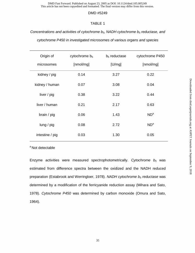
TABLE 1
Concentrations and activities of cytochrome b5, NADH cytochrome b5 reductase, and
cytochrome P450 in investigated microsomes of various organs and species
Origin of
microsomes
cytochrome b5
[nmol/mg]
b5 reductase
[U/mg]
cytochrome P450
[nmol/mg]
kidney / pig 0.14 3.27 0.22
kidney / human 0.07 3.08 0.04
liver / pig 0.38 3.22 0.44
liver / human 0.21 2.17 0.63
brain / pig 0.06 1.43 NDa
lung / pig 0.08 2.72 NDa
intestine / pig 0.03 1.30 0.05
a Not detectable
Enzyme activities were measured spectrophotometrically. Cytochrome b5 was
estimated from difference spectra between the oxidized and the NADH reduced
preparation (Estabrook and Werringloer, 1978). NADH cytochrome b5 reductase was
determined by a modification of the ferricyanide reduction assay (Mihara and Sato,
1978). Cytochrome P450 was determined by carbon monoxide (Omura and Sato,
1964).
This article has not been copyedited and formatted. The final version may differ from this version.DMD Fast Forward. Published on August 23, 2005 as DOI: 10.1124/dmd.105.005249
at ASPE
T Journals on Septem
ber 9, 2018dm
d.aspetjournals.orgD
ownloaded from
DMD #5249
36
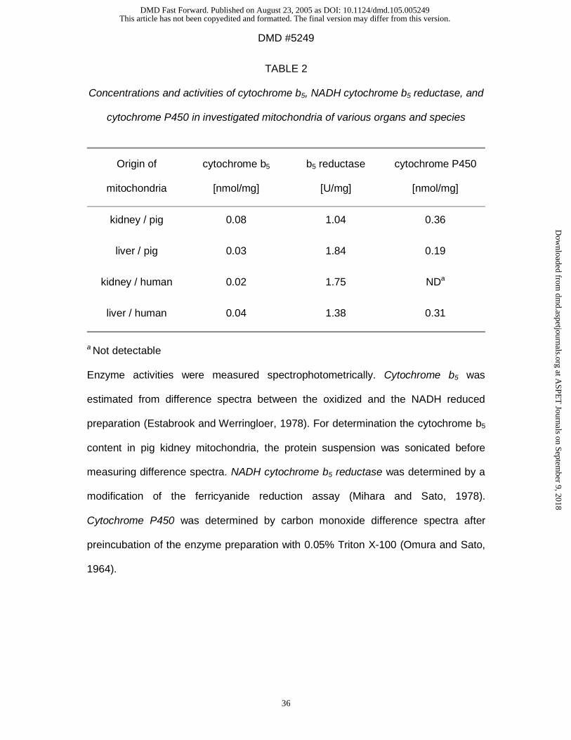
TABLE 2
Concentrations and activities of cytochrome b5, NADH cytochrome b5 reductase, and
cytochrome P450 in investigated mitochondria of various organs and species
Origin of
mitochondria
cytochrome b5
[nmol/mg]
b5 reductase
[U/mg]
cytochrome P450
[nmol/mg]
kidney / pig 0.08 1.04 0.36
liver / pig 0.03 1.84 0.19
kidney / human 0.02 1.75 NDa
liver / human 0.04 1.38 0.31
a Not detectable Enzyme activities were measured spectrophotometrically. Cytochrome b5 was
estimated from difference spectra between the oxidized and the NADH reduced
preparation (Estabrook and Werringloer, 1978). For determination the cytochrome b5
content in pig kidney mitochondria, the protein suspension was sonicated before
measuring difference spectra. NADH cytochrome b5 reductase was determined by a
modification of the ferricyanide reduction assay (Mihara and Sato, 1978).
Cytochrome P450 was determined by carbon monoxide difference spectra after
preincubation of the enzyme preparation with 0.05% Triton X-100 (Omura and Sato,
1964).
This article has not been copyedited and formatted. The final version may differ from this version.DMD Fast Forward. Published on August 23, 2005 as DOI: 10.1124/dmd.105.005249
at ASPE
T Journals on Septem
ber 9, 2018dm
d.aspetjournals.orgD
ownloaded from
DMD #5249
37
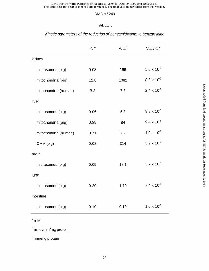
TABLE 3
Kinetic parameters of the reduction of benzamidoxime to benzamidine
Kma Vmax
b Vmax/Kmc
kidney
microsomes (pig) 0.03 166 5.0 × 10⁻¹
mitochondria (pig) 12.8 1082 8.5 × 10⁻⁵
mitochondria (human) 3.2 7.8 2.4 × 10⁻⁶
liver
microsomes (pig) 0.06 5.3 8.8 × 10⁻⁵
mitochondria (pig) 0.89 84 9.4 × 10⁻⁵
mitochondria (human) 0.71 7.2 1.0 × 10⁻⁵
OMV (pig) 0.08 314 3.9 × 10⁻³
brain
microsomes (pig) 0.05 18.1 3.7 × 10⁻⁴
lung
microsomes (pig) 0.20 1.70 7.4 × 10⁻⁶
intestine
microsomes (pig) 0.10 0.10 1.0 × 10⁻⁶
a mM
b nmol/min/mg protein
c min/mg protein
This article has not been copyedited and formatted. The final version may differ from this version.DMD Fast Forward. Published on August 23, 2005 as DOI: 10.1124/dmd.105.005249
at ASPE
T Journals on Septem
ber 9, 2018dm
d.aspetjournals.orgD
ownloaded from
DMD #5249
38
TABLE 4
Kinetic parameters of the reduction of guanoxabenz to guanabenz
Kma Vmax
b Vmax/Kmc
kidney
microsomes (pig) 0.51 181 3.6 × 10⁻⁴
mitochondria (pig) 1.04 53.2 5.1 × 10⁻⁵
mitochondria (human) 0.43 7.10 1.7 × 10⁻⁵
liver
microsomes (pig) 0.19 8.64 4.6 × 10⁻⁵
mitochondria (pig) 0.15 19.3 1.3 × 10⁻⁴
mitochondria (human) 0.19 7.10 3.7 × 10⁻⁵
brain
microsomes (pig) 0.25 0.24 9.0 × 10⁻⁶
lung
microsomes (pig) 0.15 1.46 9.7 × 10⁻⁶
intestine
microsomes (pig) 12.1 1.40 1.2 × 10⁻⁷
a mM
b nmol/min/mg protein
c min/mg protein
This article has not been copyedited and formatted. The final version may differ from this version.DMD Fast Forward. Published on August 23, 2005 as DOI: 10.1124/dmd.105.005249
at ASPE
T Journals on Septem
ber 9, 2018dm
d.aspetjournals.orgD
ownloaded from
DMD #5249
39
TABLE 5
Kinetic parameters of the reduction of Ro 48-3656 to Ro 44-3888
Kma Vmax
b Vmax/Kmc
kidney
microsomes (pig) 4.03 8.0 2.0 × 10⁻⁶
mitochondria (pig) 0.30 9.2 1.8 × 10⁻⁶
mitochondria (human) 0.51 0.3 5.5 × 10⁻⁷
liver
mitochondria (pig) 0.31 7.3 2.4 × 10⁻⁵
mitochondria (human) 0.44 1.5 3.4 × 10⁻⁶
a mM
b nmol/min/mg protein
c min/mg protein
This article has not been copyedited and formatted. The final version may differ from this version.DMD Fast Forward. Published on August 23, 2005 as DOI: 10.1124/dmd.105.005249
at ASPE
T Journals on Septem
ber 9, 2018dm
d.aspetjournals.orgD
ownloaded from
DMD #5249
40
TABLE 6
Enzyme activity of benzamidoxime reductase and marker enzymes in mitochondria
and purified OMV fraction from pig liver
BAORed CR SuccCytc CR/SuccCytcc
Homogenate
18.0 ± 1.7a
100b
170 ± 17a
100b
497 ± 23a
100b 1
OMV
256 ± 10a
1430b
1978 ± 161a
1170b
64 ± 3a
13b 90
a specific activity [nmol/min/mg protein] are means ± SD of at least seven
determinations
b enrichment [%]
c quotient of the enrichment of the enzyme markers
SD-Homogenate and OMV fraction (see Materials and Methods) were assayed for
the enzyme markers rotenone-insensitive cytochrome c reductase (CR), and
succinate cytochrome c reductase (SuccCytc) spectrophotometrically. The
enrichment of CR over SuccCytc (CR/SuccCytc) is the quotient of the enrichment of
the two enzyme markers, respectively.
Benzamidoxime reductase activity (BAORed) was determined by HPLC. A complete
incubation mixture consisted of 6-56 µg mitochondrial protein, 0.5 mM
benzamidoxime, and 0.4–1.0 mM NADH in a total volume of 150 µl 100 mM
phosphate buffer pH 6.0. Control incubations were run with denaturated enzyme.
Sample preparation and HPLC analysis were described in Materials and Methods.
Data are means ± SD of four determinations.
This article has not been copyedited and formatted. The final version may differ from this version.DMD Fast Forward. Published on August 23, 2005 as DOI: 10.1124/dmd.105.005249
at ASPE
T Journals on Septem
ber 9, 2018dm
d.aspetjournals.orgD
ownloaded from
This article has not been copyedited and formatted. The final version may differ from this version.DMD Fast Forward. Published on August 23, 2005 as DOI: 10.1124/dmd.105.005249
at ASPE
T Journals on Septem
ber 9, 2018dm
d.aspetjournals.orgD
ownloaded from
This article has not been copyedited and formatted. The final version may differ from this version.DMD Fast Forward. Published on August 23, 2005 as DOI: 10.1124/dmd.105.005249
at ASPE
T Journals on Septem
ber 9, 2018dm
d.aspetjournals.orgD
ownloaded from
This article has not been copyedited and formatted. The final version may differ from this version.DMD Fast Forward. Published on August 23, 2005 as DOI: 10.1124/dmd.105.005249
at ASPE
T Journals on Septem
ber 9, 2018dm
d.aspetjournals.orgD
ownloaded from
This article has not been copyedited and formatted. The final version may differ from this version.DMD Fast Forward. Published on August 23, 2005 as DOI: 10.1124/dmd.105.005249
at ASPE
T Journals on Septem
ber 9, 2018dm
d.aspetjournals.orgD
ownloaded from
This article has not been copyedited and formatted. The final version may differ from this version.DMD Fast Forward. Published on August 23, 2005 as DOI: 10.1124/dmd.105.005249
at ASPE
T Journals on Septem
ber 9, 2018dm
d.aspetjournals.orgD
ownloaded from
This article has not been copyedited and formatted. The final version may differ from this version.DMD Fast Forward. Published on August 23, 2005 as DOI: 10.1124/dmd.105.005249
at ASPE
T Journals on Septem
ber 9, 2018dm
d.aspetjournals.orgD
ownloaded from