Embed Size (px)
Citation preview
Cellular Signalling 20 (2008) 1521–1527
Contents lists available at ScienceDirect
Cellular Signalling
j ourna l homepage: www.e lsev ie r.com/ locate /ce l l s ig
Sphingosine 1-phosphate induces platelet/endothelial cell adhesion molecule-1phosphorylation in human endothelial cells through cSrc and Fyn☆
Yu-Ting Huang b, Shee-Uan Chen c, Chia-Hong Chou c, Hsinyu Lee a,b,⁎a Department of Life Science, National Taiwan University, No 1, Sec. 4, Roosevelt Rd., Taipei, 10617, Taiwan, ROCb Institute of Zoology, National Taiwan University, No 1, Sec. 4, Roosevelt Rd., Taipei, 10617, Taiwan, ROCc Department of Obstetrics and Gynecology, National Taiwan University Hospital, No 7, Chung-Shan South Rd., Taipei, 10002, Taiwan, ROC
☆ This work was supported by grant 95-2311-B-00Science Council of ROC to H. Lee.⁎ Corresponding author. Department of Life Science, N
Sec. 4, Roosevelt Rd., Taipei, 10617, Taiwan, ROC. Tel.: +23636837.
E-mail address: [email protected] (H. Lee).
0898-6568/$ – see front matter © 2008 Elsevier Inc. Aldoi:10.1016/j.cellsig.2008.04.008
A B S T R A C T
A R T I C L E I N F OArticle history:
Sphingosine 1-phosphate (S Received 23 January 2008Received in revised form 21 March 2008Accepted 7 April 2008Available online 18 April 2008Keywords:Sphingosine 1-phosphatePECAM-1cSrcFynHUVEC
1P) is a multifunctional phospholipid which acts through a specific family of Gprotein-coupled receptors. Platelet/endothelial cell adhesion molecule-1 (PECAM-1) form trans-homophilicbinding at lateral cell border. Upon stimulation, its cytoplasmic tyrosine residues could be phosphorylatedand interact with various downstream signaling molecules. In this study, we demonstrated that S1P inducedPECAM-1 tyrosine phosphorylation in human umbilical cord vein cells (HUVECs). By pharmacologicalinhibitors, it was suggested that Gi and Src family kinases were involved in PECAM-1 phosphorylation.Moreover, cSrc and Fyn siRNA significantly suppressed S1P-induced PECAM-1 phosphorylation. These resultssuggested that S1P-induced PECAM-1 phosphorylation through Gi and subsequent cSrc and Fyn. Our findingsprovide further understanding of S1P and PECAM-1 signaling as well as their functions in endothelial cells.
© 2008 Elsevier Inc. All rights reserved.
1. Introduction
Sphingosine 1-phosphate (S1P) is a low-molecular-weight phospho-lipidmainly released from stimulated platelets [1,2]. Platelet-released S1Pbinds to S1P receptors on surrounding endothelial cells through aparacrine route [3,4]. To date, five S1P receptors (S1P1–5) have beenidentified [5–10]. These S1P receptors couple to various G proteins,including Gi, G12/13, and Gq [11,12], and activate extracellular signal-regulated kinase (ERK), phospholipase C (PLC), phosphoinositide 3-kinase(PI3K), adenylyl cyclase (AC), Rho, Cdc42, and c-jun N-terminal kinase(JNK) [11–15]. In endothelial cells, S1P regulates various cellular functions,including proliferation,migration, and adhesion [16–19]. Besides, S1P alsomediates re-assembly of cytoskeleton and adherens junction, as well asenhancement of endothelial barrier integrity [19–21].
Platelet/endothelial cell adhesion molecule-1 (PECAM-1) is a 130-kDatransmembrane glycoprotein expressed in endothelial cells, platelets,monocytes, and leukocytes [22,23]. Inendothelial cells, PECAM-1 ismainlylocated at lateral cell border. Its extracellular domain forms trans-homophilic interaction between adjacent cells [24–26]. Moreover,
2-018-MY2 from the National
ational Taiwan University, No 1,886 2 33662499; fax: +886 2
l rights reserved.
cytoplasmic domain of PECAM-1 contains several tyrosine residues.Upon stimulations, these tyrosine residues become phosphorylated andinteract with SH2 domain-containing molecules, such as SHP-2 [27–29].Association of SHP-2 with phosphorylated PECAM-1 was shown tostabilize adherens junctions through de-phosphorylation of beta-catenin[29–31]. Furthermore, studies on PECAM-1-deleted cells and knockoutmice also indicated the association between PECAM-1 phosphorylationand endothelial permeability maintenance [32,33].
Both S1P and PECAM-1 play critical roles in endothelial functions.However, the relationship between these two molecules remainsunclear. In the present study, we first demonstrated that S1P-inducedPECAM-1 tyrosine phosphorylation in human endothelial cells. Byusing pharmacological inhibitors, we demonstrated that activation ofGi and Src family kinases (SFK) involves in the phosphorylation event.In addition, transfection of cSrc and Fyn siRNA attenuated S1P-induced PECAM-1 phosphorylation in a synergistic manner. Theseresults provide a connection between S1P and PECAM-1 signaling.
2. Materials and methods
2.1. Reagents
Collagenase IV, gelatin, sphingosine 1-phosphate, fatty acid-free bovine serumalbumin (BSA), NP-40, horseradish peroxidase molecules (Type VI-A, 44kD), guaiacolsubstrate assay, and protease inhibitors (leupeptin, sodium orthovanadate, phenyl-methanesulfonyl fluoride, and β-glycerophosphate) were purchased from Sigma-Aldrich (St. Louis, MO). PP2 and protein G plus-agarose suspension were obtainedfrom Calbiochem (San Diego, CA). Most of the antibodies, including PECAM-1 (sc1505),cSrc (sc18), Fyn (sc16), actin (sc1616), goat anti-mouse IgG-HRP (sc2005), goat anti-rabbit
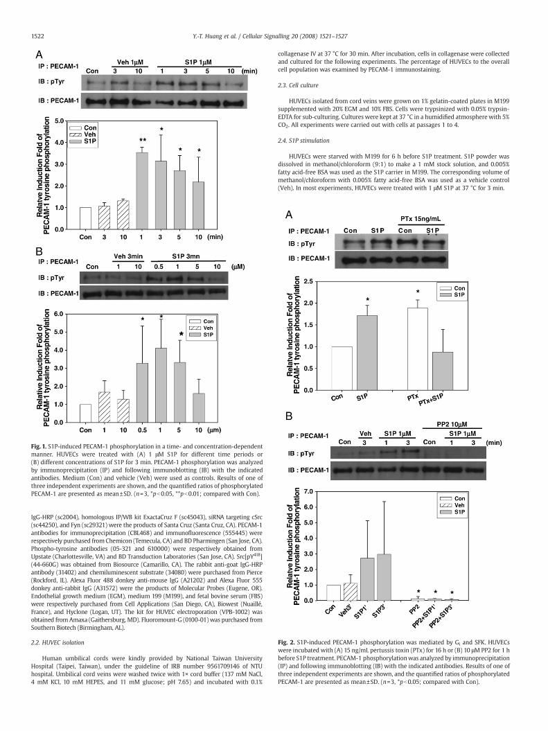
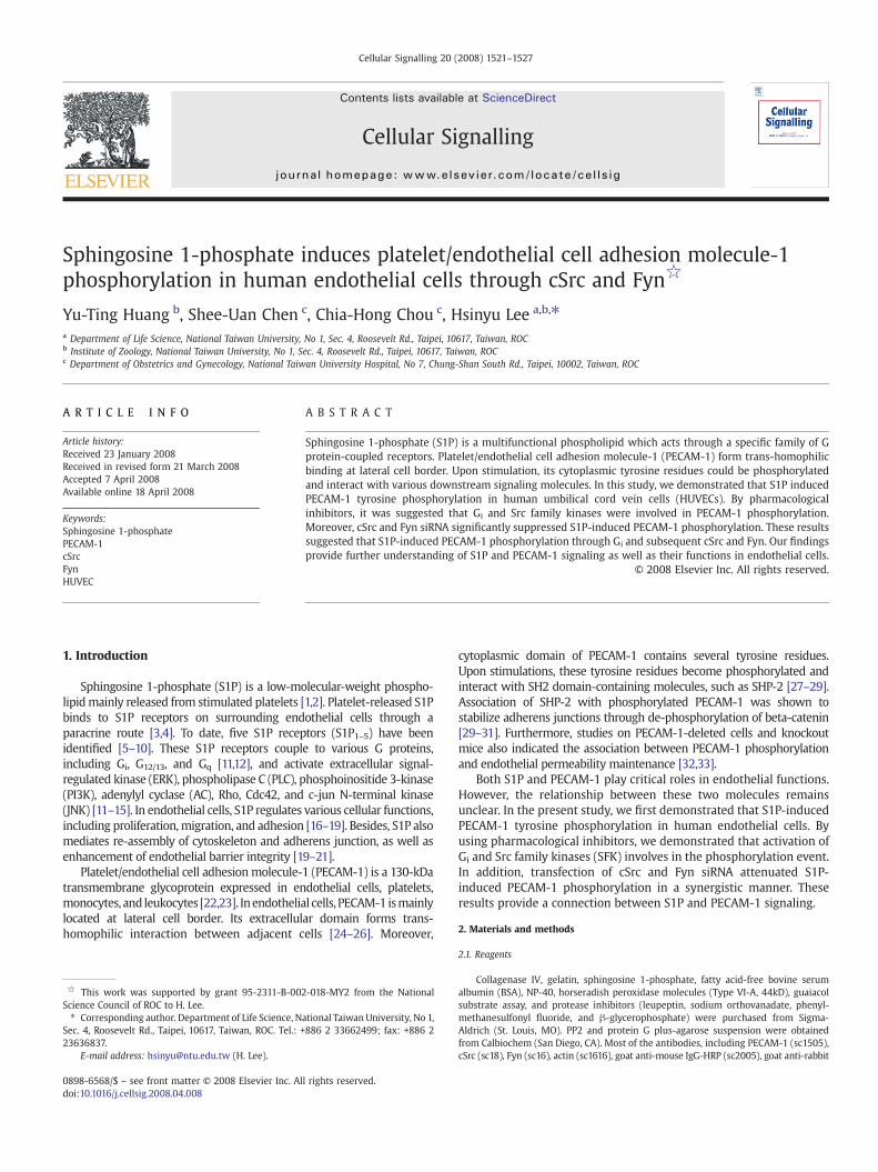
Fig. 1. S1P-induced PECAM-1 phosphorylation in a time- and concentration-dependentmanner. HUVECs were treated with (A) 1 μM S1P for different time periods or(B) different concentrations of S1P for 3 min. PECAM-1 phosphorylation was analyzedby immunoprecipitation (IP) and following immunoblotting (IB) with the indicatedantibodies. Medium (Con) and vehicle (Veh) were used as controls. Results of one ofthree independent experiments are shown, and the quantified ratios of phosphorylatedPECAM-1 are presented as mean±SD. (n=3, *pb0.05, **pb0.01; compared with Con).
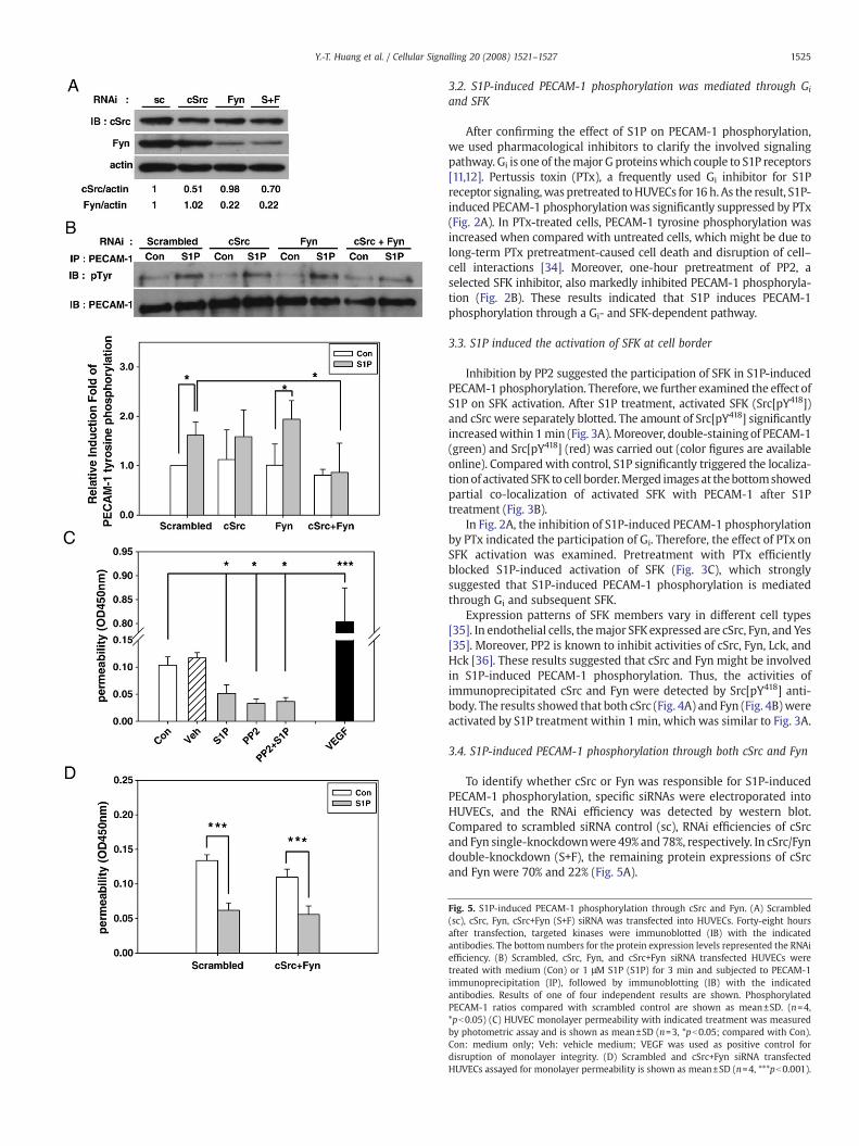
Fig. 2. S1P-induced PECAM-1 phosphorylation was mediated by Gi and SFK. HUVECswere incubated with (A) 15 ng/mL pertussis toxin (PTx) for 16 h or (B) 10 μM PP2 for 1 hbefore S1P treatment. PECAM-1 phosphorylationwas analyzed by immunoprecipitation(IP) and following immunoblotting (IB) with the indicated antibodies. Results of one ofthree independent experiments are shown, and the quantified ratios of phosphorylatedPECAM-1 are presented as mean±SD. (n=3, *pb0.05; compared with Con).
1522 Y.-T. Huang et al. / Cellular Signalling 20 (2008) 1521–1527
IgG-HRP (sc2004), homologous IP/WB kit ExactaCruz F (sc45043), siRNA targeting cSrc(sc44250), and Fyn (sc29321) were the products of Santa Cruz (Santa Cruz, CA). PECAM-1antibodies for immunoprecipitation (CBL468) and immunofluorescence (555445) wererespectively purchased fromChemicon (Temecula, CA) and BD Pharmingen (San Jose, CA).Phospho-tyrosine antibodies (05-321 and 610000) were respectively obtained fromUpstate (Charlottesville, VA) and BD Transduction Laboratories (San Jose, CA). Src[pY418](44-660G) was obtained from Biosource (Camarillo, CA). The rabbit anti-goat IgG-HRPantibody (31402) and chemiluminescent substrate (34080) were purchased from Pierce(Rockford, IL). Alexa Fluor 488 donkey anti-mouse IgG (A21202) and Alexa Fluor 555donkey anti-rabbit IgG (A31572) were the products of Molecular Probes (Eugene, OR).Endothelial growth medium (EGM), medium 199 (M199), and fetal bovine serum (FBS)were respectively purchased from Cell Applications (San Diego, CA), Biowest (Nuaillé,France), and Hyclone (Logan, UT). The kit for HUVEC electroporation (VPB-1002) wasobtained from Amaxa (Gaithersburg, MD). Fluoromount-G (0100-01)was purchased fromSouthern Biotech (Birmingham, AL).
2.2. HUVEC isolation
Human umbilical cords were kindly provided by National Taiwan UniversityHospital (Taipei, Taiwan), under the guideline of IRB number 9561709146 of NTUhospital. Umbilical cord veins were washed twice with 1× cord buffer (137 mM NaCl,4 mM KCl, 10 mM HEPES, and 11 mM glucose; pH 7.65) and incubated with 0.1%
collagenase IV at 37 °C for 30 min. After incubation, cells in collagenase were collectedand cultured for the following experiments. The percentage of HUVECs to the overallcell population was examined by PECAM-1 immunostaining.
2.3. Cell culture
HUVECs isolated from cord veins were grown on 1% gelatin-coated plates in M199supplemented with 20% EGM and 10% FBS. Cells were trypsinized with 0.05% trypsin-EDTA for sub-culturing. Cultures were kept at 37 °C in a humidified atmospherewith 5%CO2. All experiments were carried out with cells at passages 1 to 4.
2.4. S1P stimulation
HUVECs were starved with M199 for 6 h before S1P treatment. S1P powder wasdissolved in methanol/chloroform (9:1) to make a 1 mM stock solution, and 0.005%fatty acid-free BSA was used as the S1P carrier in M199. The corresponding volume ofmethanol/chloroform with 0.005% fatty acid-free BSA was used as a vehicle control(Veh). In most experiments, HUVECs were treated with 1 μM S1P at 37 °C for 3 min.
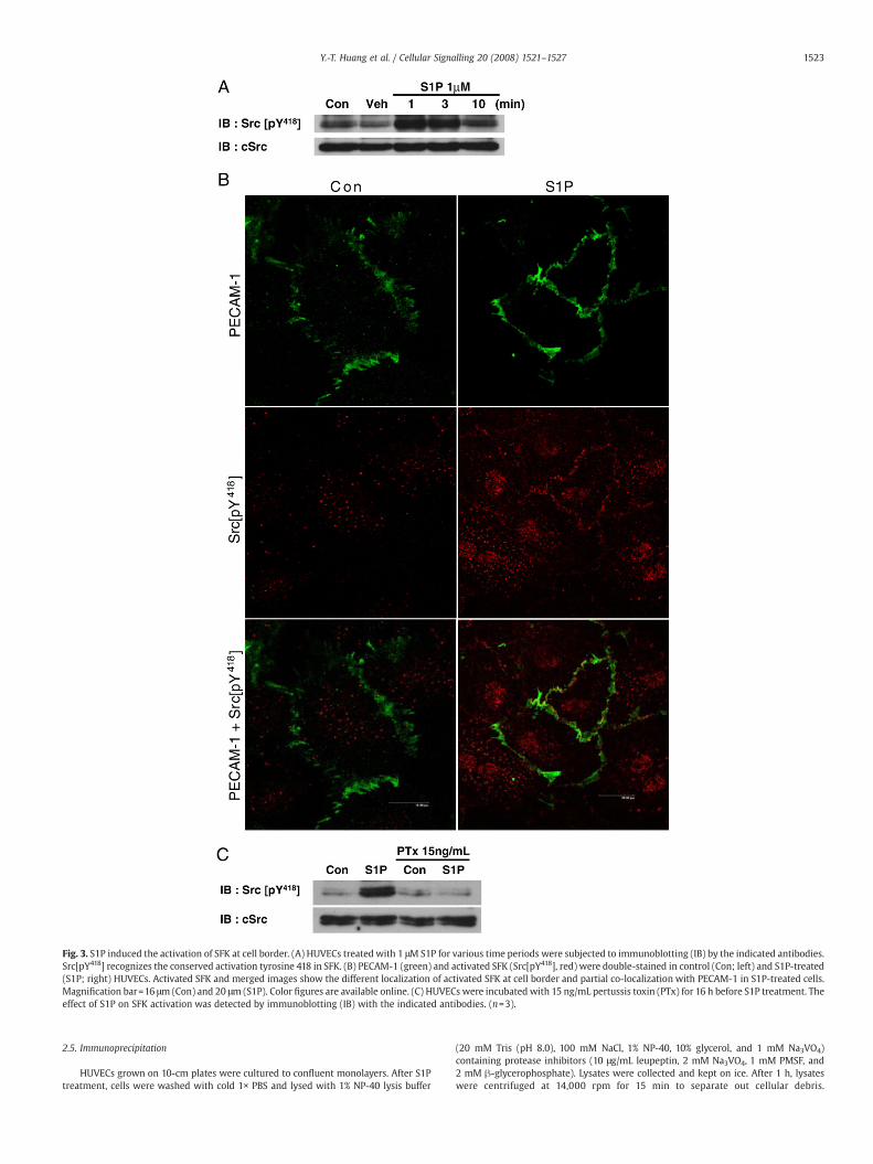
Fig. 3. S1P induced the activation of SFK at cell border. (A) HUVECs treated with 1 μM S1P for various time periods were subjected to immunoblotting (IB) by the indicated antibodies.Src[pY418] recognizes the conserved activation tyrosine 418 in SFK. (B) PECAM-1 (green) and activated SFK (Src[pY418], red) were double-stained in control (Con; left) and S1P-treated(S1P; right) HUVECs. Activated SFK and merged images show the different localization of activated SFK at cell border and partial co-localization with PECAM-1 in S1P-treated cells.Magnification bar=16 μm (Con) and 20 μm (S1P). Color figures are available online. (C) HUVECs were incubated with 15 ng/mL pertussis toxin (PTx) for 16 h before S1P treatment. Theeffect of S1P on SFK activation was detected by immunoblotting (IB) with the indicated antibodies. (n=3).
1523Y.-T. Huang et al. / Cellular Signalling 20 (2008) 1521–1527
2.5. Immunoprecipitation
HUVECs grown on 10-cm plates were cultured to confluent monolayers. After S1Ptreatment, cells were washed with cold 1× PBS and lysed with 1% NP-40 lysis buffer
(20 mM Tris (pH 8.0), 100 mM NaCl, 1% NP-40, 10% glycerol, and 1 mM Na3VO4)containing protease inhibitors (10 μg/mL leupeptin, 2 mM Na3VO4, 1 mM PMSF, and2 mM β-glycerophosphate). Lysates were collected and kept on ice. After 1 h, lysateswere centrifuged at 14,000 rpm for 15 min to separate out cellular debris.
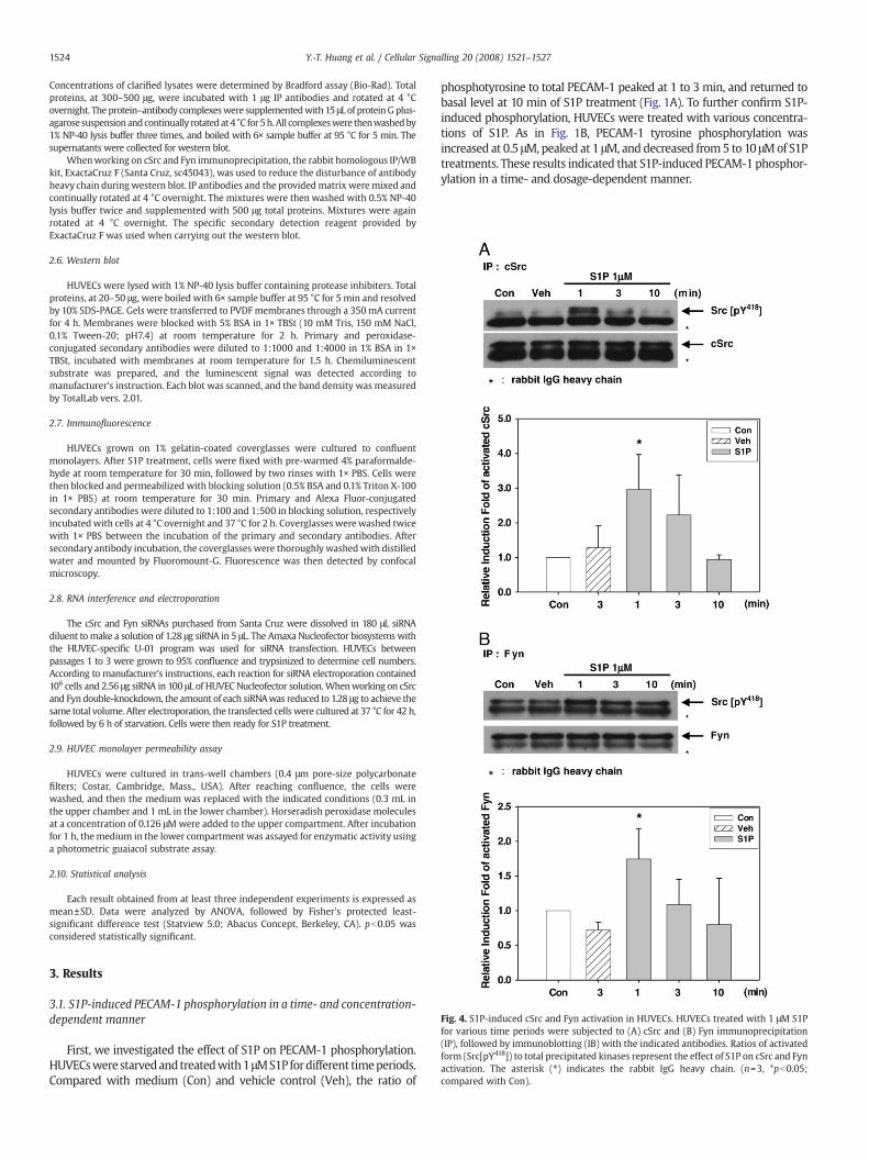
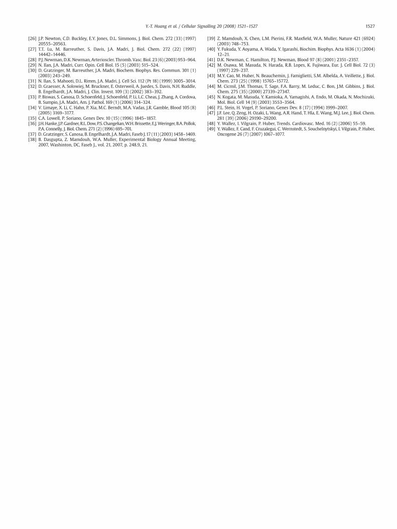
Fig. 4. S1P-induced cSrc and Fyn activation in HUVECs. HUVECs treated with 1 μM S1Pfor various time periods were subjected to (A) cSrc and (B) Fyn immunoprecipitation(IP), followed by immunoblotting (IB) with the indicated antibodies. Ratios of activatedform (Src[pY418]) to total precipitated kinases represent the effect of S1P on cSrc and Fynactivation. The asterisk (⁎) indicates the rabbit IgG heavy chain. (n=3, *pb0.05;compared with Con).
1524 Y.-T. Huang et al. / Cellular Signalling 20 (2008) 1521–1527
Concentrations of clarified lysates were determined by Bradford assay (Bio-Rad). Totalproteins, at 300–500 μg, were incubated with 1 μg IP antibodies and rotated at 4 °Covernight. The protein–antibody complexeswere supplementedwith 15 μL of proteinGplus-agarose suspensionandcontinually rotatedat4 °C for5h.All complexeswere thenwashedby1% NP-40 lysis buffer three times, and boiled with 6× sample buffer at 95 °C for 5 min. Thesupernatants were collected for western blot.
Whenworking on cSrc and Fyn immunoprecipitation, the rabbit homologous IP/WBkit, ExactaCruz F (Santa Cruz, sc45043), was used to reduce the disturbance of antibodyheavy chain during western blot. IP antibodies and the providedmatrix weremixed andcontinually rotated at 4 °C overnight. The mixtures were then washed with 0.5% NP-40lysis buffer twice and supplemented with 500 μg total proteins. Mixtures were againrotated at 4 °C overnight. The specific secondary detection reagent provided byExactaCruz F was used when carrying out the western blot.
2.6. Western blot
HUVECs were lysed with 1% NP-40 lysis buffer containing protease inhibiters. Totalproteins, at 20–50 μg, were boiled with 6× sample buffer at 95 °C for 5 min and resolvedby 10% SDS-PAGE. Gels were transferred to PVDF membranes through a 350mA currentfor 4 h. Membranes were blocked with 5% BSA in 1× TBSt (10 mM Tris, 150 mM NaCl,0.1% Tween-20; pH7.4) at room temperature for 2 h. Primary and peroxidase-conjugated secondary antibodies were diluted to 1:1000 and 1:4000 in 1% BSA in 1×TBSt, incubated with membranes at room temperature for 1.5 h. Chemiluminescentsubstrate was prepared, and the luminescent signal was detected according tomanufacturer's instruction. Each blot was scanned, and the band density was measuredby TotalLab vers. 2.01.
2.7. Immunofluorescence
HUVECs grown on 1% gelatin-coated coverglasses were cultured to confluentmonolayers. After S1P treatment, cells were fixed with pre-warmed 4% paraformalde-hyde at room temperature for 30 min, followed by two rinses with 1× PBS. Cells werethen blocked and permeabilized with blocking solution (0.5% BSA and 0.1% Triton X-100in 1× PBS) at room temperature for 30 min. Primary and Alexa Fluor-conjugatedsecondary antibodies were diluted to 1:100 and 1:500 in blocking solution, respectivelyincubatedwith cells at 4 °C overnight and 37 °C for 2 h. Coverglasses werewashed twicewith 1× PBS between the incubation of the primary and secondary antibodies. Aftersecondary antibody incubation, the coverglasses were thoroughly washedwith distilledwater and mounted by Fluoromount-G. Fluorescence was then detected by confocalmicroscopy.
2.8. RNA interference and electroporation
The cSrc and Fyn siRNAs purchased from Santa Cruz were dissolved in 180 μL siRNAdiluent to make a solution of 1.28 μg siRNA in 5 μL. The Amaxa Nucleofector biosystemswiththe HUVEC-specific U-01 program was used for siRNA transfection. HUVECs betweenpassages 1 to 3 were grown to 95% confluence and trypsinized to determine cell numbers.According to manufacturer's instructions, each reaction for siRNA electroporation contained106 cells and 2.56 μg siRNA in 100 μL of HUVEC Nucleofector solution.Whenworking on cSrcand Fyn double-knockdown, the amount of each siRNAwas reduced to 1.28 μg to achieve thesame total volume. After electroporation, the transfected cells were cultured at 37 °C for 42 h,followed by 6 h of starvation. Cells were then ready for S1P treatment.
2.9. HUVEC monolayer permeability assay
HUVECs were cultured in trans-well chambers (0.4 μm pore-size polycarbonatefilters; Costar, Cambridge, Mass., USA). After reaching confluence, the cells werewashed, and then the medium was replaced with the indicated conditions (0.3 mL inthe upper chamber and 1 mL in the lower chamber). Horseradish peroxidase moleculesat a concentration of 0.126 μMwere added to the upper compartment. After incubationfor 1 h, themedium in the lower compartment was assayed for enzymatic activity usinga photometric guaiacol substrate assay.
2.10. Statistical analysis
Each result obtained from at least three independent experiments is expressed asmean±SD. Data were analyzed by ANOVA, followed by Fisher's protected least-significant difference test (Statview 5.0; Abacus Concept, Berkeley, CA). pb0.05 wasconsidered statistically significant.
3. Results
3.1. S1P-induced PECAM-1 phosphorylation in a time- and concentration-dependent manner
First, we investigated the effect of S1P on PECAM-1 phosphorylation.HUVECswerestarvedandtreatedwith1μMS1P fordifferent timeperiods.Compared with medium (Con) and vehicle control (Veh), the ratio of
phosphotyrosine to total PECAM-1 peaked at 1 to 3 min, and returned tobasal level at 10 min of S1P treatment (Fig. 1A). To further confirm S1P-induced phosphorylation, HUVECs were treated with various concentra-tions of S1P. As in Fig. 1B, PECAM-1 tyrosine phosphorylation wasincreased at 0.5 μM, peaked at 1 μM, and decreased from5 to 10 μMof S1Ptreatments. These results indicated that S1P-induced PECAM-1 phosphor-ylation in a time- and dosage-dependent manner.
1525Y.-T. Huang et al. / Cellular Signalling 20 (2008) 1521–1527
3.2. S1P-induced PECAM-1 phosphorylation was mediated through Gi
and SFK
After confirming the effect of S1P on PECAM-1 phosphorylation,we used pharmacological inhibitors to clarify the involved signalingpathway. Gi is one of themajor G proteinswhich couple to S1P receptors[11,12]. Pertussis toxin (PTx), a frequently used Gi inhibitor for S1Preceptor signaling,was pretreated toHUVECs for 16 h. As the result, S1P-induced PECAM-1 phosphorylationwas significantly suppressed by PTx(Fig. 2A). In PTx-treated cells, PECAM-1 tyrosine phosphorylation wasincreased when compared with untreated cells, which might be due tolong-term PTx pretreatment-caused cell death and disruption of cell–cell interactions [34]. Moreover, one-hour pretreatment of PP2, aselected SFK inhibitor, also markedly inhibited PECAM-1 phosphoryla-tion (Fig. 2B). These results indicated that S1P induces PECAM-1phosphorylation through a Gi- and SFK-dependent pathway.
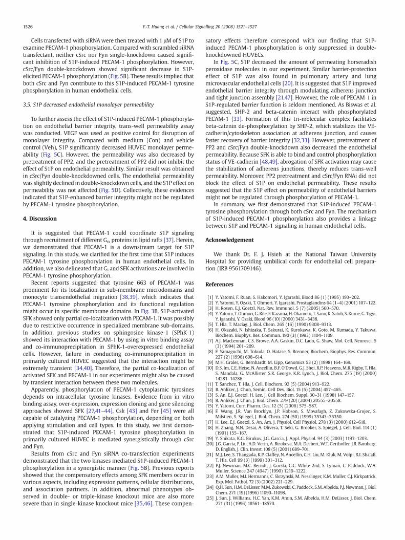
3.3. S1P induced the activation of SFK at cell border
Inhibition by PP2 suggested the participation of SFK in S1P-inducedPECAM-1 phosphorylation. Therefore, we further examined the effect ofS1P on SFK activation. After S1P treatment, activated SFK (Src[pY418])and cSrc were separately blotted. The amount of Src[pY418] significantlyincreasedwithin 1min (Fig. 3A).Moreover, double-staining of PECAM-1(green) and Src[pY418] (red) was carried out (color figures are availableonline). Comparedwith control, S1P significantly triggered the localiza-tionof activated SFK to cell border.Merged images at thebottomshowedpartial co-localization of activated SFK with PECAM-1 after S1Ptreatment (Fig. 3B).
In Fig. 2A, the inhibition of S1P-induced PECAM-1 phosphorylationby PTx indicated the participation of Gi. Therefore, the effect of PTx onSFK activation was examined. Pretreatment with PTx efficientlyblocked S1P-induced activation of SFK (Fig. 3C), which stronglysuggested that S1P-induced PECAM-1 phosphorylation is mediatedthrough Gi and subsequent SFK.
Expression patterns of SFK members vary in different cell types[35]. In endothelial cells, themajor SFKexpressed are cSrc, Fyn, and Yes[35]. Moreover, PP2 is known to inhibit activities of cSrc, Fyn, Lck, andHck [36]. These results suggested that cSrc and Fyn might be involvedin S1P-induced PECAM-1 phosphorylation. Thus, the activities ofimmunoprecipitated cSrc and Fyn were detected by Src[pY418] anti-body. The results showed that both cSrc (Fig. 4A) and Fyn (Fig. 4B)wereactivated by S1P treatment within 1 min, which was similar to Fig. 3A.
3.4. S1P-induced PECAM-1 phosphorylation through both cSrc and Fyn
To identify whether cSrc or Fyn was responsible for S1P-inducedPECAM-1 phosphorylation, specific siRNAs were electroporated intoHUVECs, and the RNAi efficiency was detected by western blot.Compared to scrambled siRNA control (sc), RNAi efficiencies of cSrcand Fyn single-knockdownwere 49% and 78%, respectively. In cSrc/Fyndouble-knockdown (S+F), the remaining protein expressions of cSrcand Fyn were 70% and 22% (Fig. 5A).
Fig. 5. S1P-induced PECAM-1 phosphorylation through cSrc and Fyn. (A) Scrambled(sc), cSrc, Fyn, cSrc+Fyn (S+F) siRNA was transfected into HUVECs. Forty-eight hoursafter transfection, targeted kinases were immunoblotted (IB) with the indicatedantibodies. The bottom numbers for the protein expression levels represented the RNAiefficiency. (B) Scrambled, cSrc, Fyn, and cSrc+Fyn siRNA transfected HUVECs weretreated with medium (Con) or 1 μM S1P (S1P) for 3 min and subjected to PECAM-1immunoprecipitation (IP), followed by immunoblotting (IB) with the indicatedantibodies. Results of one of four independent results are shown. PhosphorylatedPECAM-1 ratios compared with scrambled control are shown as mean±SD. (n=4,*pb0.05) (C) HUVEC monolayer permeability with indicated treatment was measuredby photometric assay and is shown as mean±SD (n=3, *pb0.05; compared with Con).Con: medium only; Veh: vehicle medium; VEGF was used as positive control fordisruption of monolayer integrity. (D) Scrambled and cSrc+Fyn siRNA transfectedHUVECs assayed for monolayer permeability is shown as mean±SD (n=4, ***pb0.001).
1526 Y.-T. Huang et al. / Cellular Signalling 20 (2008) 1521–1527
Cells transfected with siRNAwere then treated with 1 μM of S1P toexamine PECAM-1 phosphorylation. Compared with scrambled siRNAtransfectant, neither cSrc nor Fyn single-knockdown caused signifi-cant inhibition of S1P-induced PECAM-1 phosphorylation. However,cSrc/Fyn double-knockdown showed significant decrease in S1P-elicited PECAM-1 phosphorylation (Fig. 5B). These results implied thatboth cSrc and Fyn contribute to this S1P-induced PECAM-1 tyrosinephosphorylation in human endothelial cells.
3.5. S1P decreased endothelial monolayer permeability
To further assess the effect of S1P-induced PECAM-1 phosphoryla-tion on endothelial barrier integrity, trans-well permeability assaywas conducted. VEGF was used as positive control for disruption ofmonolayer integrity. Compared with medium (Con) and vehiclecontrol (Veh), S1P significantly decreased HUVEC monolayer perme-ability (Fig. 5C). However, the permeability was also decreased bypretreatment of PP2, and the pretreatment of PP2 did not inhibit theeffect of S1P on endothelial permeability. Similar result was obtainedin cSrc/Fyn double-knockdowned cells. The endothelial permeabilitywas slightly declined in double-knockdown cells, and the S1P effect onpermeability was not affected (Fig. 5D). Collectively, these evidencesindicated that S1P-enhanced barrier integrity might not be regulatedby PECAM-1 tyrosine phosphorylation.
4. Discussion
It is suggested that PECAM-1 could coordinate S1P signalingthrough recruitment of different Gα proteins in lipid rafts [37]. Herein,we demonstrated that PECAM-1 is a downstream target for S1Psignaling. In this study, we clarified for the first time that S1P inducesPECAM-1 tyrosine phosphorylation in human endothelial cells. Inaddition, we also delineated that Gi and SFK activations are involved inPECAM-1 tyrosine phosphorylation.
Recent reports suggested that tyrosine 663 of PECAM-1 wasprominent for its localization in sub-membrane microdomains andmonocyte transendothelial migration [38,39], which indicates thatPECAM-1 tyrosine phosphorylation and its functional regulationmight occur in specific membrane domains. In Fig. 3B, S1P-activatedSFK showed only partial co-localizationwith PECAM-1. It was possiblydue to restrictive occurrence in specialized membrane sub-domains.In addition, previous studies on sphingosine kinase-1 (SPhK-1)showed its interaction with PECAM-1 by using in vitro binding assayand co-immunoprecipitation in SPhK-1-overexpressed endothelialcells. However, failure in conducting co-immunoprecipitation inprimarily cultured HUVEC suggested that the interaction might beextremely transient [34,40]. Therefore, the partial co-localization ofactivated SFK and PECAM-1 in our experiments might also be causedby transient interaction between these two molecules.
Apparently, phosphorylation of PECAM-1 cytoplasmic tyrosinesdepends on intracellular tyrosine kinases. Evidence from in vitrobinding assay, over-expression, expression cloning and gene silencingapproaches showed SFK [27,41–44], Csk [43] and Fer [45] were allcapable of catalyzing PECAM-1 phosphorylation, depending on bothapplying stimulation and cell types. In this study, we first demon-strated that S1P-induced PECAM-1 tyrosine phosphorylation inprimarily cultured HUVEC is mediated synergistically through cSrcand Fyn.
Results from cSrc and Fyn siRNA co-transfection experimentsdemonstrated that the two kinases mediated S1P-induced PECAM-1phosphorylation in a synergistic manner (Fig. 5B). Previous reportsshowed that the compensatory effects among SFK members occur invarious aspects, including expression patterns, cellular distributions,and association partners. In addition, abnormal phenotypes ob-served in double- or triple-kinase knockout mice are also moresevere than in single-kinase knockout mice [35,46]. These compen-
satory effects therefore correspond with our finding that S1P-induced PECAM-1 phosphorylation is only suppressed in double-knockdowned HUVECs.
In Fig. 5C, S1P decreased the amount of permeating horseradishperoxidase molecules in our experiment. Similar barrier-protectioneffect of S1P was also found in pulmonary artery and lungmicrovascular endothelial cells [20]. It is suggested that S1P improvedendothelial barrier integrity through modulating adherens junctionand tight junction assembly [21,47]. However, the role of PECAM-1 inS1P-regulated barrier function is seldom mentioned. As Biswas et al.suggested, SHP-2 and beta-catenin interact with phosphorylatedPECAM-1 [33]. Formation of this tri-molecular complex facilitatesbeta-catenin de-phosphorylation by SHP-2, which stabilizes the VE-cadherin/cytoskeleton association at adherens junction, and causesfaster recovery of barrier integrity [32,33]. However, pretreatment ofPP2 and cSrc/Fyn double-knockdown also decreased the endothelialpermeability. Because SFK is able to bind and control phosphorylationstatus of VE-cadherin [48,49], abrogation of SFK activation may causethe stabilization of adherens junctions, thereby reduces trans-wellpermeability. Moreover, PP2 pretreatment and cSrc/Fyn RNAi did notblock the effect of S1P on endothelial permeability. These resultssuggested that the S1P effect on permeability of endothelial barriersmight not be regulated through phosphorylation of PECAM-1.
In summary, we first demonstrated that S1P-induced PECAM-1tyrosine phosphorylation through both cSrc and Fyn. The mechanismof S1P-induced PECAM-1 phosphorylation also provides a linkagebetween S1P and PECAM-1 signaling in human endothelial cells.
Acknowledgement
We thank Dr. F. J. Hsieh at the National Taiwan UniversityHospital for providing umbilical cords for endothelial cell prepara-tion (IRB 9561709146).
References
[1] Y. Yatomi, F. Ruan, S. Hakomori, Y. Igarashi, Blood 86 (1) (1995) 193–202.[2] Y. Yatomi, Y. Ozaki, T. Ohmori, Y. Igarashi, Prostaglandins 64 (1–4) (2001) 107–122.[3] H. Rosen, E.J. Goetzl, Nat. Rev. Immunol. 5 (7) (2005) 560–570.[4] Y. Yatomi, T.Ohmori, G. Rile, F. Kazama,H.Okamoto, T. Sano, K. Satoh, S. Kume,G.Tigyi,
Y. Igarashi, Y. Ozaki, Blood 96 (10) (2000) 3431–3438.[5] T. Hla, T. Maciag, J. Biol. Chem. 265 (16) (1990) 9308–9313.[6] H. Okazaki, N. Ishizaka, T. Sakurai, K. Kurokawa, K. Goto, M. Kumada, Y. Takuwa,
Biochem. Biophys. Res. Commun. 190 (3) (1993) 1104–1109.[7] A.J. MacLennan, C.S. Browe, A.A. Gaskin, D.C. Lado, G. Shaw, Mol. Cell. Neurosci. 5
(3) (1994) 201–209.[8] F. Yamaguchi, M. Tokuda, O. Hatase, S. Brenner, Biochem. Biophys. Res. Commun.
227 (2) (1996) 608–614.[9] M.H. Graler, G. Bernhardt, M. Lipp, Genomics 53 (2) (1998) 164–169.[10] D.S. Im, C.E. Heise, N. Ancellin, B.F. O'Dowd, G.J. Shei, R.P. Heavens,M.R. Rigby, T. Hla,
S. Mandala, G. McAllister, S.R. George, K.R. Lynch, J. Biol. Chem. 275 (19) (2000)14281–14286.
[11] T. Sanchez, T. Hla, J. Cell. Biochem. 92 (5) (2004) 913–922.[12] B. Anliker, J. Chun, Semin. Cell Dev. Biol. 15 (5) (2004) 457–465.[13] S. An, E.J. Goetzl, H. Lee, J. Cell Biochem. Suppl. 30–31 (1998) 147–157.[14] B. Anliker, J. Chun, J. Biol. Chem. 279 (20) (2004) 20555–20558.[15] Y. Yatomi, Curr. Pharm. Des. 12 (5) (2006) 575–587.[16] F. Wang, J.R. Van Brocklyn, J.P. Hobson, S. Movafagh, Z. Zukowska-Grojec, S.
Milstien, S. Spiegel, J. Biol. Chem. 274 (50) (1999) 35343–35350.[17] H. Lee, E.J. Goetzl, S. An, Am. J. Physiol. Cell Physiol. 278 (3) (2000) 612–618.[18] H. Zhang, N.N. Desai, A. Olivera, T. Seki, G. Brooker, S. Spiegel, J. Cell. Biol. 114 (1)
(1991) 155–167.[19] Y. Shikata, K.G. Birukov, J.G. Garcia, J. Appl. Physiol. 94 (3) (2003) 1193–1203.[20] J.G. Garcia, F. Liu, A.D. Verin, A. Birukova, M.A. Dechert, W.T. Gerthoffer, J.R. Bamberg,
D. English, J. Clin. Invest. 108 (5) (2001) 689–701.[21] M.J. Lee, S. Thangada, K.P. Claffey, N. Ancellin, C.H. Liu, M. Kluk, M. Volpi, R.I. Sha'afi,
T. Hla, Cell 99 (3) (1999) 301–312.[22] P.J. Newman, M.C. Berndt, J. Gorski, G.C. White 2nd, S. Lyman, C. Paddock, W.A.
Muller, Science 247 (4947) (1990) 1219–1222.[23] A.M. Muller, M.I. Hermanns, C. Skrzynski, M. Nesslinger, K.M. Muller, C.J. Kirkpatrick,
Exp. Mol. Pathol. 72 (3) (2002) 221–229.[24] Q.H. Sun, H.M. DeLisser,M.M. Zukowski, C. Paddock, S.M. Albelda, P.J. Newman, J. Biol.
Chem. 271 (19) (1996) 11090–11098.[25] J. Sun, J. Williams, H.C. Yan, K.M. Amin, S.M. Albelda, H.M. DeLisser, J. Biol. Chem.
271 (31) (1996) 18561–18570.
1527Y.-T. Huang et al. / Cellular Signalling 20 (2008) 1521–1527
[26] J.P. Newton, C.D. Buckley, E.Y. Jones, D.L. Simmons, J. Biol. Chem. 272 (33) (1997)20555–20563.
[27] T.T. Lu, M. Barreuther, S. Davis, J.A. Madri, J. Biol. Chem. 272 (22) (1997)14442–14446.
[28] P.J. Newman, D.K. Newman, Arterioscler. Thromb. Vasc. Biol. 23 (6) (2003) 953–964.[29] N. Ilan, J.A. Madri, Curr. Opin. Cell Biol. 15 (5) (2003) 515–524.[30] D. Gratzinger, M. Barreuther, J.A. Madri, Biochem. Biophys. Res. Commun. 301 (1)
(2003) 243–249.[31] N. Ilan, S. Mahooti, D.L. Rimm, J.A. Madri, J. Cell Sci. 112 (Pt 18) (1999) 3005–3014.[32] D. Graesser, A. Solowiej, M. Bruckner, E. Osterweil, A. Juedes, S. Davis, N.H. Ruddle,
B. Engelhardt, J.A. Madri, J. Clin. Invest. 109 (3) (2002) 383–392.[33] P. Biswas, S. Canosa, D. Schoenfeld, J. Schoenfeld, P. Li, L.C. Cheas, J. Zhang, A. Cordova,
B. Sumpio, J.A. Madri, Am. J. Pathol. 169 (1) (2006) 314–324.[34] V. Limaye, X. Li, C. Hahn, P. Xia, M.C. Berndt, M.A. Vadas, J.R. Gamble, Blood 105 (8)
(2005) 3169–3177.[35] C.A. Lowell, P. Soriano, Genes Dev. 10 (15) (1996) 1845–1857.[36] J.H.Hanke, J.P.Gardner,R.L.Dow,P.S. Changelian,W.H.Brissette,E.J.Weringer, B.A.Pollok,
P.A. Connelly, J. Biol. Chem. 271 (2) (1996) 695–701.[37] D. Gratzinger, S. Canosa, B. Engelhardt, J.A.Madri, Faseb J.17 (11) (2003) 1458–1469.[38] B. Dasgupta, Z. Mamdouh, W.A. Muller, Experimental Biology Annual Meeting,
2007, Washinton, DC, Faseb J., vol. 21, 2007, p. 248.9, 21.
[39] Z. Mamdouh, X. Chen, L.M. Pierini, F.R. Maxfield, W.A. Muller, Nature 421 (6924)(2003) 748–753.
[40] Y. Fukuda, Y. Aoyama, A. Wada, Y. Igarashi, Biochim. Biophys. Acta 1636 (1) (2004)12–21.
[41] D.K. Newman, C. Hamilton, P.J. Newman, Blood 97 (8) (2001) 2351–2357.[42] M. Osawa, M. Masuda, N. Harada, R.B. Lopes, K. Fujiwara, Eur. J. Cell Biol. 72 (3)
(1997) 229–237.[43] M.Y. Cao, M. Huber, N. Beauchemin, J. Famiglietti, S.M. Albelda, A. Veillette, J. Biol.
Chem. 273 (25) (1998) 15765–15772.[44] M. Cicmil, J.M. Thomas, T. Sage, F.A. Barry, M. Leduc, C. Bon, J.M. Gibbins, J. Biol.
Chem. 275 (35) (2000) 27339–27347.[45] N. Kogata, M. Masuda, Y. Kamioka, A. Yamagishi, A. Endo, M. Okada, N. Mochizuki,
Mol. Biol. Cell 14 (9) (2003) 3553–3564.[46] P.L. Stein, H. Vogel, P. Soriano, Genes Dev. 8 (17) (1994) 1999–2007.[47] J.F. Lee, Q. Zeng, H. Ozaki, L. Wang, A.R. Hand, T. Hla, E. Wang, M.J. Lee, J. Biol. Chem.
281 (39) (2006) 29190–29200.[48] Y. Wallez, I. Vilgrain, P. Huber, Trends. Cardiovasc. Med. 16 (2) (2006) 55–59.[49] Y. Wallez, F. Cand, F. Cruzalegui, C. Wernstedt, S. Souchelnytskyi, I. Vilgrain, P. Huber,
Oncogene 26 (7) (2007) 1067–1077.


![Review Article Clinical Impact of Sphingosine-1-Phosphate in ...mouse models and human patient samples [6, 61–64]. However, endothelial cells can also synthesize and release endogenousS1P[45]](https://img.pdfslide.net/doc/110x75/60ecb00cf927e5546d482d8d/review-article-clinical-impact-of-sphingosine-1-phosphate-in-mouse-models-and.jpg)