Embed Size (px)
Citation preview

Therapeutics, Targets, and Chemical Biology
Sphingosine-1-Phosphate Produced by SphingosineKinase 1 Promotes Breast Cancer Progression byStimulating Angiogenesis and Lymphangiogenesis
Masayuki Nagahashi1,2, Subramaniam Ramachandran1,2, Eugene Y. Kim2, Jeremy C. Allegood2,Omar M. Rashid1,2, Akimitsu Yamada1,2, Renping Zhao3, Sheldon Milstien2, Huiping Zhou3,Sarah Spiegel2, and Kazuaki Takabe1,2
AbstractSphingosine-1-phosphate (S1P) is a pleiotropic bioactive lipid mediator that promotes breast cancer pro-
gression by diverse mechanisms that remain somewhat unclear. Here we report pharmacologic evidence of acritical role for sphingosine kinase 1 (SphK1) in producing S1P and mediating tumor-induced hemangiogenesisand lymphangiogenesis in amurinemodel of breast cancermetastasis. S1P levels increased both in the tumor andthe circulation. In agreement, serum S1P levels were significantly elevated in stage IIIA human breast cancerpatients, compared with age/ethnicity-matched healthy volunteers. However, treatment with the specific SphK1inhibitor SK1-I suppressed S1P levels, reducedmetastases to lymphnodes and lungs, and decreased overall tumorburden of our murine model. Both S1P and angiopoietin 2 (Ang2) stimulated hemangiogenesis and lymphan-giogenesis in vitro, whereas SK1-I inhibited each process. We quantified both processes in vivo from the samespecimen by combining directed in vivo angiogenesis assays with fluorescence-activated cell sorting, therebyconfirming the results obtained in vitro. Notably, SK1-I decreased both processes not only at the primary tumorbut also in lymph nodes, with peritumoral lymphatic vessel density reduced in SK1-I–treated animals. Takentogether, our findings show that SphK1-produced S1P is a crucial mediator of breast cancer–induced heman-giogenesis and lymphangiogenesis. Our results implicate SphK1 along with S1P as therapeutic targets in breastcancer. Cancer Res; 72(3); 726–35. �2012 AACR.
Introduction
Breast cancer is the most commonly diagnosed canceramong women accounting for 30% of all new cancer cases,and close to 40,000 breast cancer deaths are expected to occuramong U.S. women in 2011 (1). Themajority of deaths of breastcancer patients occur after it metastasizes and becomes asystemic disease (2). To date, most systemic therapies, includ-ing cytotoxic chemotherapy, target the tumor itself and not thehost tumor microenvironment, which is known to play animportant role in the progression of cancer (3, 4).
It is well established that hemangiogenesis is one of the keytumor microenvironmental factors in cancer progression.Tumor growth beyond a diameter of a few millimeters requireshemangiogenesis which contributes to the development ofmetastatic disease (5, 6). Although hemangiogenesis is knownto be an acquired capability of cancer (7), results of multiplerandomized clinical trials that targeted hemangiogenesis usingbevacizumab, an anti-VEGFmonoclonal antibody (mAb), failedto show an overall survival benefit in breast cancer (8–10).
Breast cancer first metastasizes to its sentinel lymph node,and the level of lymph node metastasis is a major determinantof staging and prognosis of breast cancer (2, 11). Recently,lymphangiogenesis, a process that generates new lymphaticvessels from preexisting ones, was found to be mediated byseveral factors including VEGF-C, D, and angiopoietins (Ang1and Ang2), which provided new insights into how the lym-phatic vessels grow and affect metastasis (12, 13). Althoughclinical and experimental evidence suggest a role for lymphan-giogenesis in lymph node metastasis, this process is far lessunderstood than hemangiogenesis (13, 14).
The pleiotropic bioactive lipid mediator sphingosine-1-phosphate (S1P) is now emerging as a key regulatory moleculein cancer through its ability to promote cell proliferation,migration, invasion, and hemangiogenesis (15, 16). S1P is gene-rated intracellularly by 2 sphingosine kinases, sphingosinekinase 1 (SphK1) and SphK2, and is exported out of the cells
Authors' Affiliations: 1Division of Surgical Oncology, 2Department ofBiochemistry and Molecular Biology, and the Massey Cancer Center, and3Department of Microbiology and Immunology, Virginia CommonwealthUniversity School of Medicine, Richmond, Virginia
Note: Supplementary data for this article are available at Cancer ResearchOnline (http://cancerres.aacrjournals.org/).
Corresponding Author: Kazuaki Takabe, Division of Surgical Oncology,Virginia CommonwealthUniversity School ofMedicine andMasseyCancerCenter, West Hospital 7-402, 1200 East Broad Street, PO Box 980011,Richmond, VA 23298. Phone: 804-828-9322; Fax: 804-828-4808; E-mail:[email protected]
doi: 10.1158/0008-5472.CAN-11-2167
�2012 American Association for Cancer Research.
CancerResearch
Cancer Res; 72(3) February 1, 2012726

where it regulates many functions by binding to and signalingthrough a family of 5 G protein-coupled receptors (S1P1–5).This process known as "inside-out" signaling explains theautocrine and paracrine actions of S1P (15). We previouslyshowed that SphK1, but not SphK2, is involved in S1P exportfrom breast cancer cells mediated by the ATP-binding cassettetransporters, ABCC1 and ABCG2 (17). Expression of SphK1 isupregulated in breast cancer (18, 19), correlates with poorprognosis (20), and is associated with resistance to chemo-therapy (18). Furthermore, abundant evidence implicates theSphK1/S1P/S1P1 axis in hemangiogenesis and vasculogenesis(21). In contrast, only a few studies so far have examined theinvolvement of SphK1 and S1P in lymphangiogenesis. S1P hasbeen shown to induce in vitro lymphangiogenesis via theS1P1 receptor (22), and it was recently reported that SphK1and S1P in lymphatic endothelial cells (LEC) are required forthe proper development of lymphatic vessels (23). However,nothing is yet known of the role of SphK1 and S1P in tumor-induced lymphangiogenesis in vivo.In this study, we utilized an improved syngeneic breast
cancer cell implantation method, which models human breastcancer biology better than conventional xenograft subcutane-ous implantation, to explore the role of SphK1 and S1P inhemangiogenesis and lymphangiogenesis. We found not onlythat circulating S1P levels correlated with tumor burden butalso that targeting SphK1 with a specific inhibitor reducedtumor growth, lymph node and lungmetastasis, and decreasedhemangiogenesis and lymphangiogenesis around the primarytumor and in the lymph nodes. Our results suggest thattargeting SphK1 and S1P may be a useful additional modalityfor the treatment of metastatic breast cancer.
Materials and Methods
Cell culture4T1-luc2 cells, a mouse mammary adenocarcinoma cell
line that has been engineered to express luciferase (PerkinEl-mer), were cultured in RPMI Medium 1640 with 10% FBS.Green fluorescent protein (GFP)-expressing human umbilicalvein endothelial cells (HUVEC) and GFP-expressing humanlymphatic endothelial cells (HLEC) purchased from Angio-Proteomie were maintained in endothelial cell medium sup-plemented with 5% FBS and endothelial cell growth supple-ment (ScienCell Research Laboratories).
Construction of lentiviral shRNAs for mouse SphK1Three sense sequences of siRNA cassettes specifically tar-
geting the nucleotides of mouse SphK1 (accession number:NM_011451.1) were designed using siRNA Target Finder(Ambion). The specificity of the selected sequences was con-firmed by BLAST search. The lentiviral short hairpin RNAs(shRNA) were constructed using a pLL3.7 expression vector asdescribed previously (24). The sequences of the shRNAs formouse SphK1 are as follows: shRNA 1, 50- GCACCCAAAC-TACCTTTGGAT-30; shRNA 2, 50- GCACCTTCTTTCGCCTAG-CAA-30; shRNA 3, 50-GAGGCAGAGATAACCTTTAAA-30.Recombinant lentivirus was produced by cotransfection of293FT cells with lentiviral vector and the packaging vectors
using calcium phosphate. The viruses were collected from theculture medium and purified by polyethylene glycol precipi-tation. The transduction efficiency of lentivirus in 4T1 cells wasdetermined by fluorescence microscopy. The silencing effi-ciency was determined using quantitative PCR (qPCR) andWestern blot analysis.
Syngeneic tumor modelAll animal studies were conducted in the Animal Research
Core Facility at VCUSchool ofMedicine in accordancewith theinstitutional guidelines. All procedures were approved by theVCU Institutional Animal Care and Use Committee (IACUC)that is accredited by the Association for Assessment andAccreditation of Laboratory Animal Care (AAALAC). FemaleBALB/cmice, 8 to 12weeks of age, weighing approximately 20 gwere obtained from Harlan. 4T1-luc2 cells (1 � 105 cells in 10mL RPMI) were surgically implanted in the upper fat pad underdirect vision. One day after implantation, mice were random-ized by tumor size determined by bioluminescence with theIVIS Imaging System (Xenogen). SK1-I in PBS was injectedintraperitoneally, as indicated, every day at a dose of 20mg/kg.At the indicated times, the animals were sacrificed by exsan-guination, blood was collected, tumors excised, weighed, fixedin formalin, and embedded in paraffin or frozen in liquidnitrogen.
Bioluminescent quantification of tumor burdenD-Luciferin (0.2 mL of 15 mg/mL stock; PerkinElmer) was
injected intraperitoneally into mice previously implanted with4T1-luc2 cells. Living Image Software (Xenogen) was used toquantify the photons/sec emitted by the cells. Biolumines-cence was measured and quantified at 5-minute intervals over30 minutes using a subject height of 1.5 cm, medium binningand an exposure time of 0.5 seconds to 1 minutes. Biolumi-nescence was then determined by the peak number ofphotons/sec calculated over this time frame. Axillary lymphnode metastasis was quantified in vivo after intraperitonealinjection of luciferin and primary tumor resection. Lungmetastasis was quantified ex vivo after the lungswere removed.
Quantification of sphingolipids by mass spectrometryLipids were extracted from serum and tissues and sphingo-
lipids quantified by liquid chromatography–electrospray ion-ization–tandem mass spectrometry (LC-ESI-MS/MS; 4000QTRAP, AB Sciex) as described (25).
Directed in vivo angiogenesis assays and cell isolationAngioreactors [directed in vivo angiogenesis assays (DIVAA)
kit; Trevigen] were implanted into the subcutaneous layer ofthe back of 8-week-old nude mice under anesthesia accordingto the manufacturer's instructions. Mice were euthanized withCO2 at day 11 after implantation and the angioreactors wereremoved under stereomicroscopy (SZ51, Olympus).
Fluorescence-activated cell sortingSingle-cell suspensions were obtained from lymph nodes as
described (26). For DIVAA/FACS (fluorescence-activated cellsorting) analyses, Matrigel and fibrotic reactive tissue in the
S1P Promotes Hemangiogenesis and Lymphangiogenesis
www.aacrjournals.org Cancer Res; 72(3) February 1, 2012 727

open end of the angioreactors were removed with Dulbecco'smodified Eagle's medium and digested with a mixture of 9 mLof Cell Sparse (Trevigen) containing 0.1% collagenase type 2(Gibco), 1 mL of 0.25 U/mL dispase (Gibco), and 75 mL of 0.1%DNase (Invitrogen) for 30minutes to obtain single-cell suspen-sions. Tumors were minced and digested similarly. Cell sus-pensions were blocked with anti-CD16/CD32 (Mouse BDFc Block; BD Biosciences) and then stained with the follow-ing antibodies as indicated: Alexa 488–conjugated LYVE-1(eBioscience); PE-conjugated podoplanin, PerCP-Cy5.5–con-jugated CD45, APC-conjugated CD31, Alexa 700–conjugatedTER-119 (Biolegend); or appropriate matched, fluorochrome-labeled isotype control mAb. The LIVE/DEAD Viability Assaykit (Invitrogen) was used to eliminate dead cells. Cells wereanalyzed by FACS using BD FACSCanto II and BD FACSAria II(BD Biosciences) and corollary data assessed with BD FACS-Diva Software version 6.1.3 (BD Biosciences).
Histopathologic analysis and vessel densitydetermination
Immediately after the sacrifice of the animals, tumor sam-ples were fixed in 10% neutral buffered formalin for immuno-histochemical analyses of cell proliferation and apoptosis.4T1-luc2 cell proliferation in the tumor was determined bystaining histologic sections with mAbs against Ki67 (Dako), anuclear protein expressed in proliferating cells. Apoptosis wasdetermined by terminal uridine deoxynucleotidyl transferasedUTP nick end labeling (TUNEL) assay using the ApopTagPeroxidase In Situ Apoptosis Detection Kit S7100 (Millipore).
The tumors were also embedded in optimal cutting medi-um (OCT 4583; Sakura Finetek) and frozen for immunofluo-rescent analysis for hemangiogenesis and lymphangiogenesis.The sections were stained with the following antibodies asindicated: CD31 (BD), LYVE-1 (Abcam) and F4/80 (kindlyprovided by Bin-Zhi Qian in Jeffrey Pollard laboratory at Albert
Figure 1. The SphK1/S1P axis in breast cancer progression. A, 4T1 tumors were established by surgical implantation of 4T1-luc2 cells into the chestmammary fat pad under direct vision. The tumor burden was determined by bioluminescence technology. Representative IVIS images (left) andbioluminescent quantification (right) on the indicated days are shown. B, axillary lymph nodemetastases weremeasured by bioluminescence at the indicatedtimes when mammary tumor was removed for accurate measurement (left). Lung metastases were measured separately ex vivo (middle). The incidence ofmetastasis to the axillary lymph nodes and the lung was determined on the indicated days (right). C, mRNA was isolated from mammary fat pads ofnaive BALB/c mice (light gray filled bars), cultured 4T1-luc2 cells (black cross hatched bars), and primary tumors in the chest mammary fat pads formed by4T1-luc2 cells (dark gray filled bars) 10 days after implantation. Expression of SphK1 and SphK2 was determined by qPCR and normalized to levels ofglyceraldehyde-3-phosphate dehydrogenase (GAPDH)mRNA.Data are expressed as fold increases�SD. D, S1P levelswere determinedby LC-ESI-MS/MSin the serumof shamsurgerymice and inmicewith 4T1-luc2 xenograft tumors 15 days after implantation. Data are expressed as fold increases�SEM. E, S1Pwas measured in serum from stage IIIA breast cancer patients and age/ethnicity-matched healthy volunteers (n ¼ 5). �, P < 0.001; ��, P < 0.05.
Nagahashi et al.
Cancer Res; 72(3) February 1, 2012 Cancer Research728
Einstein College of Medicine; ref. 27). The stained sectionswere examined with LSM510 laser scanning confocal micro-scopes (Zeiss) and microvessel density was determined aspreviously described (28).
Patient samplesAll studies using patient samples were conducted in accor-
dance with the institutional guidelines after approval by VCUInstitutional Review Board. Human sera were collected by theTissue andData Acquisition and Analysis Core of VCU and S1Plevels determined in serum from stage IIIA breast cancerpatients and age/ethnicity-matched healthy volunteers.
In vitro assaysCell number of 4T1-luc2 cells was determined by mea-
surement of luciferase activity with the luciferin substrate(PerkinElmer) using a VICTOR X4 Multilabel Plate Reader(PerkinElmer). qPCR (15) and tube formation assay wereconducted as described previously (29).
Statistical analysisResults were analyzed for statistical significance with a 2-
tailed Student's t test, with P < 0.05 considered significant.Experiments were repeated at least 3 times in triplicate with
consistent results. In vivo experiments were repeated 3 timesand each experimental group consisted of at least 6 mice.
Results
Upregulation of SphK1 and increased S1P levels in 4T1-luc2 tumor progression
Previous studies showed that S1P and SphK1, the enzymethat produces it, regulate many processes important for breastcancer (15). To examine their involvement in breast cancerprogression in vivo, we utilized an enhanced syngeneic mousemetastatic breast cancer model in which 4T1-luc2 murinemammary cancer cells were orthotopically implanted underdirect vision into the chest mammary fat pad of immune-competent mice. We have found that this chest orthotopicmodel more accurately mimics human breast cancer progres-sion than subcutaneous models. Indeed, 4T1-luc2 cells pro-duced large tumors in the chest mammary fat pad that rapidlymetastasized and increased total tumor burden (Fig. 1A).Metastatic spread to the regional axillary lymph nodes andthe lungs was quantified by bioluminescence after removal ofthe 4T1-luc2 tumors from the implantation sites (primarytumors). As can be seen in Fig. 1B, lymph node metastasesemerged earlier than lung metastases, with a rapid increase in
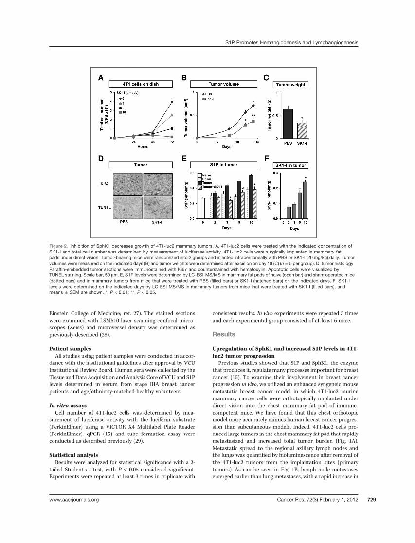
Figure 2. Inhibition of SphK1 decreases growth of 4T1-luc2 mammary tumors. A, 4T1-luc2 cells were treated with the indicated concentration ofSK1-I and total cell number was determined by measurement of luciferase activity. 4T1-luc2 cells were surgically implanted in mammary fatpads under direct vision. Tumor-bearing mice were randomized into 2 groups and injected intraperitoneally with PBS or SK1-I (20 mg/kg) daily. Tumorvolumes were measured on the indicated days (B) and tumor weights were determined after excision on day 18 (C) (n¼ 5 per group). D, tumor histology.Paraffin-embedded tumor sections were immunostained with Ki67 and counterstained with hematoxylin. Apoptotic cells were visualized byTUNEL staining. Scale bar, 50 mm. E, S1P levels were determined by LC-ESI-MS/MS in mammary fat pads of naive (open bar) and sham operated mice(dotted bars) and in mammary tumors from mice that were treated with PBS (filled bars) or SK1-I (hatched bars) on the indicated days. F, SK1-Ilevels were determined on the indicated days by LC-ESI-MS/MS in mammary tumors from mice that were treated with SK1-I (filled bars), andmeans � SEM are shown. �, P < 0.01; ��, P < 0.05.
S1P Promotes Hemangiogenesis and Lymphangiogenesis
www.aacrjournals.org Cancer Res; 72(3) February 1, 2012 729
the incidence of metastasis to the lymph nodes, similar tohuman breast cancer progression.
Expressions of both SphK1 and SphK2were very low in naivemammary fat pads, whereas 4T1-luc2 cells express muchhigher levels of SphK1 mRNA than SphK2 (Fig. 1C). However,after implantation in mammary fat pads, SphK1 mRNA levelsin breast tumors were significantly increased (Fig. 1C), withconcomitantly increased S1P levels in the tumors (Fig. 2E).Interestingly, levels of S1P in serum from these mice were alsosignificantly elevated (Fig. 1D), suggesting that overexpressionof SphK1 in the tumors may be responsible for the increasedcirculating S1P.
Because it has been shown that overexpression of SphK1correlates with poor prognosis of breast cancer patients (20),it was of interest to measure serum levels of S1P in breastcancer patients. We found that the serum S1P levels weresignificantly elevated in stage IIIA breast cancer patients whohave lymph node metastases, compared with age/ethnicity-matched healthy volunteers (Fig. 1E).
Growth of primary mammary tumors, tumor burden,and lymph node and lung metastases are reduced byinhibition of SphK1
We next examined the effect of inhibition of SphK1 withSK1-I [(2R,3S,4E)-N-methyl-5-(4-pentylphenyl)-2-aminopent-4-ene-1,3-diol (BML-258); ref. 30]. SKI-I is a potent, water-
soluble, isoenzyme-specific inhibitor of SphK1 that in contrastto pan-SphK inhibitors does not inhibit SphK2, protein kinaseC, or numerous other protein kinases (30). Consistent withprevious studies in other types of cancer cells (30, 31), SK1-Iinhibited growth of 4T1-luc2 cells in a dose-dependentmanner(Fig. 2A). A significant effect was observed at a concentration of3 mmol/L and complete inhibition of growth at 10 mmol/L (Fig.2A). We confirmed that SK1-I decreased the enzyme activity ofSphK1 and that downregulation of SphK1 with specific siRNA,similar to SK1-I, also suppressed the growth of these cells (datanot shown). We first examined circulating levels of SK1-Ifollowing a single intraperitoneal injection of 20mg/kg. PlasmaSK1-I levels reached a maximum concentration of 0.6 mmol/Lwithin 2 hours but still could be detected up to 12 hours.Concomitantly, plasma levels of S1Pwere significantly reducedup to 12 hours (Supplementary Fig. S1). We then investigatedthe effect of SK1-I on the growth of 4T1-luc2 tumors in mousemammary fat pads. Intraperitoneal injections of SK1-I signif-icantly reduced both tumor volume andweight (Fig. 2B and C).The tumors from animals treated with SK1-I also showedreduced mitotic activity compared with those from vehicle-treated animals, as shownbyKi67 immunohistochemistry (Fig.2D). Conversely, TUNEL staining revealed a large increase inapoptotic cells in tumors from SK1-I–treated mice (Fig. 2D).Consistent with the upregulation of SphK1 in tumors (Fig. 1C),S1P levels in tumors gradually increased compared with naive
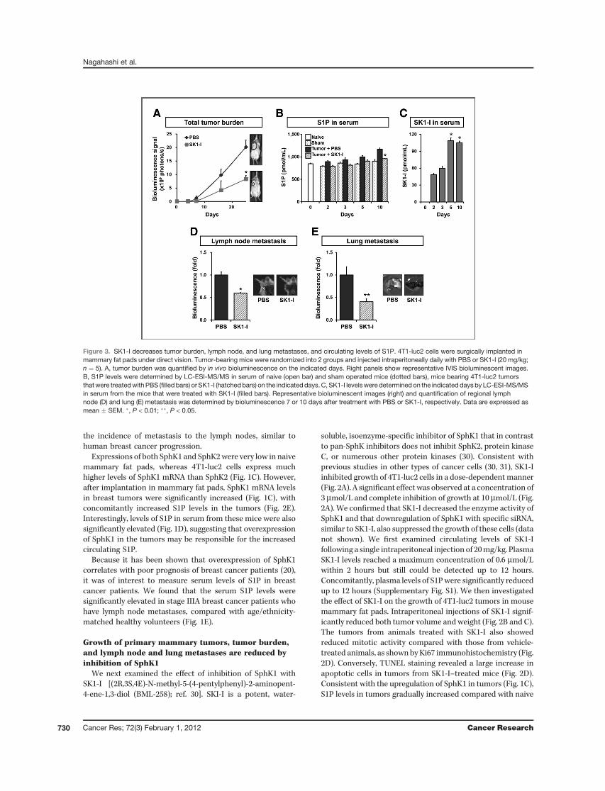
Figure 3. SK1-I decreases tumor burden, lymph node, and lung metastases, and circulating levels of S1P. 4T1-luc2 cells were surgically implanted inmammary fat pads under direct vision. Tumor-bearing mice were randomized into 2 groups and injected intraperitoneally daily with PBS or SK1-I (20 mg/kg;n ¼ 5). A, tumor burden was quantified by in vivo bioluminescence on the indicated days. Right panels show representative IVIS bioluminescent images.B, S1P levels were determined by LC-ESI-MS/MS in serum of naive (open bar) and sham operated mice (dotted bars), mice bearing 4T1-luc2 tumorsthatwere treatedwith PBS (filled bars) or SK1-I (hatchedbars) on the indicateddays. C, SK1-I levelswere determined on the indicated days by LC-ESI-MS/MSin serum from the mice that were treated with SK1-I (filled bars). Representative bioluminescent images (right) and quantification of regional lymphnode (D) and lung (E) metastasis was determined by bioluminescence 7 or 10 days after treatment with PBS or SK1-I, respectively. Data are expressed asmean � SEM. �, P < 0.01; ��, P < 0.05.
Nagahashi et al.
Cancer Res; 72(3) February 1, 2012 Cancer Research730
or sham operated mammary fat pads as measured by LC-ESI-MS/MS. This increase of S1P in the tumors was prevented bytreatment with SK1-I (Fig. 2E). Notably, SK1-I was taken up bythe mammary tumors and its levels gradually increased withtime (Fig. 2F).The total tumor burden determined by the IVIS Imaging
System was also significantly suppressed by SK1-I treat-ment (Fig. 3A). Serum S1P levels, which are elevated in thetumor-bearing mice, were also reduced by SK1-I treatment(Fig. 3B). Serum levels of SK1-I in these mice increased withtime (Fig. 3C). Importantly, SK1-I significantly reducedlymph node and lung metastasis (Fig. 3D and E). Takentogether, these results suggested that S1P produced bySphK1 in the mammary tumors is involved in lymph nodeand lung metastasis.
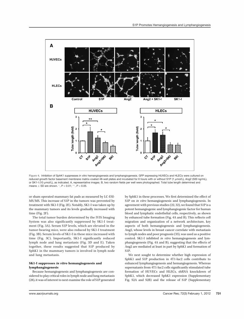
SK1-I suppresses in vitro hemangiogenesis andlymphangiogenesisBecause hemangiogenesis and lymphangiogenesis are con-
sidered to play critical roles in lymph node and lungmetastasis(28), it was of interest to next examine the role of S1P generated
by SphK1 in these processes. We first determined the effect ofS1P on in vitro hemangiogenesis and lymphangiogenesis. Inagreement with previous studies (22, 32), we found that S1P is apotent hemangiogenic and lymphangiogenic factor for humanblood and lymphatic endothelial cells, respectively, as shownby enhanced tube formation (Fig. 4A and B). This reflects cellmigration and organization of a network architecture, keyaspects of both hemangiogenesis and lymphangiogenesis.Ang2, whose levels in breast cancer correlate with metastasisto lymph nodes and poor prognosis (33), was used as a positivecontrol. SK1-I inhibited in vitro hemangiogenesis and lym-phangiogenesis (Fig. 4A and B), suggesting that the effects ofAng2 are mediated at least in part by SphK1 and formation ofS1P.
We next sought to determine whether high expression ofSphK1 and S1P production in 4T1-luc2 cells contribute toenhanced lymphangiogenesis and hemangiogenesis. Whereassupernatants from 4T1-luc2 cells significantly stimulated tubeformation of HUVECs and HLECs, shRNA knockdown ofSphK1, which decreased SphK1 expression (SupplementaryFig. S2A and S2B) and the release of S1P (Supplementary
Figure 4. Inhibition of SphK1 suppresses in vitro hemangiogenesis and lymphangiogenesis. GFP expressing HUVECs and HLECs were cultured onreduced growth factor basement membrane matrix–coated 48-well plates and incubated for 6 hours with or without S1P (1 mmol/L), Ang2 (500 ng/mL),or SK1-I (10 mmol/L), as indicated. A, representative images. B, two random fields per well were photographed. Total tube length determined andmeans � SD are shown. �, P < 0.01; ��, P < 0.05.
S1P Promotes Hemangiogenesis and Lymphangiogenesis
www.aacrjournals.org Cancer Res; 72(3) February 1, 2012 731
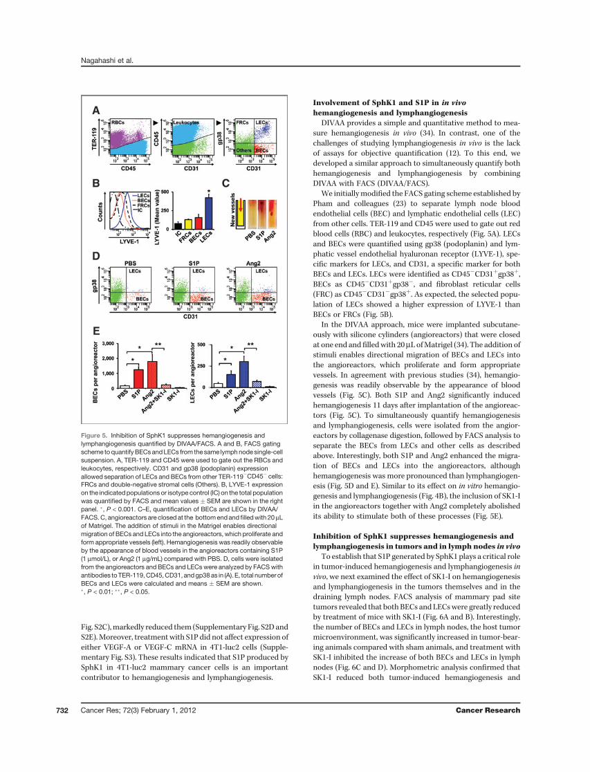
Fig. S2C),markedly reduced them (Supplementary Fig. S2DandS2E).Moreover, treatmentwith S1P did not affect expression ofeither VEGF-A or VEGF-C mRNA in 4T1-luc2 cells (Supple-mentary Fig. S3). These results indicated that S1P produced bySphK1 in 4T1-luc2 mammary cancer cells is an importantcontributor to hemangiogenesis and lymphangiogenesis.
Involvement of SphK1 and S1P in in vivohemangiogenesis and lymphangiogenesis
DIVAA provides a simple and quantitative method to mea-sure hemangiogenesis in vivo (34). In contrast, one of thechallenges of studying lymphangiogenesis in vivo is the lackof assays for objective quantification (12). To this end, wedeveloped a similar approach to simultaneously quantify bothhemangiogenesis and lymphangiogenesis by combiningDIVAA with FACS (DIVAA/FACS).
We initiallymodified the FACS gating scheme established byPham and colleagues (23) to separate lymph node bloodendothelial cells (BEC) and lymphatic endothelial cells (LEC)from other cells. TER-119 and CD45 were used to gate out redblood cells (RBC) and leukocytes, respectively (Fig. 5A). LECsand BECs were quantified using gp38 (podoplanin) and lym-phatic vessel endothelial hyaluronan receptor (LYVE-1), spe-cific markers for LECs, and CD31, a specific marker for bothBECs and LECs. LECs were identified as CD45�CD31þgp38þ,BECs as CD45�CD31þgp38�, and fibroblast reticular cells(FRC) as CD45�CD31�gp38þ. As expected, the selected popu-lation of LECs showed a higher expression of LYVE-1 thanBECs or FRCs (Fig. 5B).
In the DIVAA approach, mice were implanted subcutane-ously with silicone cylinders (angioreactors) that were closedat one end andfilledwith 20mL ofMatrigel (34). The addition ofstimuli enables directional migration of BECs and LECs intothe angioreactors, which proliferate and form appropriatevessels. In agreement with previous studies (34), hemangio-genesis was readily observable by the appearance of bloodvessels (Fig. 5C). Both S1P and Ang2 significantly inducedhemangiogenesis 11 days after implantation of the angioreac-tors (Fig. 5C). To simultaneously quantify hemangiogenesisand lymphangiogenesis, cells were isolated from the angior-eactors by collagenase digestion, followed by FACS analysis toseparate the BECs from LECs and other cells as describedabove. Interestingly, both S1P and Ang2 enhanced the migra-tion of BECs and LECs into the angioreactors, althoughhemangiogenesis was more pronounced than lymphangiogen-esis (Fig. 5D and E). Similar to its effect on in vitro hemangio-genesis and lymphangiogenesis (Fig. 4B), the inclusion of SK1-Iin the angioreactors together with Ang2 completely abolishedits ability to stimulate both of these processes (Fig. 5E).
Inhibition of SphK1 suppresses hemangiogenesis andlymphangiogenesis in tumors and in lymph nodes in vivo
To establish that S1P generated by SphK1 plays a critical rolein tumor-induced hemangiogenesis and lymphangiogenesis invivo, we next examined the effect of SK1-I on hemangiogenesisand lymphangiogenesis in the tumors themselves and in thedraining lymph nodes. FACS analysis of mammary pad sitetumors revealed that bothBECs andLECswere greatly reducedby treatment of mice with SK1-I (Fig. 6A and B). Interestingly,the number of BECs and LECs in lymph nodes, the host tumormicroenvironment, was significantly increased in tumor-bear-ing animals compared with sham animals, and treatment withSK1-I inhibited the increase of both BECs and LECs in lymphnodes (Fig. 6C and D). Morphometric analysis confirmed thatSK1-I reduced both tumor-induced hemangiogenesis and
Figure 5. Inhibition of SphK1 suppresses hemangiogenesis andlymphangiogenesis quantified by DIVAA/FACS. A and B, FACS gatingscheme toquantifyBECsandLECs from the same lymphnode single-cellsuspension. A, TER-119 and CD45 were used to gate out the RBCs andleukocytes, respectively. CD31 and gp38 (podoplanin) expressionallowed separation of LECs and BECs from other TER-119�CD45� cells:FRCs and double-negative stromal cells (Others). B, LYVE-1 expressionon the indicatedpopulationsor isotype control (IC) on the total populationwas quantified by FACS and mean values � SEM are shown in the rightpanel. �, P < 0.001. C–E, quantification of BECs and LECs by DIVAA/FACS.C, angioreactors are closed at the bottomendand filledwith 20mLof Matrigel. The addition of stimuli in the Matrigel enables directionalmigration of BECs and LECs into the angioreactors, which proliferate andform appropriate vessels (left). Hemangiogenesis was readily observableby the appearance of blood vessels in the angioreactors containing S1P(1 mmol/L), or Ang2 (1 mg/mL) compared with PBS. D, cells were isolatedfrom the angioreactors and BECs and LECs were analyzed by FACSwithantibodies toTER-119,CD45,CD31, andgp38as in (A). E, total numberofBECs and LECs were calculated and means � SEM are shown.�, P < 0.01; ��, P < 0.05.
Nagahashi et al.
Cancer Res; 72(3) February 1, 2012 Cancer Research732
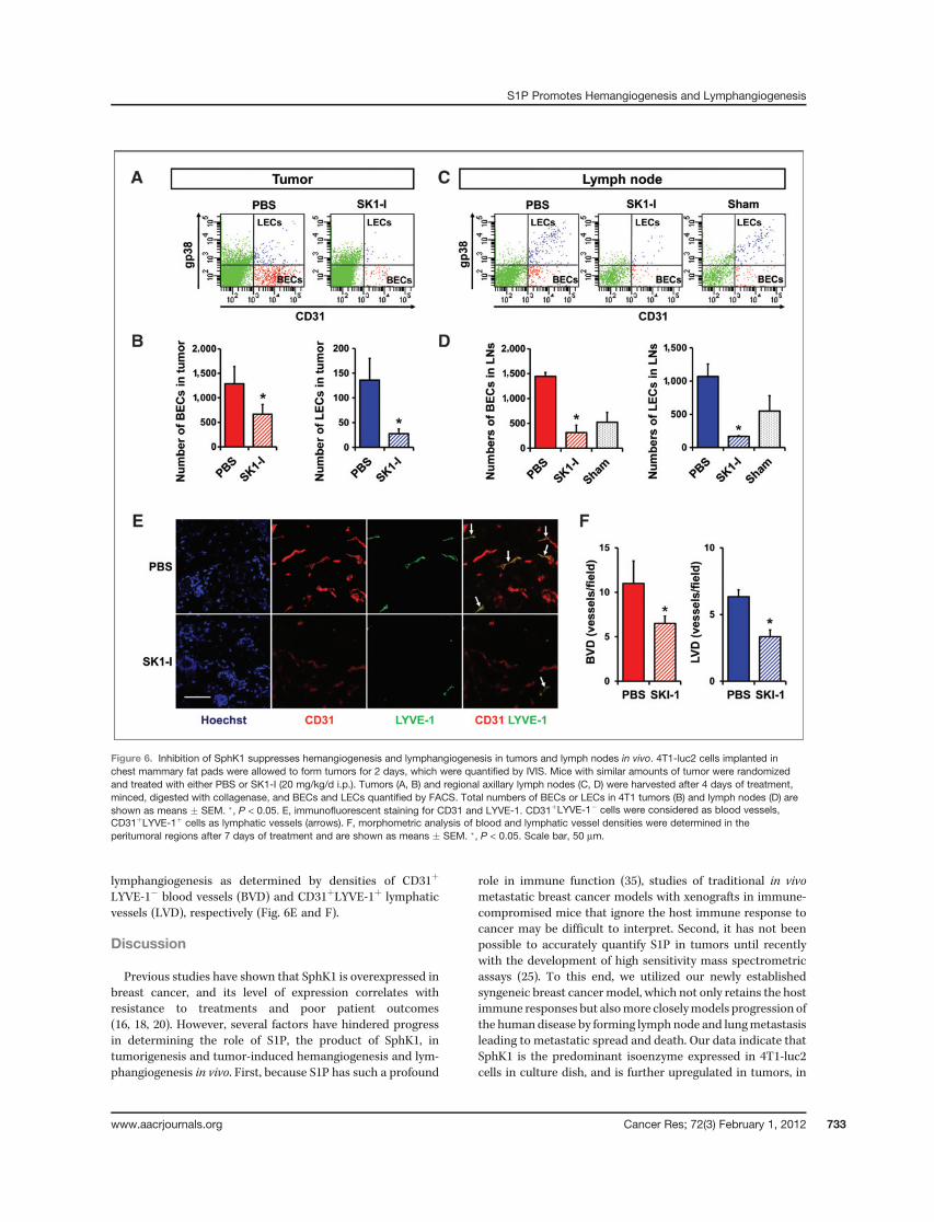
lymphangiogenesis as determined by densities of CD31þ
LYVE-1� blood vessels (BVD) and CD31þLYVE-1þ lymphaticvessels (LVD), respectively (Fig. 6E and F).
Discussion
Previous studies have shown that SphK1 is overexpressed inbreast cancer, and its level of expression correlates withresistance to treatments and poor patient outcomes(16, 18, 20). However, several factors have hindered progressin determining the role of S1P, the product of SphK1, intumorigenesis and tumor-induced hemangiogenesis and lym-phangiogenesis in vivo. First, because S1P has such a profound
role in immune function (35), studies of traditional in vivometastatic breast cancer models with xenografts in immune-compromised mice that ignore the host immune response tocancer may be difficult to interpret. Second, it has not beenpossible to accurately quantify S1P in tumors until recentlywith the development of high sensitivity mass spectrometricassays (25). To this end, we utilized our newly establishedsyngeneic breast cancer model, which not only retains the hostimmune responses but alsomore closelymodels progression ofthe human disease by forming lymph node and lungmetastasisleading to metastatic spread and death. Our data indicate thatSphK1 is the predominant isoenzyme expressed in 4T1-luc2cells in culture dish, and is further upregulated in tumors, in
Figure 6. Inhibition of SphK1 suppresses hemangiogenesis and lymphangiogenesis in tumors and lymph nodes in vivo. 4T1-luc2 cells implanted inchest mammary fat pads were allowed to form tumors for 2 days, which were quantified by IVIS. Mice with similar amounts of tumor were randomizedand treated with either PBS or SK1-I (20 mg/kg/d i.p.). Tumors (A, B) and regional axillary lymph nodes (C, D) were harvested after 4 days of treatment,minced, digested with collagenase, and BECs and LECs quantified by FACS. Total numbers of BECs or LECs in 4T1 tumors (B) and lymph nodes (D) areshown as means � SEM. �, P < 0.05. E, immunofluorescent staining for CD31 and LYVE-1. CD31þLYVE-1� cells were considered as blood vessels,CD31þLYVE-1þ cells as lymphatic vessels (arrows). F, morphometric analysis of blood and lymphatic vessel densities were determined in theperitumoral regions after 7 days of treatment and are shown as means � SEM. �, P < 0.05. Scale bar, 50 mm.
S1P Promotes Hemangiogenesis and Lymphangiogenesis
www.aacrjournals.org Cancer Res; 72(3) February 1, 2012 733
contrast to SphK2. This is in agreement with previous reportscorrelating expression of SphK1, but not of SphK2, with poorprognosis in breast cancer (20). We found that levels of S1Pgradually increased both in tumors and in serum determinedby LC-ESI-MS/MS and correlated with tumor burden. More-over, treatment of tumor-bearing animals with the specificSphK1 inhibitor SK1-I reduced S1P levels in the tumor and incirculation, and greatly reduced the size of the primary tumor,lymph node, and lung metastasis. This is in agreement with aprevious report that blood and tumor S1P levels were increasedin mice with colon cancer (36).
Quantification of tumor-induced hemangiogenesis and lym-phangiogenesis has remained a challenge (12). This is usuallyexamined by histologic determinations of microvessel densityor LVD, which rely on selective morphometric analysis (e.g.,vessel counts, vascular morphology, etc.; ref. 12). The strengthsof the morphologic approach are that it can evaluate thelocation of the vessels in relationship to the tumor andidentify/quantify morphologic changes that lymphatic vesselsundergo during tumor progression. The limitations includevariable sites of tissue sectioning, variable immunostainingtechniques, different vessel density quantification methods,and the lack of standardization in the estimation of heman-giogenesis and lymphangiogenesis (12). To compliment thisapproach and overcome some of these limitations, we devel-oped a flow cytometrymethod to quantify both BECs and LECsin the same sample, to simultaneously evaluate both heman-giogenesis and lymphangiogenesis. Flow cytometric analysiscan provide supportive data to quantify the changes in BECsand LECs.
Consistentwith both the in vitro tube formation andDIVAA/FACS assays, we showed that exogenous S1P enhanced heman-giogenesis and lymphangiogenesis. In this regard, Anelli andcolleagues showed that downregulation of SphK1 did notchange the levels of VEGF-A or VEGF-C secretion in gliomacells, and downregulation of SphK1, but not VEGF-A or VEGF-C, suppressed glioma-induced hemangiogenesis (37). UtilizingshSphK1, we confirmed that tumor-derived S1P mediatestumor-induced hemangiogenesis and lymphangiogenesiswithout altering VEGF-A or VEGF-C (Supplementary Fig.S3), which is in agreement with Anelli and colleagues (37).Taken together, S1P is an important stimulator of hemangio-genesis and lymphangiogenesis in vitro.
SK1-I decreased hemangiogenesis and lymphangiogenesisnot only around the primary tumor but also in lymph nodesthat are distant from the tumor. Furthermore, we haveobserved by F4/80 immunofluorescent staining that SK1-Ialso reduced macrophage recruitment surrounding the tumor(Supplementary Fig. S4). Macrophages are cellular compo-nents of the tumor microenvironment that promote heman-giogenesis and lymphangiogenesis (38, 39). Thus, in addition todirect effects on hemangiogenesis and lymphangiogenesis, S1Pcan indirectly affect the tumor microenvironment to furtherenhance these processes.
In sum, S1P generated by SphK1 is important not only fortumor progression but also for tumor-induced hemangiogen-esis and lymphangiogenesis and, therefore, targeting SphK1and its product S1P would be a multipronged attack againstbreast cancer.
Disclosure of Potential Conflicts of Interest
No potential conflicts of interest were disclosed.
Acknowledgments
The authors thank the Tissue and Data Acquisition and Analysis Core(TDAAC) of VCU for collection of human samples, and the AnatomicPathology Research Services (APRS) Director, Dr. Jorge A. Almenara, andthe histotechnologists for technical assistance with tissue processing, sec-tioning, and staining. The authors also thank Dr. Bin-Zhi Qian in JeffreyPollard laboratory at Albert Einstein College of Medicine, Bronx, NY, for anti-F4/80 antibody.
Grant Support
This work was supported by NCI grant R01CA61774 to S. Spiegel and NIHgrant K12HD055881 and Susan G. Komen for the Cure Research FoundationCareer Catalyst Research Grant KG090510 to K. Takabe. M. Nagahashi wassupported by the SUMITOMO Life Social Welfare Services Foundation.
The Flow Cytometry and Confocal microscopy Shared Resource Cores weresupported in part by NIH Grant P30CA16059 to the Massey Cancer Center andNINDS Center core Grant 5P30NS047463, respectively.
The costs of publication of this article were defrayed in part by thepayment of page charges. This article must therefore be hereby markedadvertisement in accordance with 18 U.S.C. Section 1734 solely to indicatethis fact.
Received July 5, 2011; revised November 30, 2011; accepted December 1, 2011;published online February 1, 2012.
References1. Siegel R, Ward E, Brawley O, Jemal A. Cancer statistics, 2011: the
impact of eliminating socioeconomic and racial disparities on prema-ture cancer deaths. CA Cancer J Clin 2011;61:212–36.
2. Mumprecht V, Honer M, Vigl B, Proulx ST, Trachsel E, Kaspar M, et al.In vivo imaging of inflammation- and tumor-induced lymph nodelymphangiogenesis by immuno-positron emission tomography. Can-cer Res 2010;70:8842–51.
3. Hanahan D, Weinberg RA. Hallmarks of cancer: the next generation.Cell 2011;144:646–74.
4. Nagahashi M, Shirai Y, Wakai T, Sakata J, Ajioka Y, Nomura T, et al.Depth of invasion determines the postresectional prognosis forpatients with T1 extrahepatic cholangiocarcinoma. Cancer 2010;116:400–5.
5. Folkman J. Tumor angiogenesis: therapeutic implications. N Engl JMed 1971;285:1182–6.
6. Holmgren L, O'Reilly MS, Folkman J. Dormancy of micrometastases:balanced proliferation and apoptosis in the presence of angiogenesissuppression. Nat Med 1995;1:149–53.
7. Hanahan D, Weinberg RA. The hallmarks of cancer. Cell 2000;100:57–70.
8. Miller K, Wang M, Gralow J, Dickler M, Cobleigh M, Perez EA, et al.Paclitaxel plus bevacizumab versus paclitaxel alone for metastaticbreast cancer. N Engl J Med 2007;357:2666–76.
9. Miles DW, Chan A, Dirix LY, Cortes J, Pivot X, Tomczak P, et al.Phase III study of bevacizumab plus docetaxel compared withplacebo plus docetaxel for the first-line treatment of human
Nagahashi et al.
Cancer Res; 72(3) February 1, 2012 Cancer Research734
epidermal growth factor receptor 2-negative metastatic breastcancer. J Clin Oncol 2010;28:3239–47.
10. Robert NJ, Dieras V, Glaspy J, Brufsky AM, Bondarenko I, Lipatov ON,et al. RIBBON-1: randomized, double-blind, placebo-controlled,phase III trial of chemotherapy with or without bevacizumab forfirst-line treatment of human epidermal growth factor receptor2-negative, locally recurrent or metastatic breast cancer. J Clin Oncol2011;29:1252–60.
11. Edge SB, Byrd DR, Compton CC, Fritz AG, Greene FL, Trotti A, et al.AJCC cancer staging manual (7th ed). New York, NY: Springer; 2010.
12. Nagahashi M, Ramachandran S, Rashid OM, Takabe K. Lymphangio-genesis: a new player in cancer progression. World J Gastroenterol2010;16:4003–12.
13. Tammela T, Alitalo K. Lymphangiogenesis: Molecular mechanismsand future promise. Cell 2010;140:460–76.
14. Cueni LN, Hegyi I, Shin JW, Albinger-Hegyi A, Gruber S, Kunstfeld R,et al. Tumor lymphangiogenesis and metastasis to lymph nodesinduced by cancer cell expression of podoplanin. Am J Pathol 2010;177:1004–16.
15. Takabe K, Paugh SW, Milstien S, Spiegel S. "Inside-out" signaling ofsphingosine-1-phosphate: therapeutic targets. Pharmacol Rev 2008;60:181–95.
16. Pyne NJ, Pyne S. Sphingosine 1-phosphate and cancer. Nat RevCancer 2010;10:489–503.
17. TakabeK, KimRH, Allegood JC,Mitra P, Ramachandran S, NagahashiM, et al. Estradiol induces export of sphingosine 1-phosphate frombreast cancer cells via ABCC1 and ABCG2. J Biol Chem 2010;285:10477–86.
18. Shida D, Takabe K, Kapitonov D, Milstien S, Spiegel S. TargetingSphK1 as a new strategy against cancer. Curr Drug Targets 2008;9:662–73.
19. Kim RH, Takabe K, Milstien S, Spiegel S. Export and functions ofsphingosine-1-phosphate. Biochim Biophys Acta 2009;1791:692–6.
20. Ruckhaberle E, Rody A, Engels K, Gaetje R, von Minckwitz G, Schiff-mann S, et al. Microarray analysis of altered sphingolipid metabolismreveals prognostic significance of sphingosine kinase 1 in breastcancer. Breast Cancer Res Treat 2008;112:41–52.
21. Milstien S, Spiegel S. Targeting sphingosine-1-phosphate: a novelavenue for cancer therapeutics. Cancer Cell 2006;9:148–50.
22. Yoon CM, Hong BS, Moon HG, Lim S, Suh PG, Kim YK, et al.Sphingosine-1-phosphate promotes lymphangiogenesis by stimulat-ing S1P1/Gi/PLC/Ca2þ signaling pathways. Blood 2008;112:1129–38.
23. Pham TH, Baluk P, Xu Y, Grigorova I, Bankovich AJ, Pappu R, et al.Lymphatic endothelial cell sphingosine kinase activity is required forlymphocyte egress and lymphatic patterning. J ExpMed2010;207:17–27, S1–4.
24. Zhou H, Jarujaron S, Gurley EC, Chen L, Ding H, Studer E, et al. HIVprotease inhibitors increase TNF-alpha and IL-6 expression in macro-
phages: involvement of the RNA-binding protein HuR. Atherosclerosis2007;195:e134–43.
25. Hait NC, Allegood J, Maceyka M, Strub GM, Harikumar KB, Singh SK,et al. Regulation of histone acetylation in the nucleus by sphingosine-1-phosphate. Science 2009;325:1254–7.
26. van Beijnum JR, RouschM, Castermans K, van der Linden E, GriffioenAW. Isolation of endothelial cells from fresh tissues. Nat Protoc2008;3:1085–91.
27. Qian BZ, Li J, Zhang H, Kitamura T, Zhang J, Campion LR, et al. CCL2recruits inflammatory monocytes to facilitate breast-tumour metasta-sis. Nature 2011;475:222–5.
28. Nagahashi M, Shirai Y, Wakai T, Sakata J, Ajioka Y, Hatakeyama K.Perimuscular connective tissue contains more and larger lymphaticvessels than the shallower layers in human gallbladders. World JGastroenterol 2007;13:4480–3.
29. Wang F, Van Brocklyn JR, Hobson JP, Movafagh S, Zukowska-GrojecZ, Milstien S, et al. Sphingosine 1-phosphate stimulates cell migrationthrough a G(i)-coupled cell surface receptor. Potential involvement inangiogenesis. J Biol Chem 1999;274:35343–50.
30. PaughSW,PaughBS,RahmaniM,KapitonovD, Almenara JA,KordulaT, et al. A selective sphingosine kinase 1 inhibitor integrates multiplemolecular therapeutic targets in human leukemia. Blood 2008;112:1382–91.
31. Kapitonov D, Allegood JC, Mitchell C, Hait NC, Almenara JA, AdamsJK, et al. Targeting sphingosine kinase 1 inhibits Akt signaling, inducesapoptosis, and suppresses growth of human glioblastoma cells andxenografts. Cancer Res 2009;69:6915–23.
32. Lee MJ, Thangada S, Claffey KP, Ancellin N, Liu CH, Kluk M, et al.Vascular endothelial cell adherens junction assembly and morpho-genesis induced by sphingosine-1-phosphate. Cell 1999;99:301–12.
33. Sfiligoi C, de Luca A, Cascone I, Sorbello V, Fuso L, Ponzone R, et al.Angiopoietin-2 expression in breast cancer correlateswith lymphnodeinvasion and short survival. Int J Cancer 2003;103:466–74.
34. Guedez L, Rivera AM, Salloum R, Miller ML, Diegmueller JJ, BungayPM, et al. Quantitative assessment of angiogenic responses by thedirected in vivo angiogenesis assay. Am J Pathol 2003;162:1431–9.
35. Spiegel S,MilstienS. The outs and the ins of sphingosine-1-phosphatein immunity. Nat Rev Immunol 2011;11:403–15.
36. Kawamori T, Kaneshiro T, Okumura M, Maalouf S, Uflacker A, Bie-lawski J, et al. Role for sphingosine kinase 1 in colon carcinogenesis.FASEB J 2009;23:405–14.
37. Anelli V, Gault CR, Snider AJ, Obeid LM. Role of sphingosine kinase-1in paracrine/transcellular angiogenesis and lymphangiogenesis invitro. FASEB J 2010;24:2727–38.
38. Qian BZ, Pollard JW. Macrophage diversity enhances tumor progres-sion and metastasis. Cell 2010;141:39–51.
39. Ji RC. Macrophages are important mediators of either tumor- orinflammation-induced lymphangiogenesis. Cell Mol Life Sci 2011 Oct8 [Epub ahead of print].
S1P Promotes Hemangiogenesis and Lymphangiogenesis
www.aacrjournals.org Cancer Res; 72(3) February 1, 2012 735