Embed Size (px)
Citation preview
Julia R. Crim 1
Lawrence W. Bassett 1
Richard H. Gold1
Joseph M. Mirra2
Mark Mikulics3
Edgar G. Dawson4
Jeffrey J. Eckhardt4
Received May 6, 1987; accepted after revision September 1, 1987.
1 Department of Radiology, UCLA Medical Center, Los Angeles, CA 90024. Address reprint requests to L. W. Bassett.
2 Department of Pathology, UCLA Medical Center, Los Angeles, CA 90024.
3 Division of Orthopedic Surgery, Hospital of the University of Pennsylvania, 34th and Spruce, Philadelphia, PA 19104.
, Division of Orthopedic Surgery, UCLA Medical Center. Los Anoeles. CA 90024.
AJNR 9:359-362, March/April 1988 0195-6108/88/0902-0359 © American Society of Neuroradiology
Spinal Neuroarthropathy After Traumatic Paraplegia
359
Spinal neuroarthropathy is a little-known complication of traumatic paraplegia. Four cases of this syndrome are described, with emphasis on the characteristic radiographic findings of severe juxta-articular bone destruction, dense appOSitional new bone formation, large osteophytosis, and soft-tissue bony debris. The factors predisposing patients to develop a neuropathic joint are diminished pain and proprioceptive sensations with maintained mobility. When a paraplegic patient transfers in or out of a wheelchair or moves his upper torso, he exerts force on an insensate spine. Repeated trauma increases joint mobility beyond the normal limits, and this leads to further damage, with the process culminating in severe instability and bone destruction. The other causes of neuropathic joints in the spine-tertiary syphilis, syringomyelia, and diabetes-must be ruled out on clinical grounds.
Neuropathic changes in the spine are often silent, delaying treatment, or may be mistaken for infection or degenerative disease. Their true prevalence is difficult to determine, but the possibility should be considered in paraplegic patients with the characteristic radiographic findings.
Traumatic paraplegia was first reported as a cause of spinal neuroarthropathy in 1978, when Slabaugh and Smith [1] described a case of Charcot joint at T11-T12 in a patient who had been paraplegic below T7 for 13 years and who was seronegative for syphilis . In 1985, Sobel et al. [2] described five patients, also seronegative for syphilis , who developed spinal neuroarthropathy 7-34 years after paralysis. Two of their patients had radiologic findings but were asymptomatic, while the others complained of a grinding sensation in the spine with movement, or difficulty in maintaining balance when seated.
We report the radiologic findings in four cases of Charcot spine secondary to traumatic paraplegia of 4-34 years ' duration. Each patient had a negative serologic test for syphilis and negative bacteriologic cultures. None had diabetes mellitus or syringomyelia.
Case Reports
Case 1
A 54-year-old man had been paraplegic since a gunshot wound to T1 0 at age 22. Treatment at the time of injury included a laminectomy. His health remained good except for recurrent urinary tract infections and a sacral decubitus ulcer. Early changes of neuropathic spine at the L2-L3 level were radiographically documented 24 years after injury (Fig . 1 A). Thirty-two years after injury, he complained of an increasing lumbar deformity and an audible, nonpainful crunch in the lumbar spine with movement. Physical examination revealed a lumbar scoliosis , flaccid paralysis , and no sensation below T10 . Serial radiographs over a 6-month period showed rapidly progressive destruction and sclerosis at L2- L3 (Figs. 1 Band 1 C). A serologic test for syphilis was negative, and there was no history of diabetes mellitus or syringomyelia. A needle aspirate of the L2-L3 disk space was negative on aerobic and anaerobic bacterio-
360 GRIM ET EL. AJNR:9, March/April 1988
A 8
c o
logic cultures. Harrington rod fixation and posterior fusion of L 1-L4 led to healing of the spondyloarthropathy (Fig. 1 D).
Case 2
A 60-year-old man had spastic paraplegia below T8 due to a T7-T8 dislocation sustained in a motor vehicle accident at the age of 26. A laminectomy was performed at the time of injury. His general health remained good, except for recurrent urinary tract infections. Thirtyfour years after injury , he presented with a 10-year history of progressive "tilting" of the spine. For 18 months he had suffered from vague, deep, spine pain originating at a level below that at which he retained sensation. For '9 months he had experienced grinding sensations in the spine, paras pinal muscle spasms, and increasing spasticity 'of the lower extremities. Physical examination disclosed an acute kyphosis of the lower thoracic 'spine and absence of motor function and sensation below T8. There was no history of diabetes mellitus or syringomyelia, and a serologic test for syphilis was negative. Radiographs showed a florid Charcot jOint at T11-T12 and less severe changes at T8-T9 (Fig. 2). He was treated with Harrington rod fixation and posterior fusion from T8- L2. At surgery, striking
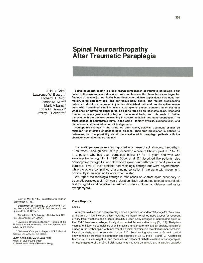
Fig. 1.-Charcot spine, case 1. A, Radiograph 24 years after injury to thoracic
spine. Osteophytosis is present at L2-L3, with blurring of superior end plate of L3. Disk space is narrowed on the right.
Anteroposterior (8) and lateral (C) radiographs 32 years after injury. Periarticular portions of L2 and L3 vertebral bodies show progressive destruction, with dense, fluffy appositional new bone formation. There is anterolateral dislocation of L3 in relation to L2. The osteophytes are exuberant, and there is extensive periarticular bony debris. Milder changes are present at L 1-L2.
D, Radiograph 5 years after surgery shows that the spine has healed.
instability was found at the affected levels. Aerobic and anaerobic cultures were negative. Six months later, one of the Harrington rods fractured and the other became dislocated. Nevertheless, the fusion progressed to complete healing.
Case 3
A 32-year-old woman was paraplegic due to a T12-L 1 fracture dislocation 15 years previously. She had been treated initially with fusion and Harrington rod fixation. Her health was good postoperatively, except for recurrent urinary tract infections and decubitus ulcers. Routine radiographs of the lumbosacral spine, obtained during evaluation of an episode of urinary sepsis, showed extensive destruction of the sacrum (Fig. 3A). CT defined an associated soft-tissue mass (Fig. 3B). The patient was asymptomatic. Multiple open biopsy specimens from throughout the lesion were obtained. Bacteriologic cultures were negative. Pathologic examination disclosed granulation tissue and shards of bone, and no evidence of neoplasia or infection (Fig . 3C). The patient had a negative serologic test for syphilis, and no history of diabetes mellitus or syringomyelia. She is now awaiting treatment.
AJNR:9, March/April 1988 SPINAL NEUROARTHROPATHY 361
Case 4
A 36-year-old man was paraplegic due to an L 1 fracture 4 years previously. He had been treated initially with laminectomy and fusion of T12-L2, with Harrington rod fixation from T1-L3. He did well postoperatively and assumed an active lifestyle, practicing weight lifting and gymnastics. Two years after onset of paralysis he began to have gradually increasing, painless, grinding and snapping sensations in the lower lumbar spine when transferring in and out of his wheelchair. Two years later, the physical examination was negative but radiographs disclosed Charcot arthropathy at L3-L4. There was no history of diabetes mellitus or syringomyelia, and a serologic test for syphilis was negative. His spinal fusion was extended to L4. At surgery, there was no evidence of infection, and aerobic and anaerobic cultures were negative. The fusion healed well and his symptoms resolved.
Discussion
Charcot described the neuropathic joint in 1868 [3] , recognizing that the painless but severe degenerative changes
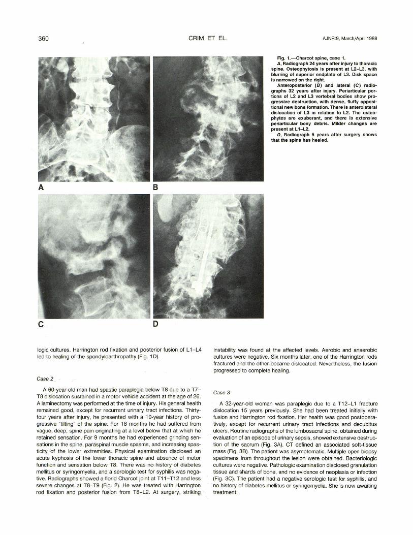
Fig. 2.-Charcot spine, case 2. Radiograph 34 years after injury shows that peridiskal portions of T11 and T12 vertebral bodies have been destroyed, and that subjacent bone is dense. Early neuropathic changes are seen at higher spinal levels.
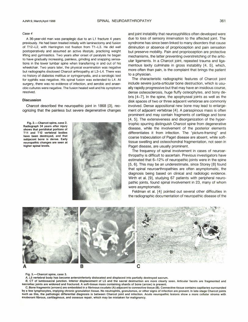
Fig. 3.-Charcot spine, case 3.
and joint instability that neurosyphilitics often developed were due to loss of sensory innervation to the affected joint. The syndrome has since been linked to many disorders that cause diminution or absence of proprioception and pain sensation but preserve mobility. Pain and proprioception are protective mechanisms, the latter preventing overstretching of the articular ligaments. In a Charcot joint, repeated trauma and ligamentous laxity culminate in gross instability [4, 5] , which, more often than pain, is the complaint that brings the patient to a physician .
The characteristic radiographic features of Charcot jOint include severe juxta-articular bone destruction, which is usually rapidly progressive but that may have an insidious course, dense osteosclerosis, huge fluffy osteophytes , and bony debris [4-7]. In the spine, the apophyseal jOints as well as the disk spaces of two or three adjacent vertebrae are commonly involved. Dense appositional new bone may lead to enlargement of adjacent vertebrae [4] . A paraspinous mass is often prominent and may contain fragments of cartilage and bone [4, 5] . The extensiveness and disorganization of the hypertrophic spurring distinguish Charcot spine from degenerative disease, while the involvement of the posterior elements differentiates it from infection. The "picture-framing" and coarse trabeculation of Paget disease are absent, while softtissue swelling and osteochondral fragmentation , not seen in Paget disease, are usually prominent.
The frequency of spinal involvement in cases of neuroarthropathy is difficult to ascertain. Previous investigators have estimated that 6-12% of neuropathic jOints were in the spine [5 , 6]. This may be an underestimate, since Storey [8] found that spinal neuroarthropathies are often asymptomatic, the diagnosis being based on clinical and radiologic evidence . Wirth et al. [9], studying 67 patients with peripheral neuropathic jOints, found spinal involvement in 23, many of whom were asymptomatic.
Feldman et al. [4] pOinted out several other difficulties in the radiographic documentation of neuropathic disease of the
A, L5 vertebral body has become anteroinferiorly dislocated and displaced into partially destroyed sacrum. B, CT of lumbosacral junction. Inferior displacement of L5 and the sacral destruction are more clearly seen. Articular facets are fragmented and
sacroiliac joints are widened and fractured. A soft-tissue mass containing shards of bone (arrow) is present. C, Bone fragments (arrows) are embedded in a fibrinous exudate (A) adjacent to connective tissue (B). Connective tissue contains capillaries surrounded
by a few lymphocytes, implying chronic granulation tissue. No neutrophils, granulomas, or other signs of infection are present. In late-stage Charcot joints such as this, the pathologic differential diagnosis is between Charcot joint and infection. Acute neuropathic lesions show a more cellular stroma with exuberant fibrous, cartilaginous, and osseous repair, which may be mistaken for malignancy.
362 CRIM ET EL. AJNR:9, March/April 1988
spine: the spine is not often routinely radiographed; the great thickness of the surrounding soft tissues leads to extensive scattered radiation and, therefore, to decreased bony detail, causing subtle radiographic abnormalities to be less apparent in the spine than in peripheral joints; and moderate and even advanced neuropathic changes, when detected, are often mistakenly attributed to simple degenerative disease.
The majority of reported cases of spinal neuroarthropathy are due to neurosyphilis. Feldman et al. [4] found that of 14 patients with Charcot spine, seven had syphilis, one had syphilis and diabetes, two had syringomyelia, two had syringomyelia and diabetes, and two had diabetes alone. Of 23 cases of Charcot spine, Wirth et al. [9] found that 17 resulted from syphilis and one each from diabetes, syringomyelia, ankylosing spondylitis, neuropathy of unknown origin, radiation, and trauma. The case of traumatic origin was not discussed. The case of radiation myelopathy involved a woman with paraplegia following radiotherapy for Hodgkin disease. She was treated with laminectomy, and later developed a Charcot spine. This case might have been more correctly attributed to paraplegia.
Dejerine et al. [10], in 1919, emphasized the lack of destructive joint changes in patients with traumatic paraplegia, and later investigators concurred [11-14]. Lodge [14] postulated that Charcot jOints could not be a complication of paraplegia because paraplegics were inactive. However, a more cogent reason for previous failure to associate spinal neuropathy with paraplegia is that the patients were evaluated too soon after their injuries. When more long-term studies were performed, spinal abnormalities were found. Wright et al. [15] evaluated the spine radiographs of 26 patients whose paraplegia had been present for a period of several months to 10 years, looking for evidence of spondylitis. Although spondylitis was not found, one 64-year-old man was noted to have "marked osteophytosis. " Bhate et al. [16] studied the anteroposterior spine radiographs of 200 men with traumatic paralysis of 1-34 years ' duration. Twelve percent had spine abnormalities, including interspinous ossification, disk calcification, large bridging and non bridging osteophytes, and apophyseal joint changes. The severity of the abnormalities was directly proportional to the duration of the paralysis and the age of the patient. None of the abnormalities, however, was identified as neuropathic.
Development of Charcot spine requires the coexistence of certain articular phenomena: diminshed pain and proprioceptive sensation and maintenance of mobility. In traumatic paraplegia, sensation is absent below the level of the injury, while mobility is maintained to a varying degree. Paraplegics exert stress on their spines when they sit upright and when they make any significant movement using their upper limbs and torso. The development of Charcot changes could be expected to correlate with the level of physical activity as well as the duration of paralysis. All of our patients were physically active, and the most active patient developed the earliest lesion. Quadriplegics also stress an insensate spine when in
a sitting position [2] , and one of Sobel 's cases of spinal neuropathy was quadriplegic.
Although laminectomy was performed at the time of injury in all our cases, in the one reported by Slabaugh and Smith [1] and in four of the five reported by Sobel et al. [2], the hypothesis of a causal relationship between laminectomy and the development of Charcot spine is tenuous, since most cases of severe spinal trauma are treated with laminectomy. Nevertheless, a laminectomy may decrease stability, predisposing the spine to neuropathic changes.
Pyogenic, fungal, or tuberculous infection of the spine may occasionally manifest many of the same radiographic signs as Charcot spine. Moreover, the devitalized shards of bone associated with Charcot joints predispose the joint to infection . When infection is suspected, percutaneous needle biopsy is useful to exclude that diagnosis.
Reports of spinal neuroarthropathy in paraplegic patients are rare . However, neuropathic changes in the spine, regardless of their origin, are more likely to be silent and more difficult to detect than those of peripheral joints. The possibility of a Charcot spine should be considered in paraplegic patients with characteristic radiographic findings , even in the absence of other predisposing conditions.
REFERENCES
1. Slabaugh P, Smith T. Neuropathic spine after spinal cord injury. J Bone Joint Surg [Am] 1978;60-A(7): 1 005-1 006
2. Sobel JW, Bohlman HH, Freehafer AA. Charcot's arthropathy of the spine following spinal cord injury. J Bone Joint Surg [Am] 1985;67-A(5) :771-776
3. Charcot JM. Sur quelques arthropathies qui paraissent dependre d'une lesion du cerveau ou de la moelle epiniere. Arch Physiol Norm Path 1868;1: 161 -178
4. Feldman F, Johnson AM , Walter JF. Acute axial neuroarthropathy. Radiology 1974;111 :1- 16
5. Goldman AB, Freiberger RH. Localized infections and neuropathic diseases. Semin Roentgenol 1979;XIV(1 ): 19-32
6. McNeel DP, Ehni G. Charcot joints of the lumbar spine. J Neurosurg 1969;30:55-61
7. Theros EG, Harris JH, eds. Bone disease syllabus , third series. Chicago ACR , 1980 :242-243
8. Storey G. Charcot jOints. Br J Venereal Dis 1964;40: 1 09-117 9. Wirth CR, Jacobs RL, Rolander SO. Neuropathic spinal arthropathy: a
review of the Charcot spine. Spine 1980;5(6):558-568 10. Oejerine Mme, Ceillier A, Oejerine Y. Paraosteoarthropathies des para ple
giques par lesions medullaires (etude clinique et radiographique). Ann de Med 1919;5:497-535
11. Abel MS. Sacroiliac joint changes in traumatic paraplegics. Radiology 1950;55 : 233-239
12. Abramson OJ, Kamberg S. Spondylitis, pathological ossification and calcification associated with spinal cord injury. J Bone Joint Surg [Am] 1949;31-A(2):275-283
13. Heilbrun N, Kuhn WG. Erosive bone lesions and soft tissue ossifications associated with spinal cord injuries (paraplegia). Radiology 1947;48 :579-593
14. Lodge T. Bone, joint and soft tissue changes following paraplegia. Acta Radiol [Diagn] (Stockh) 1956;46:435-445
15. Wright V, Catterall RO , Cook JB. Bone and jOint changes in paraplegic men. Ann Rheum Dis 1965;24 :419-431
16. Bhate OV, Pizarro AJ , Seitam A, Mak EB. Axial skeletal changes in paraplegics. Radiology 1979;133:55-58