Embed Size (px)
Citation preview

11/13/13
1
Spirometry: Introduction
Dr. Badri Paudel GMC
Spirometry
! Spirometry is a method of assessing lung function by measuring the volume of air the patient can expel from the lungs after a maximal expiration.
! Conventionally, a spirometer is a device used to measure timed expired and inspired volumes,
! From these we can calculate how effectively and how quickly the lungs can be emptied and filled.
Pulmonary Function Test
11/13/13
Progression of symptoms in COPD reflected by spirometry, arterial blood gas studies, and CXR
Objectives of Spirometry Diagnosis
! Screening for persons at risk of having pulmonary disease.
! Evaluating symptoms of respiratory impairment
! Pre-op assessment
! Pre-employment screening
Monitoring
! Occ Health: monitor those exposed to hazardous agents
! Determine effectiveness of medication
! Monitor for adverse reactions of other drugs e.g. chemotherapy
! Follow the course of disease – helps predict mortality and morbidity.
Disability and Impairment
! Assessing changes during respiratory rehab
! Insurance risk
! Employment/environmental risk
Research
! Large population studies - predicted (reference equations),
! Pharmaceutical trials
11/13/13
2
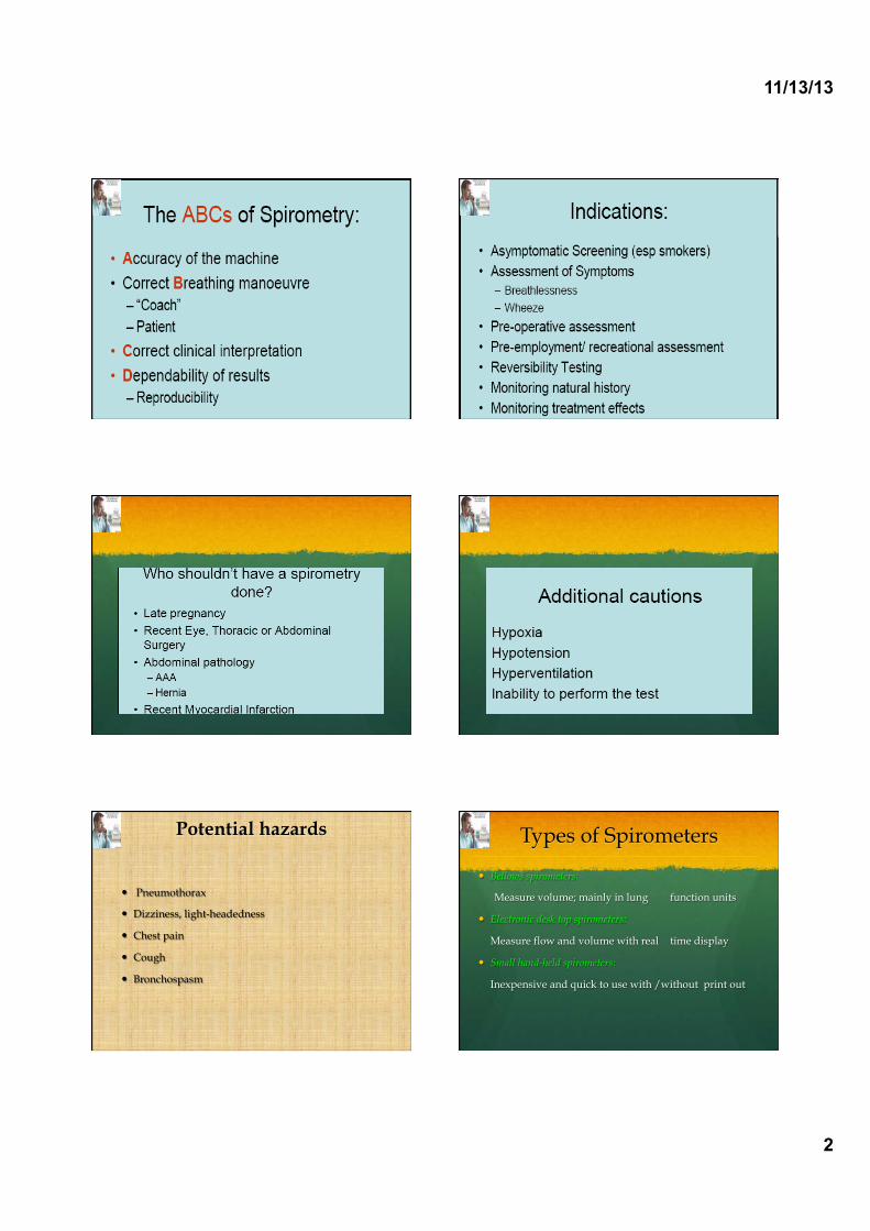
Potential hazards
! Pneumothorax
! Dizziness, light-headedness
! Chest pain
! Cough
! Bronchospasm
Types of Spirometers
! Bellows spirometers:
Measure volume; mainly in lung function units
! Electronic desk top spirometers:
Measure flow and volume with real time display
! Small hand-held spirometers:
Inexpensive and quick to use with /without print out

11/13/13
3
Water-sealed Spirometry
11/13/13
Volume Measuring Spirometer
Flow Measuring Spirometer Desktop Electronic
Spirometers
Small Hand-held Spirometers RESPIRATORY MANOEUVRE
!! Maximal breath in
"" Maximal breath out
!!
""
11/13/13
4
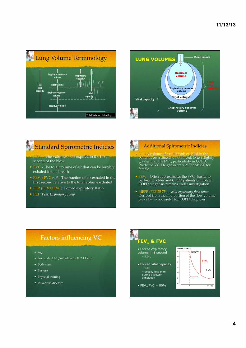
Total lung
capacity
Tidal volume
Inspiratory reserve volume
Expiratory reserve volume
Residual volume
Inspiratory capacity
Vital capacity
Lung Volume Terminology
Tidal Volume: 6-8ml/kg
Tidal volume
Residual Volume
Dead space
Total lung capacity
Vital capacity Tidal volume
Inspiratory reserve volume
Expiratory reserve volume
LUNG VOLUMES
Standard Spirometric Indicies ! FEV1 - The volume of air expired in the first
second of the blow
! FVC - The total volume of air that can be forcibly exhaled in one breath
! FEV1/FVC ratio: The fraction of air exhaled in the first second relative to the total volume exhaled
! FER (FEV1/FVC): Forced expiratory Ratio
! PEF: Peak Expiratory Flow
Additional Spirometric Indicies
! VC - A volume of a full breath exhaled in the patient’s own time and not forced. Often slightly greater than the FVC, particularly in COPD. Predicted VC: Height in cm x 25 for M; x20 for female
! FEV6 – Often approximates the FVC. Easier to
perform in older and COPD patients but role in COPD diagnosis remains under investigation
! MEFR (FEF 25-75 ) – Mid-expiratory flow rates:
Derived from the mid portion of the flow volume curve but is not useful for COPD diagnosis
Factors influencing VC
! Age
! Sex: male: 2.6 L/m2 while for F: 2.1 L/m2
! Body size
! Posture
! Physcial training
! In Various diseases
FEV1 & FVC
Forced expiratory volume in 1 second
– 4.0 L
Forced vital capacity – 5.0 L – usually less than during a slower exhalation
FEV1/FVC = 80%
FEV1
FVC

11/13/13
5
Flow Volume Curve
! Standard on most desk-top spirometers
! Adds more information than volume time curve
! Less understood but not too difficult to interpret
! Better at demonstrating mild airflow obstruction
Spirometry
Flow-Volume curve
Time-Volume curve
11/13/13
Flow Volume Curve
Expiratory flow rate L/sec
Volume (L)
FVC
Maximum expiratory flow (PEF)
Inspiratory flow rate
L/sec
RV TLC
Normal Trace Showing FEV1 and FVC
1 2 3 4 5 6
1
2
3
4
Volu
me,
lite
rs
Time, seconds
FVC 5
1
FEV1 = 4L
FVC = 5L
FEV1/FVC = 0.8
Spirometry(Time-volume curve)
11/13/13
Forced expiratory flows
Spirometry
11/13/13

11/13/13
6
Techniques
Rapid upslope Smooth down slope Complete expiration
Summary of Standards
! A min. of 3 technically satisfactory tests.
! A max. of 8 attempts.
! The two best FVC and FEV1’s should have a variance of less than 150mls.
! Exhaled for at least 6 seconds (adults) or reached a plateau on the volume-time graph. (No change of volume for at least one second.)
! Graph traces are smooth and free from irregularity--Smooth take-off without hesitation

11/13/13
7
in respiratory patients
# FVC # FEV1
Restrictive disease – # expansion of the lung – e.g., interstitial fibrosis
Obstructive disease – $ resistance to airflow – e.g., COPD, asthma
FLOW-VOLUME CURVE Bronchodilator Reversibility Testing
! Provides the best achievable FEV1 (and FVC)
! Helps to differentiate COPD from asthma
Must be interpreted with clinical history - neither asthma nor COPD are diagnosed on spirometry alone
Bronchodilator Reversibility Testing
! Can be done on first visit if no diagnosis has been made
! Best done as a planned procedure: pre- and post-bronchodilator tests require a minimum of 3-20 minutes
! Post-bronchodilator only saves time but does not help confirm if asthma is present
! Short-acting bronchodilators need to be withheld for at least 4 hours prior to test
Bronchodilator Reversibility Testing
Bronchodilator*
Dose FEV1 before and after
Salbutamol 200 – 400 µg via large volume spacer
15 minutes
Terbutaline 500 µg via Turbohaler® 15 minutes
Ipratropium 160 µg** via spacer
45 minutes
* Some guidelines suggest nebulised bronchodilators can be given but the doses are not standardised. “There is no consensus on the drug, dose or mode of administering a bronchodilator in the laboratory.” Ref: ATS/ERS Task Force : Interpretive strategies for Lung Function Tests ERJ 2005;26:948
** Usually 8 puffs of 20 µg

11/13/13
8
Bronchodilator Reversibility
Testing in COPD
GOLD Report (2006)
Flow Volume Curve Patterns Obstructive and Restrictive
Obstructive Severe obstructive Restrictive
Volume (L) Ex
pira
tory
flo
w ra
te
Expi
rato
ry f
low
rate
Exp
irat
ory
flow
rat
e Volume (L) Volume (L) Steeple pattern,
reduced peak flow, rapid fall off
Normal shape, normal peak flow, reduced volume
Reduced peak flow, scooped out mid-
curve
Spirometry: Obstructive Disease
Volu
me,
lite
rs
Time, seconds
5
4
3
2
1
1 2 3 4 5 6
FEV1 = 1.8L
FVC = 3.2L
FEV1/FVC = 0.56
Normal
Obstructive
Diseases Associated With Airflow Obstruction
! COPD ! Asthma ! Bronchiectasis ! Cystic Fibrosis ! Post-tuberculosis ! Lung cancer (greater risk in COPD) ! Obliterative Bronchiolitis
11/13/13
9
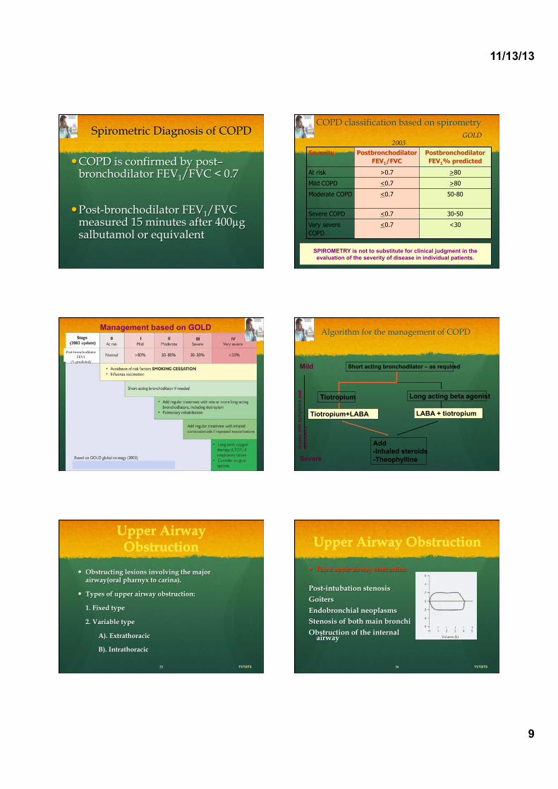
Spirometric Diagnosis of COPD
! COPD is confirmed by post–bronchodilator FEV1/FVC < 0.7
! Post-bronchodilator FEV1/FVC measured 15 minutes after 400µg salbutamol or equivalent
COPD classification based on spirometry GOLD
2003
SPIROMETRY is not to substitute for clinical judgment in the evaluation of the severity of disease in individual patients.
Severity Postbronchodilator FEV1/FVC
Postbronchodilator FEV1% predicted
At risk >0.7 >80
Mild COPD <0.7 >80
Moderate COPD <0.7 50-80
Severe COPD <0.7 30-50
Very severe COPD
<0.7 <30
Post-bronchodilator FEV1
(% predicted)
Management based on GOLD Algorithm for the management of COPD
Short acting bronchodilator – as required
Tiotropium
Tiotropium+LABA
Long acting beta agonist
LABA + tiotropium
Add -Inhaled steroids -Theophylline
Mild
Severe
asse
ss w
ith s
ympt
oms
and
spiro
met
ry
Upper Airway Obstruction
! Obstructing lesions involving the major airway(oral pharnyx to carina).
! Types of upper airway obstruction:
1. Fixed type
2. Variable type
A). Extrathoracic
B). Intrathoracic
11/13/13
Upper Airway Obstruction
! Fixed upper airway obstruction
Post-intubation stenosis
Goiters
Endobronchial neoplasms Stenosis of both main bronchi
Obstruction of the internal airway
11/13/13
11/13/13
10
Upper Airway Obstruction! Variable extrathoracic upper airway obstruction
11/13/13
Upper Airway Obstruction! Variable intrathoracic upper airway obstruction
11/13/13
Upper Airway Obstruction
Extrathoracic Intrathoracic
Bilateral vocal cord Obstruction of lower
paralysis trachea
Unilateral vocal cord Obstruction of a main
paralysis bronchus
Adhesions of vocal cord
Vocal cord constriction
Obstructive sleep apnea
Burns of nasopharynx
11/13/13
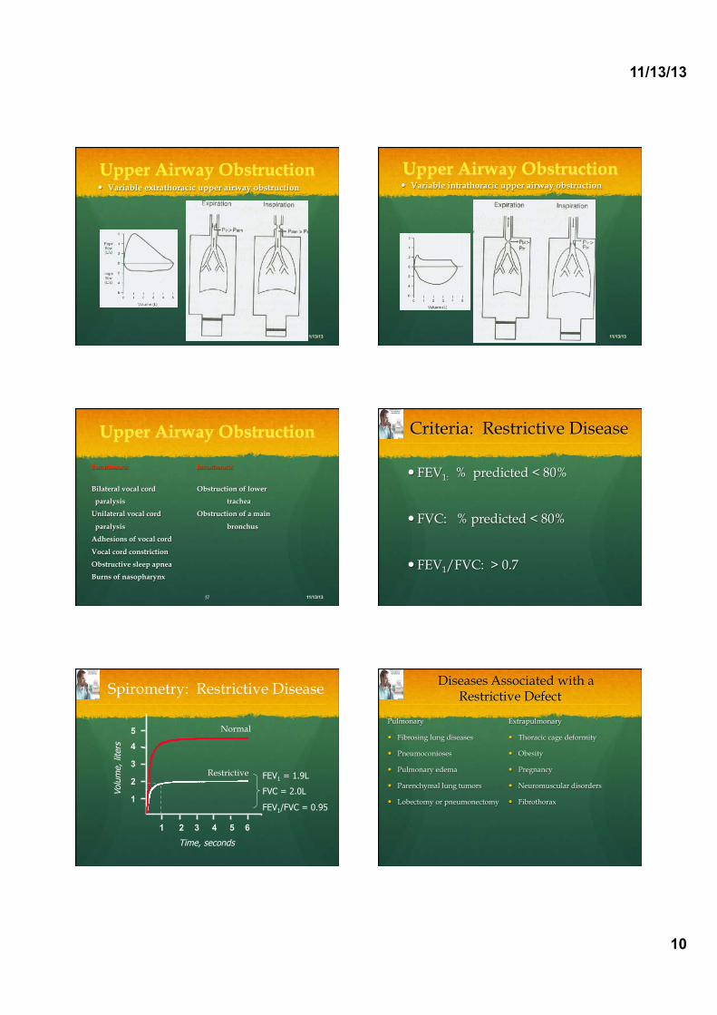
Criteria: Restrictive Disease
! FEV1: % predicted < 80%
! FVC: % predicted < 80%
! FEV1/FVC: > 0.7
Volu
me,
lite
rs
Time, seconds
FEV1 = 1.9L
FVC = 2.0L
FEV1/FVC = 0.95
1 2 3 4 5 6
5
4
3
2
1
Spirometry: Restrictive Disease
Normal
Restrictive
Diseases Associated with a Restrictive Defect
Pulmonary
! Fibrosing lung diseases
! Pneumoconioses
! Pulmonary edema
! Parenchymal lung tumors
! Lobectomy or pneumonectomy
Extrapulmonary
! Thoracic cage deformity
! Obesity
! Pregnancy
! Neuromuscular disorders
! Fibrothorax

11/13/13
11
Mixed Obstructive/Restrictive
! FEV1: % predicted < 80%
! FVC: % predicted < 80%
! FEV1 /FVC: < 0.7
Mixed Obstructive and Restrictive
Volu
me,
lite
rs
Time, seconds Restrictive and mixed obstructive-restrictive are difficult to diagnose by
spirometry alone; full respiratory function tests are usually required (e.g., body plethysmography, etc)
FEV1 = 0.5L
FVC = 1.5L
FEV1/FVC = 0.30
Normal
Obstructive - Restrictive
Flow Volume Curve Patterns Obstructive and Restrictive
Obstructive Severe obstructive Restrictive
Volume (L) Ex
pira
tory
flo
w ra
te
Expi
rato
ry f
low
rate
Exp
irat
ory
flow
rat
e Volume (L) Volume (L) Steeple pattern,
reduced peak flow, rapid fall off
Normal shape, normal peak flow, reduced volume
Reduced peak flow, scooped out mid-
curve
Obstructive Restrictive Mixed
Time Time
Time
Vo
lum
e
Vo
lum
e
Volu
me
Spirometry: Abnormal Patterns
Slow rise, reduced volume expired;
prolonged time to full expiration
Fast rise to plateau at reduced
maximum volume
Slow rise to reduced maximum volume; measure static lung
volumes and full PFT’s to confirm

11/13/13
12
BRONCHIAL PROVOCATION TESTS
Exposure of the airways to a stimulus – allergen – exercise – pharmacological bronchoconstrictive agent
Response of the smooth muscle ? – baseline FEV1 – post-exposure FEV1
% Airway hyperresponsiveness
Lung CapacityMeasuring methods of FRC:
! Nitrogen washout
! Inert gas( Helium ) dilution
! Body plethysmography
11/13/13
Functional Residual Capacity(FRC) Nitrogen Washout Method
! Duration of test: 7 minutes.
! Test duration of obstructive disease: 15-20 minutes.
11/13/13
Functional Residual Capacity(FRC)Inert Gas Dilution Method
! Helium(He) Argon(Ar) or Neon(Ne).
! O2 is added to prevent hypoxia and CO2 is absorbed to prevent hypercarbia.
11/13/13
Functional Residual Capacity(FRC)
Body Plethysmography
! The Boyle’s law, P1V1=P2V2.
! Test duration of COPD patients: 10-20 minutes.
11/13/13
Body Plethysmography
11/13/13
11/13/13
13
11/13/13
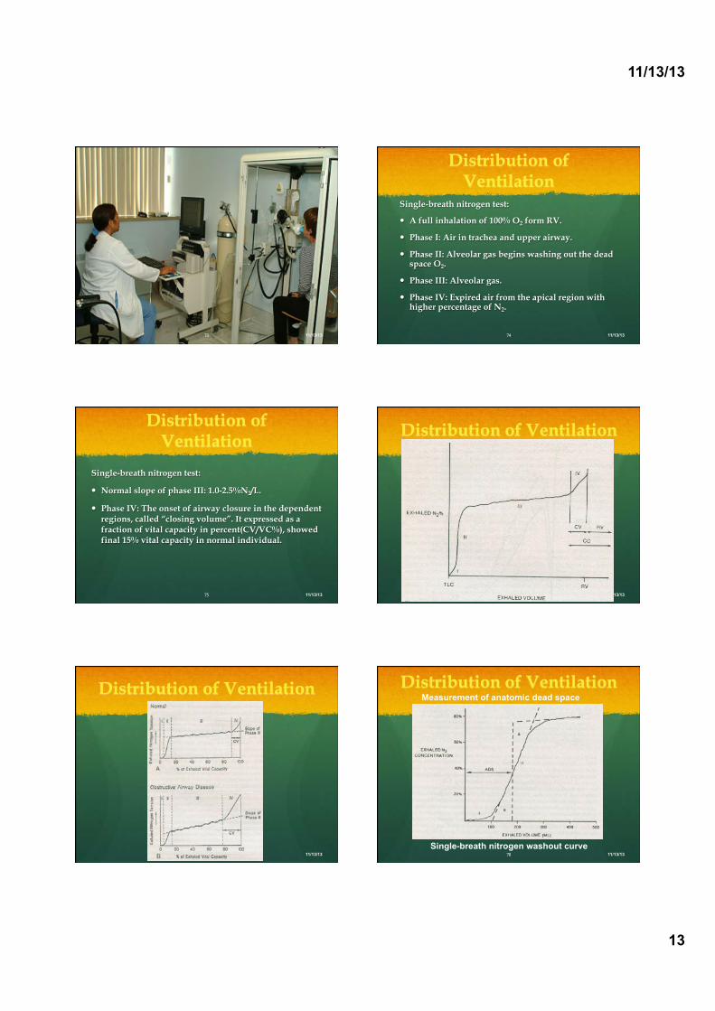
Distribution of Ventilation
Single-breath nitrogen test:
! A full inhalation of 100% O2 form RV.
! Phase I: Air in trachea and upper airway.
! Phase II: Alveolar gas begins washing out the dead space O2.
! Phase III: Alveolar gas.
! Phase IV: Expired air from the apical region with higher percentage of N2.
11/13/13
Distribution of Ventilation
Single-breath nitrogen test:
! Normal slope of phase III: 1.0-2.5%N2/L.
! Phase IV: The onset of airway closure in the dependent regions, called “closing volume”. It expressed as a fraction of vital capacity in percent(CV/VC%), showed final 15% vital capacity in normal individual.
11/13/13
Distribution of Ventilation
11/13/13
Distribution of Ventilation
11/13/13
Distribution of Ventilation
11/13/13
Measurement of anatomic dead space
Single-breath nitrogen washout curve

11/13/13
14
Gas exchange in the lungs
11/13/13
Diffusing Capacity
11/13/13
Diffusing Capacity
11/13/13
Time course of O2 transfer by diffusion into pulmonary capillary blood
Diffusing Capacity
! A single-breath(SB) method(SBDLCO).
! A He-CO-O2 mixed gas(0.3% CO, 10% He, 20% O2, 69.7% N2).
! A 10 seconds breath-holding, and should be a minimum of 5 seconds.
! The vital capacity should exceed 1.5 L for results to be acceptable.
11/13/13
Diffusing Capacity
11/13/13
Pulmonary Function Test
Interpretation of gas transfer
DLCO(Diffusing capacity) Severity
Normal 81-140%
Mild 61-80%
Moderate 41-60%
Severe <41% 11/13/13

11/13/13
15
Diffusing Capacity ! It estimates the patient’s ability to absorb alveolar
gases.
! Reduced DLCO:
Disorders of the pulmonary parenchyma, vascular abnormalities, reductions in effective alveolar units, and anemia.
! Elevated DLCO:
Left-to-right intracardiac shunt, polycythemia, and post-exercise physiology.
11/13/13
Diffusing Capacity
11/13/13
Causes of a decreased diffusing capacity
Thanks for your attention
11/13/13
The END PRACTICAL SESSION
Performing Spirometry

11/13/13
16
Spirometry Training ! Training is essential for operators to learn correct
performance and interpretation of results
! Training for competent performance of spirometry requires a minimum of 3 hours
! Acquiring good spirometry performance and interpretation skills requires practice, evaluation, and review
! Spirometry performance (who, when and where) should be adapted to local needs and resources
! Training for spirometry should be evaluated
Obtaining Predicted Values
Independent of the type of spirometer Choose values that best represent the
tested population Check for appropriateness if built into
the spirometer
Optimally, subjects should rest 10 minutes before performing spirometry
Withholding Medications
Before performing spirometry, withhold: & Short acting 2-agonists for 6 hours
& Long acting 2-agonists for 12 hours
& Ipratropium for 6 hours
& Tiotropium for 24 hours
Optimally, subjects should avoid caffeine and cigarette smoking for 30 minutes before
performing spirometry
Performing Spirometry - Preparation
1. Explain the purpose of the test and demonstrate the procedure
2. Record the patient’s age, height and gender and enter on the spirometer
3. Note when bronchodilator was last used
4. Have the patient sitting comfortably
5. Loosen any tight clothing
6. Empty the bladder beforehand if needed
Performing Spirometry
! Breath in until the lungs are full
! Hold the breath and seal the lips tightly around a clean mouthpiece
! Blast the air out as forcibly and fast as possible. Provide lots of encouragement!
! Continue blowing until the lungs feel empty
! Watch the patient during the blow to assure the lips are sealed around the mouthpiece
! Check to determine if an adequate trace has been achieved
! Repeat the procedure at least twice more until ideally 3 readings within 100 ml or 5% of each other are obtained
Performing Spirometry

11/13/13
17
Three times FVC within 5% or 0.1 litre (100 ml)
Reproducibility - Quality of Results
Volu
me,
lite
rs
Time, seconds
Spirometry - Possible Side Effects
! Feeling light-headed
! Headache
! Getting red in the face
! Fainting: reduced venous return or vasovagal attack (reflex)
! Transient urinary incontinence
Spirometry should be avoided after recent heart attack or stroke
Spirometry - Quality Control ! Most common cause of inconsistent readings is poor
patient technique
& Sub-optimal inspiration & Sub-maximal expiratory effort & Delay in forced expiration & Shortened expiratory time & Air leak around the mouthpiece
! Subjects must be observed and encouraged throughout the procedure
Spirometry – Common Problems & Inadequate or incomplete blow
& Lack of blast effort during exhalation
& Slow start to maximal effort
& Lips not sealed around mouthpiece
& Coughing during the blow
& Extra breath during the blow
& Glottic closure or obstruction of mouthpiece
by tongue or teeth
& Poor posture – leaning forwards
Equipment Maintenance ! Most spirometers need regular calibration to
check accuracy
! Calibration is normally performed with a 3 litre syringe
! Some electronic spirometers do not require daily/weekly calibration
! Good equipment cleanliness and anti-infection control are important; check instruction manual
! Spirometers should be regularly serviced; check manufacturer’s recommendations
Troubleshooting
Examples - Unacceptable Traces

11/13/13
18
Unacceptable Trace - Poor Effort
Volu
me,
lite
rs
Time, seconds
May be accompanied by a slow start
Inadequate sustaining of effort
Variable expiratory effort Normal
Volu
me,
lite
rs
Time, seconds
Unacceptable Trace – Stop Early
Normal
Volu
me,
lite
rs
Time, seconds
Unacceptable Trace – Slow Start
Normal
Volu
me,
lite
rs
Time, seconds
Unacceptable Trace - Coughing
Normal
Volu
me,
lite
rs
Time, seconds
Unacceptable Trace – Extra Breath
Normal