Embed Size (px)
Citation preview
Spondylolysis inAmerican FootballPlayers: Etiology,Symptoms, andImplications for Strengthand ConditioningSpecialistsJason P. Shurley, PhD, ATC, CSCS1 and Justin K. Newman, PT, DPT, ATC, CSCS2
1Department of Health, Physical Education, Recreation, and Coaching, The University of Wisconsin—Whitewater,Whitewater, Wisconsin; and 2AOK Medical Center, Physical Therapy Department, Houston, Texas
A B S T R A C T
SPONDYLOLYSIS IS A STRESS
FRACTURE, TYPICALLY OCCUR-
RING IN THE LUMBAR SPINE. IT IS
THE LEADING CAUSE OF BACK
PAIN IN ADOLESCENTS, WITH A
HIGHER INCIDENCE IN ATHLETES
THAN IN THE GENERAL POPULA-
TION. AMERICAN FOOTBALL
PLAYERS DEVELOP THE CONDI-
TION AT A HIGHER RATE THAN
MOST OTHER SPORTS, AND THE
CONDITION CAN CAUSE SEVERAL
MONTHS OF MISSED PLAYING
TIME. THIS INCREASED INCIDENCE
MAY BE DUE TO THE SPINE
LOADING INHERENT IN FOOTBALL,
BUT IS LIKELY EXACERBATED BY
OTHER FACTORS. THIS ARTICLE
DESCRIBES A SPONDYLOLYSIS,
DISCUSSES THE POTENTIAL
CAUSES, AND CONCLUDES WITH
A SERIES OF EXERCISES
INTENDED TO ADDRESS LIKELY
RISK FACTORS.
INTRODUCTION
Low-back pain (LBP) is a com-mon complaint among adoles-cent athletes, affecting up to
36% annually (65). In this populationone of the most common causes ofLBP is a spondylolysis, which is a frac-ture of the pars interarticularis; an areaon the posterior aspect of the vertebraebetween the facet joints (87). Indeed,a frequently cited study by Micheliand Wood (59) demonstrated that 47of 100 adolescent patients complainingof LBP had a spondylolysis. The pur-pose of this article is to describe a spon-dylolysis, discuss mechanisms of injuryand predisposing factors as they relateto American football players, andthen to detail specific activities whichmay help offset acquired anatomical
alignments likely to play a role in thedevelopment of this condition.
DESCRIPTION OFSPONDYLOLYSIS
Anatomical discussions of the spineoften subdivide it into anterior and pos-terior columns (31). The anterior columncomprises the vertebral body, interverte-bral disc, and associated anterior andposterior longitudinal ligaments. Theposterior column is the focus of this arti-cle, and includes the pedicles, lamina, spi-nous and transverse processes, and thefacet joints. The pedicles and lamina col-lectively form the neural arch. Betweenthe facet joints is a narrow region of theneural arch called the pars interarticularis.A common defect in this region, typicallydescribed as a stress fracture and depictedin Figure 1, is spondylolysis. The term is
Address correspondence to Jason P. Shurley,[email protected].
KEY WORDS :
pelvic stability; stress fracture; parsinterarticularis
VOLUME 38 | NUMBER 5 | OCTOBER 2016 Copyright � National Strength and Conditioning Association40
Copyright ª National Strength and Conditioning Association. Unauthorized reproduction of this article is prohibited.
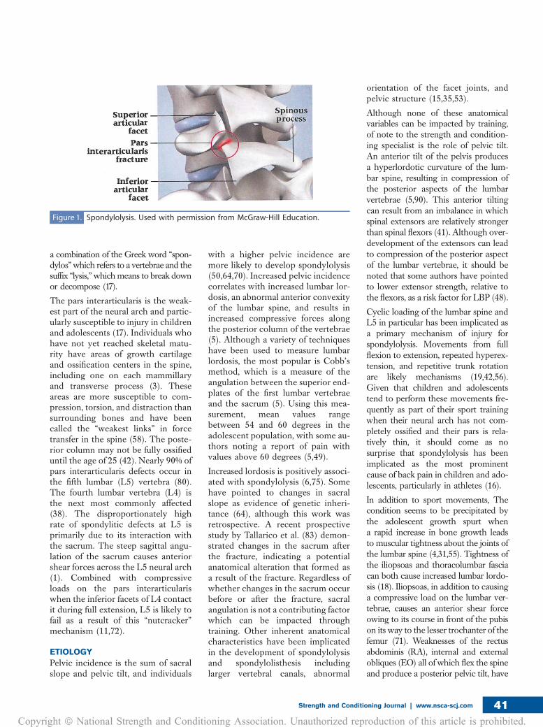
a combination of the Greek word “spon-dylos” which refers to a vertebrae and thesuffix “lysis,” whichmeans to break downor decompose (17).
The pars interarticularis is the weak-est part of the neural arch and partic-ularly susceptible to injury in childrenand adolescents (17). Individuals whohave not yet reached skeletal matu-rity have areas of growth cartilageand ossification centers in the spine,including one on each mammillaryand transverse process (3). Theseareas are more susceptible to com-pression, torsion, and distraction thansurrounding bones and have beencalled the “weakest links” in forcetransfer in the spine (58). The poste-rior column may not be fully ossifieduntil the age of 25 (42). Nearly 90% ofpars interarticularis defects occur inthe fifth lumbar (L5) vertebra (80).The fourth lumbar vertebra (L4) isthe next most commonly affected(38). The disproportionately highrate of spondylitic defects at L5 isprimarily due to its interaction withthe sacrum. The steep sagittal angu-lation of the sacrum causes anteriorshear forces across the L5 neural arch(1). Combined with compressiveloads on the pars interarticulariswhen the inferior facets of L4 contactit during full extension, L5 is likely tofail as a result of this “nutcracker”mechanism (11,72).
ETIOLOGY
Pelvic incidence is the sum of sacralslope and pelvic tilt, and individuals
with a higher pelvic incidence aremore likely to develop spondylolysis(50,64,70). Increased pelvic incidencecorrelates with increased lumbar lor-dosis, an abnormal anterior convexityof the lumbar spine, and results inincreased compressive forces alongthe posterior column of the vertebrae(5). Although a variety of techniqueshave been used to measure lumbarlordosis, the most popular is Cobb’smethod, which is a measure of theangulation between the superior end-plates of the first lumbar vertebraeand the sacrum (5). Using this mea-surement, mean values rangebetween 54 and 60 degrees in theadolescent population, with some au-thors noting a report of pain withvalues above 60 degrees (5,49).
Increased lordosis is positively associ-ated with spondylolysis (6,75). Somehave pointed to changes in sacralslope as evidence of genetic inheri-tance (64), although this work wasretrospective. A recent prospectivestudy by Tallarico et al. (83) demon-strated changes in the sacrum afterthe fracture, indicating a potentialanatomical alteration that formed asa result of the fracture. Regardless ofwhether changes in the sacrum occurbefore or after the fracture, sacralangulation is not a contributing factorwhich can be impacted throughtraining. Other inherent anatomicalcharacteristics have been implicatedin the development of spondylolysisand spondylolisthesis includinglarger vertebral canals, abnormal
orientation of the facet joints, andpelvic structure (15,35,53).
Although none of these anatomicalvariables can be impacted by training,of note to the strength and condition-ing specialist is the role of pelvic tilt.An anterior tilt of the pelvis producesa hyperlordotic curvature of the lum-bar spine, resulting in compression ofthe posterior aspects of the lumbarvertebrae (5,90). This anterior tiltingcan result from an imbalance in whichspinal extensors are relatively strongerthan spinal flexors (41). Although over-development of the extensors can leadto compression of the posterior aspectof the lumbar vertebrae, it should benoted that some authors have pointedto lower extensor strength, relative tothe flexors, as a risk factor for LBP (48).
Cyclic loading of the lumbar spine andL5 in particular has been implicated asa primary mechanism of injury forspondylolysis. Movements from fullflexion to extension, repeated hyperex-tension, and repetitive trunk rotationare likely mechanisms (19,42,56).Given that children and adolescentstend to perform these movements fre-quently as part of their sport trainingwhen their neural arch has not com-pletely ossified and their pars is rela-tively thin, it should come as nosurprise that spondylolysis has beenimplicated as the most prominentcause of back pain in children and ado-lescents, particularly in athletes (16).
In addition to sport movements, Thecondition seems to be precipitated bythe adolescent growth spurt whena rapid increase in bone growth leadsto muscular tightness about the joints ofthe lumbar spine (4,31,55). Tightness ofthe iliopsoas and thoracolumbar fasciacan both cause increased lumbar lordo-sis (18). Iliopsoas, in addition to causinga compressive load on the lumbar ver-tebrae, causes an anterior shear forceowing to its course in front of the pubison its way to the lesser trochanter of thefemur (71). Weaknesses of the rectusabdominis (RA), internal and externalobliques (EO) all of which flex the spineand produce a posterior pelvic tilt, have
Figure 1. Spondylolysis. Used with permission from McGraw-Hill Education.
Strength and Conditioning Journal | www.nsca-scj.com 41
Copyright ª National Strength and Conditioning Association. Unauthorized reproduction of this article is prohibited.
been suggested as contributors toincreased lordosis (71,85). This is morelikely in the presence of stiffness of therectus femoris or tensor fasciae latae,both of which originate on the pelvis,insert onto the tibia, and can produce ananterior pelvic tilt resulting in lumbarlordosis (71,84). Similarly, iliopsoascan cause an anterior pelvic tilt, andtightness of the hip flexors has beenassociated with LBP (19,45). Stiffnessor overdevelopment of the erector spi-nae group can also cause an anteriorpelvic tilt (41). It has also been postu-lated that the latissimus dorsi, throughits origin on the thoracolumbar fascia,can cause lumbar hyperextension andanterior pelvic tilt (71). This anteriortilting is resisted not only by the rectusabdominis and the obliques, but also bythe hamstrings (71). The finding of tighthamstrings in patients with spondyloly-sis may well be due to the chronicstretch produced by an anteriorly tiltedpelvis.
SPONDYLOLYSIS AND AMERICANFOOTBALL
Epidemiological studies have shownan incidence of spondylolysis in thegeneral adult population between6 and 11% (25,38). In high-level ath-letes the incidence is slightly higher,observed in up to 14% of individualsin this population (69). In Americanfootball players, the incidence may beeven higher with reports ranging from15–21% of all players and someevidence that as many as 50% of line-men have spondylolysis (24,54,76,82).Iwamoto et al. (34) found radiographicabnormalities in the spines of 63% ofhigh school and 60% of college footballplayers, with the presence of spondy-lolysis as the most important predictorfor back pain.
Football players have a higher likeli-hood of developing spondylolysisbased on both the nature of the sportand the training typically employedbefore competition. Football linemenoften start in a 3 or 4-point stance with1 or 2 hands on the ground and thelumbar spine in a flexed position. Atthe snap of the ball, they move intoa neutral or extended lumbar spine
and engage players from the opposingteam. At this point, there is an axialload on the spine with the athletelikely to be forced into hyperextension.Gatt et al. (26) noted that loadingof the lumbar spine in collegiate line-men hitting a blocking sled exceededloads previously demonstrated to causepathologic changes to both the parsinterarticularis and intervertebral disc.With college teams running 60–90plays per game, the volume of loading,along with the magnitude, creates anincreased risk of degenerative changesin the lumbar spine, particularly in line-men (27). Furthermore, it should benoted that football linemen also tendto be the largest players on the team,with one study on high school footballlinemen classifying 45% of them asbeing in the highest fifth percentile inage-specific body mass index (BMI)(46). This is significant because a highBMI has been correlated with increasedlumbar lordosis (62).
In addition to the mechanical loadingof the spine inherent to the game, mostfootball players engage in preseasonconditioning to prepare for their sport,and the most common activity used inthat preparation is weight training(86). Contemporary weight-trainingprograms for football typically includesome combination of weightlifting var-iants such as power cleans, push jerks,or snatch squats, and powerlifting ex-ercises such as squats and deadlifts.There is some evidence that weight-lifters are at an increased risk of spon-dylolysis, with one report showing anincidence of just more than 30% (44). Itshould be noted that this study useddata from before the elimination ofthe press which, when performed asa competitive lift, often involved signif-icant hyperextension of the spine.Nonetheless, a more recent study byYang et al. (89) found radiographic evi-dence of spondylolysis and spondylo-listhesis in nearly 29% of weightliftersthey examined. Additionally, Yanget al. found that weightlifters demon-strated increased lumbar lordosis rela-tive to a control group and speculatedthat the altered posture might result
from the role of the erector spinaegroup in resisting anterior shear forcesduring the lifts. Although not specificto spondylolysis, other works (13,68)have pointed to the low-back as themost frequently injured area in weight-lifters. In a study of junior high andhigh school athletes injured inweight-training programs, 67% of theinjuries were to the low back area (7).Similarly, a study of high schoolpowerlifters in Michigan demonstratedthat the low back was the most likely tosustain injuries, accounting for morethan 50% of injuries (10). A 2011 studyon elite powerlifters found that morethan 40% of participants surveyedcomplained of back injuries (77).
SPONDYLOLYSIS SIGNS,SYMPTOMS, AND OUTCOMES
Athletes with spondylolysis typicallypresent with LBP, described as a diffusedull ache, and no known specific mech-anism of injury (31). Resting and lyingdown typically decrease pain, whereasactivity tends to increase it. The painmay radiate into the buttocks and pos-terior thigh and is exacerbated by lum-bar hyperextension, particularly whencombined with a single-leg stance (31).Muscular spasm of the spinal erectors iscommonly noted, as is hamstring tight-ness (55). Neurologic symptoms, such astingling or burning pain along a derma-tome or lower extremity weakness areunusual. Spondylolysis is often bilateraland may predispose the athlete to thedevelopment of a spondylolisthesis,which is an anterior translation of thevertebrae relative to the next inferiorvertebral segment (58).
Once a spondylolysis has been diag-nosed, athletes typically respond wellto conservative treatment (60). Surgerymay be considered if pain does notresolve after 9–12 months of conserva-tive treatment or if there has been a slip-page (spondylolisthesis) of greater than50% of the superior vertebral body overthe inferior vertebrae (55). There issome controversy regarding the useof bracing with conservative treatment,with some authors advocating the useof a modified Boston brace to limitextension (57). The brace is worn 23
Spondylolysis in American Football
VOLUME 38 | NUMBER 5 | OCTOBER 201642
Copyright ª National Strength and Conditioning Association. Unauthorized reproduction of this article is prohibited.
hours per day for 3–4 months withweaning thereafter based on evidenceof bony healing (31,55). Some authorshave pointed out that the brace prob-ably acts more to restrict activity thanstabilize the spine and that bony heal-ing has been shown with or withouta brace (79). Whether or not bracingis used, rest is the primary componentof all protocols. Athletes who stoppedsport participation for at least 3 monthswere 16 times more likely to return totheir previous level of play with no painthan those who did not rest as long(21). After this rest period, physicaltherapy is performed for 2–4 monthswith athletes eligible to return to playonce they have demonstrated full pain-free range of motion (ROM), spinalawareness, appropriate sport-specificconditioning, and no pain while per-forming sport movements (79). In theauthors’ experience, the total return toplay time is typically 5–7 months.
In addition to missed time, there issome evidence that LBP and injuriesthat are due to overuse can recurin up to 26% of males (81). This makessense when one considers that LBPmaylead to the development of decreasedproprioception in the lumbar spineand altered lumbar muscle activationpatterns (28,29). For athletes with pros-pects of playing at the highest levels,a recent study by Schroeder et al. (74)suggests that football players with a lum-bar spine diagnosis, including spondylol-ysis or spondylolisthesis, were less likelyto be drafted and had shorter playingcareers in the National Football League(NFL) than matched controls. Brophyet al. (9) found that spondylolysisreduced the likelihood of running backsplaying in the NFL and a trend towardfewer receivers with the diagnosis play-ing in the league. It should be noted thatstudies on spondylolysis in collegiatefootball players have not found the con-dition to adversely affect their playingcareers (54,76).
POTENTIAL MITIGATION THROUGHSTRENGTH AND CONDITIONING
As previously discussed, spondyliticfractures are a multifactorial condition.From the perspective of the strength
and conditioning specialist, it shouldbe noted that lumbar hyperlordosis isa key contributing factor, which can becaused by an anterior tilt of the pelvis.The strength and conditioning special-ist may be able to play a role indecreasing the risk that a footballplayer develops a spondylolysis byincluding activities which help miti-gate anterior pelvic tilt. This could beaccomplished by incorporating a com-bination of core endurance work, my-ofascial release, and static stretching. Arecent study by Lee and McGill (47)demonstrated that isometric exerciseswere more effective at producing stiff-ness of the torso when compared withdynamic exercises over a 6-week train-ing period. When one considers thatspondylitic fractures are precipitatedby cyclic flexion, extension, and rota-tion of the spine, isometric exercisesseem to offer an additional benefit ofincreasing core endurance withoutadding spinal stresses at end-rangesof motion. Furthermore, Lee andMcGill argue that low loads on thespine experienced during isometricexercises allow performance of the ex-ercises almost daily. Given that there isa dose-response relationship betweenthe volume of training and muscularendurance, it stands to reason thata higher total volume of work may
confer additional protective bene-fits (67).
Myofascial release, in the form of foamrolling, has been demonstrated toacutely increase ROM in young adultsand resistance-trained adolescent ath-letes (12,51,52,61,78). Although theexact mechanism for this increase inROM is unknown, some have specu-lated that it might be due to a combi-nation of decreased viscosity of thefascia, increased blood flow to the mus-cle, and decreased adhesions betweenlayers of fascia (23,73). The increase inROM seems to be greater when foamrolling is combined with static stretch-ing (61,78). In addition to increasedROM, 2 studies have demonstratedpositive effects on vertical jump heightand muscular force (30,52), whereasanother showed a reduction in percep-tion of fatigue after foam rolling (32).Several studies failed to demonstrateenhancement of power, agility, orROM (32,36). Although static stretch-ing has been demonstrated to increaseROM, there is evidence that it de-creases expression of muscular strengthand power (8,66). Although a recentreview article concluded that the det-rimental effects of static stretching arelargely limited to longer-durationstretches of more than 60 seconds, itseems prudent to incorporate foamrolling into the pre-exercise routine
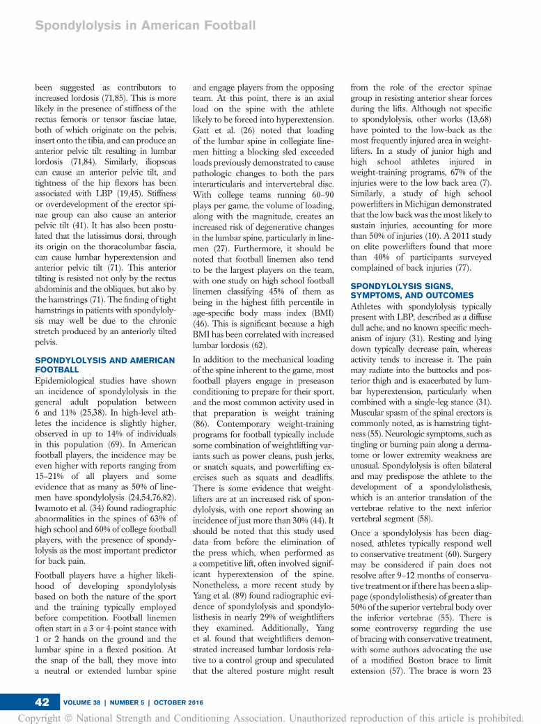
Figure 2. Foam rolling for the rectus femoris. Should be performed with the kneeboth flexed and extended.
Strength and Conditioning Journal | www.nsca-scj.com 43
Copyright ª National Strength and Conditioning Association. Unauthorized reproduction of this article is prohibited.
as it has been shown to increase ROMwithout deleterious effects on muscularstrength or power (23,40). Becausethere seems to be an additive effectbetween foam rolling and staticstretching at increasing ROM, bothmay be combined in the postexerciseroutine (61,78). Moreover, foam rollinghas been demonstrated to reduce mus-cle soreness and fatigue, making ita potentially useful recovery tool afterexercise (51,73).
FOAM ROLLING
Based on research on the utility offoam rolling at acutely increasingROM, the authors recommend foamrolling for 2–3 sets of 1 minute in dura-tion for each of the following muscles/groups.
Rectus femoris. The athlete lies ina prone position with the foam rollerin contact with the anterior thigh, sup-porting the body in push-up positionwith upper extremity. They are in-structed to perform passes of the foamroller with the knee both extended andflexed (Figure 2).
Tensor fasciae latae. Although still ina prone position with the foam rollerin contact with the superior-lateralanterior thigh and supporting the bodyin push-up position, the athlete per-forms passes with the hip both slightlyflexed and extended.
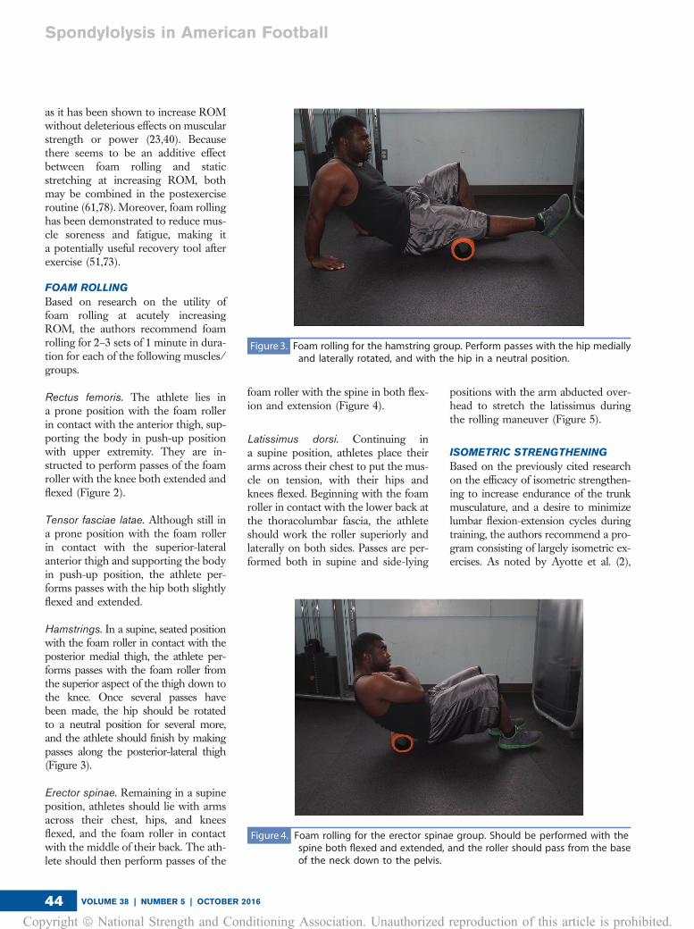
Hamstrings. In a supine, seated positionwith the foam roller in contact with theposterior medial thigh, the athlete per-forms passes with the foam roller fromthe superior aspect of the thigh down tothe knee. Once several passes havebeen made, the hip should be rotatedto a neutral position for several more,and the athlete should finish by makingpasses along the posterior-lateral thigh(Figure 3).
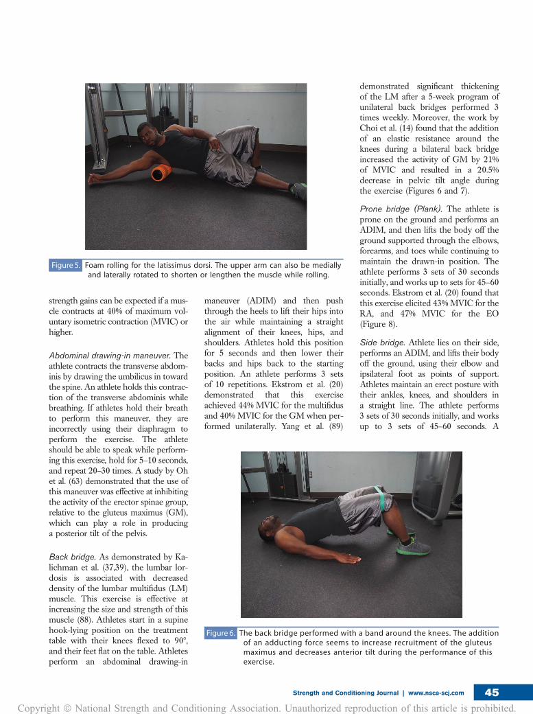
Erector spinae. Remaining in a supineposition, athletes should lie with armsacross their chest, hips, and kneesflexed, and the foam roller in contactwith the middle of their back. The ath-lete should then perform passes of the
foam roller with the spine in both flex-ion and extension (Figure 4).
Latissimus dorsi. Continuing ina supine position, athletes place theirarms across their chest to put the mus-cle on tension, with their hips andknees flexed. Beginning with the foamroller in contact with the lower back atthe thoracolumbar fascia, the athleteshould work the roller superiorly andlaterally on both sides. Passes are per-formed both in supine and side-lying
positions with the arm abducted over-head to stretch the latissimus duringthe rolling maneuver (Figure 5).
ISOMETRIC STRENGTHENING
Based on the previously cited researchon the efficacy of isometric strengthen-ing to increase endurance of the trunkmusculature, and a desire to minimizelumbar flexion-extension cycles duringtraining, the authors recommend a pro-gram consisting of largely isometric ex-ercises. As noted by Ayotte et al. (2),
Figure 3. Foam rolling for the hamstring group. Perform passes with the hip mediallyand laterally rotated, and with the hip in a neutral position.
Figure 4. Foam rolling for the erector spinae group. Should be performed with thespine both flexed and extended, and the roller should pass from the baseof the neck down to the pelvis.
Spondylolysis in American Football
VOLUME 38 | NUMBER 5 | OCTOBER 201644
Copyright ª National Strength and Conditioning Association. Unauthorized reproduction of this article is prohibited.
strength gains can be expected if a mus-cle contracts at 40% of maximum vol-untary isometric contraction (MVIC) orhigher.
Abdominal drawing-in maneuver. Theathlete contracts the transverse abdom-inis by drawing the umbilicus in towardthe spine. An athlete holds this contrac-tion of the transverse abdominis whilebreathing. If athletes hold their breathto perform this maneuver, they areincorrectly using their diaphragm toperform the exercise. The athleteshould be able to speak while perform-ing this exercise, hold for 5–10 seconds,and repeat 20–30 times. A study by Ohet al. (63) demonstrated that the use ofthis maneuver was effective at inhibitingthe activity of the erector spinae group,relative to the gluteus maximus (GM),which can play a role in producinga posterior tilt of the pelvis.
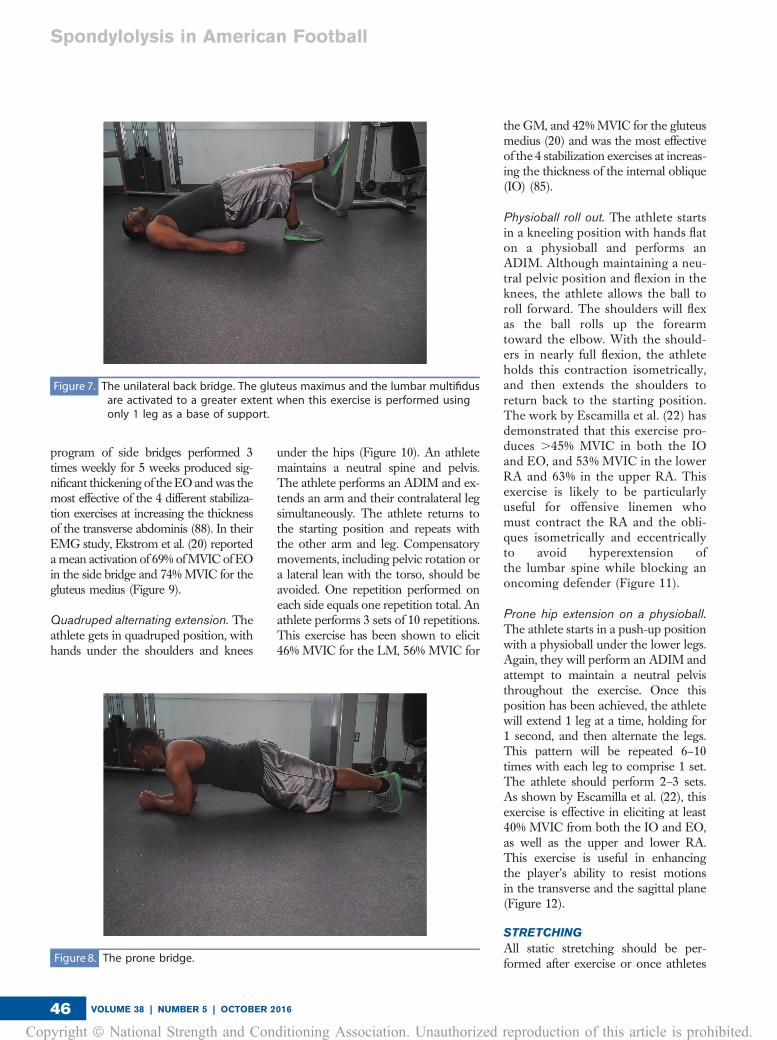
Back bridge. As demonstrated by Ka-lichman et al. (37,39), the lumbar lor-dosis is associated with decreaseddensity of the lumbar multifidus (LM)muscle. This exercise is effective atincreasing the size and strength of thismuscle (88). Athletes start in a supinehook-lying position on the treatmenttable with their knees flexed to 908,and their feet flat on the table. Athletesperform an abdominal drawing-in
maneuver (ADIM) and then pushthrough the heels to lift their hips intothe air while maintaining a straightalignment of their knees, hips, andshoulders. Athletes hold this positionfor 5 seconds and then lower theirbacks and hips back to the startingposition. An athlete performs 3 setsof 10 repetitions. Ekstrom et al. (20)demonstrated that this exerciseachieved 44% MVIC for the multifidusand 40% MVIC for the GM when per-formed unilaterally. Yang et al. (89)
demonstrated significant thickeningof the LM after a 5-week program ofunilateral back bridges performed 3times weekly. Moreover, the work byChoi et al. (14) found that the additionof an elastic resistance around theknees during a bilateral back bridgeincreased the activity of GM by 21%of MVIC and resulted in a 20.5%decrease in pelvic tilt angle duringthe exercise (Figures 6 and 7).
Prone bridge (Plank). The athlete isprone on the ground and performs anADIM, and then lifts the body off theground supported through the elbows,forearms, and toes while continuing tomaintain the drawn-in position. Theathlete performs 3 sets of 30 secondsinitially, and works up to sets for 45–60seconds. Ekstrom et al. (20) found thatthis exercise elicited 43% MVIC for theRA, and 47% MVIC for the EO(Figure 8).
Side bridge. Athlete lies on their side,performs an ADIM, and lifts their bodyoff the ground, using their elbow andipsilateral foot as points of support.Athletes maintain an erect posture withtheir ankles, knees, and shoulders ina straight line. The athlete performs3 sets of 30 seconds initially, and worksup to 3 sets of 45–60 seconds. A
Figure 5. Foam rolling for the latissimus dorsi. The upper arm can also be mediallyand laterally rotated to shorten or lengthen the muscle while rolling.
Figure 6. The back bridge performed with a band around the knees. The additionof an adducting force seems to increase recruitment of the gluteusmaximus and decreases anterior tilt during the performance of thisexercise.
Strength and Conditioning Journal | www.nsca-scj.com 45
Copyright ª National Strength and Conditioning Association. Unauthorized reproduction of this article is prohibited.
program of side bridges performed 3times weekly for 5 weeks produced sig-nificant thickening of the EO andwas themost effective of the 4 different stabiliza-tion exercises at increasing the thicknessof the transverse abdominis (88). In theirEMG study, Ekstrom et al. (20) reportedamean activation of 69% ofMVIC of EOin the side bridge and 74%MVIC for thegluteus medius (Figure 9).
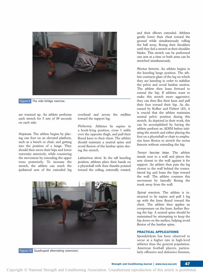
Quadruped alternating extension. Theathlete gets in quadruped position, withhands under the shoulders and knees
under the hips (Figure 10). An athletemaintains a neutral spine and pelvis.The athlete performs an ADIM and ex-tends an arm and their contralateral legsimultaneously. The athlete returns tothe starting position and repeats withthe other arm and leg. Compensatorymovements, including pelvic rotation ora lateral lean with the torso, should beavoided. One repetition performed oneach side equals one repetition total. Anathlete performs 3 sets of 10 repetitions.This exercise has been shown to elicit46% MVIC for the LM, 56% MVIC for
the GM, and 42%MVIC for the gluteusmedius (20) and was the most effectiveof the 4 stabilization exercises at increas-ing the thickness of the internal oblique(IO) (85).
Physioball roll out. The athlete startsin a kneeling position with hands flaton a physioball and performs anADIM. Although maintaining a neu-tral pelvic position and flexion in theknees, the athlete allows the ball toroll forward. The shoulders will flexas the ball rolls up the forearmtoward the elbow. With the should-ers in nearly full flexion, the athleteholds this contraction isometrically,and then extends the shoulders toreturn back to the starting position.The work by Escamilla et al. (22) hasdemonstrated that this exercise pro-duces .45% MVIC in both the IOand EO, and 53% MVIC in the lowerRA and 63% in the upper RA. Thisexercise is likely to be particularlyuseful for offensive linemen whomust contract the RA and the obli-ques isometrically and eccentricallyto avoid hyperextension ofthe lumbar spine while blocking anoncoming defender (Figure 11).
Prone hip extension on a physioball.The athlete starts in a push-up positionwith a physioball under the lower legs.Again, they will perform an ADIM andattempt to maintain a neutral pelvisthroughout the exercise. Once thisposition has been achieved, the athletewill extend 1 leg at a time, holding for1 second, and then alternate the legs.This pattern will be repeated 6–10times with each leg to comprise 1 set.The athlete should perform 2–3 sets.As shown by Escamilla et al. (22), thisexercise is effective in eliciting at least40% MVIC from both the IO and EO,as well as the upper and lower RA.This exercise is useful in enhancingthe player’s ability to resist motionsin the transverse and the sagittal plane(Figure 12).
STRETCHING
All static stretching should be per-formed after exercise or once athletes
Figure 7. The unilateral back bridge. The gluteus maximus and the lumbar multifidusare activated to a greater extent when this exercise is performed usingonly 1 leg as a base of support.
Figure 8. The prone bridge.
Spondylolysis in American Football
VOLUME 38 | NUMBER 5 | OCTOBER 201646
Copyright ª National Strength and Conditioning Association. Unauthorized reproduction of this article is prohibited.
are warmed up. An athlete performseach stretch for 3 sets of 30 secondson each side.
Iliopsoas. The athlete begins by plac-ing one foot on an elevated platform,such as a bench or chair, and gettinginto the position of a lunge. Theyshould then move their hips and lowerextremity anteriorly, while counteringthe movement by extending the uppertorso posteriorly. To increase thestretch, the athlete can reach theipsilateral arm of the extended leg
overhead and across the midlinetoward the support leg.
Piriformis. Athletes lie supine ina hook-lying position, cross 1 ankleover the opposite thigh, and pull theirflexed knee to their chest. The athleteshould maintain a neutral spine andavoid flexion of the lumbar spine dur-ing the stretch.
Latissimus dorsi. In the tall kneelingposition, athletes place their hands ona physioball with their thumbs pointedtoward the ceiling, externally rotated,
and their elbows extended. Athletesgently lower their chest toward theground while simultaneously rollingthe ball away, flexing their shouldersuntil they feel a stretch at their shoulderblades. This stretch can be performedone arm at a time or both arms can bestretched simultaneously.
Rectus femoris. An athlete begins inthe kneeling lunge position. The ath-lete contracts glute of the leg on whichthey are kneeling in order to stabilizethe pelvis and avoid lumbar motion.The athlete then leans forward toextend the hip. If athletes want tomake this stretch more aggressive,they can then flex their knee and pulltheir foot toward their hip. As dis-cussed by Kolber and Fiebert (43), itis crucial that the athlete maintainsneutral pelvic position during thisstretch. As depicted in their work, thiscan be accomplished by having theathlete perform an ADIM before initi-ating the stretch and either placing thefoot on a physioball or chair to empha-size knee flexion to stretch the rectusfemoris without extending the hip.
Tensor fasciae latae. The athletestands next to a wall and places thearm closest to the wall against it forsupport. An athlete then puts the legclosest to the wall behind the contra-lateral leg and leans the hips towardthe wall. The athlete counters thismovement by laterally flexing thetrunk away from the wall.
Spinal erectors. The athlete is in-structed to lie supine and pull 1 legup with the knee flexed toward thechest. The athlete then applies anoverpressure on the knee, further flex-ing the hip. A neutral spine should bemaintained by attempting to keep thehip down on the surface, helping avoidflexion of the lumbar spine.
PRACTICAL APPLICATIONS
Spondylolysis has been observed tooccur at a higher rate in high-levelathletes than the general population.American football players, particu-larly offensive and defensive linemen,
Figure 9. The side bridge exercise.
Figure 10. Quadruped alternating extension.
Strength and Conditioning Journal | www.nsca-scj.com 47
Copyright ª National Strength and Conditioning Association. Unauthorized reproduction of this article is prohibited.

seem to have an even higher inci-dence of the condition than otherathletes. In addition to pain and dis-ability, the condition is likely to causemissed time for athletes and mayreoccur. Although inherent charac-teristics of the athlete, such as sacralslope and vertebral configurationplay important roles in the develop-ment of spondylolysis, hyperlordosisof the lumbar spine has also beencorrelated with the condition. Byincorporating a combination of foamrolling, pelvic stabilization exercises,and static stretching, the strengthand conditioning professional may
be able to play a role in decreasinganterior tilt of the pelvis andhyperlordosis of the lumbar spine.As a result, athletes should havea decreased risk of developingspondylolysis. Although the timecommitment of this program is notinsubstantial, it is worth noting thata program consisting of pelvic stabi-lization exercises performed by Aus-tralian Rules football players resultedin increased player availability anda significant perception of benefitamong players (33). Keeping playerson the field is a primary goal of thestrength and conditioning specialist
and will lead to better outcomes forthe players, the team, and the coach.
Conflicts of Interest and Source of Funding:The authors report no conflicts of interestand no source of funding.
Jason P.
Shurley is anAssistant Profes-sor in theDepartment ofHealth, PhysicalEducation, Rec-reation, andCoaching, at the
University of Wisconsin—Whitewater.
Justin Newman
is the Director ofPhysical Ther-apy, AOK Med-ical Center.
REFERENCES1. Alexander MJ. Biomechanical aspects of
lumbar injuries in athletes: A review. Can J
Appl Sport Sci 10: 1–19, 1985.
2. Ayotte NW, Stetts DM, Keenan G, and
Greenway EH. Electromyographical analysis
of selected lower extremity muscles during
Figure 11. The physioball roll out. This exercise builds upon the prone bridge and requires maintenance of a neutral pelvic position whilethe upper extremities are in motion. A is the beginning and ending position of the movement. B is the position of isometriccontraction of RA, EO, and IO. The athlete should stop rolling the ball forward once a neutral pelvis can no longer be maintained.
Figure 12. Prone hip extension on a physioball. Another exercise building on the pronebridge. Successful performance requires minimizing lumbar rotation andisolated hip extension.
Spondylolysis in American Football
VOLUME 38 | NUMBER 5 | OCTOBER 201648
Copyright ª National Strength and Conditioning Association. Unauthorized reproduction of this article is prohibited.

five unilateral weight-bearing exercises.
J Orthop Sport Phys 37: 48–55, 2007.
3. Baker BJ, Dupras TL, and Tocheri MW. The
Osteology of Infants and Children. Vol. 81.
College Station, TX: Texas A&M University
Press, 2005.
4. Baranto A, Hellstrom M, Cederlund CG,
Nyman R, and Sward L. Back pain and MRI
changes in the thoraco-lumbar spine of top
athletes in four different sports: A 15-year
follow-up study. Knee Surg Sports
Traumatol Arthrosc 17: 1125–1134, 2009.
5. Been E and Kalichman L. Lumbar lordosis.
Spine J 14: 87–97, 2014.
6. Been E, Li L, Hunter DJ, and Kalichman L.
Geometry of the vertebral bodies and the
intervertebral discs in lumbar segments
adjacent to spondylolysis and
spondylolisthesis: A pilot study. Eur Spine J
20: 1159–1165, 2011.
7. Brady TA, Cahill BR, and Bodnar LM.
Weight training-related injuries in the high
school athlete. Am J Sport Med 10: 1–5,
1982.
8. Brandenburg JP. Duration of stretch does
not influence the degree of force loss
following static stretching. J Sport Med
Phys Fit 46: 526–534, 2006.
9. Brophy RH, Lyman S, Chehab EL,
Barnes RP, Rodeo SA, and Warren RF.
Predictive value of prior injury on career in
professional American football is affected
by player position. Am J Sport Med 37:
768–775, 2009.
10. Brown EW and Kimball RG. Medical
history associated with adolescent
powerlifting. Pediatrics 72: 636–644,
1983.
11. Bugg WH, Lewis M, Juette A, Cahir JG,
and Toms AP. Lumbar lordosis and pars
interarticularis fractures. Skeletal Radiol
41: 817–822, 2012.
12. Bushell JE, Dawson SM, andWebster MM.
Clinical relevance of foam rolling hip hip
extension angle in a functional lunge
position. J Strength Cond Res 29: 2397–
2403, 2015.
13. Calhoon G and Fry AC. Injury rates and
profiles of elite competitive weightlifters.
J Athl Train 34: 232–238, 1999.
14. Choi SA, Cynn HS, Yi CH, Kwon OY,
Yoon TL, Choi WJ, and Lee JH. Isometric
hip abduction using a thera-band alters
gluteus maximus muscle activity and the
anterior pelvic tilt angle during bridging
exercise. J Electromyogr Kinesiol 25: 310–
315, 2015.
15. Cinotti G, Postacchini F, Fassari F, and
Urso S. Predisposing factors in
degenerative spondylolisthesis. Int Orthop
21: 337–342, 1997.
16. Cohen E and Stuecker RD. Magnetic
resonance imaging in diagnosis and
follow-up of impending spondylolysis in
children and adolescents. J Pediatr Orthop
B 14: 63–67, 2005.
17. Congeni J. Evaluating spondylolysis in
adolescent athletes. J Musculoskelet Med
17: 123–129, 2000.
18. d’Hemecourt PA, Gerbino PG, and
Micheli LJ. Back injuries in the young athlete.
Clin Sport Med 19: 663–679, 2000.
19. Donatelli R, Dimond D, and Holland M.
Sport-specific biomechanics of spinal
injuries in the athlete. Clin Sport Med 31:
381–396, 2012.
20. Ekstrom RA, Donatelli RA, and Carp KC.
Electromyographic analysis of core trunk,
hip, and thigh muscles during 9
rehabilitation exercises. J Orthop Sport
Phys 37: 754–762, 2007.
21. El Rassi G, Takemitsu M, Glutting J, and
Shah SA. Effect of sports modification on
clinical outcome in children and adolescent
athletes with symptomatic lumbar
spondylolysis. Am J Phys Med Rehab 92:
1070–1074, 2013.
22. Escamilla R, Lewis C, Bell D, Bramblet G,
Daffron J, Lambert S, Pecson A,
Imamura R, Paulos L, and Andrews J. Core
muscle activation during Swiss ball and
traditional abdominal exercises. J Orthop
Sport Phys 40: 265–276, 2010.
23. Feldbauer CM, Smith BA, and Van
Lunen B. The effects of self-myofascial
release on flexibility of the lower extremity.
Int J Athl Ther Train 20: 14–19, 2015.
24. Ferguson RJ, McMaster JH, and
Stanitski CL. Low back pain in college
football linemen. Am J Sport Med 2: 63–
69, 1974.
25. Fredrickson BE, Baker D, McHolick WJ,
Yuan HA, and Lubicky JP. The natural
history of spondylolysis and
spondylolisthesis. J Bone Joint Surg Am
66: 699–707, 1984.
26. Gatt CJ, Hosea TM, Palumbo RC, and
Zawadsky JP. Impact loading of the lumbar
spine during football blocking. Am J Sport
Med 25: 317–321, 1997.
27. Gerbino PG and d’Hemecourt PA. Does
football cause an increase in degenerative
disease of the lumbar spine? Curr Sport
Med Rep 1: 47–51, 2002.
28. Gill KP and Callaghan MJ. The
measurement of lumbar proprioception in
individuals with and without low back pain.
Spine 23: 371–377, 1998.
29. Grabiner MD, Koh TJ, and Al Ghazawi A.
Decoupling of bilateral paraspinal
excitation in subjects with low back pain.
Spine 17: 1219–1223, 1992.
30. Halperin I, Aboodarda SJ, Button DC,
Andersen LL, and Behm DG. Roller
massager improves range of motion of
plantar flexor muscles without subsequent
decreases in force parameters. Int J Sport
Phys Ther 9: 92–102, 2014.
31. Haus BM and Micheli LJ. Back pain in the
pediatric and adolescent athlete. Clin
Sport Med 31: 423–440, 2012.
32. Healey KC, Hatfield DL, Blanpied P,
Dorfman LR, and Riebe D. The effects of
myofascial release with foam rolling on
performance. J Strength Cond Res 28:
61–68, 2014.
33. Hides JA, Stanton W, Mendis MD, and
Gildea J. Effect of stabilisation training on
trunk muscle size, motor control, low back
pain and player availability among elite
Australian Rules football players. Br J
Sports Med 45: 320, 2011.
34. Iwamoto J, Abe H, Tsukimura Y, and
Wakano K. Relationship between
radiographic abnormalities of lumbar spine
and incidence of low back pain in high
school and college football players. Am J
Sport Med 32: 781–786, 2004.
35. Jackson RP, Phipps T, Hales C, and Surber J.
Pelvic lordosis and alignment in
spondylolisthesis.Spine 28: 151–160, 2003.
36. Jay K, Sundstrup E, Sondergaard SD,
Behm D, Brandt M, Saervoll CA,
Jakobsen MD, and Andersen LL. Specific
and cross over effects of massage for
muscle soreness: Randomized control trial.
Int J Sport Phys Ther 9: 82–91, 2014.
37. Kalichman L, Hodges P, Li L, Guermazi A,
and Hunter DJ. Changes in paraspinal
muscles and their association with low
back pain and spinal degeneration: CT
study. Eur Spine J 19: 1136–1144, 2010.
38. Kalichman L, Kim DH, Li L, Guermazi A,
Berkin V, and Hunter DJ. Spondylolysis and
spondylolisthesis: Prevalence and
association with low back pain in the adult
community-based population. Spine 34:
199–205, 2009.
39. Kalichman L, Li L, Hunter DJ, and Been E.
Association between computed
tomography-evaluated lumbar lordosis and
features of spinal degeneration, evaluated
in the supine position. Spine J 11: 308–
315, 2011.
40. Kay AD and Blazevich AJ. Effect of acute
static stretch on maximal muscle
performance: A systematic review.Med Sci
Sport Exer 44: 154–164, 2012.
Strength and Conditioning Journal | www.nsca-scj.com 49
Copyright ª National Strength and Conditioning Association. Unauthorized reproduction of this article is prohibited.
41. Kim HJ, Chung S, Kim S, Shin H, Lee J,
Kim S, and Song MY. Influences of
trunk muscles on lumbar lordosis and
sacral angle. Eur Spine J 15: 409–414,
2006.
42. Kim HJ and Green DW. Spondylolysis in
the adolescent athlete. Curr Opin Pediatr
23: 68–72, 2011.
43. Kolber MJ and Fiebert IM. Addressing
flexibility of the rectus femoris in the athlete
with low back pain. Strength Cond J 27:
66–73, 2005.
44. Kotani PT, Ichikawa N, Wakabayashi W,
Yoshii T, and Koshimune M. Studies of
spondylolysis found among weight lifters.
Br J Sport Med 6: 4–8, 1971.
45. Kujala U, Salminen J, Taimela S,
Oksanen A, and Jaakkola L. Subject
characteristics and low back pain in young
athletes and nonathletes. Med Sci Sport
Exer 24: 627–632, 1992.
46. Laurson KR and Eisenmann JC. The
prevalence of overweight among high
school football linemen. J Am Med Assoc
297: 363–364, 2007.
47. Lee BC and McGill SM. Effect of long-term
isometric training on core/torso stiffness.
J Strength Cond Res 29: 1515–1526,
2015.
48. Lee JH, Hoshino Y, Nakamura K, Kariya Y,
Saita K, and Ito K. Trunk muscle weakness
as a risk factor for low back pain. Spine 24:
54–57, 1999.
49. Lee JH and Yoo WG. Application of
posterior pelvic tilt taping for the treatment
of chronic low back pain with sacroiliac
dysfunction and increased sacral horizontal
angle. Phys Ther Sport 13: 279–285,
2012.
50. Legaye J, Duval-Beaupere G, Hecquet J,
and Marty C. Pelvic incidence: The
fundamental pelvic parameter for three-
dimensional regulation of spinal sagittal
curves. Eur Spine J 7: 99–103, 1998.
51. MacDonald GZ, Button DC, Drinkwater EJ,
and Behm DG. Foam rolling as a recovery
tool after an intense bout of physical
activity. Med Sci Sport Exer 46: 131–142,
2014.
52. MacDonald GZ, Penney MD, Mullaley ME,
Cuconato AL, Drake CD, Behm DG, and
Button DC. An acute bout of self-
myofascial release increases range of
motion without a subsequent decrease in
muscle activation or force. J Strength Cond
Res 27: 812–821, 2013.
53. Masharawi Y. Lumbar shape
characterization of the neural arch and
vertebral body in spondylolysis. Clin Anat
25: 224–230, 2012.
54. McCarroll JR, Miller JM, and Ritter MA.
Lumbar spondylolysis and
spondylolisthesis in college football
players. Am J Sport Med 14: 404–406,
1986.
55. McCleary MD and Congeni JA. Current
concepts in the diagnosis and treatment of
spondylolysis in young athletes. Curr Sport
Med Rep 6: 62–66, 2007.
56. McGill SM. Low Back Disorders:
Evidence-Based Prevention and
Rehabilitation. Vol. 51. Champaign, IL:
Human Kinetics, 2002. pp. 217.
57. McNeely ML, Torrance G, and Magee DJ. A
systemic review of physiotherapy for
spondylolysis and spondylolisthesis. Man
Ther 8: 80–91, 2003.
58. Micheli L and Curtis C. Stress fractures in
the spine and sacrum. Clin Sport Med 25:
75–88, 2005.
59. Micheli L and Wood R. Back pain in young
athletes. Arch Pediatr Adolesc Med 149:
15–18, 1995.
60. Miller SF, Congeni JA, and Swanson K.
Long-term functional and anatomical
follow-up of early detected spondylolysis in
young athletes. Am J Sport Med 32: 928–
933, 2004.
61. Mohr AR, Long BC, and Goad CL. Effect of
foam rolling and static stretching on
passive hip-flexion range of motion. J Sport
Rehabil 23: 296–299, 2014.
62. Murrie VL, Dixon AK, Hollingworth W,
Wilson H, and Doyle TA. Lumbar lordosis:
A study of patients with and without low
back pain. Clin Anat 16: 144–147, 2003.
63. Oh JS, Cynn HS, Won JH, Kwon OY, and
Yi CH. Effects of performing an abdominal
drawing-in maneuver during prone hip
extension exercises on hip and back
extensor muscle activity and amount of
anterior pelvic tilt. J Orthop Sports Phys
Ther 37: 320–324, 2007.
64. Oh YM, Choi HA, and Eun JP. The
comparison of sagittal spinopelvic
parameters between young adult patients
with L5 spondylolysis and age-matched
control group. J Korean Neurosurg Soc 54:
207–210, 2013.
65. Olsen TL, Anderson RL, Dearwater SR,
Kriska AM, Cauley JA, Aaron DJ, and
LaPorte RE. The epidemiology of low back
pain an in adolescent population. Am J
Public Health 82: 606–608, 1992.
66. Pinto MD, Wilhelm EN, Tricoli V,
Pinto RS, and Blazevich AJ. Differential
effects of 30- vs. 60-second static
muscle stretching on vertical jump
performance. J Strength Cond Res 28:
3440–3446, 2014.
67. Radaelli R, Fleck SJ, Leite T, Leite RD,
Pinto RS, Fernandes L, and Simao R.
Dose-response of 1, 3, and 5 sets of
resistance exercise on strength, local
muscular endurance, and hypertrophy.
J Strength Cond Res 29: 1349–1366,
2015.
68. Raske A and Norlin R. Injury incidence
among elite weight and power lifters. Am J
Sport Med 30: 248–256, 2002.
69. Rossi F and Dragoni S. The prevalence of
spondylolysis and spondylolisthesis in
symptomatic elite athletes: Radiographic
findings. Radiography 7: 37–42, 2001.
70. Roussouly P, Gollogly S, Bertonnaud E,
Labelle H, and Weidenbaum M. Sagittal
alignment of the spine and pelvis in the
presence of L5-S1 isthmic lysis and low-
grade spondylolisthesis. Spine 31: 2484–
2490, 2006.
71. Sahrmann S. Diagnosis and Treatment of
Movement Impairment Syndromes. Vol.
31. St Louis, MO: Mosby, 2002. pp. 63–
68, 136.
72. Sakai T, Sairyo K, Suzue N, Kosaka H, and
Yasui N. Incidence and etiology of lumbar
spondylolysis. J Orthop Sci 15: 281–288,
2010.
73. Schroeder AN and Best TM. Is self
myofascial release an effective preexercise
and recovery strategy? Curr Sport Med
Rep 14: 200–208, 2015.
74. Schroeder GD, Lynch TS, Gibbs DB,
LaBelle M, Patel AA, Savage JW, Hsu WK,
and Nuber GW. Pre-existing lumbar spine
diagnosis as a predictor of outcomes in
National Football League athletes. Am J
Sport Med 43: 972–978, 2015.
75. Schuller S, Charles YP, and Steib JP.
Sagittal spinopelvic alignment and body
mass index in patients with degenerative
spondylolisthesis. Eur Spine J 20: 713–
719, 2011.
76. Semon RL and Spengler D. Significance of
lumbar spondylolysis in college football
players. Spine 6: 172, 1981.
77. Siewe J, Rudat J, Rollinghoff M,
Schlegel UH, Eysel P, and Michael JW.
Injuries and overuse syndromes in
powerlifting. Int J Sport Med 32: 703–711,
2011.
78. Skarabot J, Beardsley C, and Stirn I.
Comparing the effects of self-myofascial
release with static stretching on ankle
range-of-motion in adolescent athletes. Int
J Sport Phys Ther 10: 203–212, 2015.
79. Standaert CJ and Herring SA. Expert
opinion and controversies in sports and
musculoskeletal medicine: The diagnosis
and treatment of spondylolysis in
Spondylolysis in American Football
VOLUME 38 | NUMBER 5 | OCTOBER 201650
Copyright ª National Strength and Conditioning Association. Unauthorized reproduction of this article is prohibited.
adolescent athletes. Arch Phys Med
Rehab 88: 537–540, 2007.
80. Sutton JH, Guin PD, and Theiss SM. Acute
lumbar spondylolysis in intercollegiate
athletes. J Spinal Disord Tech 25: 422–
425, 2012.
81. Taimela S, Kujala UM, Salinen JJ, and
Viljanen T. The prevalence of low back pain
among children and adolescents: A
nationwide cohort-based questionnaire
survey in Finland. Spine 22: 1132–1136,
1997.
82. Tall RL and DeVault W. Spinal injury in
sport: Epidemiologic considerations. Clin
Sport Med 12: 441–446, 1993.
83. Tallarico RA, Fredrickson BE,
Whitesides TE, and Lavelle WF. The
association of sacral table angle
measurements with spondylolytic and
spondylolisthetic defects at the
lumbosacral articulation: A radiographic
analysis. Spine Def 3: 372–397, 2015.
84. Tateuchi H, Taniguchi M, Mori N, and
Ichihashi N. Balance of hip and trunk muscle
activity is associated with increased anterior
pelvic tilt during prone hip extension.
J Electromyogr Kinesiol 22: 391–397, 2012.
85. Waryasz GR. Exercise strategies to
prevent the development of the anterior
pelvic tilt: Implications for possible
prevention of sports hernias and osteitis
pubis. Strength Cond J 32: 56–65, 2010.
86. Wilkerson GB, Giles JL, and Seibel DK.
Prediction of core and lower extremity
strains and sprains in collegiate football
players. J Athl Train 47: 264–272, 2012.
87. Wiltse LL, Widell EH, and Jackson DW.
Fatigue fracture: The basic lesion in isthmic
spondylolisthesis. J Bone Joint Surg Am
57: 17–22, 1975.
88. Yang HS, Lee YS, and Jin SA. Effect of
evidence-based trunk stability exercises on
the thickness of trunk muscles. J Phys Ther
Sci 27: 473–475, 2015.
89. Yang JH, Barani R, Bhandarkar AW,
Suh SW, Hong JY, and Modi HN.
Changes in spinopelvic parameters of
elite weightlifters. Clin J Sport Med 24:
343–350, 2014.
90. Youdas JW, Garrett TR, Egan KS, and
Therneau TM. Lumbar lordosis and
pelvic inclination in adults with chronic
low back pain. Phys Ther 80: 261–275,
2000.
Strength and Conditioning Journal | www.nsca-scj.com 51
Copyright ª National Strength and Conditioning Association. Unauthorized reproduction of this article is prohibited.