Embed Size (px)
Citation preview

Korean Journal of UrologyⒸ The Korean Urological Association, 2011 865 Korean J Urol 2011;52:865-867
www.kjurology.orghttp://dx.doi.org/10.4111/kju.2011.52.12.865
Case Report
Spontaneously Ruptured Renal Cell Carcinoma During Hemodialysis in Two Patients with End-Stage Renal Disease Woong Bin Kim, Eui Sang Lee, Seung Whan Doo, Won Jae Yang, Yun Seob Song, Hyunjin Noh1
Departments of Urology, 1Nephrology, Soonchunhyang University College of Medicine, Seoul, Korea
Spontaneously ruptured renal cell carcinoma (RCC) in end-stage kidney disease is very rare. Preoperative diagnosis is difficult because of the relatively small tumor size, asso-ciated hematoma, and surrounding acquired cysts. Two middle-aged men who were maintained on hemodialysis (HD) for over 10 years suddenly developed flank pain dur-ing HD. Computed tomography scans revealed an enhancing ruptured renal mass in one patient, and no obvious tumor lesion except for a hematoma in the other, both of which were later confirmed as RCCs by pathologic specimens.
Key Words: Renal cell carcinoma; Rupture; Spontaneous
This is an Open Access article distributed under the terms of the Creative Commons Attribution Non-Commercial License (http://creativecommons.org/licenses/by-nc/3.0) which permits unrestricted non-commercial use, distribution, and reproduction in any medium, provided the original work is properly cited.
Article History:received 10 June, 2011accepted 2 August, 2011
Corresponding Author:Won Jae YangDepartment of Urology, College of Medicine, Soonchunhyang University College of Medicine, 59, Daesagwan- ro, Yongsan-gu, Seoul 140-743, KoreaTEL: +82-2-709-9376FAX: +82-2-709-9378 E-mail: [email protected]
FIG. 1. Case 1. Enhanced Computed tomography scan revealed a 3.2x2.5 cm sized ill-defined enhanced mass in the lower pole of the left kidney (arrow), which had acute extravasation of contrast material.
Spontaneously ruptured renal cell carcinoma (RCC) is a well-known entity; however, such occurrences in patients with end-stage kidney disease are very rare [1]. To our knowledge, only 5 cases have been reported in the liter-ature, all of which were associated with acquired cystic dis-ease of the kidney (ACDK) [2]. Preoperative diagnosis is difficult because of the relatively small tumor size, asso-ciated hematoma, and surrounding acquired cysts. Herein, we report two cases of spontaneously ruptured RCC in pa-tients with end-stage kidney disease, with a review of the current medical literature. In the first case, a ruptured en-hancing tumor could be identified on the preoperative com-puted tomography (CT) scan that was not associated with ACDK. In the second case, only a hematoma with ACDK changes was detected by CT scan, but the patient was later confirmed to have RCC on microscopic examination of the pathologic specimen.
CASE REPORTS
1. Case 1A 47-year-old male with end-stage renal disease (ESRD) who was maintained on regular hemodialysis (HD) for over 15 years presented with acute left flank pain during HD. His vital signs were stable, and a physical exam showed left costovertebral angle tenderness. Laboratory evaluation revealed a hemoglobin value of 7.3 g/dl, which was de-
creased by 2 points compared with his baseline value. No anti-coagulatory agents were included in his outpatient medications. An enhanced abdominal CT scan revealed a 3.2x2.5 cm sized ill-defined lobulated mass in the lower pole of the left kidney, which had acute extravasation of con-
Korean J Urol 2011;52:865-867
866 Kim et al
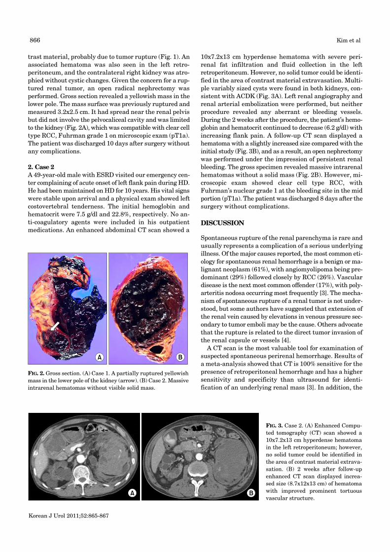
FIG. 2. Gross section. (A) Case 1. A partially ruptured yellowish mass in the lower pole of the kidney (arrow). (B) Case 2. Massive intrarenal hematomas without visible solid mass.
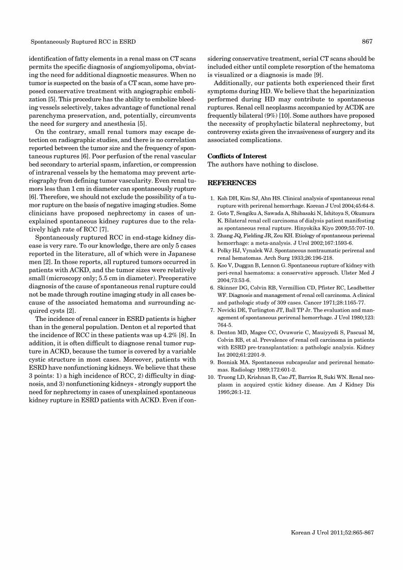
FIG. 3. Case 2. (A) Enhanced Compu-ted tomography (CT) scan showed a 10x7.2x13 cm hyperdense hematoma in the left retroperitoneum; however, no solid tumor could be identified in the area of contrast material extrava-sation. (B) 2 weeks after follow-up enhanced CT scan displayed increa-sed size (8.7x12x13 cm) of hematoma with improved prominent tortuous vascular structure.
trast material, probably due to tumor rupture (Fig. 1). An associated hematoma was also seen in the left retro-peritoneum, and the contralateral right kidney was atro-phied without cystic changes. Given the concern for a rup-tured renal tumor, an open radical nephrectomy was performed. Gross section revealed a yellowish mass in the lower pole. The mass surface was previously ruptured and measured 3.2x2.5 cm. It had spread near the renal pelvis but did not involve the pelvocaliceal cavity and was limited to the kidney (Fig. 2A), which was compatible with clear cell type RCC, Fuhrman grade 1 on microscopic exam (pT1a). The patient was discharged 10 days after surgery without any complications.
2. Case 2 A 49-year-old male with ESRD visited our emergency cen-ter complaining of acute onset of left flank pain during HD. He had been maintained on HD for 10 years. His vital signs were stable upon arrival and a physical exam showed left costovertebral tenderness. The initial hemoglobin and hematocrit were 7.5 g/dl and 22.8%, respectively. No an-ti-coagulatory agents were included in his outpatient medications. An enhanced abdominal CT scan showed a
10x7.2x13 cm hyperdense hematoma with severe peri-renal fat infiltration and fluid collection in the left retroperitoneum. However, no solid tumor could be identi-fied in the area of contrast material extravasation. Multi-ple variably sized cysts were found in both kidneys, con-sistent with ACDK (Fig. 3A). Left renal angiography and renal arterial embolization were performed, but neither procedure revealed any aberrant or bleeding vessels. During the 2 weeks after the procedure, the patient’s hemo-globin and hematocrit continued to decrease (6.2 g/dl) with increasing flank pain. A follow-up CT scan displayed a hematoma with a slightly increased size compared with the initial study (Fig. 3B), and as a result, an open nephrectomy was performed under the impression of persistent renal bleeding. The gross specimen revealed massive intrarenal hematomas without a solid mass (Fig. 2B). However, mi-croscopic exam showed clear cell type RCC, with Fuhrman’s nuclear grade 1 at the bleeding site in the mid portion (pT1a). The patient was discharged 8 days after the surgery without complications.
DISCUSSION
Spontaneous rupture of the renal parenchyma is rare and usually represents a complication of a serious underlying illness. Of the major causes reported, the most common eti-ology for spontaneous renal hemorrhage is a benign or ma-lignant neoplasm (61%), with angiomyolipoma being pre-dominant (29%) followed closely by RCC (26%). Vascular disease is the next most common offender (17%), with poly-arteritis nodosa occurring most frequently [3]. The mecha-nism of spontaneous rupture of a renal tumor is not under-stood, but some authors have suggested that extension of the renal vein caused by elevations in venous pressure sec-ondary to tumor emboli may be the cause. Others advocate that the rupture is related to the direct tumor invasion of the renal capsule or vessels [4].
A CT scan is the most valuable tool for examination of suspected spontaneous perirenal hemorrhage. Results of a meta-analysis showed that CT is 100% sensitive for the presence of retroperitoneal hemorrhage and has a higher sensitivity and specificity than ultrasound for identi-fication of an underlying renal mass [3]. In addition, the
Korean J Urol 2011;52:865-867
Spontaneously Ruptured RCC in ESRD 867
identification of fatty elements in a renal mass on CT scans permits the specific diagnosis of angiomyolipoma, obviat-ing the need for additional diagnostic measures. When no tumor is suspected on the basis of a CT scan, some have pro-posed conservative treatment with angiographic emboli-zation [5]. This procedure has the ability to embolize bleed-ing vessels selectively, takes advantage of functional renal parenchyma preservation, and, potentially, circumvents the need for surgery and anesthesia [5].
On the contrary, small renal tumors may escape de-tection on radiographic studies, and there is no correlation reported between the tumor size and the frequency of spon-taneous ruptures [6]. Poor perfusion of the renal vascular bed secondary to arterial spasm, infarction, or compression of intrarenal vessels by the hematoma may prevent arte-riography from defining tumor vascularity. Even renal tu-mors less than 1 cm in diameter can spontaneously rupture [6]. Therefore, we should not exclude the possibility of a tu-mor rupture on the basis of negative imaging studies. Some clinicians have proposed nephrectomy in cases of un-explained spontaneous kidney ruptures due to the rela-tively high rate of RCC [7].
Spontaneously ruptured RCC in end-stage kidney dis-ease is very rare. To our knowledge, there are only 5 cases reported in the literature, all of which were in Japanese men [2]. In those reports, all ruptured tumors occurred in patients with ACKD, and the tumor sizes were relatively small (microscopy only; 5.5 cm in diameter). Preoperative diagnosis of the cause of spontaneous renal rupture could not be made through routine imaging study in all cases be-cause of the associated hematoma and surrounding ac-quired cysts [2].
The incidence of renal cancer in ESRD patients is higher than in the general population. Denton et al reported that the incidence of RCC in these patients was up 4.2% [8]. In addition, it is often difficult to diagnose renal tumor rup-ture in ACKD, because the tumor is covered by a variable cystic structure in most cases. Moreover, patients with ESRD have nonfunctioning kidneys. We believe that these 3 points: 1) a high incidence of RCC, 2) difficulty in diag-nosis, and 3) nonfunctioning kidneys - strongly support the need for nephrectomy in cases of unexplained spontaneous kidney rupture in ESRD patients with ACKD. Even if con-
sidering conservative treatment, serial CT scans should be included either until complete resorption of the hematoma is visualized or a diagnosis is made [9].
Additionally, our patients both experienced their first symptoms during HD. We believe that the heparinization performed during HD may contribute to spontaneous ruptures. Renal cell neoplasms accompanied by ACDK are frequently bilateral (9%) [10]. Some authors have proposed the necessity of prophylactic bilateral nephrectomy, but controversy exists given the invasiveness of surgery and its associated complications.
Conflicts of InterestThe authors have nothing to disclose.
REFERENCES
1. Koh DH, Kim SJ, Ahn HS. Clinical analysis of spontaneous renal rupture with perirenal hemorrhage. Korean J Urol 2004;45:64-8.
2. Goto T, Sengiku A, Sawada A, Shibasaki N, Ishitoya S, Okumura K. Bilateral renal cell carcinoma of dialysis patient manifesting as spontaneous renal rupture. Hinyokika Kiyo 2009;55:707-10.
3. Zhang JQ, Fielding JR, Zou KH. Etiology of spontaneous perirenal hemorrhage: a meta-analysis. J Urol 2002;167:1593-6.
4. Polky HJ, Vynalek WJ. Spontaneous nontraumatic perirenal and renal hematomas. Arch Surg 1933;26:196-218.
5. Koo V, Duggan B, Lennon G. Spontaneous rupture of kidney with peri-renal haematoma: a conservative approach. Ulster Med J 2004;73:53-6.
6. Skinner DG, Colvin RB, Vermillion CD, Pfister RC, Leadbetter WF. Diagnosis and management of renal cell carcinoma. A clinical and pathologic study of 309 cases. Cancer 1971;28:1165-77.
7. Novicki DE, Turlington JT, Ball TP Jr. The evaluation and man-agement of spontaneous perirenal hemorrhage. J Urol 1980;123: 764-5.
8. Denton MD, Magee CC, Ovuworie C, Mauiyyedi S, Pascual M, Colvin RB, et al. Prevalence of renal cell carcinoma in patients with ESRD pre-transplantation: a pathologic analysis. Kidney Int 2002;61:2201-9.
9. Bosniak MA. Spontaneous subcapsular and perirenal hemato-mas. Radiology 1989;172:601-2.
10. Truong LD, Krishnan B, Cao JT, Barrios R, Suki WN. Renal neo-plasm in acquired cystic kidney disease. Am J Kidney Dis 1995;26:1-12.