Embed Size (px)
Citation preview

J. Neurol. Neurosurg. Psychiat., 1971, 34, 82-85
Posterior fossa subdural haematoma associated withanticoagulant therapy
T. CAPISTRANT,1 R. GOLDBERG, H. SHIBASAKI, AND D. CASTLE
From the Department of Neurology, St. Paul-Ramsey Hospital, St. Paul, Minnesota, U.S.A.
SUMMARY This is a report of spontaneous posterior fossa subdural haematoma associated withanticoagulation therapy. The possibility of posterior fossa lesions related to spontaneous haemor-rhage is suggested by the combination of severe headache and increasing disturbance of conscious-ness associated with signs of brain-stem decompensation. A thorough neurological evaluationincluding appropriate contrast studies will help rule out a supratentorial lesion. This is a neurologicalemergency which can be successfully treated by early detection and prompt surgical decompression.This is the second reported case of spontaneous subdural haematoma of the posterior fossa occurringduring anticoagulant therapy.
The physician who prescribes anticoagulant drugsstrives for a point of balance in bleeding and clottingmechanisms. A significant decrease in the efficiencyof these mechanisms must be achieved if therapy isto serve its purpose, but this must not proceed tothe point of inducing haemorrhages.
This paper is concerned with a patient whodeveloped a spontaneous subdural haematoma whilereceiving anticoagulant therapy. We are reportingthis case because the lesion occurred in the posteriorfossa, which is an uncommon site of subduralhaematoma (Ciarla, 1913; Munro, 1934; McKissock,Richardson, and Bloom, 1960; Ciembroniewicz,1965; Wright, 1966).A previous case of posterior fossasubdural haematoma complicating anticoagulanttherapy was reported by Zenteno-Alanis, Corvera,and Mateos (1968). Diagnosis was difficult andevolved from a combination of clinical observationsand emergency contrast studies. Ultimately diagnosiswas confirmed by neurosurgical intervention.
CASE HISTORY
A 50 year old Mexican male laundry worker was admittedto St. Paul-Ramsey Hospital on 24 July 1969. He com-plained of sudden chest pain and blood-tinged sputum.The patient had noted swelling in the left leg and anklethe day before admission. He contracted and wastreated for tuberculosis at 36 years of age. There was nohistory of head injury or alcoholism. The family andsocial histories were non-contributory. Physical exam-ination was normal except for tenderness and swelling of
'Reprint requests to above address.
82
the left ankle and stasis skin changes in the lower left leg.There were no neurological abnormalities. Laboratoryexamination was within normal limits except for in-creased serum GOT to 230 (normal 10 to 40). Radiographof the chest and electrocardiogram were within normallimits. A diagnosis of thrombophlebitis of the left legand probable pulmonary embolism was made.
Anticoagulant treatment was started on 25 July withintravenous administration of 6,000 u. heparin every fourhours. On 28 July prothrombin time was 25-9 seconds inthe patient and 11-8 seconds in the control. On 29 July,the dosage of 6,000 u. was decreased to 5,000 u. heparinevery four hours. Coumadin was initiated with 15 mg on2 August and 10 mg on 3 August.On the morning of 2 August, the patient complained
of a severe, bitemporal, throbbing headache. He vomitedonce that afternoon. A complete neurological examinationat that time was normal. The headache persisted. On3 August, he seemed to fall asleep after lunch but washard to arouse. He became progressively unresponsiveand developed a high fever. At 2 a.m. on 4 August, thepatient responded only to deep pain with slight jerkingmovements of both feet. The pulse rate was 76 perminute and regular; blood pressure was 150/56 mmHg;and the body temperature was 103° F (39 4'C). Breathingwas normal. The optic discs appeared normal, butvenous pulsations were absent. The left pupil was 3 mmand the right 2 mm in diameter, and both were non-reactive to light stimulation. The eyes were slightlydivergent, and oculocephalic and ciliospinal reflexes wereabsent. Corneal reflexes were present. Muscle tone on theright was slightly reduced. There were no involuntarymovements. Deep tendon reflexes and abdominal reflexeswere absent. There w-ere marked extensor toe signsbilaterally. Complete blood count, blood urea nitrogen,blood glucose, and serum electrolytes were within normallimits. Prothrombin time was 14 8 seconds with a control
group.bmj.com on March 26, 2018 - Published by http://jnnp.bmj.com/Downloaded from

Posterior fossa subdural haematoma associated with anticoagulant therapy
of 11 5 seconds; partial thromboplastin time was 38seconds with a control of 31 seconds.At this time a subarachnoid or intracerebral haemor-
rhage was suspected. Radiographs of the skull were
normal. An echoencephalogram indicated no shift ofmidline structures. Heparin and coumadin were dis-continued. The patient was given Decadron, 2 mg
intravenously, followed by 4 mg intramuscularly.At 8 a.m. on 4 August, the patient responded only with
decerebrate posturing on supraorbital compression.Corneal and caloric responses were now absent. The leftpupil continued to be slightly larger, and there was a milddecrease of tone on the right side. A left carotid angio-gram (Fig. 1) was performed at 10 a.m. The lateral viewshowed moderate unrolling of the pericallosal arteriessuggesting ventricular enlargement. There was no focalabnormality and no midline shift of the anterior cerebralvessels or the vein of Galen. The tentative diagnoses were
intraventricular haemorrhage or haemorrhagic mass
lesion in the posterior fossa. Air ventriculography was
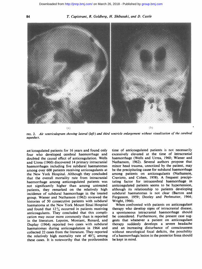
performed through bilateral occipital burr holes. Theventricular fluid was clear and colourless, ruling outintraventricular haemorrhage. As shown in Figs. 2a and2b, there was moderate enlargement of the lateralventricles and the third ventricle. The cerebral aqueductand the fourth ventricle were not visualized.An occipital craniotomy was performed and revealed
that the dura mater was bulging posteriorly and had a
brownish discolouration. Approximately 15 ml. ofsemiclotted blood was removed from the subdural space.The right cerebellar hemisphere was compressed forward,but there was no intracerebellar haemorrhage. The surfaceof the cerebellar hemispheres appeared normal and withadequate circulation. The dura mater was suturedprimarily.
FIG. 1. Left percutaneous carotid angiogram showingunrolling of the pericallosal arteries suggesting ventricularenlargement.
Three days after surgery, the patient opened his eyes andmoved his hands and legs to verbal commands. A right-sided extensor toe sign was present. One week aftersurgery the patient was alert and his speech was appropri-ate. At this time he had a fever, and pseudomonasaeruginosa was cultured from the sputum. Pseudomonaspneumonia was the first of a series of complicationswhich eventually resulted in the patient's death. Thepulmonary infection was followed by pseudomonasmeningitis. The patient was treated with intrathecalpolymyxin B and parenteral colymycin. Activation ofpulmonary tuberculosis secondary to the general debili-tation and high dosages of steroids was suspected. Therewas no change in neurological status during this time,except for somnolence associated with periods of highfever. On 2 September 1969, the 29th postoperative day,the patient suddenly expired. Permission for necropsycould not be obtained.
COMMENT
A 50 year old man developed headache and pro-gressive comaone week afterbeing given intravenousheparin for thrombophlebitis and probable pulmon-ary embolism. Although the neurological exam-ination revealed some lateralization with anisocoriaand diminished tone in the right extremities, neitherechoencephalography nor left carotid angiographyshowed any evidence of a supratentorial mass lesion.Enlarged lateral ventricles were suggested by theangiographic appearance of the pericallosal arteries.
This could be secondary to (1) intraventricularhaemorrhage, or (2) compression of the brain-stemby a posterior fossa mass. The first possibility wasruled out by the clear appearance of the cerebro-spinal fluid obtained during air ventriculography. Aposterior fossa subdural haematoma was confirmedby occipital craniotomy. The patient initially made agood recovery, but a series of infectious andmetabolic complications resulted in his death on the29th postoperative day.The diagnosis of a haemorrhagic lesion in the
posterior fossa is often difficult The paucity ofneurological localizing signs and the relative lack ofspecific diagnostic assistance provided by EEG,radiographs of the skull, echoencephalography,brain scan, and carotid angiography all contributeto the problem. A most important asset in makingthe diagnosis of a posterior fossa haemorrhageappears to be simply keeping the possibility of itsexistence in mind. The rapidity with which un-consciousness deepens necessitates swift action toavoid death or irreparable brain dysfunction.Although an increased haemorrhagic tendency
during anticoagulation is a well-known phenom-enon, there still exists a controversy about theassociation of anticoagulation and central nervoussystem haemorrhage. Coogan (1965) followed 142
83
group.bmj.com on March 26, 2018 - Published by http://jnnp.bmj.com/Downloaded from
T. Capistrant, R. Goldberg, H. Shibasaki, and D. Castle
FIG. 2. Air ventriculogram showing lateral (left) and third ventricle enlargement without visualization of the cerebralaqueduct.
an'icoagulated patients for 16 years and found onlyfour who developed cerebral haemorrhage anddoubted the causal effect of anticoagulation. Wellsand Urrea (1960) discovered 14 primary intracranialhaemorrhages including five subdural haematomasamong over 600 patients receiving anticoagulants atthe New York Hospital. Although they concludedthat the overall mortality rate from intracranialhaemorrhage among anticoagulated patients was
not significantly higher than among untreatedpatients, they remarked on the relatively highincidence of subdural haemorrhage in the treatedgroup. Wiener and Nathanson (1962) reviewed thehistories of 50 consecutive patients with subduralhaematoma at the New York Mount Sinai Hospitaland found that 12% occurred in patients receivinganticoagulants. They concluded that this compli-cation may occur more commonly than is reportedin the literature. Lepoire, Montant, Renard, andDuplay (1964) reported two cases with subduralhaematomas during anticoagulation in 1964 andcollected 22 cases from the literature. They reportedthe relatively high mortality rate of 40% among
these cases. It is noteworthy that the prothrombin
time of anticoagulated patients is not necessarilyexcessively elevated at the time of intracranialhaemorrhage (Wells and Urrea, 1960; Wiener andNathanson, 1962). Several authors propose thatminor head trauma, unnoticed by the patient, maybe the precipitating cause for subdural haemorrhageamong patients on anticoagulants (Nathanson,Cravioto, and Cohen, 1958). A frequent precipi-tating factor for intracerebral haemorrhage inanticoagulated patients seems to be hypertension,although its relationship to patients developingsubdural haematomas is not clear (Barron andFergusson, 1959; Dooley and Perlmutter, 1964;Wright, 1966).When confronted with patients on anticoagulant
therapy who develop signs of intracranial disease,a spontaneous intracranial haemorrhage shouldbe considered. Furthermore, the present case sug-gests that whenever a patient on anticoagulanttherapy suddenly develops a severe headacheand an increasing disturbance of consciousnesswithout neurological focal deficits, the possibilityof a haemorrhagic lesion in the posterior fossa shouldbe kept in mind.
84
group.bmj.com on March 26, 2018 - Published by http://jnnp.bmj.com/Downloaded from
Posterior fossa subdural haematoma associated with anticoagulant therapy
REFERENCES
Barron, K. D., and Fergusson, G. (1959). Intracranialhemorrhage as a complication of anticoagulant therapy.Neurology (Minneap.), 9, 447-455.
Ciarla, E. (1913). Beitrag zum pathologisch-anatomischenund klinischen Studium der Pachymeningitis cerebralishaemorrhagica. Arch. Psychiat. Nervenkr., 52, 439-491.
Ciembroniewicz, J. E. (1965). Subdural hematoma of theposterior fossa: review of the literature with addition ofthree cases. J. Neurosurg., 22, 465-473.
Coogan, T. J. (1965). The danger of hemorrhage in long-termanticoagulant therapy. In Anticoagulant Therapy inIschemic Heart Disease. Edited by E. S. Nichol. Pp. 415-418. Grune & Stratton: New York.
Dooley, D. M., and Perlmutter, I. (1964). Spontaneousintracranial hematomas in patients receiving anti-coagulant ion therapy. Surgical treatment. J. Amer. med.Ass., 187, 396-398.
Lepoire, J., Montaut, J., Renard, M., and Duplay, J. (1964).Les hematomes sous-duraux spontanes au cours destraitements anticoagulants prolonges. Neurochirurgia, 7,184-193.
McKissock, W., Richardson, A., and Bloom, W. H. (1960).Subdural haematoma: a review of 389 cases. Lancet, 1,1365-1369.
Munro, D. (1934). The diagnosis and treatment of subduralhematoma: a report of sixty-two cases. New Engi. J. Med.,210, 1145-1160.
Nathanson, M., Cravioto, H., and Cohen, B. (1958). Sub-dural hematoma related to anticoagulation therapy.Ann. intern. Med., 49, 1368-1372.
Wells, C. E., and Urrea, D. (1960). Cerebrovascular accidentsin patients receiving anticoagulant drugs. Arch. Neurol.(Chic.), 3, 553-558.
Wiener, L. M., and Nathanson, M. (1962). The relationshipof subdural hematoma to anticoagulant therapy. Arch.Neurol. (Chic.), 6, 282-286.
Wright, R. L. (1966). Traumatic hematomas of the posteriorcranial fossa. J. Neurosurg., 25, 402-409.
Zenteno-Alanis, G. H., Corvera, J., and Mateos, J. H. (1968).Subdural hematoma of the posterior fossa as a com-plication of anticoagulant therapy. Presentation of a case.Neurology (Minneap.), 18, 1133-1136.
85
group.bmj.com on March 26, 2018 - Published by http://jnnp.bmj.com/Downloaded from

anticoagulant therapyhaematoma associated with Posterior fossa subdural
T. Capistrant, R. Goldberg, H. Shibasaki and D. Castle
doi: 10.1136/jnnp.34.1.821971 34: 82-85 J Neurol Neurosurg Psychiatry
http://jnnp.bmj.com/content/34/1/82Updated information and services can be found at:
These include:
serviceEmail alerting
online article. article. Sign up in the box at the top right corner of the Receive free email alerts when new articles cite this
Notes
http://group.bmj.com/group/rights-licensing/permissionsTo request permissions go to:
http://journals.bmj.com/cgi/reprintformTo order reprints go to:
http://group.bmj.com/subscribe/To subscribe to BMJ go to:
group.bmj.com on March 26, 2018 - Published by http://jnnp.bmj.com/Downloaded from