Embed Size (px)
Citation preview

Sports Related Hip Disorders in
Children and Adolescents
Sarah D. Bixby
Department of Radiology
Boston Children’s Hospital
Boston, MA
1

Imaging the Painful Hip
• Plain radiographs of pelvis and hips
• AP and frog-leg views
• No pelvic shield on first AP radiograph
• Cross-table lateral radiograph
(if patient unable to externally rotate)

Lateral Hip Radiographs
3
Frog-leg
Dunn
False Profile
Cross-table

Lateral Hip Radiographs
• Frog-leg: SCFE, LCP, femoral head
lesions, screening
• False Profile: DDH, Pincer
impingement
• Cross-table: Femoral neck lesions
and fractures
• Dunn: Cam-type femoroacetabular
impingement (FAI)
4
Frog
Dunn
FP
CT

Sports (+/-) Related Hip Injuries
• Slipped Capital Femoral Epiphysis
• Traumatic Hip Dislocation
• Femoral Neck Stress Fracture
• Avulsion Fractures
• Athletic Pubalgia
• Femoroacetabular Impingement
• Mimics

14 yo male with anterior hip pain x 1 month

Slipped Capital Femoral Epiphysis
• THE “Don’t Miss” diagnosis!
• Most common adolescent hip disorder
• Salter I fracture
• Risk factors:
– Obesity
– Hypothyroidism
– Growth spurt

8
Slipped Capital Femoral Epiphysis
• Radiographs may be normal in
a “Pre-Slip” (especially if
contralateral hip not available
as a control)
– MRI useful
• Physeal widening
• Metaphyseal marrow edema
• Joint effusion
• Synovitis
Left hip pain; Normal or Abnormal?

9
Slipped Capital Femoral Epiphysis
10
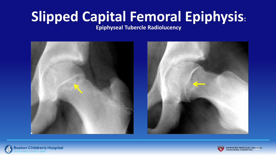
Slipped Capital Femoral Epiphysis: Epiphyseal Tubercle Radiolucency
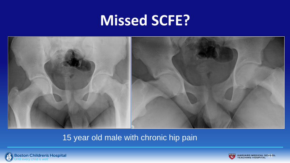
Missed SCFE?
11
15 year old male with chronic hip pain

Slipped Capital Femoral Epiphysis
12
In situ screw fixation Modified Dunn osteotomy

13
Modifed Dunn osteotomy Avascular Necrosis • 26% risk of AVN
• Technically challenging (compared to in situ pinning). Tertiary care centers
Slipped Capital Femoral Epiphysis

14
15
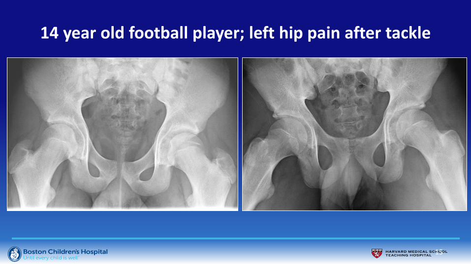
14 year old football player; left hip pain after tackle

16
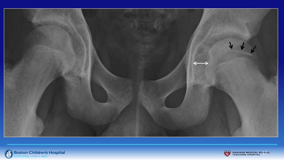
14 year old football player; left hip pain after tackle
2 years later, persistent limp
17

18
Posterior Hip Dislocation

Posterior Hip Dislocation
19

Posterior Hip Dislocation
• Top 3 Causes of Posterior Hip
Dislocation in Children at BCH
– Football injuries
– Snow sports (falls while skiing)
– MVC
20

Posterior Hip Dislocation with Labral Entrapment
21

Posterior Hip Dislocation with Labral Entrapment
22

Posterior Hip Dislocation with Labral Entrapment
23
• Presence of entrapped labrum changes
management
• Surgical dislocation may be necessary to
repair labrum
• If posterior dislocation clinically suspected,
MRI to evaluate for both osseous and soft
tissue injury
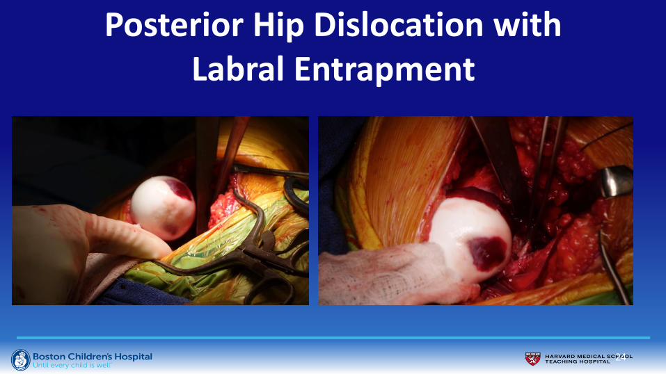
Posterior Hip Dislocation with Labral Entrapment
24

Complications of Posterior Hip Dislocation
25
• Arthrosis
– Most common complication (up to 24%)
– Related to chondrocyte damage at time of injury
• Avascular Necrosis
– Increased risk with >6 hours to reduction
– Rare in children
• Sciatic Nerve Injury

26

Femoral Neck Stress Fracture • Uncommon location for stress injury
(tibia, metatarsals)
• Female > male
• Female athlete triad: low-energy
availability, functional hypothalamic
amenorrhea, and osteoporosis
• High risk for progression
• Radiographs usually 1st diagnostic
test ordered
• Sensitivity 10% in early stage
• Usually only seen on AP radiograph

• MRI more sensitive than
radiographs
• MRI grade correlates with time to
return to sport:
• Grade 1: mild marrow edema
with no fracture line
• Grade 2: moderate edema, no
fracture line
• Grade 3: severe edema
• Grade 4: edema + fracture
Femoral Neck Stress Fracture
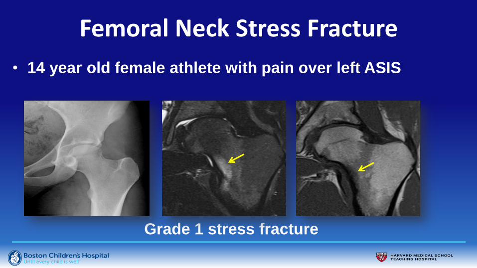
• 14 year old female athlete with pain over left ASIS
Grade 1 stress fracture
Femoral Neck Stress Fracture

• 20 year old female runner
with right hip pain
Grade 1 stress fracture
Femoral Neck Stress Fracture

Grade 3 stress fracture
Femoral Neck Stress Fracture

Grade 4 stress fracture
Femoral Neck Stress Fracture

Athletic Pubalgia
• “Sports hernia”
• Strain or tear of soft
tissue in the lower
abdomen/groin
• Most common in
sports with twisting at
waist

• Specific MRI protocol
angled to pubic
symphysis:
• Adductor tears
• Osteitis pubis
• Sports hernia
Athletic Pubalgia

18 year old male with right hip pain
Right adductor longus tendinopathy
Athletic Pubalgia

• Osteitis Pubis:
• Pubic symphysis has numerous musculotendinous connections
• Chronic repetitive shear and distraction injuries
• Sclerosis, osteolysis, physeal widening
Athletic Pubalgia

• Osteitis Pubis:
– Pubic symphysis has
numerous musculotendinous
connections
– Chronic repetitive shear and
distraction injuries
– Sclerosis, osteolysis, physeal
widening
Athletic Pubalgia

38

Ischial Avulsion Fracture
• Ischial growth plate open
until age ~20-25 years
• Nondisplaced fractures
treated conservatively
• Displacement > 2 cm
require surgical fixation
14 year old male with right hip pain
Ischial apophyseal avulsion at semimembranosus attachment

14 year old male with right hip pain
Ischial apophyseal avulsion at semimembranosus attachment
Ischial Avulsion Fracture

16 year old male with football injury and left hip pain
Ischial apophyseal avulsion at hamstrings attachment
Ischial Avulsion Fracture

Ischial Avulsion Fracture
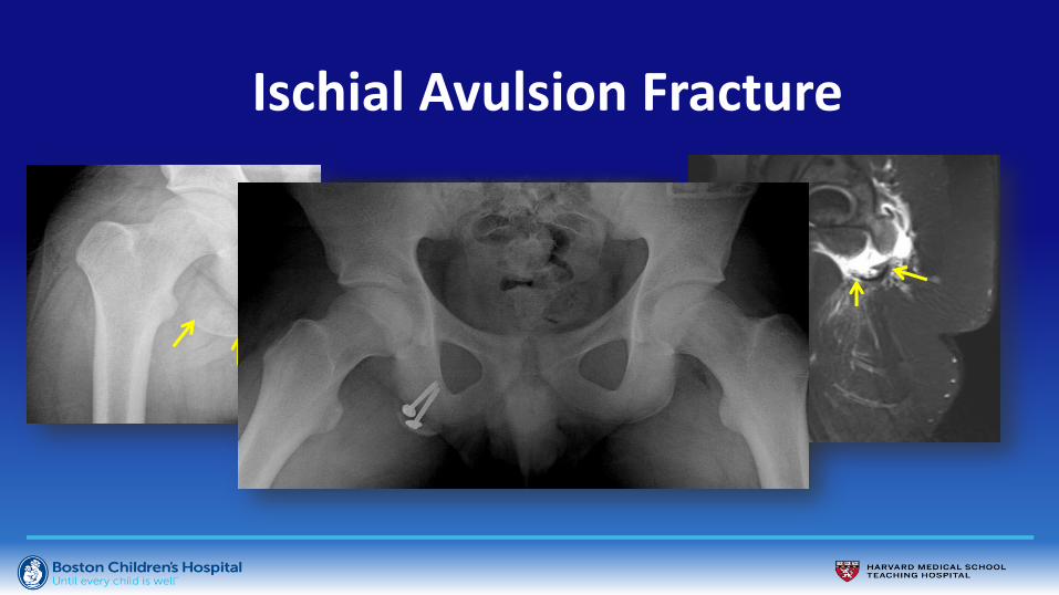
Ischial Avulsion Fracture

AIIS Avulsion Extra-articular FAI
15 year old male with right hip pain and s/sx FAI

15 year old male with right hip pain and s/sx FAI
Extra-articular/Subspinous impingement
AIIS Avulsion Extra-articular FAI
Post-op

13 year old female with right hip pain
Date of injury 1 year later
AIIS Avulsion Extra-articular FAI

Arthrogram findings:
• Direct impingement of FN on AIIS
• Lever effect with hip abduction
Contrast pool in fossa
Subluxation of FH
Abutment of FN on AIIS
AIIS Avulsion Extra-articular FAI

13 year old female with right hip pain after soccer injury
Date of injury Healing
AIIS Avulsion Extra-articular FAI

Femoroacetabular Impingement (FAI)
– Repetitive “collisions”
between femur and
acetabular rim
• Morphologic alterations femur
and/or acetabulum
• Extremes of hip motion/activity
Pfirrmann CW et al. Radiology 2008;249(1):236-241
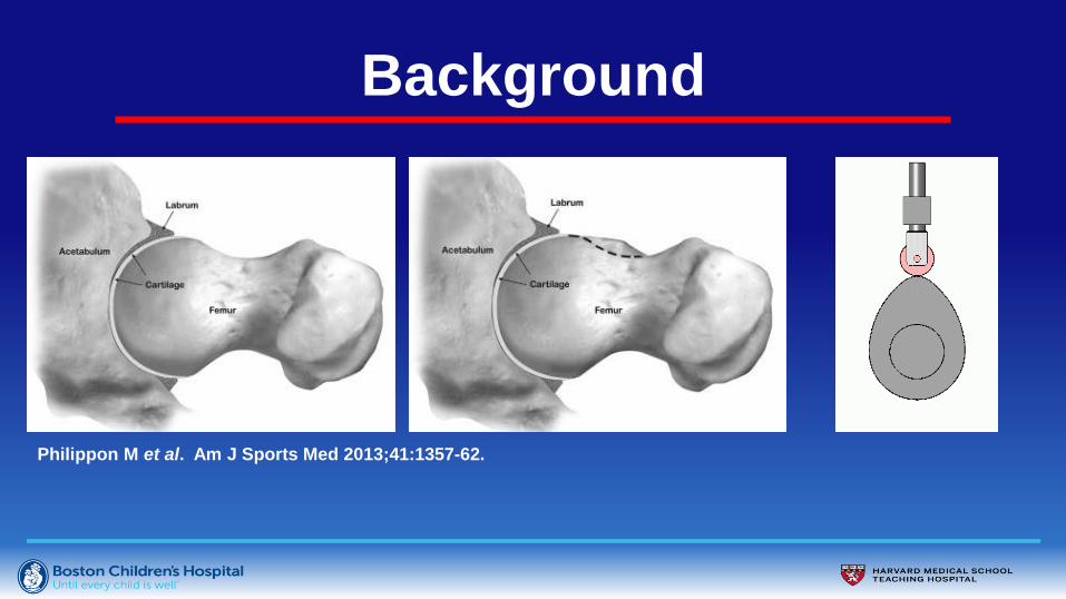
Background
Philippon M et al. Am J Sports Med 2013;41:1357-62.

Cam FAI
51

Alpha Angle Abnormal values
• > 50 º – Notzli HP et al. JBJS Br 2002;84:556-560.
– Tannast M et al. Am J Roentgenol 2007;188:154-1552.
• > 50.5 º – Beaulé PE et al. J Orthop Res 2005;23:1286-1292.
– Hack K et al. JBJS Am 2010;92:2436-2444.
• > 60 º – Sutter R et al. Radiology 2012;264:514-521.
• > 63 º – Pollard TC. Acta Orthopaedica 2010;81:134-141
• > 83 º – Gosvig KK et al. Acta Radiol 2008;49:436-441
– Jung KA et al. JBJS Br 2011;93:1303-1307
FH
FN
FH
FN

Cam FAI
53

Alpha Angle
Artwork courtesy of Andrew Phelps MD

Radial MRI Imaging
Imaging planes rotate around a fixed central point
Creates
images
orthogonal to
points on the
circumference
of a circular
surface

Radial MRI Imaging
Labrum: Planes rotate around acetabulum
ACETABULUM
Anterior Posterior
Useful in MR
arthrography
Transverse ligament

Radial MRI Imaging
Labrum: Planes rotate around acetabulum
ACETABULUM
Anterior Posterior
Useful in MR
arthrography
Transverse ligament
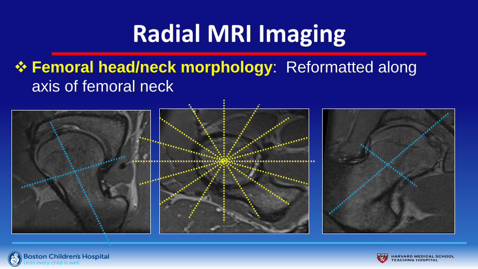
Radial MRI Imaging Femoral head/neck morphology: Reformatted along
axis of femoral neck

Radial MRI Imaging
3D SPACE 3D True Fisp
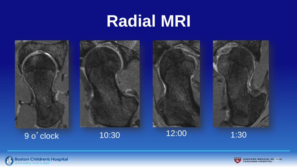
Radial MRI
60
9 o’clock 10:30 12:00 1:30
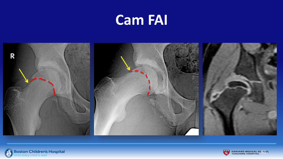
Cam FAI
61

Cam FAI
• Etiology:
– Developmental?
– Traumatic?
– Genetic?
10 year old male with abdominal pain
AP scout image from abdominal CT

Cam FAI
10 year old male with abdominal pain
AP scout image from abdominal CT
Same patient at age 18 years
AP scout image from abdominal CT

Cam FAI
• MRI for FAI:
– Acetabular
cartilage damage
– Labral tear
1. Cartilage delamination
2. Labral tear
Right hip
Left hip

What do these patients have in common?
65
14 year old
female with
left hip pain
16 year old
male with
left hip pain

Acetabular “rim fracture” • May be present in both DDH and FAI
• 3.6% of FAI patients have rim fracture
• Related to abnormal acetabular development and/or
stress fractures of the acetabular rim
• Can be excised in their entirety, partially excised, or
left in situ
• Treatment dilemma: rim fracture with intact
articular cartilage that contributes to joint stability
66

Rim Fragment – Fracture vs. Ossicle?
67
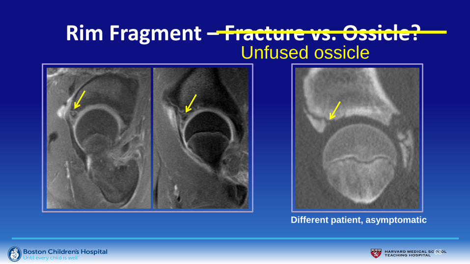
68
Different patient, asymptomatic
Rim Fragment – Fracture vs. Ossicle? Unfused ossicle

69
Rim Fragment - Healing
Healed “fracture”
after correcting
the dysplasia
with PAO

Rim Fragment - DDH
70
Articular cartilage spans fragment Healed “fracture” s/p PAO

Rim Fragment- FAI
• Os acetabuli/rim fractures
described in 3.6% of patients with
FAI
• May be excised in their entirety,
partial resection, or internal fixation
(versus left alone).
71

Rim Fracture - FAI
72
Corrected LCE angle <<< 20 degrees
LCE 50˚ LCE 16˚
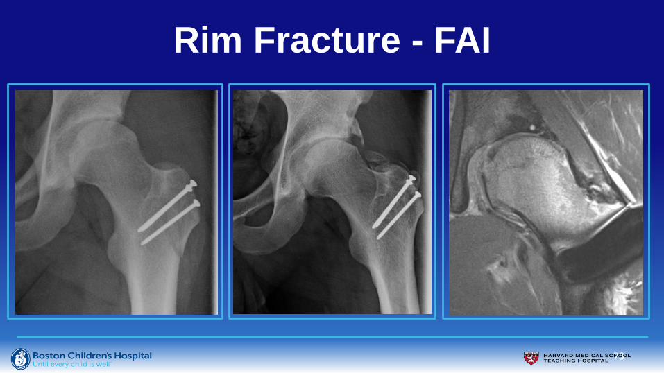
73
Rim Fracture - FAI
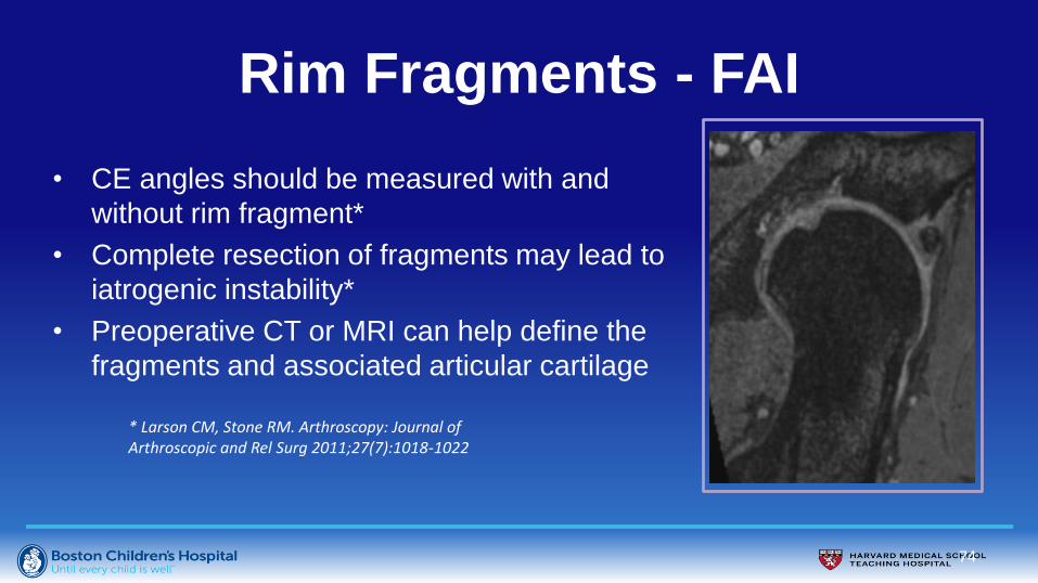
Rim Fragments - FAI
• CE angles should be measured with and
without rim fragment*
• Complete resection of fragments may lead to
iatrogenic instability*
• Preoperative CT or MRI can help define the
fragments and associated articular cartilage
74
* Larson CM, Stone RM. Arthroscopy: Journal of Arthroscopic and Rel Surg 2011;27(7):1018-1022

Rim Fracture - FAI
75
14 year old male competitive soccer player with progressive hip pain

76
Rim Fracture - FAI 17 year old male soccer and lacrosse player with right hip pain

77
Rim Fracture - FAI 17 year old male soccer and lacrosse player with right hip pain
s/p Head/neck osteoplasty Healed Rim Fracture
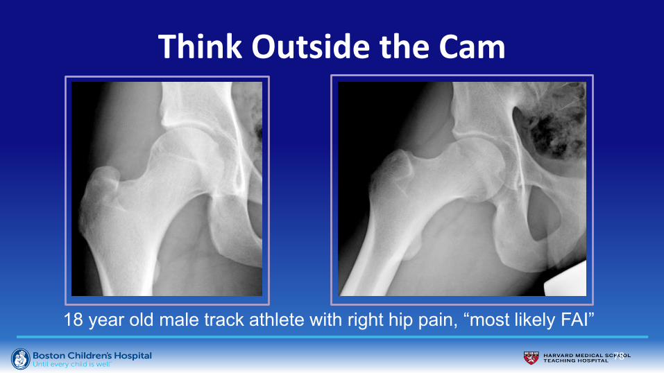
Think Outside the Cam
78
18 year old male track athlete with right hip pain, “most likely FAI”

Think Outside the Cam
79
18 year old male track athlete with right hip pain, “most likely FAI”

Think Outside the Cam
80

Spondyloarthropathy
81

82
15 year old male with left hip pain c/w FAI
Think Outside the Cam

83
15 year old male with left hip pain c/w FAI
Think Outside the Cam

84
15 year old male with left hip pain c/w FAI
Prior outside study: MRI arthrogram
Think Outside the Cam

85
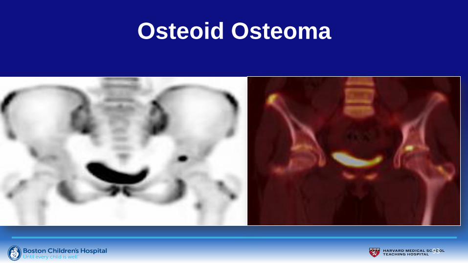
Patient went to surgery for head/neck arthroscopy
Think Outside the Cam

86
Patient went to surgery for head/neck arthroscopy
Osteoid osteoma
Think Outside the Cam

87
13 year old female with right hip pain

88
13 year old female with right hip pain
T2 fs T2 fs

89
Osteoid Osteoma
• Radiolucent nidus
• Central calcification
• Geographic marrow edema
• Thickened medial retinaculum
• Joint effusion

90
Osteoid Osteoma
Klontzas ME et al.
Osteoid osteoma of the femoral neck:
use of the half-moon sign in MRI
diagnosis.
Am J Roentgenol 2015;205(2):353-7.

91
Companion Case:
14 year old female with left hip pain

92
93
Osteoid Osteoma

94
Osteoid Osteoma– Example 1
Initial MR Diagnosis: Stress Fracture

95
Osteoid Osteoma-Example 2
Initial PE and radiographs c/w FAI

96
Osteoid Osteoma- Example 3

Conclusion • No single imaging algorithm for sports-related pediatric hip pain
– history guides imaging
• Radiographs nearly always useful (even when negative); MRI is
useful when radiographs “normal”
– Optimize protocols for indication
• Growth disturbances resulting from injury may be more problematic
than original trauma (e.g SCFE, avulsion fractures, FAI, etc.)
• FAI common; overdiagnosis may mask other disease
• Patients with sports injuries may have unrelated diagnosis (e.g.
osteoid osteoma)
97