Embed Size (px)
Citation preview
SRHisto: Public deliverable for the ATTRACT Final Conference
SRHisto: Public deliverable for the ATTRACT Final Conference
SRS histopathology (SRHisto)
Ingo Rimke,1* and Hervé Rigneault,2
1APE Angewandte Physik \& Elektronik GmbH, Berlin,Germany; 2Aix Marseille Univ, CNRS, Centrale Marseille, Institut Fresnel,
Marseille, France.
*Corresponding authors: [email protected]; [email protected]
ABSTRACT
The SRS hitopathology project aims at developing a novel technology able to generate histology images that are used by medical doctors
to detect and diagnose cancer tissues. The technology is based on the stimulated Raman effect, a nonlinear optical technique that can
visualize the chemical bonds present in tissues. In this project, we have developed the building blocks (laser source and detection system)
and demonstrated the ability to generate histology images in few minutes only, as compared to the 12h-24h commonly required by
standard histology protocols. These results open the road further develop the technology for its transfer and evaluation in the hospital.
Keywords: Stimulated Raman imaging, nonlinear microscopy, laser sources, histology, oncology
1. INTRODUCTION
Every year, approximately 14 million cancers are
diagnosed and 8 million people die of cancer worldwide
(13% of all deaths every year). This makes invasive
cancer the leading cause of death in the developed world
and the second leading cause of death in the developing
world. Histology plays a vital role in the diagnosis,
staging, and treatment of cancers and is the ‘gold
standard’ method recognized by the medical community
and legal authorities. Histology is a labor intensive
technique that requires the removal of small regions of
suspect tissues (biopsies) that are later sectioned and
stained with hematoxylin, eosin and saffron (HES) [1].
Standard HES protocol requires the excised samples to
undergo several preparation steps (dehydration, fixation,
wax, staining, slicing, mounting) that are time consuming,
typically in the range of 10 hours up to 3 days. There is an
urgent need for an intra-operative diagnostic technique
that avoids HES staining and delivers immediate high
quality histology images in the operatory room to assist in
surgical and decision making.
Histopathologic diagnostic requires the visualization of
cell nuclei and cell cytoplasm. Although many imaging
approaches have been developed to address histologic
needs such and confocal reflectance [2], fluorescence [3]
or multiphoton [4, 5] microscopies, none of them provide
direct and unambiguous tissue components identification
based of their chemical signature. An elegant approach to
identify these components from their chemical signature,
in a label free manner and at high resolution is to use
coherent Raman scattering (CRS) [6]. CRS encompasses
coherent anti-Stokes Raman scattering (CARS) and
stimulated Raman scattering (SRS). Both are optical
nonlinear wave mixing processes [7] that can probe the
presence of CH2 and CH3 chemical bonds found in cell
cytoplasm and cell nuclei using their distinctive bond
vibrations, respectively. This ability of SRS was shown
very recently to provide label free microscopic images of
healthy and cancer tissues sections in near-perfect
concordance with standard histology [8, 9]. This major
step forwards opens the new field of label free stimulated
Raman histology (SRH) for real-time intra-operative
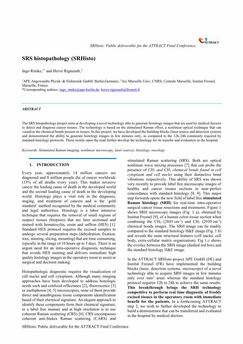
surgical cancer tissue resections and treatments. Figure 1
shows SRH microscopy images (Fig. 1 a), obtained by
Institut Fresnel [9], of a human colon tissue section when
combining the CH2 (2845 cm-1) and CH3 (2930 cm-1)
chemical bonds images. The SRH image can be readily
compared to the standard histology H&E image (Fig. 1 b)
and reveals the same structural features (cell nuclei, cell
body, extra-cellular matrix organization). Fig 1.c shows
the overlay between the SRH image (dashed red box) and
the standard histology H&E image.
In the ATTRACT SRHisto project APE GmbH (DE) and
Institut Fresnel (FR) have implemented the building
blocks (laser, detection systems, microscope) of a novel
technology able to acquire SRH images in few minutes
only over mm2 areas whereas the standard histology
protocol requires 12h to 24h to achieve the same results.
This breakthrough brings the SRH technology
competitive to perform real time diagnostic of freshly
excised tissues in the operatory room with immediate
benefit for the patients. In a forthcoming ATTRACT
step 2, we wish to further developed the technology to
build a demonstrator that can be transferred and evaluated
in the hospital by medical doctors.
2
SRHisto
Figure 1: Stimulated Raman histology (SRH) in human colon
tissue (Institut Fresnel work [9]). (a) SRH image obtained in 5
minutes, (b) standard H&E histology image, (c) overlay between
the SRH image (red dashed box) and the standard H&E
histology image.
2. STATE OF THE ART
Up to now the SRH imaging technology has used noisy
fiber laser sources that required complex balanced
detections scheme [8], or bulky laser systems [9].
Furthermore, these systems acquired sequentially the two
images corresponding to the CH2 and CH3 chemical bonds
that are necessary to generate SRH images. This
sequential acquisition limits the acquisition speed of SRH
images on freshly excised tissues that are prone to move
during the image acquisition. A dual-colour approach
enable to acquire simultaneously the CH2 and CH3
incorporating a grating together with an acousto-optic
deflector employed as angle-to-wavelength pulse shaper
was recently proposed [10]. As a disadvantages the largest
part of the fs-laser is wasted. Detecting two non-
neighbouring Raman bands (off- or in-resonance),
spectral focusing [11] in combination with two different
time delays was implemented for dual-color SRS imaging
[12]. However, any time delay between pump and Stokes
pulses affect the optimal temporal overlap of the pulses
and leads to a reduced signal generation. Alternatively,
and to circumvent major drawbacks of the previously
mentioned techniques, Institut Fresnel recently introduced
a dual-color SRS-microscopy approach combining the
output of a fiber-laser at 1030 nm and two optical
parametric oscillators (OPOs) which are modulated at two
distinct frequencies to enable cross-talk-free SRS-images
at two independently tunable Raman-shifts within the
range of 500cm-1 – 5000cm-1 [13]. The performance of the
latter is compromised by the utilization of a pump fiber-
laser that is 6 dB(W) above the shot-noise limit resulting
in the reduction of the acquisition speed by a factor of
approximately 4 compared to a suitable shot-noise limited
system.
3. BREAKTHROUGH CHARACTER OF
THE PROJECT
In the ATTRACT SRHisto project we have breakthrough
these limitations and built, for the first time, a low noise
compact laser system able to acquire the CH2 and CH2
images simultaneously. Contrary to [13], the system is
shot noise limited and shows x4 reduction of the footprint.
We have also developed an optimized detection system
and a H&E virtual colouring software that provides SRH
images over mm2 areas in few minutes only. We have
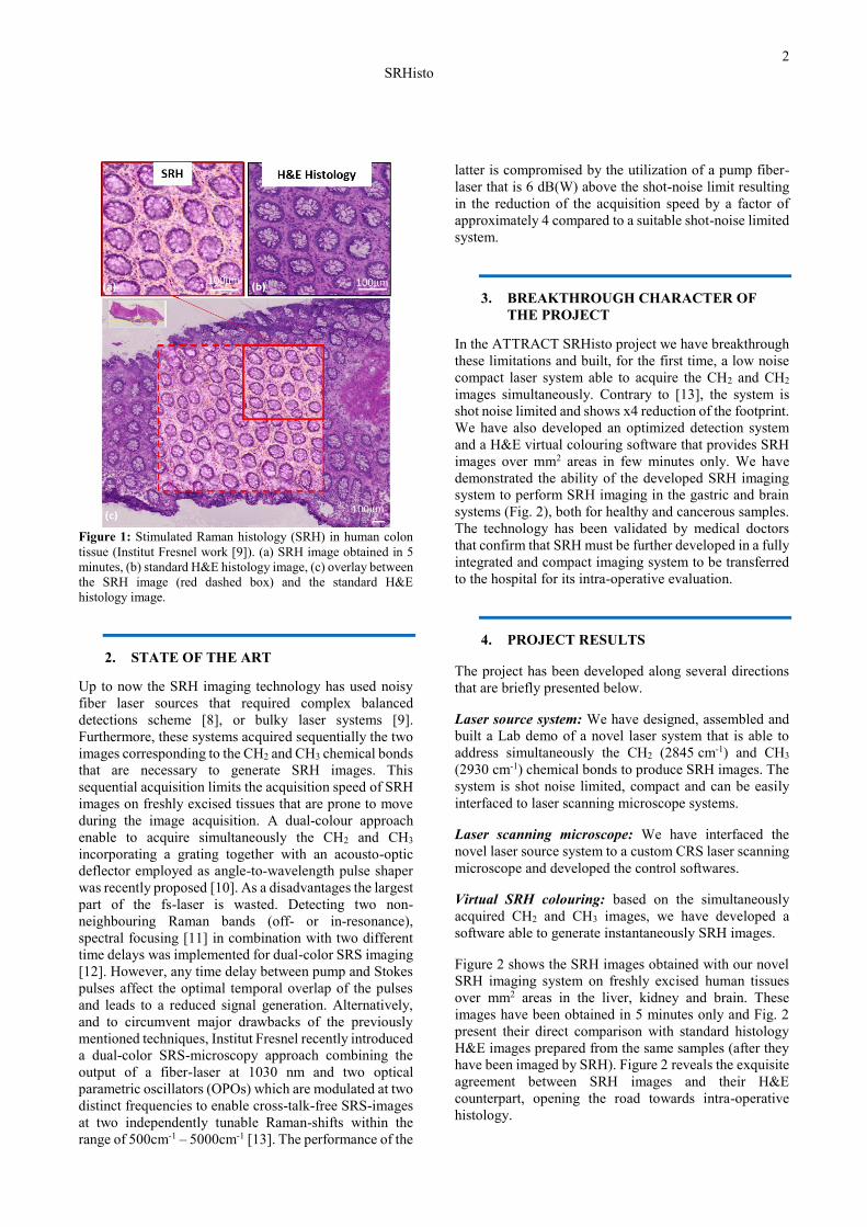
demonstrated the ability of the developed SRH imaging
system to perform SRH imaging in the gastric and brain
systems (Fig. 2), both for healthy and cancerous samples.
The technology has been validated by medical doctors
that confirm that SRH must be further developed in a fully
integrated and compact imaging system to be transferred
to the hospital for its intra-operative evaluation.
4. PROJECT RESULTS
The project has been developed along several directions
that are briefly presented below.
Laser source system: We have designed, assembled and
built a Lab demo of a novel laser system that is able to
address simultaneously the CH2 (2845 cm-1) and CH3
(2930 cm-1) chemical bonds to produce SRH images. The
system is shot noise limited, compact and can be easily
interfaced to laser scanning microscope systems.
Laser scanning microscope: We have interfaced the
novel laser source system to a custom CRS laser scanning
microscope and developed the control softwares.
Virtual SRH colouring: based on the simultaneously
acquired CH2 and CH3 images, we have developed a
software able to generate instantaneously SRH images.
Figure 2 shows the SRH images obtained with our novel
SRH imaging system on freshly excised human tissues
over mm2 areas in the liver, kidney and brain. These
images have been obtained in 5 minutes only and Fig. 2
present their direct comparison with standard histology
H&E images prepared from the same samples (after they
have been imaged by SRH). Figure 2 reveals the exquisite
agreement between SRH images and their H&E
counterpart, opening the road towards intra-operative
histology.
3
SRHisto
Figure 2: Validated SRH images and their direct comparison
with H&E classical histology images. SRH acquisition time 5
minutes; scale bar: 100m. The samples are coming from AP-
HM and IPC hospitals in Marseille.
5- FUTURE PROJECT VISION
The aim of the SRHisto ATTRACT phase 2 project is to
develop new optical imaging instruments using label
free stimulated Raman histology (SRH) to generate
images of histological quality from freshly excised
biopsies, in real time, without labelling or any sample
preparation. The device will be a compact microscope
including the ATTRACT phase 1 laser source and
detection schemes that can be used close to the operatory
room to examine sections of freshly excised tissue to
evaluate their cancerous nature in few minutes only. The
microscope will be transferred to partner hospital for its
direct evaluation by medical doctors (histo-pathologists
and surgeons).
5.1. Technology Scaling
In SRHisto ATTRACT phase 2 we aim at bringing the
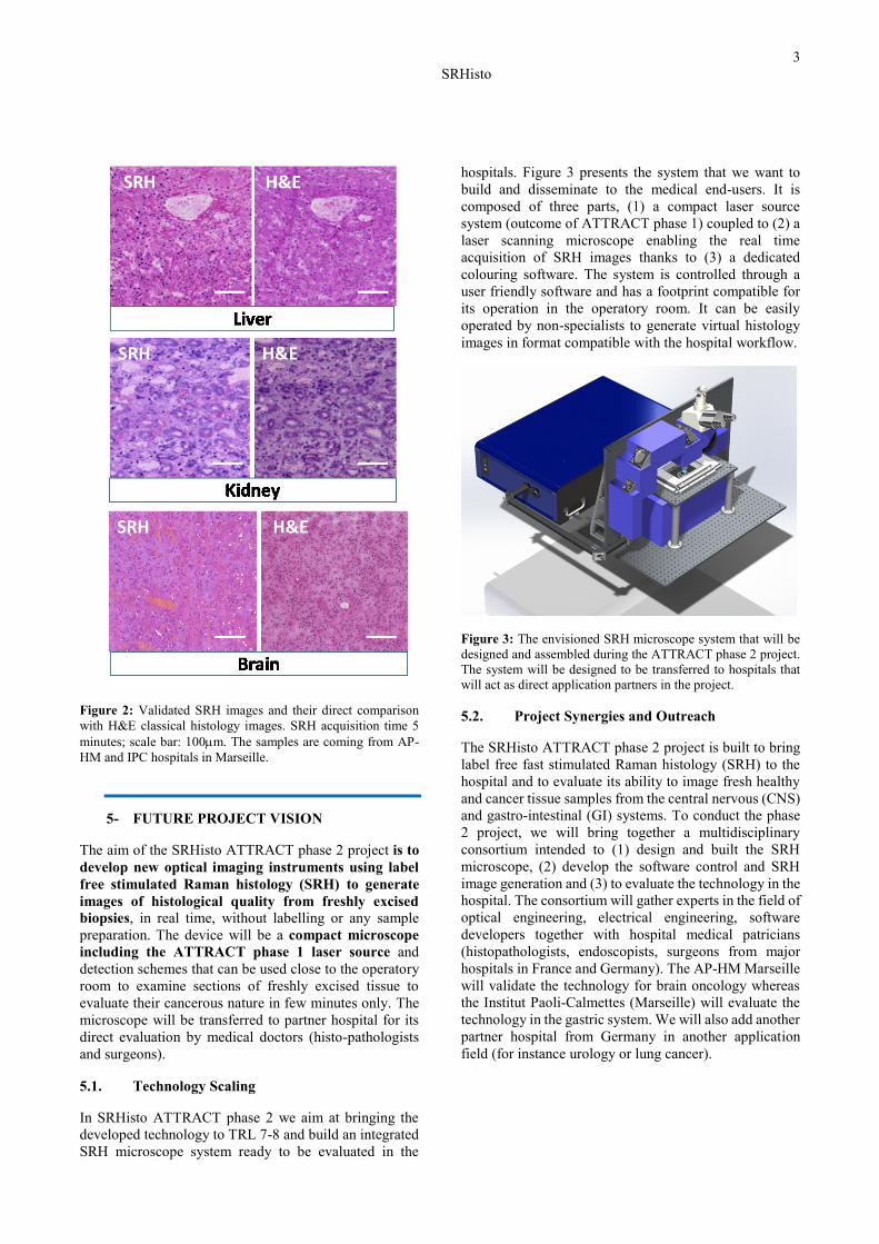
developed technology to TRL 7-8 and build an integrated
SRH microscope system ready to be evaluated in the
hospitals. Figure 3 presents the system that we want to
build and disseminate to the medical end-users. It is
composed of three parts, (1) a compact laser source
system (outcome of ATTRACT phase 1) coupled to (2) a
laser scanning microscope enabling the real time
acquisition of SRH images thanks to (3) a dedicated
colouring software. The system is controlled through a
user friendly software and has a footprint compatible for
its operation in the operatory room. It can be easily
operated by non-specialists to generate virtual histology
images in format compatible with the hospital workflow.
Figure 3: The envisioned SRH microscope system that will be
designed and assembled during the ATTRACT phase 2 project.
The system will be designed to be transferred to hospitals that
will act as direct application partners in the project.
5.2. Project Synergies and Outreach
The SRHisto ATTRACT phase 2 project is built to bring
label free fast stimulated Raman histology (SRH) to the
hospital and to evaluate its ability to image fresh healthy
and cancer tissue samples from the central nervous (CNS)
and gastro-intestinal (GI) systems. To conduct the phase
2 project, we will bring together a multidisciplinary
consortium intended to (1) design and built the SRH
microscope, (2) develop the software control and SRH
image generation and (3) to evaluate the technology in the
hospital. The consortium will gather experts in the field of
optical engineering, electrical engineering, software
developers together with hospital medical patricians
(histopathologists, endoscopists, surgeons from major
hospitals in France and Germany). The AP-HM Marseille
will validate the technology for brain oncology whereas
the Institut Paoli-Calmettes (Marseille) will evaluate the
technology in the gastric system. We will also add another
partner hospital from Germany in another application
field (for instance urology or lung cancer).
4
SRHisto
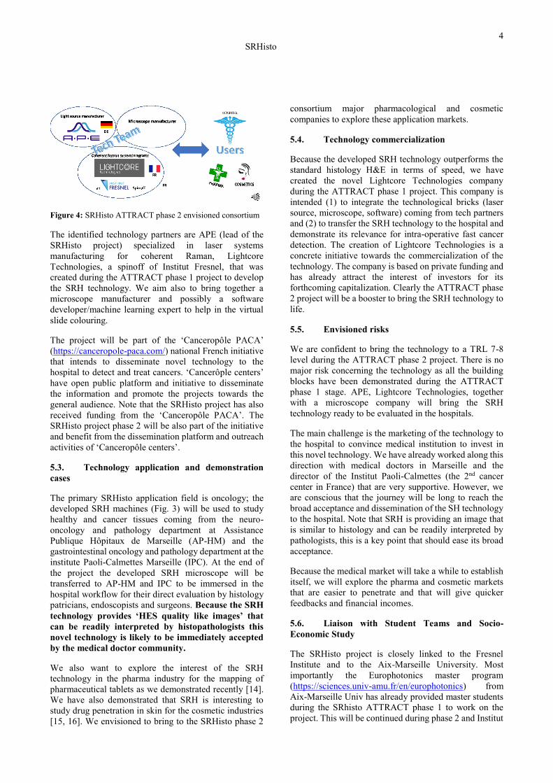
Figure 4: SRHisto ATTRACT phase 2 envisioned consortium
The identified technology partners are APE (lead of the
SRHisto project) specialized in laser systems
manufacturing for coherent Raman, Lightcore
Technologies, a spinoff of Institut Fresnel, that was
created during the ATTRACT phase 1 project to develop
the SRH technology. We aim also to bring together a
microscope manufacturer and possibly a software
developer/machine learning expert to help in the virtual
slide colouring.
The project will be part of the ‘Canceropôle PACA’
(https://canceropole-paca.com/) national French initiative
that intends to disseminate novel technology to the
hospital to detect and treat cancers. ‘Cancerôple centers’
have open public platform and initiative to disseminate
the information and promote the projects towards the
general audience. Note that the SRHisto project has also
received funding from the ‘Canceropôle PACA’. The
SRHisto project phase 2 will be also part of the initiative
and benefit from the dissemination platform and outreach
activities of ‘Canceropôle centers’.
5.3. Technology application and demonstration
cases
The primary SRHisto application field is oncology; the
developed SRH machines (Fig. 3) will be used to study
healthy and cancer tissues coming from the neuro-
oncology and pathology department at Assistance
Publique Hôpitaux de Marseille (AP-HM) and the
gastrointestinal oncology and pathology department at the
institute Paoli-Calmettes Marseille (IPC). At the end of
the project the developed SRH microscope will be
transferred to AP-HM and IPC to be immersed in the
hospital workflow for their direct evaluation by histology
patricians, endoscopists and surgeons. Because the SRH
technology provides ‘HES quality like images’ that
can be readily interpreted by histopathologists this
novel technology is likely to be immediately accepted
by the medical doctor community.
We also want to explore the interest of the SRH
technology in the pharma industry for the mapping of
pharmaceutical tablets as we demonstrated recently [14].
We have also demonstrated that SRH is interesting to
study drug penetration in skin for the cosmetic industries
[15, 16]. We envisioned to bring to the SRHisto phase 2
consortium major pharmacological and cosmetic
companies to explore these application markets.
5.4. Technology commercialization
Because the developed SRH technology outperforms the
standard histology H&E in terms of speed, we have
created the novel Lightcore Technologies company
during the ATTRACT phase 1 project. This company is
intended (1) to integrate the technological bricks (laser
source, microscope, software) coming from tech partners
and (2) to transfer the SRH technology to the hospital and
demonstrate its relevance for intra-operative fast cancer
detection. The creation of Lightcore Technologies is a
concrete initiative towards the commercialization of the
technology. The company is based on private funding and
has already attract the interest of investors for its
forthcoming capitalization. Clearly the ATTRACT phase
2 project will be a booster to bring the SRH technology to
life.
5.5. Envisioned risks
We are confident to bring the technology to a TRL 7-8
level during the ATTRACT phase 2 project. There is no
major risk concerning the technology as all the building
blocks have been demonstrated during the ATTRACT
phase 1 stage. APE, Lightcore Technologies, together
with a microscope company will bring the SRH
technology ready to be evaluated in the hospitals.
The main challenge is the marketing of the technology to
the hospital to convince medical institution to invest in
this novel technology. We have already worked along this
direction with medical doctors in Marseille and the
director of the Institut Paoli-Calmettes (the 2nd cancer
center in France) that are very supportive. However, we
are conscious that the journey will be long to reach the
broad acceptance and dissemination of the SH technology
to the hospital. Note that SRH is providing an image that
is similar to histology and can be readily interpreted by
pathologists, this is a key point that should ease its broad
acceptance.
Because the medical market will take a while to establish
itself, we will explore the pharma and cosmetic markets
that are easier to penetrate and that will give quicker
feedbacks and financial incomes.
5.6. Liaison with Student Teams and Socio-
Economic Study
The SRHisto project is closely linked to the Fresnel
Institute and to the Aix-Marseille University. Most
importantly the Europhotonics master program
(https://sciences.univ-amu.fr/en/europhotonics) from
Aix-Marseille Univ has already provided master students
during the SRhisto ATTRACT phase 1 to work on the
project. This will be continued during phase 2 and Institut
5
SRHisto
Fresnel will open several MSc training position every year
on the SRHisto project. It is likely that PhD research topic
will follow, they will be funded through Aix-Marseille
Univ or using the CIFRE mechanism open for small
companies in France.
Lightcore Technologies is closely followed by the
Marseille SATT (http://www.sattse.com/qui-sommes-
nous/), SATT is intended to promote, help and follow
startup companies in the south-east of France. SATT
provides a full service to startup companies in terms of
initial funding, marketing, public relationships. During
the ATTRACT phase 2 project Lightcore Technologies
will allocate a SATT representative to help in the
dissemination and the referencing of the technology
impact.
.
1. ACKNOWLEDGEMENT
The authors thanks Sandro Heuke (I Fresnel), Barbara
Sarri (I Fresnel), Romain Appay (AP-HM), Flora Poizat
(IPC), Edlef Butner (APE) that have contributed to the
SRHisto project.
This project has received funding from the ATTRACT
project funded by the EC under Grant Agreement
777222. This project has also received funding from
SATT Sud-Est and Canceropôle Provence.
7. REFERENCES
1. A. H. Fischer, K. A. Jacobson, J. Rose, and R.
Zeller, "Hematoxylin and Eosin Staining of Tissue and Cell
Sections," Cold Spring Harbor Protocols 2008, pdb.prot4986
(2008).
2. E. S. Flores, M. Cordova, K. Kose, W. Phillips, A.
Rossi, K. S. N. M.D., and M. Rajadhyaksha, "Intraoperative
imaging during Mohs surgery with reflectance confocal
microscopy: initial clinical experience," Journal of
Biomedical Optics 20, 1-11, 11 (2015).
3. B. Larson, S. Abeytunge, E. Seltzer, M.
Rajadhyaksha, and K. Nehal, "Detection of skin cancer
margins in Mohs excisions with high-speed strip mosaicing
confocal microscopy: a feasibility study," British Journal of
Dermatology 169, 922-926 (2013).
4. S.-J. Lin, S.-H. Jee, C.-J. Kuo, R. Wu, Jr., W.-C.
Lin, J.-S. Chen, Y.-H. Liao, C.-J. Hsu, T.-F. Tsai, Y.-F. Chen,
and C.-Y. Dong, "Discrimination of basal cell carcinoma from
normal dermal stroma by quantitative multiphoton imaging,"
Optics Letters 31, 2756-2758 (2006).
5. Y. K. Tao, D. Shen, Y. Sheikine, O. O. Ahsen, H.
H. Wang, D. B. Schmolze, N. B. Johnson, J. S. Brooker, A. E.
Cable, J. L. Connolly, and J. G. Fujimoto, "Assessment of
breast pathologies using nonlinear microscopy," Proceedings
of the National Academy of Sciences 111, 15304-15309
(2014).
6. J.-X. Cheng, and X. S. Xie, "Vibrational
spectroscopic imaging of living systems: An emerging
platform for biology and medicine," Science 350 (2015).
7. H. Rigneault, and P. Berto, "Tutorial: Coherent
Raman light matter interaction processes," APL Photonics 3,
091101 (2018).
8. D. A. Orringer, B. Pandian, Y. S. Niknafs, T. C.
Hollon, J. Boyle, S. Lewis, M. Garrard, S. L. Hervey-Jumper,
H. J. L. Garton, C. O. Maher, J. A. Heth, O. Sagher, D. A.
Wilkinson, M. Snuderl, S. Venneti, S. H. Ramkissoon, K. A.
McFadden, A. Fisher-Hubbard, A. P. Lieberman, T. D.
Johnson, X. S. Xie, J. K. Trautman, C. W. Freudiger, and S.
Camelo-Piragua, "Rapid intraoperative histology of
unprocessed surgical specimens via fibre-laser-based
stimulated Raman scattering microscopy," Nature Biomedical
Engineering 1, 0027 (2017).
9. B. Sarri, R. Canonge, X. Audier, E. Simon, J.
Wojak, F. Caillol, C. Cador, D. Marguet, F. Poizat, M.
Giovannini, and H. Rigneault, "Fast stimulated Raman and
second harmonic generation imaging for intraoperative gastro-
intestinal cancer detection," Scientific Reports 9, 10052
(2019).
10. D. Zhang, M. N. Slipchenko, D. E. Leaird, A. M.
Weiner, and J.-X. Cheng, "Spectrally modulated stimulated
Raman scattering imaging with an angle-to-wavelength pulse
shaper," Optics Express 21, 13864-13874 (2013).
11. T. Hellerer, A. Enejder, and A. Zumbusch, "Spectral
focusing: High spectral resolution spectroscopy with
broadbandwidth laser pulses," Appl Phys Lett 85, 25-27
(2004).
12. R. He, Y. Xu, L. Zhang, S. Ma, X. Wang, D. Ye,
and M. Ji, "Dual-phase stimulated Raman scattering
microscopy for real-time two-color imaging," Optica 4, 44-47
(2017).
13. S. Heuke, B. Sarri, X. Audier, and H. Rigneault,
"Simultaneous dual-channel stimulated Raman scattering
microscopy demultiplexed at distinct modulation
frequencies," Optics Letters 43, 3582-3585 (2018).
14. B. Sarri, R. Canonge, X. Audier, V. Lavastre, G.
Pénarier, J. Alie, and H. Rigneault, "Discriminating
polymorph distributions in pharmaceutical tablets using
stimulated Raman scattering microscopy," Journal of Raman
Spectroscopy 50, 1896-1904 (2019).
15. X. Chen, S. Grégoire, F. Formanek, J.-B. Galey, and
H. Rigneault, "Quantitative 3D molecular cutaneous
absorption in human skin using label free nonlinear
microscopy," Journal of Controlled Release 200, 78-86
(2015).
16. B. Sarri, X. Chen, R. Canonge, S. Grégoire, F.
Formanek, J.-B. Galey, A. Potter, T. Bornschlögl, and H.
Rigneault, "In vivo quantitative molecular absorption of
glycerol in human skin using coherent anti-Stokes Raman
scattering (CARS) and two-photon auto-fluorescence,"
Journal of Controlled Release 308, 190-196 (2019).