Embed Size (px)
Citation preview
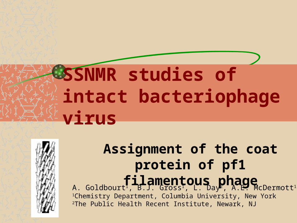
SSNMR studies of intact bacteriophage virus
Assignment of the coat protein of pf1 filamentous phage
A. Goldbourt1, B.J. Gross1, L. Day2, A.E. McDermott1 1Chemistry Department, Columbia University, New York2The Public Health Recent Institute, Newark, NJ

Filamentous bacteriophage
Pf1 belongs to the filamentous Bacteriophage (Inovirus) family of organisms known to attack bacteria.Members of the family include Pf1, Pf3 and Xf (Class-II) as well as M13, fd, f1, If1, and IKe (Class-I). The host bacteria for Pf1 (and Pf3) are Pseudomonas aeruginosa of different strains (strain K for Pf1).Most virions consist of a long single stranded circular Most virions consist of a long single stranded circular DNA encapsulated by multiple copies of protein DNA encapsulated by multiple copies of protein subunits. The DNA loop is stretched from one end of subunits. The DNA loop is stretched from one end of the virus to the other in an unknown conformation. the virus to the other in an unknown conformation. On the surface of the coat protein, several additional functional proteins are docked. These proteins are crucial for the bacterial infection and for the reassembly process of the virion.
2000nm
6nmPf1 cartoon modelG. Stubbs, Rep. Prog. Phys. (2001) 64, 1389
Pf1 is the longest known virionPf1 is the longest known virion
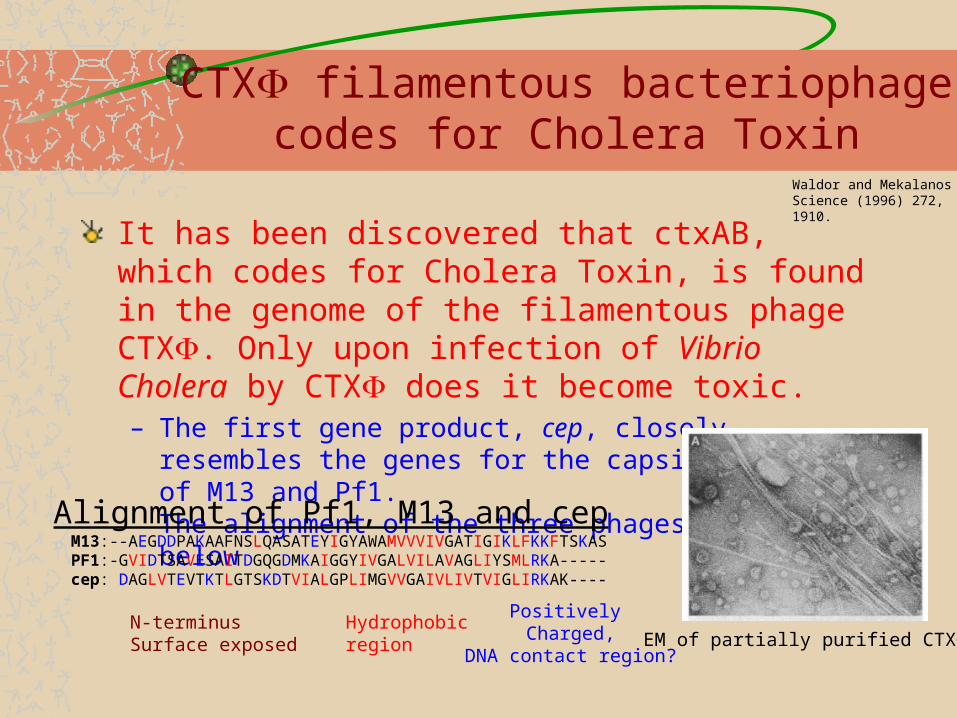
CTX filamentous bacteriophage codes for Cholera Toxin
It has been discovered that ctxAB, which codes for Cholera Toxin, is found in the genome of the filamentous phage CTX. Only upon infection of Vibrio Cholera by CTX does it become toxic. – The first gene product, cep, closely resembles the genes for the capsid
proteins of M13 and Pf1. The alignment of the three phages is shown below
Waldor and Mekalanos Science (1996) 272, 1910.
EM of partially purified CTX
M13:--AEGDDPAKAAFNSLQASATEYIGYAWAMVVVIVGATIGIKLFKKFTSKASPF1:-GVIDTSAVESAITDGQGDMKAIGGYIVGALVILAVAGLIYSMLRKA-----cep: DAGLVTEVTKTLGTSKDTVIALGPLIMGVVGAIVLIVTVIGLIRKAK----
Hydrophobicregion
Positively Charged,
DNA contact region?
Alignment of Pf1, M13 and cep
N-terminusSurface exposed

Uses of filamentous phages
Pf1 readily aligns in a magnetic field and is regularly used for protein partial alignment in order to obtain RDC constrains.M13 and T7 are used as sequencing primers.Virions like fd, f1 and M13 are used for peptide phage display.

pf1 virion
Longest known phage, 2000nm.Coat protein with 46 amino acids, all-helicalNucleotide/subunit ratio of 1:1 – smallest knownUndergoes a phase transition at 10oC
GVIDTSAVESAITDGQGDMKAIGGYIVGALVILAVAGLIYSMLRKA
Top view of 1PFI, DNA in the center
Side view of1PJFShowing the repeating subunits
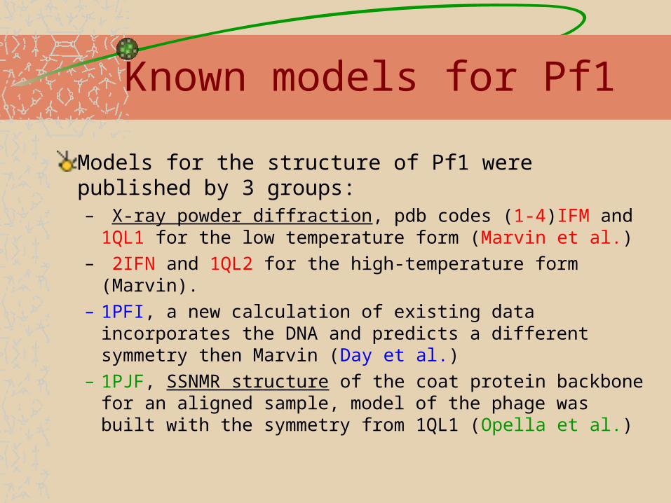
Known models for Pf1
Models for the structure of Pf1 were published by 3 groups: – X-ray powder diffraction, pdb codes (1-4)IFM and 1QL1 for the
low temperature form (Marvin et al.) – 2IFN and 1QL2 for the high-temperature form (Marvin).– 1PFI, a new calculation of existing data incorporates the DNA and
predicts a different symmetry then Marvin (Day et al.)– 1PJF, SSNMR structure of the coat protein backbone for an
aligned sample, model of the phage was built with the symmetry from 1QL1 (Opella et al.)

Structure of Pf1 The helix is assumed to have a kink in the center, or to be gently curvedthe N-terminus can be a loop (1PJF,1QL1,4IFM) or helical (1PFI, 1IFM) and probably depends on the solvent (surface exposed)The locations of the sidechains are unresolved in the x-ray data and are partially obtained from alternative spectroscopic experiments.The helical bundle symmetry around the DNA is on debate – 27 units in 5 turns (1QL1/2) or 71 subunits in 13 turns (1PFI) or alternatively, 71 dimers in 26 turns (Day, unpublished). And perhaps, a non-rational ratio.
1QL11QL1
SSNMR dipolar waves (opella, 1PJF1PJF)experimentalexperimentalgently curved helix 1QL1
15N-H dipolar coupling

Advantages of MAS NMR
What is the coat protein’s exact structure? – Refinement of X-ray diffraction data requires a fitting of the protein
structure + the whole helical bundle.– Different coat protein models can give different results for the helical
bundle arrangement.– A more accurate structure of the protein will directly contribute to the
solution of the whole virus structure by reducing the number of fit parameters for the X-ray powder diffraction data.
Is it an inside out DNA?– Model 1PFI suggests that the structure is ‘inside out’: The bases point
outside towards the coat protein and the phosphates point towards the inside.
– Hopefully, 31P/13C NMR experiments …
Site specific information
What is the nature of the phase transition?– The pf1 virus undergoes a reversible phase transition at 10oC. It is
known that the overall length of the virus increases slightly (~100nm ; 15 turns). A model assumes reorganization of the helix bundle.
– Site-specific information will be obtained with ssNMR experiments.
What kind of contacts exist with the DNA?– Mainly qualitative data exist: Quenching of Tyr40 fluorescence signal,
Lys45 & Arg44 compensate for phosphate charges etc.– With site specific information contacts to the DNA can be obtained
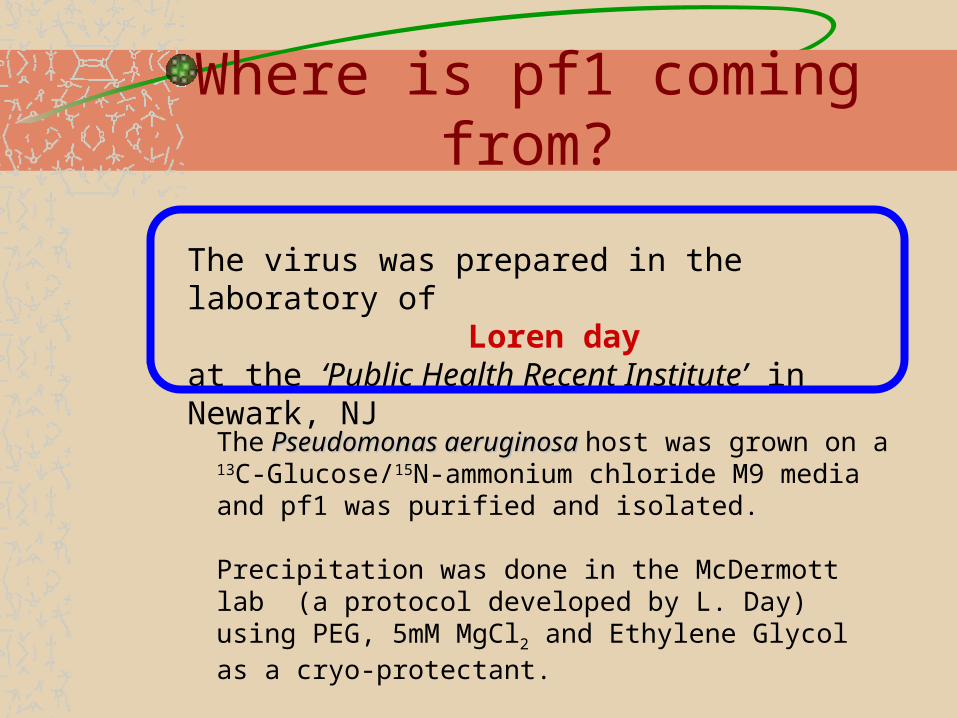
Where is pf1 coming from?
The virus was prepared in the laboratory of Loren day
at the ‘Public Health Recent Institute’ in Newark, NJ
The Pseudomonas aeruginosa Pseudomonas aeruginosa host was grown on a 13C-Glucose/15N-ammonium chloride M9 media and pf1 was purified and isolated.
Precipitation was done in the McDermott lab (a protocol developed by L. Day) using PEG, 5mM MgCl2 and Ethylene Glycol as a cryo-protectant.
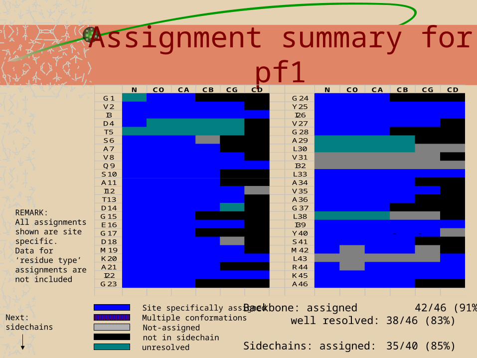
Assignment summary for pf1N CO CA CB CG CD N CO CA CB CG CD
G1 - - G24 - -V2 Y25I3 I26D4 V27T5 G28S6 A29A7 L30V8 V31Q9 I32S10 L33A11 A34I12 V35T13 A36D14 G37 - -G15 - - L38E16 I39G17 - - Y40 - -D18 S41M19 M42K20 L43A21 R44I22 K45
G23 - - A46
Backbone: assigned 42/46 (91%)well resolved: 38/46 (83%)
Sidechains: assigned: 35/40 (85%)
Site specifically assigned
not in sidechainunresolved
Not-assignedMultiple conformations
REMARK:All assignments shown are site specific.Data for ‘residue type’ assignments are not included
Next:sidechains
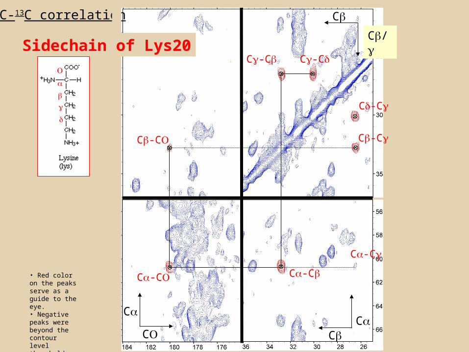
CC
C
C
C/
C13C-13C correlation
• Red color on the peaks serve as a guide to the eye.• Negative peaks were beyond the contour level threshold
C-CC-C
C-C C-C
C-C
C-C
C-C
C-C
Sidechain of Lys20
C-C
C-CC-C
C-C C-C
C-C
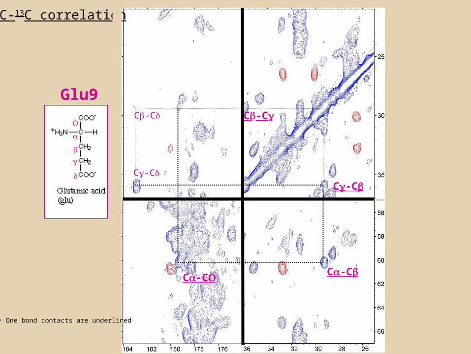
Glu9
13C-13C correlation
• One bond contacts are underlined

13C-13C correlation
(C-C
Val2
C-C
C-C
C-C
C-C

Strip plots from 3D
STRIP PLOTS FROM 750MHz 3D EXPERIMENTS
29
31
33
35
37
39
41
43
45
47
49
51
55
55
57
59
61
C (
pp
m)
RED: NCACXBLUE: NCOCX
Similar 13CO/13CA shifts, sequential 15N
i-1i
176.9 177.0
G17 D18G17
i-1i
Similar 15N shifts 121.9 121.8 121.7
Q16 Q16G15
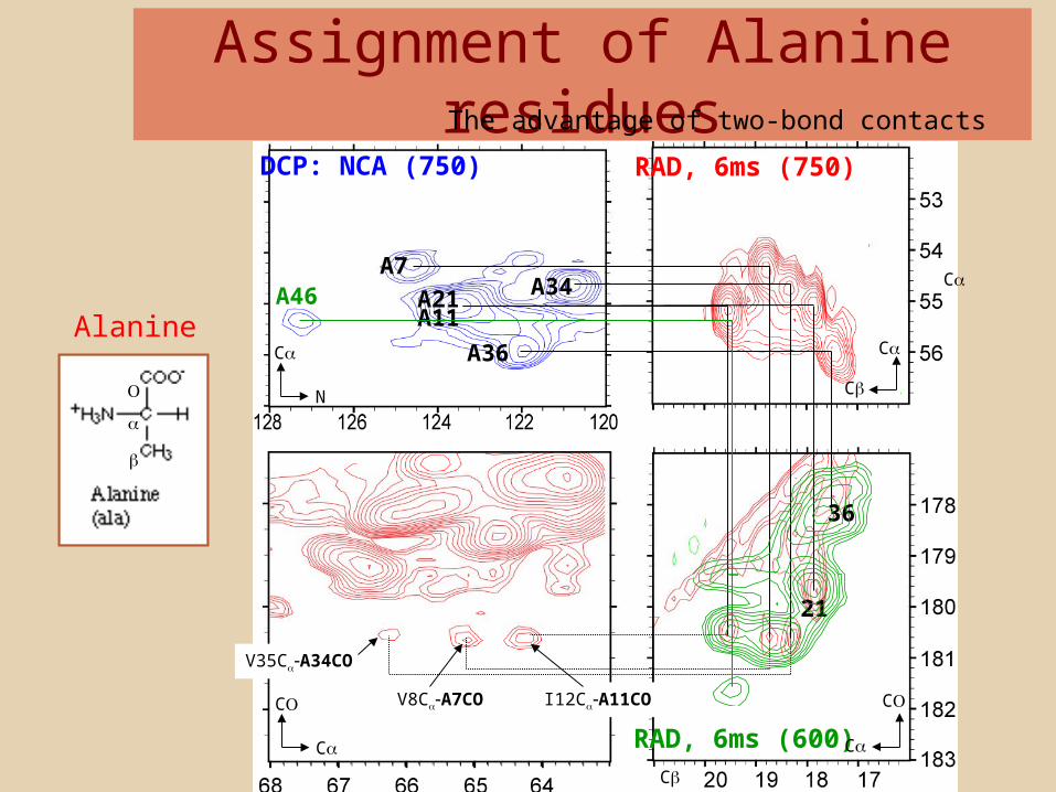
Assignment of Alanine residuesThe advantage of two-bond contacts
DCP: NCA (750) RAD, 6ms (750)
A34A21A11
A36
21
36
V35CA34CO
V8CA7CO I12CA11CO
A7C
C
C
C
C
C
N
C
A46
RAD, 6ms (600)C
C
Alanine

Initial structural information
The TALOS derived secondary structure
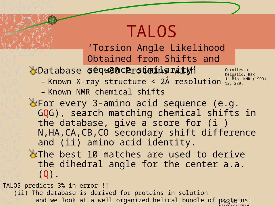
TALOS
Database of ~80 Proteins with– Known X-ray structure < 2Å resolution– Known NMR chemical shifts
For every 3-amino acid sequence (e.g. GQG), search matching chemical shifts in the database, give a score for (i ) N,HA,CA,CB,CO secondary shift difference and (ii) amino acid identity.The best 10 matches are used to derive the dihedral angle for the center a.a. (Q).
‘Torsion Angle Likelihood Obtained from Shifts and sequence similarity’
Weights : HA>CO>CA~CB>N
Remark : (i) TALOS predicts 3% in error !! (ii) The database is derived for proteins in solution and we look at a well organized helical bundle of proteins!
Cornilescu, Delgalio, Bax, J. Bio. NMR (1999) 13, 289.

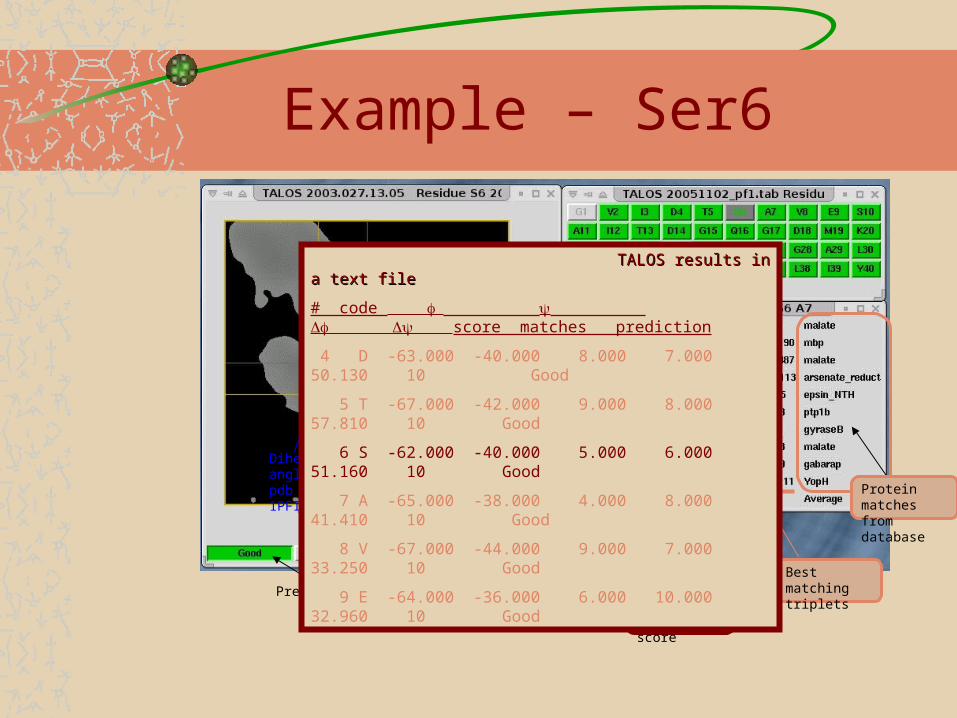
Example – Ser6
Dihedral angle in pdb file 1PFI
10 best predictions
Prediction is good of 9/10 results agree
Protein matches from database
Best matching triplets
TALOS score
Example – Ser6
Dihedral angle in pdb file 1PFI
10 best predictions
Prediction is good of 9/10 results agree
Protein matches from database
Best matching triplets
TALOS average score
result
TALOS results in a text fileTALOS results in a text file
# code score matches prediction
4 D -63.000 -40.000 8.000 7.000 50.130 10 Good
5 T -67.000 -42.000 9.000 8.000 57.810 10 Good
6 S -62.000 -40.000 5.000 6.000 51.160 10 Good
7 A -65.000 -38.000 4.000 8.000 41.410 10 Good
8 V -67.000 -44.000 9.000 7.000 33.250 10 Good
9 E -64.000 -36.000 6.000 10.000 32.960 10 Good
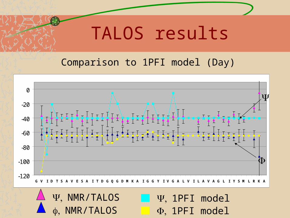
TALOS results
-120
-100
-80
-60
-40
-20
0
G V I D T S A V E S A I T D G Q G D M K A I G G Y I V G A L V I L A V A G L I Y S M L R K A
Comparison to 1PFI model (Day)
NMR/TALOSNMR/TALOS
1PFI model1PFI model
-120
-100
-80
-60
-40
-20
0
G V I D T S A V E S A I T D G Q G D M K A I G G Y I V G A L V I L A V A G L I Y S M L R K A
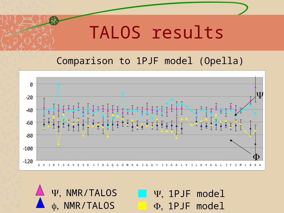
TALOS resultsComparison to 1PJF model (Opella)
NMR/TALOSNMR/TALOS
1PJF model1PJF model
-120
-100
-80
-60
-40
-20
0
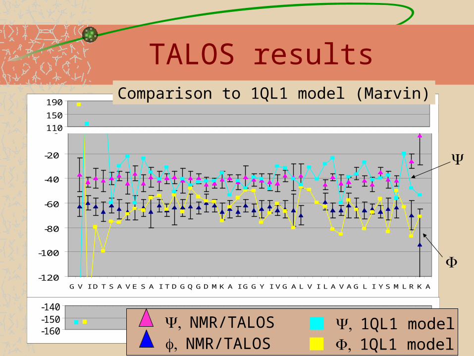
G V I D T S A V E S A I T D GQ GD MK A I G G Y I V G A L V I L A V A G L I Y S M L R K A
110
150
190
-160-150-140
TALOS resultsComparison to 1QL1 model (Marvin)
NMR/TALOSNMR/TALOS
1QL1 model1QL1 model

Summary
Almost complete assignment of pf1, a filamentous bacteriophage virus, 35MD molecular weight, has been obtained.Weak DNA sugar-peaks have been observed.TALOS results suggest a completely helical structure. No significant loop/kink region has been predicted. Current work in progress:– Structural distance constrains will be obtained with emphasis on inter-
molecular contacts.– Site specific information will enable to probe the phase transition.– DNA-protein contacts will be obtained by low-temperature experiments.
Thanks to
$$$ Columbia University $$$Loren Day Initiated the project, prepares constant flow of samples and enlightens us with all we know about pf1!!Ben Gross – from him I inherited this project, thanks and good luck!! (of course he also work hard – 600 3D’s, 400 experiments, assignments!!!)NYSBC (and Boris Itin!!)2D NCA/NCO experiments – J. Lorieau Ann McDermott and the wonderful group at Havemeyer 3rd floor.

DNA signals at the 2D 13C-13C spectra
C3
C1/4
Additional expected regionsFor C5 (65ppm) and C2 (41ppm)
RAD mixing, 6msObtained with 60Hz line broadening in both dimensions
C1/4C1/4
C3C3
C5C5
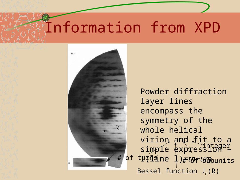
Powder diffraction layer lines encompass the symmetry of the whole helical virion and fit to a simple expression – I(line l)=tn+um
Information from XPD
# of turns # of subunits
integer
Bessel function Jn(R)
R
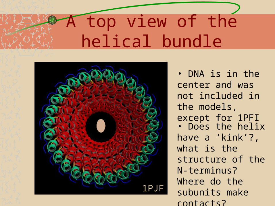
A top view of the helical bundle
• DNA is in the center and was not included in the models, except for 1PFI
• Does the helix have a ‘kink’?, what is the structure of the N-terminus? Where do the subunits make contacts?
1PJF
What do we have so far?
Almost complete assignment has been achieved (for the high-temperature form) with 2D and 3D experiment on 600 and 750 MHz spectrometers.DNA signals from the sugars have been detected but they are weak. Low temperature experiment are underway in order to try and obtain the DNA-coat protein specific contacts. 31P-31P DQ and 31P/13C REDOR experiments failed to produce any correlations but 1D spectra suggest strong DNA dynamics. These also will require low temperaturesThe low temperature form was precipitated and spectra will be used to probe the site-specific changes of the phase transition