Embed Size (px)
Citation preview

CELL JOURNAL(Yakhteh), Vol 15, No 4, Winter 2014 364
Short Communication
Stem Cell Isolation from Human Wharton’s Jelly: A Study of Their Differentiation Ability into Lens Fiber Cells
Seyedeh Mahsa Khatami, M.Sc.1, Saber Zahri, Ph.D.1, Masoud Maleki, Ph.D.2*, Kamaloddin Hamidi, Ph.D.1
1. Department of Biology, Cell and Molecular Laboratory, Faculty of Science, University of Mohaghegh Ardabili, Ardabil, Iran 2. Department of Biology, East Azarbaijan Science and Research Branch, Islamic Azad University, Tabriz, Iran
* Corresponding Address: P.O.Box: 5166857513, Department of Biology, East Azarbaijan Science and Research Branch, Islamic Azad University, Tabriz, IranEmail: [email protected]
Received: 11/Jul/2012, Accepted: 22/Jan/2013 AbstractRecently, the use of stem cells has expanded into numerous areas including cell therapy. In this study, we investigated the differentiation capacity of human Wharton’s jelly stem cells (hWJSCs) into lens fiber cells. Morphological changes and expressions of four crystallin genes (αA, αB, βB1 and βB3) were studied. The bovine vitreous body has been shown to induce expression of crystallin genes in hWJSCs. By using the vitreous as a lens fiber cell inducer, we showed that αB-, βB1- and βB3-crystallin genes expressed in hWJSCs. Keywords: Wharton’s jelly, Mesenchymal Stem Cells, Crystallin, Differentiation Cell Journal(Yakhteh), Vol 15, No 4, Winter 2014, Pages: 364-371
Citation: Khatami SM, Zahri S, Maleki M, Hamidi K. Stem cell isolation from human wharton’s jelly: a study of their differentiation ability into lens fiber cells. Cell J. 2014; 15(4): 364-371.
Lens induction is a multistep process that in-volves competence, bias, specification, inhibition and differentiation (1). The early stages of lens morphogenesis are specified by a close physical connection between the presumptive lens and the optic vesicle. Then, the presumptive lens thickens to form the placode and invaginates together with the optic vesicle to organize the lens pit and optic cup, respectively (2). Cells in the posterior half of the vesicle elongate and differentiate to form the primary fibers, whereas anterior cells differentiate into the epithelium. The lens rapidly grows by cell division during late embryonic and early postnatal stages (3). Lens polarity is maintained throughout its lifetime; evidence exists that it is regulated by the ocular environment.
The Pax6 gene is located at the head of the regu-latory system in lens induction. Fibroblast growth factor (FGF) and bone morphogenetic protein 7 (BMP7) are required for lens induction and these molecules coordinate with Pax6 expression. In the posterior half of the lens, fiber cells contact with the vitreous body. FGF-1 and FGF-2 in the vitre-ous body are necessary to induce lens epithelial
cells to lens fiber cells and molecular changes that include elongation, structural specialization, and the onset of specialized crystallin gene expres-sion occurs in these cells (4, 5). All vertebrate lenses express crystallins that belong to the α-and βγ-crystallin protein families. αA and αB are lens fiber cell markers (6, 7).
Due to the unique characteristics of mesenchy-mal stem cells (MSCs), they have been considered for therapeutic applications by many researchers (8). The main source for MSCs is the bone mar-row but recently umbilical cord Wharton’s jelly has been recognized as an excellent source for the isolation of MSCs. Wharton’s jelly stem cells (WJSCs) can differentiate into different cell types such as osteoblasts (9), chondrocytes (10), cardio-myocytes (11), skeletal myoblasts (12), hepato-cyte-like cells (13), endothelial cells (14), neural cells, adipocytes (15), dopaminergic cells (16) and lens fiber cells (17). WJSCs express surface cell markers such as CD105, CD44 (12, 18), CD68 (19), CD13 and CD95, yet are negative for hemat-opoitic stem cells markers CD34, CD45, CD38 and CD71. WJSCs are fibroblast-like and multipo-

CELL JOURNAL(Yakhteh), Vol 15, No 4, Winter 2014 365
Cells Differentiate into Lens Fiber Cells
tent (15). In this study, WJSCs have been differen-tiated into lens fiber cells using bovine vitreous as a specific inducer. This is the first time that human WJSCs (hWJSCs) have been show to differentiate into lens fiber cells by using bovine vitreous.
In this study, umbilical cords (n=12) were ob-tained following consent of the mothers after ce-sarean section (Arta Hospital). The cords were washed with 70% alcohol and cut into 2 cm pieces in Hanks’ balanced salt solution (HBSS), after which the vein and two arteries were separated from the stroma by manual stripping. The remain-ing tissue, Wharton’s jelly, was chopped into piec-es of approximately 0.5 mm by a scalpel, then tiny tissue pieces were cultured in low glucose Dulbec-co’s modified Eagle’s medium (DMEM, Gibco, Germany) +20% fetal bovine serum (FBS, Gibco, Germany) +1% Penstrep (Sigma, USA). Culture flasks were placed in an incubator and after three days the culture medium was replaced. When the culture reached 70-80% confluency, cells were detached with 0.25% trypsin-EDTA and passaged (18). We counted the cells at passage 7 and cal-
culated the cell doubling time with doubling-time software.
Bovine eyes were immediately transferred to the laboratory from the Ardabil Industrial Slaughterhouse. The vitreous was extracted, then mashed and poured into centrifuge tubes and centrifuged at a high speed. The resultant homogenized vitreous was filtered by a sy-ringe filter (0.2 µm, Sartorius Stedim Biotech and stored at -80˚C. hWJSCs were induced by the vitreous body in three experimental groups (50% vitreous +50% DMEM + FBS; 25% vitre-ous +75% DMEM + FBS; and control) for ten days. The total hWJSCs and induced cell RNA were extracted and the total cDNA synthesized by the use of oligo (dT) 18 and specific prim-ers for CD105 and CD44 (positive markers), and CD34 (negative marker). In order to detect dif-ferentiation, we screened for expressions of the crystallin genes αA, αB, βB1 and βB3 by RT-PCR (Table 1). hWJSCs at passage 2 and the two experimental groups after ten days of induction were studied by scanning electron microscopy.
Table 1: Sequences of the primers used in RT-PCR assays Size Primer sequence (5'- 3') Gene
395 bpF: CAGCATTGTGGCATCCTTCGTGR: CCTTTTTCCGCTGTGGTGATGAG
CD105
433 bpF: ARCCACCCCAACTCCATCTGTR: TGTTTGCTCCACCTTCTTGACTC
CD44
434 bpF: GCCTGGAGCAAAATAAGACCR: ACCGTTTTCCGTGTAATAAGG
CD34
285 bpF: CGCACCCTGGGGCCCTTCTACCR: GTCGTCCTGGCGCTCGTTGTGCT
αA-crystallin
387 bpF: CTACCTTCGGCCACCCTCCTTCCR: TATTTCTTGGGGGCTGCGGTGAC
αB-crystallin
388 bpF: GCCCCAACAACCGTGCCTATTACR: CCCCCTGGATCTCTATGGTGTTGC
βB1-crystallin
433 bpF: ATGGCGGAACAGCACGGAGCACR: GGAAGCCATGAGCCCACAGG
βB3-crystallin
250 bpF: TGGAGAAATCTGGCACCACACCR: GATGGGCACAGTGTGGGTGACCC
β-actin
CELL JOURNAL(Yakhteh), Vol 15, No 4, Winter 2014 366
Khatami et al.
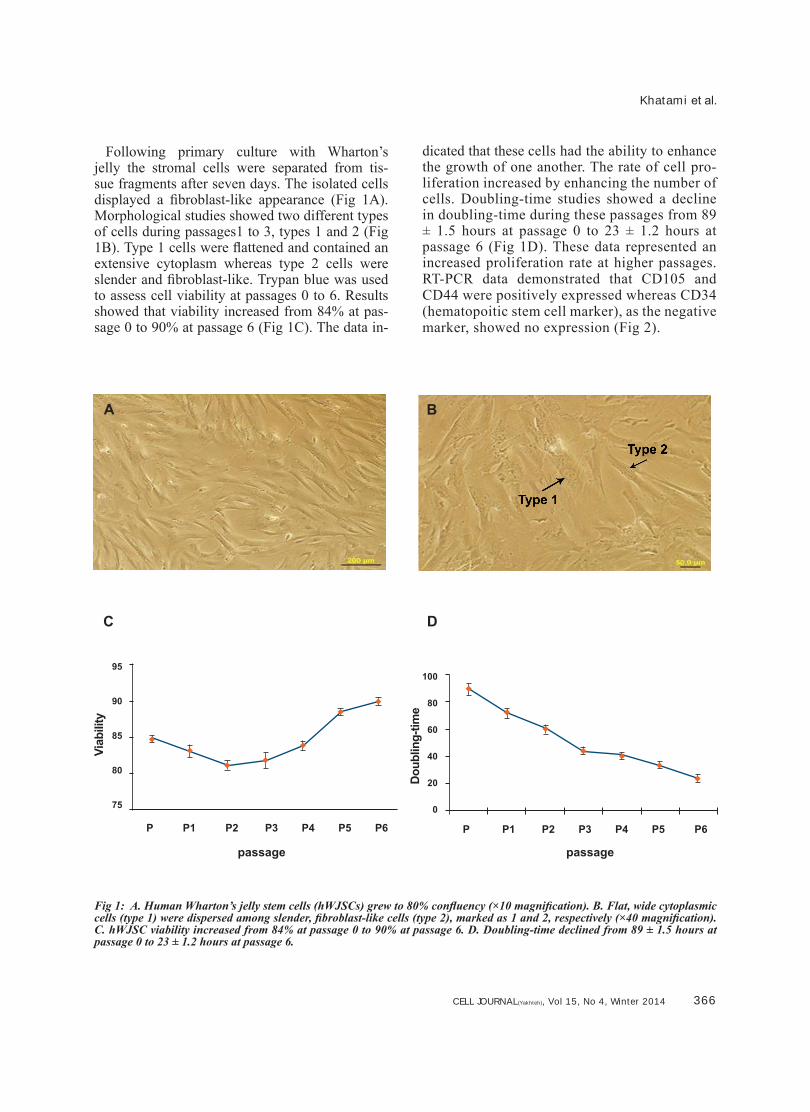
Following primary culture with Wharton’s jelly the stromal cells were separated from tis-sue fragments after seven days. The isolated cells displayed a fibroblast-like appearance (Fig 1A). Morphological studies showed two different types of cells during passages1 to 3, types 1 and 2 (Fig 1B). Type 1 cells were flattened and contained an extensive cytoplasm whereas type 2 cells were slender and fibroblast-like. Trypan blue was used to assess cell viability at passages 0 to 6. Results showed that viability increased from 84% at pas-sage 0 to 90% at passage 6 (Fig 1C). The data in-
dicated that these cells had the ability to enhance the growth of one another. The rate of cell pro-liferation increased by enhancing the number of cells. Doubling-time studies showed a decline in doubling-time during these passages from 89 ± 1.5 hours at passage 0 to 23 ± 1.2 hours at passage 6 (Fig 1D). These data represented an increased proliferation rate at higher passages. RT-PCR data demonstrated that CD105 and CD44 were positively expressed whereas CD34 (hematopoitic stem cell marker), as the negative marker, showed no expression (Fig 2).
passage
Viab
ility
95
90
85
80
75
P P1 P2 P3 P4 P5 P6
Dou
blin
g-tim
e
100
80
60
40
20
0
P P1 P2 P3 P4 P5 P6
passage
Fig 1: A. Human Wharton’s jelly stem cells (hWJSCs) grew to 80% confluency (×10 magnification). B. Flat, wide cytoplasmic cells (type 1) were dispersed among slender, fibroblast-like cells (type 2), marked as 1 and 2, respectively (×40 magnification). C. hWJSC viability increased from 84% at passage 0 to 90% at passage 6. D. Doubling-time declined from 89 ± 1.5 hours at passage 0 to 23 ± 1.2 hours at passage 6.
A B
C D

CELL JOURNAL(Yakhteh), Vol 15, No 4, Winter 2014 367
Cells Differentiate into Lens Fiber Cells
Fig 2: Qualitative RT-PCR analyses showed CD105 and CD44 expression (band of 433 and 395 bp), while CD34 was not expressed.
Morphological images showed that induced cells were extensive and parallel to each oth-er at two concentrations, 1:1 and 1:3. There were decreased cytoplasmic processes and the fiber-like cells had large nuclei with multiple nucleoli. Further studies indicated that induced cell differentiation at the 1:1 ratio (Fig 3A) was greater than the 1:3 ratio (Fig 3B). Control cells did not show any differentiation and were pas-saged due to increasing numbers (Fig 3C).
The genes αA-, αB-, βB1- and βB3-crystallin were used as lens fiber cell differentiation mark-ers. αA-crystallin showed no expression at the ex-perimental concentration as well as βB1-crystallin at the 1:3 ratio (Data not shown ). αB-crystallin (Fig 4A, B) and βB3-crystallin expressed at two concentrations (Fig 4C, D) and βB1-crystallin at the 1:1 ratio (Fig 4E).
Fig 3: A. First group (1:1 ratio). B. Second group (1:3 ratio). Images show induced cells after ten days. C. Control cells (×10 magnification).
A B
C

CELL JOURNAL(Yakhteh), Vol 15, No 4, Winter 2014 368
Khatami et al.
Fig 4: Qualitative RT-PCR analyses. A. αB-crystallin at the 1:1 ratio. B. αB-crystallin at the 1:3 ratio. C. βB3-crystallin at the 1:1ratio. D. βB3-crystallin at the 1:3 ratio. E. βB1-crystallin at the 1:1 ratio.
A B C
D E

CELL JOURNAL(Yakhteh), Vol 15, No 4, Winter 2014 369
Cells Differentiate into Lens Fiber Cells
The most prominent features of hWJSCs were their spindle shape and presence of a number of long cytoplasmic extensions (Fig 5A, B). However the lens fiber cells showed a very elongated morphology. As visualized
by electron microscopy, there were morpho-logical changes in the induced cells compared with the control group. There were extensive numbers of induced cells at both concentra-tions (Fig 5C, D, E, F).
Fig 5: A and B. Human Wharton’s jelly stem cells (hWJSCs). Long cytoplasmic extensions as shown by the arrows. C and D. Induced cells at a 1:1 ratio. E and F. Induced cells at a 1:3 ratio.
A B
C D
E F

CELL JOURNAL(Yakhteh), Vol 15, No 4, Winter 2014 370
Khatami et al.
Extensive cell markers have been found that iden-tify MSCs, the most important of which is their intrin-sic ability to adhere to uncoated plastic surfaces (8). MSCs are isolated from various sources. Although bone marrow is the most important source, umbilical cord stroma or Wharton’s jelly, has been recently con-sidered by researchers.
In this study, the hWJSCs were fibroblast-like and multipotent. As seen in figure 1B, there were two morphologies observed in culture during the early passages, one to three. Karahusyinoglu has classi-fied hWJSCs into type 1 (flat, wide cytoplasmic cells) dispersed among slender, fibroblast-like cells (type 2) (15). In this study, we observed gradually less type 1 cells at higher passages. Researchers have suggested that although there are two types of mesenchymal cells, the morphological differences between these cell types are due to the different parts of tissue from which the cells are isolated.
We observed a decrease in doubling-time, from 89 ± 1.5 hours during passage 0 to 23 ± 1.2 hours during passage 6, which was similar to results reported by Karahusyinoglu. Their study showed that doubling-time decreased from 85 ± 2.7 at passage 0 to 11 ± 2.1 at passage 7 (15). The slight difference was probably related to the difference in culture conditions and ex-perimental methods. The two MSC markers, CD105 and CD44, showed hWJSCs stemness property.
So far, extensive studies have been performed re-garding hWJSCs ability to differentiate into different types of cells such as osteoblasts, adipocytes , chon-drocytes, and cardiomyocytes. In the current study, we studied the ability of hWJSCs to differentiate into lens fiber cells. The early stages of lens cell formation are characterized by an elongation of epithelial lens cells at the anterior of the lens. One function of these epi-thelial cells is to serve as storage for cells from which the lens grow during development and throughout life. Mature lens fiber cells are long, ribbon-like cells that extend lengthwise from the posterior to the ante-rior poles of the lens (20). During the differentiation process cells lose cytoplasmic organelles, exit from the cell cycle, and crystallin proteins are expressed and accumulate in cells.
In this study, the vitreous body was used as a lens fiber cell inducer. We chose the vitreous because of its important role in lens placode formation during embryonic lens formation and the presence of lens fiber cell differentiation factors such as FGF-1 and
FGF-2 (21). The results showed that the induced cells had a fiber-like appearance and an increased number of nucleoli that were attributed to enhanced protein synthesis, most likely cytoplasmic and extracellular proteins.
Despite attempts to extract a uniform vitreous, vari-ables such as the ages of the cows were unavoidable. However the concentration of FGF most likely did not differ between the tests. To prove the existence of lens fiber cell differentiation at the molecular level, we studied the expressions of αA-, αB-, βB1- and βB3-crystallin genes. αB-crystallin transcription occurs at E 9.5 in the mouse lens placode. αB-crystallin is a stress-inducible protein that expresses at high levels in the lens and is only slightly expressed in other tissues (22). In this study, we have shown that αB-crystallin expressed in both experimental groups. Cell culture conditions in this study consisted of a humid environ-ment, 5% CO2 and a temperature of 37˚C, therefore the cultures were free from stress factors. Thus, there was no possibility of αB-crystallin gene expression in the control group. Studies have shown that αB-crystallin and Pax6 co-express during differentiation of embryonic stem cells to lens fiber cells (23) thus it was possible that Pax6 was present in the culture.
αA-crystallin expresses in the lens vesicle and at E 10.5. This gene has three DNA binding regu-latory factors, Pax6, CREB and c-Maf. The αA-crystallin promoters are regulated by three enhanc-ers, DCR1, DCR2 and DCR3. DCR1 in response to FGF-2 will begin to operate, whereas DCR3 does not respond to the FGF message. However DCR3 probably responds to the Wnt pathway and postoperate after FGF pathway and cause more differentiation. Due to the lack of expression of the αA-crystallin gene and αB-crystallin expression, the αA-crystallin gene probably expressed later than the αB-crystallin gene. Thus, if the duration of induction increases to more than ten days, αA-crystallin gene expression will most likely begin. Maleki et al. (17) have shown expression of the αA-crystallin gene in 14-day induced groups.
The β-crystallin gene family in mammals has six members (βB1, βB2, βB3, βA1/A3, βA2, βA4). The regulatory pathways are not studied completely. Most studies have been performed in mice and hu-man βB1- and rat βB2-, βB3-crystallin expression. βB1-crystallin gene expression occurred in elon-gated cells of the lens vesicle. FGF pathways studies showed that low concentrations of FGF-2
CELL JOURNAL(Yakhteh), Vol 15, No 4, Winter 2014 371
Cells Differentiate into Lens Fiber Cells
caused proliferation of lens epithelial cells, mod-erate FGF-2 concentration stimulated migration of lens epithelial cells and high levels of FGF-2 concentration induced differentiation of lens fiber cells. FGF concentration increased from the aque-ous humor into the vitreous, where the greatest amount of FGF-2 was observed in the vitreous. In this study, βB1-crystallin gene expression was 50:50, while no expression was observed at 75:25. Therefore it seemed that high concentrations of FGF-2 caused βB1-crystallin expression. On the other hand, βB1-crystallin gene expression was negatively regulated by Pax6 (22). In a study, the researchers found that more differentiated epithe-lial cells had low levels of Pax6, thus suppression of βB1-crystallin decreased and transcription of this gene began. It seemed that because of greater differentiation of the 1:1 group, βB1-crystallin ex-pression was rational. But to ensure Pax6 expres-sion must be tested. βB3-crystallin gene expressed in both experimental groups. With the increase the induction culture for more than ten days the βB3-crystallin expression may be increased. The exact regulatory mechanism of this gene in humans has not been studied.
For the first time, this study has induced hWJSCs into lens fiber cells. The specific culture for lens fiber cells induction. In addition, Pax6, c-Maf and other regulatory genes involved in the eye lens fib-er cell differentiation pathway should be studied.
Acknowledgments This work was financially supported by Mo-
hagheg Ardabili University of Iran. All authors contributed equally to this work. There is no con-flict of interest in this article.
References1. Lovicu FJ, McAvoy JW. Development of the ocular lens. In:
Lovicu FJ, Robinson ML, editors. New York: Cambridge Univ Press; 2004; 70-81.
2. Lovicu FJ, McAvoy JW. Growth factor regulation of lens de-velopment. Dev Biol. 2005; 280(1): 1-14.
3. Harding CV, Reddan JR, Unakar NJ, Bagchi M. The control of cell division in the ocular lens. Int Rev Cytol. 1971; 31(2): 215-300.
4. McAvoy JW, Chamberlain CG. Fibroblast growth factor (FGF) induces different responses in lens epithelial cells de-pending on its concentration. Development. 1989; 107(2): 221-228.
5. Kok A, Lovicu FJ, Chamberlain CG, McAvoy JW. Influence of platelet-derived growth factor on lens epithelial cell prolifera-tion and differentiation. Growth Factors. 2002; 20(1): 27-34.
6. Bloemendal H, de Jong W, Jaenicke R, Lubsen NH, Slingsby C, Tardieu A. Ageing and vision: structure, stability and func-tion of lens crystallins. Prog Biophys Mol Biol. 2004; 86(3): 407-485.
7. Wang X, Garcia CM, Shui YB, Beebe DC. Expression and regulation of alpha-, beta-, and gamma-crystallins in mam-malian lens epithelial cells. Invest Ophthalmol Vis Sci. 2004; 45(10): 3608-3619.
8. Baksh D, Song L, Tuan RS. Adult mesenchymal stem cells: characterization, differentiation, and application in cell and gene therapy. J Cell Mol Med. 2004; 8(3): 301-316.
9. Sarugaser R, Lickorish D, Baksh D, Hosseini MM, Davies JE. Human umbilical cord perivascular (HUCPV) cells: a source of mesenchymal progenitors. Stem Cells. 2005; 23(2): 220–229.
10. Wang L, Detamore MS. Insulin-like growth factor-I improves chondrogenesis of predifferentiated human umbilical cord mesenchymal stromal cells. J Orthop Res. 2009; 27(8): 1109–1115.
11. Pereira WC, Khushnooma I, Madkaikar M, Ghosh K. Repro-ducible methodology for the isolation of mesenchymal stem cells from human umbilical cord and its potential for cardio-myocyte generation. J Tissue Eng Regen Med. 2008; 2(7): 394-399.
12. Conconi MT, Burra P, Di Liddo R, Calore C, Turetta M, Bellini S, et al. CD105(+) cells from Wharton’s jelly show in vitro and in vivo myogenic differentiative potential. Int J Mol Med. 2006; 18(6): 1089-1096.
13. Campard D, Lysy PA, Najimi M, Sokal EM. Native umbilical cord matrix stem cells express hepatic markers and differenti-ate into hepatocyte-like cells. Gastroenterology. 2008; 134(3): 833-848.
14. Chen MY, Lie PC, Li ZL, Wei X. Endothelial differentiation of Wharton’s jelly-derived mesenchymal stem cells in compari-son with bone marrow-derived mesenchymal stem cells. Exp Hematol. 2009; 37(5): 629-640.
15. Karahuseyinoglu S, Cinar O, Kilic E, Kara F, Akay GG, Demi-ralp DO, et al. Biology of stem cells in human umbilical cord stroma: in situ and in vitro surveys. Stem Cells. 2007; 25(2): 319 -331.
16. Fu YS, Cheng YC, Lin MY, Cheng H, Chu PM, Chou SC, et al. Conversion of human umbilical cord mesenchymal stem cells in Wharton’s jelly to dopaminergic neurons in vitro: po-tential therapeutic application for parkinsonism. Stem Cells. 2006; 24(1): 115-124.
17. Maleki M, Parivar K, Nabiyouni M, Yaghmaei P, Naji M. Induc-tion of Alpha-crystallins expression in umbilical cord mesen-chymal stem cell. IJOR. 2010; 22 (2): 67-71.
18. Wang HS, Hung SC, Peng ST, Huang CC, Wei HM, Guo YJ, et al. Mesenchymal stem cells in the Wharton’s jelly of the human umbilical cord. Stem Cells. 2004; 22(7): 1330-1337.
19. La Rocca G, Anzalone R, Farina F. The expression of CD68 in human umbilical cord mesenchymal stem cells: new evi-dences of presence in non-myeloid cell types. Scand J Im-munol. 2009; 70(2): 161-162.
20. Song S, Landsbury A, Dahm R, Liu Y, Zhang Q, Quinlan RA. Functions of the intermediate filament cytoskeleton in the eye lens. J Clin Invest. 2009; 119(7): 1837-1848.
21. Oyster CW. The human eye: structure and function. USA, Sunderland: Sinaure Associates, Inc; 1999; 430-531.
22. Cvekl A, Duncan MK. Genetic and epigenetic mechanisms of gene regulation during lens development. Prog Retin Eye Res. 2007; 26(6): 555-597.
23. Yang C, Yang Y, Brennan L, Bouhassira EE, Kantorow M, Cvekl A. Efficient generation of lens progenitor cells and len-toid bodies from human embryonic stem cells in chemically defined conditions. FASEB J. 2010; 24(9): 3274-3283.