Embed Size (px)
Citation preview
13
Stem Cell-Mediated Intervertebral Disc Regeneration
Namath S. Hussain, Vickram Tejwani and Mick Perez-Cruet Oakland University William Beaumont School of Medicine, Royal Oak, Michigan
USA
1. Introduction
Currently, degenerative disk disease (DDD) and the subsequent chronic lower back pain
that results from it represent a significant source of morbidity and mortality worldwide. The
available treatment modalities such as pain therapy and surgical interventions aim to
provide symptomatic relief; however, they do not address the underlying pathophysiology
of DDD. The disease also has high societal health care costs (Chan et al., 2006; Cassinelli et
al, 2001). Many modalities exist for symptomatic treatment of this condition, including bed
rest, massage, stretching, strengthening exercises, physical therapy, epidural injections and
other pain management therapies, and spinal surgery. Most conservative therapies are
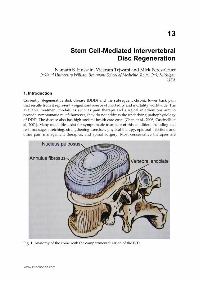
Fig. 1. Anatomy of the spine with the compartmentalization of the IVD.
www.intechopen.com
Tissue Regeneration – From Basic Biology to Clinical Application
284
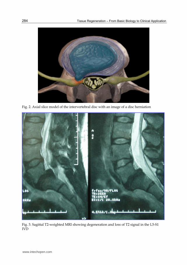
Fig. 2. Axial slice model of the intervertebral disc with an image of a disc herniation
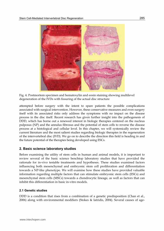
Fig. 3. Sagittal T2-weighted MRI showing degeneration and loss of T2 signal in the L5-S1 IVD
www.intechopen.com
Stem Cell-Mediated Intervertebral Disc Regeneration
285
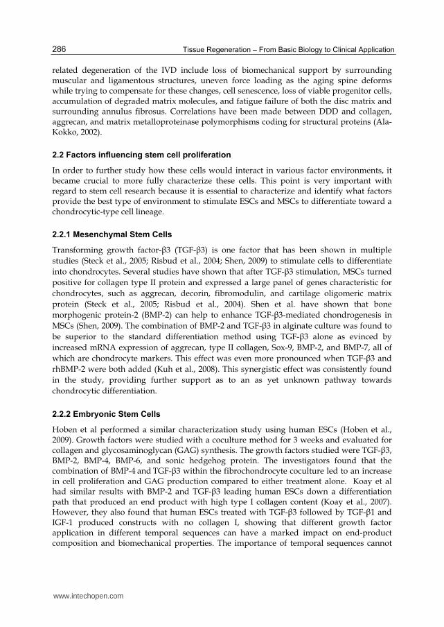
Fig. 4. Postmortem specimen and hematoxylin and eosin staining showing multilevel degeneration of the IVDs with fissuring of the actual disc structure
attempted before surgery with the intent to spare patients the possible complications associated with surgical intervention. However, these conservative measures and even surgery itself with its associated risks only address the symptoms with no impact on the disease process in the disc itself. Recent research has given further insight into the pathogenesis of DDD, which has borne out a renewed interest in biologic therapies centered on the nucleus pulposus (NP) and the annulus fibrosus and the potential of stem cells to reverse the disease process at a histological and cellular level. In this chapter, we will systemically review the current literature and the most salient studies regarding biologic therapies in the regeneration of the intervertebral disc (IVD). We go on to describe the direction this field is heading in and the future potential of the therapies being developed using ESCs.
2. Basic science laboratory studies
Before examining the utility of stem cells in human and animal models, it is important to
review several of the basic science benchtop laboratory studies that have provided the
rationale for in-vivo testable treatments and hypotheses. These studies examined factors
influencing both mesenchymal and embryonic stem cell proliferation and differentiation
towards a NP-like phenotype. We will examine how these studies have provided valuable
information regarding multiple factors that can stimulate embryonic stem cells (ESCs) and
mesenchymal stem cells (MSCs) towards a chondrocytic lineage, as well as factors that can
inhibit this differentiation in basic in-vitro models.
2.1 Genetic studies
DDD is a condition that rises from a combination of a genetic predisposition (Chan et al., 2006) along with environmental modifiers (Stokes & Iatridis, 2004). Several causes of age-
www.intechopen.com
Tissue Regeneration – From Basic Biology to Clinical Application
286
related degeneration of the IVD include loss of biomechanical support by surrounding muscular and ligamentous structures, uneven force loading as the aging spine deforms while trying to compensate for these changes, cell senescence, loss of viable progenitor cells, accumulation of degraded matrix molecules, and fatigue failure of both the disc matrix and surrounding annulus fibrosus. Correlations have been made between DDD and collagen, aggrecan, and matrix metalloproteinase polymorphisms coding for structural proteins (Ala-Kokko, 2002).
2.2 Factors influencing stem cell proliferation
In order to further study how these cells would interact in various factor environments, it became crucial to more fully characterize these cells. This point is very important with regard to stem cell research because it is essential to characterize and identify what factors provide the best type of environment to stimulate ESCs and MSCs to differentiate toward a chondrocytic-type cell lineage.
2.2.1 Mesenchymal Stem Cells
Transforming growth factor-β3 (TGF-β3) is one factor that has been shown in multiple
studies (Steck et al., 2005; Risbud et al., 2004; Shen, 2009) to stimulate cells to differentiate
into chondrocytes. Several studies have shown that after TGF-β3 stimulation, MSCs turned
positive for collagen type II protein and expressed a large panel of genes characteristic for
chondrocytes, such as aggrecan, decorin, fibromodulin, and cartilage oligomeric matrix
protein (Steck et al., 2005; Risbud et al., 2004). Shen et al. have shown that bone
morphogenic protein-2 (BMP-2) can help to enhance TGF-β3-mediated chondrogenesis in
MSCs (Shen, 2009). The combination of BMP-2 and TGF-β3 in alginate culture was found to
be superior to the standard differentiation method using TGF-β3 alone as evinced by
increased mRNA expression of aggrecan, type II collagen, Sox-9, BMP-2, and BMP-7, all of
which are chondrocyte markers. This effect was even more pronounced when TGF-β3 and
rhBMP-2 were both added (Kuh et al., 2008). This synergistic effect was consistently found
in the study, providing further support as to an as yet unknown pathway towards
chondrocytic differentiation.
2.2.2 Embryonic Stem Cells
Hoben et al performed a similar characterization study using human ESCs (Hoben et al., 2009). Growth factors were studied with a coculture method for 3 weeks and evaluated for collagen and glycosaminoglycan (GAG) synthesis. The growth factors studied were TGF-β3, BMP-2, BMP-4, BMP-6, and sonic hedgehog protein. The investigators found that the combination of BMP-4 and TGF-β3 within the fibrochondrocyte coculture led to an increase in cell proliferation and GAG production compared to either treatment alone. Koay et al had similar results with BMP-2 and TGF-β3 leading human ESCs down a differentiation path that produced an end product with high type I collagen content (Koay et al., 2007). However, they also found that human ESCs treated with TGF-β3 followed by TGF-β1 and IGF-1 produced constructs with no collagen I, showing that different growth factor application in different temporal sequences can have a marked impact on end-product composition and biomechanical properties. The importance of temporal sequences cannot
www.intechopen.com
Stem Cell-Mediated Intervertebral Disc Regeneration
287
be understated with regard to stem cell development and has important implications pertaining to harvesting and large-scale production of these cells for future potential therapeutic uses.
2.3 Stem cell growth in the native IVD microenvironment
Several groups have conducted well-designed in-vitro studies that have gone one step beyond identifying environmental factors that affect differentiation of stem cells into NP-like cells, and have actually studied how these factors may correlate to the current in-vivo microenvironment of the IVD. This was done in order to obtain a clear picture of what would happen if these stem cells were implanted into these native biological conditions. Culturing under IVD-like glucose conditions (1.0 mg/mL glucose) stimulated aggrecan and collagen I expression and deposition. IVD-like osmolarity (485 mOsm) and pH (pH = 6.8) conditions, on the other hand, strongly decreased proliferation and expression of matrix proteins. Combining these conditions resulted in decreased proliferation and gene expression of matrix proteins, demonstrating that, in this case, osmolarity and pH play a larger impact in inhibiting differentiation than glucose does in stimulating it (Wuertz et al., 2008).
Another study by the same group showed that acidity caused an inhibition of aggrecan and
collagen I expression, as well as a decrease in proliferation and cell viability. This
demonstrates that pH may be the major limitation for stem cell-based IVD repair (Wuertz,
2009). This also illustrates the importance of early intervention and the role of
predifferentiation when planning to use stem cells for reparative treatments. However,
some studies have shown that implantation of stem cells at a later stage in the DDD process
may result in a greater increase in disc height when compared to implantation at an earlier
stage (Ho et al., 2008). This finding highlights the importance of studies involving stem cell-
based intervertebral disc regeneration being carefully controlled in the context of stage of
disc degeneration. Again, this point highlights the importance of temporal sequence when
examining therapeutics with stem cells. Additionally, inflammatory processes have been
shown to inhibit the chondrogenic differentiation of stem cells, whereas hypoxic conditions
exert beneficial effects on chondrogenesis and phenotype stability of transplanted stem cells
(Felka et al., 2009).
2.4 Optimizing conditions to promote proliferation
There is currently an avid interest in using our accumulated data and knowledge of the factors influencing stem cell proliferation and the exact conditions in the native IVD microenvironment to optimize the chances for stem cell proliferation.
Multiple studies have investigated culturing MSCs with NP cells in a co-culture system, allowing for cell-to-cell contact (Yang et al., 2009; Le Maitre et al., 2009; Vadalà et al., 2009; Richardson et al., 2006; Richardson et al., 2008). This contact has been shown to stimulate these MSCs to differentiate toward a chondrocytic lineage, therefore removing the need for pre-differentiation in-vitro (Watanabe et al., 2010; Svanvik et al., 2010; Niu et al., 2009; Wei et al., 2009; Tao et al., 2008; Le Visage et al., 2006; Richardson et al., 2006). This was evidenced by mRNA expression levels of Type II collagen and aggrecan being elevated in co-cultured cells and cells undergoing morphological changes to form three-dimensional micromasses
www.intechopen.com
Tissue Regeneration – From Basic Biology to Clinical Application
288
expressing collagen-2, aggrecan, and Sox-9 at RNA and protein levels after 14 days of co-culture. These changes were unique and not detected in the samples of stem cells cultured alone (Svanvik et al., 2010; Niu et al., 2009; Wei et al., 2009). Furthermore, MSCs from older individuals differentiate spontaneously into chondrocyte-like NP cells upon insertion into NP tissue in-vitro, and thus may not require additional stimulation to induce differentiation. This is a key finding, as such a strategy would minimize the level of external manipulation required prior to insertion of these cells into the patient, thus simplifying the treatment strategy and reducing costs (Le Maitre et al., 2009).
Adipose-Derived Stem Cells (ADSCs) have also been shown to be able to differentiate into NP cells in multiple in-vitro studies (Xie et al., 2009; Tapp et al., 2008; Lu et al., 2007; Lu et al., 2008; Li et al., 2005). Soluble factors released by NP cells direct chondrogenic differentiation of ADSCs in collagen hydrogels, and combination with a nucleus-mimicking collagen type II microenvironment enhances differentiation towards a more pronounced cartilaginous lineage (Lu et al., 2007; Lu et al., 2008).
Studies using annulus fibrosus cells isolated from nondegenerated intervertebral discs have shown that these cells have the capability of differentiating into adipocytes, osteoblasts, chondrocytes, neurons, and endothelial cells in-vitro. These cells may also be induced to become more plastic, allowing them to differentiate along more mesenchymal lineages (Li et al., 2005; Feng et al., 2010; Saraiya et al., 2010). However, when annulus cells are differentiated into a chondrocyte micromass, it was not as rounded or compact as that which occurs with stem cells induced into chondrocyte differentiation (Saraiya et al., 2010). TGF-β stimulation of fetal cells cultured in high cell density led to the production of aggrecan, type I and II collagens and variable levels of type X collagen, although fetal cells had lower adipogenic and osteogenic differentiation capacity than MSCs and variability in matrix synthesis was observed between specific donors (Quintin et al., 2009; Quintin et al., 2010).
3. Animal studies
Many studies using stem cells for disc regeneration have been performed in a wide array of
animal models with promising results. Two recent studies were conducted utilizing ADSCs
in a murine (Jeong et al., 2010) and a canine model (Ganey et al., 2009). Staining in both
studies demonstrated increased Type II collagen and aggrecan in the transplantation group.
Additionally, at 6 weeks after transplantation, discs exhibited a restoration of disc hydration
and MRI T2 signal intensity and more closely resembled the healthy controls as evidenced
by matrix translucency, compartmentalization of the annulus, and increased cell density
within the nucleus pulposus. Discs also showed a significantly smaller reduction in disc
height when compared with controls.
Multiple studies have shown that MSCs are able to proliferate and survive inside the IVD, with assessments being made as far out as six months post-transplant (Tan et al., 2009; Jeong et al., 2009; Henriksson et al., 2009; Sobajima et al., 2008; Zhang et al., 2005; Crevensten et al., 2004). Additionally, these cells have been proven to differentiate into cells expressing chondrocytic phenotypes, as evidenced by positive immunostaining of collagen type II, aggrecan, and other markers (Henriksson et al., 2009; Yang et al., 2010; Wei et al., 2009; Sakai et al., 2005). Cells were also shown to exhibit NP phenotypic
www.intechopen.com
Stem Cell-Mediated Intervertebral Disc Regeneration
289
markers (Sakai et al., 2005). The injected discs had a central NP-like region which had a close similarity to the normal biconvex structure of the IVD and contained viable chondrocytes forming a matrix like that of the normal disc (Sakai et al., 2003; Revell et al., 2007). Omlor et al. studied the practical phenomenon of transplanted stem cell loss through the actual annular puncture which was used to not only simulate disc damage and herniation but also to inject the stem cells themselves. They made a logical conclusion that IVD regeneration strategies should increasingly focus on annulus reconstruction in order to reduce implant loss due to annular failure (Omlor et al., 2010). Most studies focusing on this point are still ongoing.
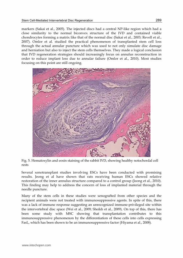
Fig. 5. Hematoxylin and eosin staining of the rabbit IVD, showing healthy notochordal cell rests
Several xenotransplant studies involving ESCs have been conducted with promising results. Jeong et al have shown that rats receiving human ESCs showed relative restoration of the inner annulus structure compared to a control group (Jeong et al., 2010). This finding may help to address the concern of loss of implanted material through the needle puncture.
Many of the stem cells in these studies were xenografted from other species and the
recipient animals were not treated with immunosuppressive agents. In spite of this, there
was a lack of immune response suggesting an unrecognized immune-privileged site within
the intervertebral disc space (Wei et al., 2009; Sheikh et al., 2009). On top of this, there has
been some study with MSC showing that transplantation contributes to this
immunosuppressive phenomenon by the differentiation of these cells into cells expressing
FasL, which has been shown to be an immunosuppressive factor (Hiyama et al., 2008).
www.intechopen.com

Tissue Regeneration – From Basic Biology to Clinical Application
290
Studies Model Intervention Results
Jeong et al. Rat model The first coccygeal disc segments of Sprague-Dawley rat were left undamaged as controls, and other two segments were damaged by needle injection. Two weeks later, stem cells or saline were injected into each of the two damaged segments.
At 6 weeks after transplantation, the experimental group showed a significantly smaller reduction in disc height than the saline-injected group and exhibited a restoration of MRI signal intensity. Hematoxylin and eosin staining revealed a greater restoration of the inner annulus structure. There was also increased collagen type II and aggrecan.
Ganey et al. Canine model 3 discs that had undergone partial nucleotomy were randomized to receive: (1) stem cells in hyaluronic acid carrier (Cells/HA); (2) HA only; or (3) No Intervention.
Disc levels receiving stem cells more closely resembled the healthy controls as evidenced in matrix translucency, compartmentalization of the annulus, and in cell density within the nucleus pulposus. Matrix analysis showed increased Type-II collagen and aggrecan.
Hiyama et al. Canine model 4 weeks after nucleotomy, MSCs were transplanted into the degeneration-induced discs. The animals were followed for 12 weeks when radiological, histological, biochemical, immunohistochemical, and RT-PCR analyses were performed.
MSC transplantation effectively led to the regeneration of degenerated discs. GFP-positive MSCs detected in the NP region 8 weeks after transplantation expressed FasL protein.
Sobajima et al. Rabbit model MSCs were isolated New Zealand White rabbits, retrovirally transduced with the lacZ marker gene, and injected into the nucleus pulposus of the L2-3, L3-4, and L4-5 lumbar discs of 12 other NZW rabbits. Rabbits each were sacrificed at 3, 6, 12, or 24 weeks after cell implantation, and staining
MSCs were detected in histological sections of rabbit discs up to 24 weeks after transplant with engraftment into the inner annulus fibrosus.
www.intechopen.com
Stem Cell-Mediated Intervertebral Disc Regeneration
291
Studies Model Intervention Results
was done to assess cell survival and localization.
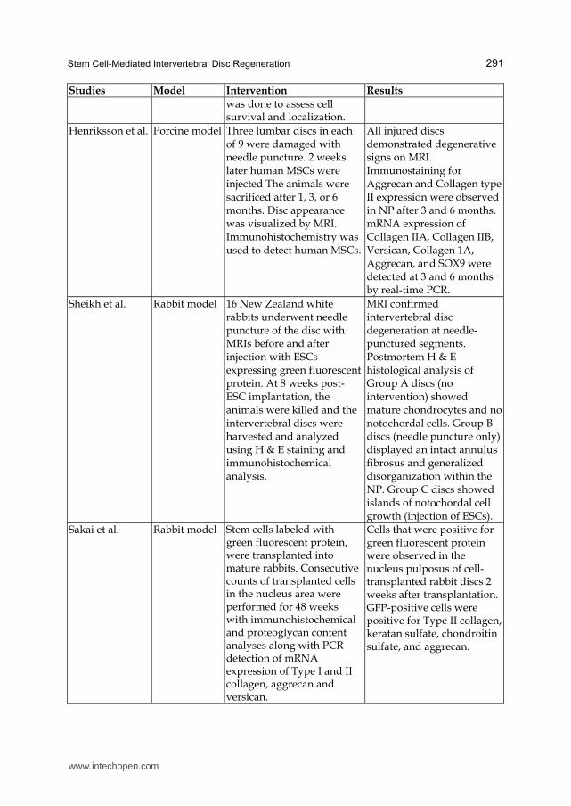
Henriksson et al. Porcine model Three lumbar discs in each of 9 were damaged with needle puncture. 2 weeks later human MSCs were injected The animals were sacrificed after 1, 3, or 6 months. Disc appearance was visualized by MRI. Immunohistochemistry was used to detect human MSCs.
All injured discs demonstrated degenerative signs on MRI. Immunostaining for Aggrecan and Collagen type II expression were observed in NP after 3 and 6 months. mRNA expression of Collagen IIA, Collagen IIB, Versican, Collagen 1A, Aggrecan, and SOX9 were detected at 3 and 6 months by real-time PCR.
Sheikh et al. Rabbit model 16 New Zealand white rabbits underwent needle puncture of the disc with MRIs before and after injection with ESCs expressing green fluorescent protein. At 8 weeks post-ESC implantation, the animals were killed and the intervertebral discs were harvested and analyzed using H & E staining and immunohistochemical analysis.
MRI confirmed intervertebral disc degeneration at needle-punctured segments. Postmortem H & E histological analysis of Group A discs (no intervention) showed mature chondrocytes and no notochordal cells. Group B discs (needle puncture only) displayed an intact annulus fibrosus and generalized disorganization within the NP. Group C discs showed islands of notochordal cell growth (injection of ESCs).
Sakai et al. Rabbit model Stem cells labeled with green fluorescent protein, were transplanted into mature rabbits. Consecutive counts of transplanted cells in the nucleus area were performed for 48 weeks with immunohistochemical and proteoglycan content analyses along with PCR detection of mRNA expression of Type I and II collagen, aggrecan and versican.
Cells that were positive for green fluorescent protein were observed in the nucleus pulposus of cell-transplanted rabbit discs 2 weeks after transplantation. GFP-positive cells were positive for Type II collagen, keratan sulfate, chondroitin sulfate, and aggrecan.
www.intechopen.com
Tissue Regeneration – From Basic Biology to Clinical Application
292
Studies Model Intervention Results
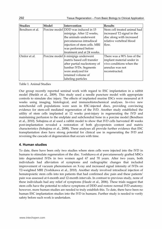
Bendtsen et al. Porcine model DDD was induced in 15 minipigs. After 12 weeks, the animals underwent percutaneous intradiscal injection of stem cells. MRI was performed before treatment and at 24 weeks.
Stem cell treated animal had increased T2 signal in the disc along with increased relative vertebral blood flow.
Omlor et al. Porcine model 6 minipigs underwent matrix based cell transfer after partial nucleotomy of lumbar IVDs. Segments were analyzed for retained volume of labeling particles
There was a 90% loss of the implant material under in vivo conditions when the annulus was not reconstructed.
Table 1. Animal Studies
Our group recently reported seminal work with regard to ESC implantation in a rabbit model (Sheikh et al., 2009). This study used a needle puncture model with appropriate controls to simulate disc injury. The effects of implanted murine ESCs were measured at 8 weeks using imaging, histological, and immunohistochemical analyses. In-vivo new notochordal cell populations were seen in ESC-injected discs, providing convincing evidence for stem-cell mediated regeneration of the IVD. Another study established the utility of stem cells implanted at 12 weeks post-injury in regenerating the IVD and maintaining perfusion to the endplate and subchondral bone in a porcine model (Bendtsen et al., 2010). Sobajima et al used a rabbit model to show that IVD cells harvested 48 weeks post-implantation revealed a restoration of both glycoprotein content and matrix characteristics (Sobajima et al., 2008). These analyses all provide further evidence that ESC transplantation does have strong potential for clinical use in regenerating the IVD and reversing the cascade of degeneration that occurs with time.
4. Human studies
To date, there have been only two studies where stem cells were injected into the IVD in
humans to stimulate regeneration of the disc. Yoshikawa et al percutaneously grafted MSCs
into degenerated IVDs in two women aged 67 and 70 years. After two years, both
individuals had alleviation of symptoms and radiographic changes that included
improvement of vacuum phenomenon on X-ray and increased signal intensity of IVDs on
T2-weighted MRI (Yoshikawa et al., 2010). Another study involved intradiscal injection of
hematopoietic stem cells into ten patients that had confirmed disc pain and these patients’
pain was assessed at 6-month and 12-month intervals. In contrast to previous study, none of
these individuals had any relief of symptoms (Haufe et al., 2006). These trials suggest that
stem cells have the potential to relieve symptoms of DDD and restore normal IVD anatomy;
however, more human studies are needed to truly establish this. To date, there have been no
human ESC implantation studies into the IVD in humans. Further study is needed to verify
safety before such work is undertaken.
www.intechopen.com
Stem Cell-Mediated Intervertebral Disc Regeneration
293
Studies Subjects Intervention Results Study Critique
Yoshikawa et al. 2 patients Percutaneous stem cell grafting
Clinical symptoms improved; increased T2 signal in the disc space on MRI
Few patients
Haufe et al. 10 patients Percutaneous stem cell grafting
No clinical symptom relief
No imaging conducted
Table 2. Human Studies
5. Future potential of ESCs
Although many laboratory and animal studies have been performed utilizing stem cells for the purposes of cell characterization and inducing chondrocyte formation, much further study is needed before human trials are undertaken on a larger scale. Several studies have already showcased the ability of ESCs to differentiate towards a chondrocytic lineage in-vitro and also to improve DDD in in-vivo animal and human trials, using a combination of imaging and histological analyses. Several benchtop lab studies have been performed to show that ESCs can be successfully stimulated to differentiate into chondrocyte-like cells (Hoben et al., 2009; Fecek et al., 2008; Hegert et al., 2002; Kawaguchi et al., 2005; zur Nieden et al., 2005; Kramer et al., 2000). Similar to the case with MSCs, different factors affect this process in ESCs, such as TGF-β3, BMP-2, and BMP-4 (Hegert et al., 2002; Kawaguchi et al., 2005; zur Nieden et al., 2005; Kramer et al., 2000; Sakai et al., 2005). Biological scaffolds seeded with chondrocytic cells derived from ESCs, when implanted in mice have been shown to generate cartilage tissue in-vivo (Kramer et al., 2000). Injection of ESCs in a DDD-induced rabbit model led to viable notochordal-type cells within the discs (Sheikh et al., 2009). These animal studies demonstrate the ability of ESCs to differentiate into a chondrocytic lineage in-vitro and in-vivo.
Our group is currently developing chondroprogenitor stem cell lines that can restore the functional capability of the IVD (Sheikh et al., 2009). Our rationale stemmed from the idea that currently there is no biologic therapy for repairing a degenerated IVD and that ESCs have a potential to fill this role based on their regenerative potential. Studies have shown that ESCs can be induced to differentiate into specific cell lineages by using selective culture media and growth environments (Kawaguchi et al., 2005).
Relying on the significant strides made by these basic science groups with regard to cell and
factor characterization, our lab proceeded for further refine these methods and develop a
protocol for both stem cell differentiation along a chondrocytic lineage and also for
examining the utility of transplantation of these cells in a rabbit model of DDD. We initially
developed a novel percutaneous animal model of disc degeneration using New Zealand
white rabbits (Figure 1) and used this model to explore the possibility of ESC implantation
for both structural regeneration and for the growth and continued presence of notochordal
stem cells in the disc space (Sheikh et al., 2009).
Previous research transplanting MSCs into degenerated rabbit discs has shown consistent biochemical and radiographic (MRI) evidence of IVD restoration (Sakai et al., 2005). Human
www.intechopen.com
Tissue Regeneration – From Basic Biology to Clinical Application
294
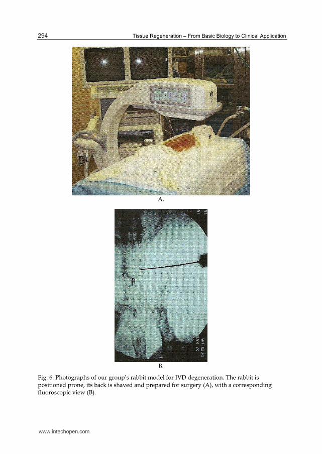
A.
B.
Fig. 6. Photographs of our group’s rabbit model for IVD degeneration. The rabbit is positioned prone, its back is shaved and prepared for surgery (A), with a corresponding fluoroscopic view (B).
www.intechopen.com
Stem Cell-Mediated Intervertebral Disc Regeneration
295
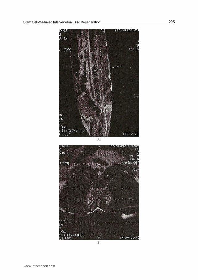
A.
B.
www.intechopen.com
Tissue Regeneration – From Basic Biology to Clinical Application
296
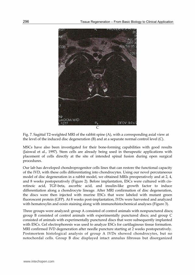
C
Fig. 7. Sagittal T2-weighted MRI of the rabbit spine (A), with a corresponding axial view at the level of the induced disc degeneration (B) and at a separate normal control level (C).
MSCs have also been investigated for their bone-forming capabilities with good results (Jaiswal et al., 1997). Stem cells are already being used in therapeutic applications with placement of cells directly at the site of intended spinal fusion during open surgical procedures.
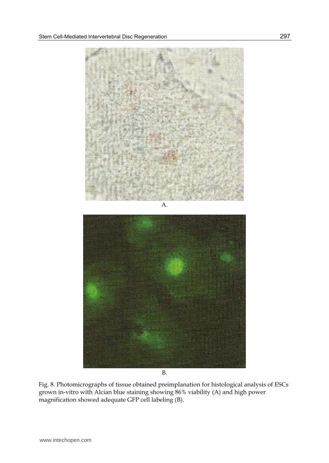
Our lab has developed chondroprogenitor cells lines that can restore the functional capacity of the IVD, with these cells differentiating into chondrocytes. Using our novel percutaneous model of disc degeneration in a rabbit model, we obtained MRIs preoperatively and at 2, 4, and 8 weeks postoperatively (Figure 2). Before implantation, ESCs were cultured with cis-retinoic acid, TGF-beta, ascorbic acid, and insulin-like growth factor to induce differentiation along a chondrocyte lineage. After MRI confirmation of disc degeneration, the discs were then injected with murine ESCs that were labeled with mutant green fluoroscent protein (GFP). At 8 weeks post-implantation, IVDs were harvested and analyzed with hematoxylin and eosin staining along with immunohistochemical analyses (Figure 3).
Three groups were analyzed: group A consisted of control animals with nonpunctured discs; group B consisted of control animals with experimentally punctured discs; and group C consisted of animals with experimentally punctured discs that were subsequently implanted with ESCs. Gel electrophoresis was used to analyze ESCs for cartilaginous tissue formation. MRI confirmed IVD degeneration after needle puncture starting at 2 weeks postoperatively. Postmortem histological analysis of group A IVDs showed chondrocytes, but no notochordal cells. Group B disc displayed intact annulus fibrosus but disorganized
www.intechopen.com
Stem Cell-Mediated Intervertebral Disc Regeneration
297
A.
B.
Fig. 8. Photomicrographs of tissue obtained preimplanation for histological analysis of ESCs grown in-vitro with Alcian blue staining showing 86% viability (A) and high power magnification showed adequate GFP cell labeling (B).
www.intechopen.com
Tissue Regeneration – From Basic Biology to Clinical Application
298
fibrous tissue in the NP. Group C discs showed new notochordal cell growth, indicating
survival and proper differentiation of the injected ESCs. Fluorescent microscopic analysis
was positive in group C tissue, confirming the viability of GFP-labeled ESCs within the
injected IVD. In addition, the notochordal cells in group C stained positive for cytokeratin
and vimentin, providing further evidence of their chondrocyte origin. There was no
inflammatory response in group C discs, indicating no cell-mediated immune response.
Our study provides a novel, reproducible model for the study of disc degeneration. New
notochordal cell populations were seen in discs injected with ESCs. The lack of an immune
response to xenograft-implanted mouse stem cells in an immune-competent rabbit suggests
an immunoprivileged site within the IVD. Although preliminary, this study highlights the
possible use of stem cells to promote IVD regeneration. Further ongoing studies are in the
process of fully elucidating the processes involved with ESC differentiation along
chondrogenic cell lines and how they may be used for new disc formation in the future.
These studies will provide a good deal of evidence with regard to the future potential of
ESCs for use in restoring the IVD in humans.
6. Summary
DDD is a high-morbidity condition with many modalities of treatment including surgery
and more conservative measures such as pain injections, which only provide symptomatic
treatment. No therapy has been developed that targets DDD at the cellular level. Recently,
many biologic therapies have emerged that may be able to restore the NP and the normal
cellular structure of the IVD. This restoration may in turn alleviate the symptoms of DDD
through restoration of foraminal height, removing the compression of nerves. In-vitro
studies have been performed to identify what cells are capable of differentiating towards
a chondrocytic lineage and to best define parameters and factors that influence this
differentiation. Multiple laboratory studies have been performed showing that MSCs,
ADSCs, fetal cartilaginous cells, and annulus fibrosus cells all have the ability to
differentiate towards a chondrocytic pathway. Factors that can induce these cells to
differentiate toward a chondrocytic lineage have been identified and include TGF-β3 and
BMP-2, which have a synergistic effect when used together. Other factors that may be
beneficial include hypoxia, IVD-like glucose conditions (1.0 mg/mL glucose), and cell-to-
cell contact with NP cells; the latter negating the need for other soluble factors (i.e. TGF-
β3). A major limiting factor may be the acidic pH (6.8) of the IVD, one that may be
especially important as acidic pH levels are typical of increasingly degenerated discs.
These studies yielded encouraging results with cells in the IVD being positive for markers
of chondrocytic differentiation such as collagen type II and aggrecan. Additionally, cells
exhibited NP phenotypic markers and had a close similarity to the normal biconvex
structure of the NP. In-vitro studies have clearly established that ESCs are capable of
differentiating into a chondrocytic lineage and have delineated some of the factors that
affect this. The optimal microenvironment needs to be more accurately characterized at
this time.
Animal studies of cell implantation have been performed in DDD-induction models. Weeks after injury, stem cells have been implanted and outcomes followed. These outcomes which
www.intechopen.com
Stem Cell-Mediated Intervertebral Disc Regeneration
299
have included radiographic analyses along with histological and immunohistochemical analyses have provided preliminary data that stem cell therapies are a viable option with regard to IVD regeneration (Sheikh et al., 2009). Human studies have further provided some preliminary evidence that stem cell therapy may be of clinical value (Haufe et al., 2006). The use of ESCs in regenerating IVD shows exciting new possibilities and further studies are needed in humans to establish its efficacy.
ESC-based regeneration of the human IVD is still in its infancy. Much progress has been made regarding laboratory research identifying the correct factors and microenvironment, and initial results from animal studies using stem cells remain promising. ESCs may be useful for repairing DDD as evidenced by their ability to differentiate into a chondrocytic lineage and yield notochordal-type cells in DDD models. ESCs need to be further studied and characterized with respect to safety, and larger human trials with appropriate clinical outcomes such as pain and disability reduction are needed to definitively establish its clinical efficacy.
7. Conclusions
The last half-century has seen an exponential rate of progress with regard to elucidating the mechanisms of degeneration of the IVD and how targeted therapies can help to alleviate this common condition. These studies have provided us with an improved understanding of the IVD and how it behaves under typical biomechanical forces and loads experienced in in-vivo conditions. Novel therapies are being studied, including stem cells with their potential regenerative capabilities in the spine. The development and action of these stem cells can be further modified through gene therapy and microenvironment manipulation. Immunologic markers are being used for more efficient targeting of these cells. With enhanced cell delivery and an improved understanding of the cell differentiation process, true regeneration of the IVD and surrounding supportive structures of the spine will become a reality that can be applied to treat patients with this common, debilitating condition.
8. References
[1] Chan D, Song Y, Sham P. Genetics of disc degeneration. Eur Spine J. 15(Suppl 3):S317-
S325. 2006.
[2] Cassinelli EH, Hall RA, Kang JD. Biochemistry of intervertebral disc degeneration and
the potential for gene therapy applications. Spine J. 1:205-214, 2001.
[3] Stokes IA, Iatridis JC. Mechanical conditions that accelerate intervertebral disc
degeneration: overload versus immobilization. Spine 29:2724-2732, 2004.
[4] Ala-Kokko L. Genetic risk factors for lumbar disc disease. Ann Med. 34:42-47, 2002.
[5] Steck E, Bertram H, Abel R, Chen B, Winter A, Richter W. Induction of intervertebral
disc-like cells from adult mesenchymal stem cells. Stem Cells. 2005 Mar;23(3):403-11.
[6] Risbud MV, Albert TJ, Guttapalli A, Vresilovic EJ, Hillibrand AS, Vaccaro AR,Shapiro
IM. Differentiation of mesenchymal stem cells towards a nucleus pulposus-like
phenotype in vitro: implications for cell-based transplantation therapy. Spine (Phila
Pa 1976). 2004 Dec 1;29(23):2627-32.
www.intechopen.com
Tissue Regeneration – From Basic Biology to Clinical Application
300
[7] Shen B, Wei A, Tao H, Diwan AD, Ma DD. BMP-2 enhances TGF-beta3-mediated
chondrogenic differentiation of human bone marrow multipotent mesenchymal
stromal cells in alginate bead culture. Tissue Eng Part A. 2009 Jun;15(6):1311-20.
[8] Kuh SU, Zhu Y, Li J, Tsai KJ, Fei Q, Hutton WC, Yoon ST. Can TGF-beta1 and rhBMP-2
act in synergy to transform bone marrow stem cells to discogenic-type cells? Acta
Neurochir (Wien). 2008 Oct;150(10):1073-9; discussion 1079.
[9] Hoben GM, Willard VP, Athanasiou KA. Fibrochondrogenesis of hESCs: growth factor
combinations and cocultures. Stem Cells Dev. 2009 Mar;18(2):283-92.
[10] Koay EJ, Hoben GM, Athanasiou KA. Tissue engineering with chondrogenically
differentiated human embryonic stem cells. Stem Cells. 2007 Sep;25(9):2183-90.
[11] Wuertz K, Godburn K, Neidlinger-Wilke C, Urban J, Iatridis JC. Behavior of
mesenchymal stem cells in the chemical microenvironment of the intervertebral
disc. Spine (Phila Pa 1976). 2008 Aug 1;33(17):1843-9.
[12] Wuertz K, Godburn K, Iatridis JC. MSC response to pH levels found in degenerating
intervertebral discs. Biochem Biophys Res Commun. 2009 Feb 20;379(4):824-9.
[13] Ho G, Leung VY, Cheung KM, Chan D. Effect of severity of intervertebral disc injury
on mesenchymal stem cell-based regeneration. Connect Tissue Res. 2008;49(1):15-
21.
[14] Felka T, Schäfer R, Schewe B, Benz K, Aicher WK. Hypoxia reduces the inhibitory effect
of IL-1beta on chondrogenic differentiation of FCS-free expanded MSC.
Osteoarthritis Cartilage. 2009 Oct;17(10):1368-76. Epub 2009 May 8.
[15] Yang F, Leung VY, Luk KD, Chan D, Cheung KM. Mesenchymal stem cells arrest
intervertebral disc degeneration through chondrocytic differentiation and
stimulation of endogenous cells. Mol Ther. 2009 Nov;17(11):1959-66.
[16] Le Maitre CL, Baird P, Freemont AJ, Hoyland JA. An in vitro study investigating the
survival and phenotype of mesenchymal stem cells following injection into nucleus
pulposus tissue. Arthritis Res Ther. 2009;11(1):R20.
[17] Vadalà G, Studer RK, Sowa G, Spiezia F, Iucu C, Denaro V, Gilbertson LG, Kang JD.
Coculture of bone marrow mesenchymal stem cells and nucleus pulposus cells
modulate gene expression profile without cell fusion. Spine (Phila Pa 1976). 2008
Apr 15;33(8):870-6.
[18] Richardson SM, Hughes N, Hunt JA, Freemont AJ, Hoyland JA. Human mesenchymal
stem cell differentiation to NP-like cells in chitosan-glycerophosphate hydrogels.
Biomaterials. 2008 Jan;29(1):85-93.
[19] Richardson SM, Curran JM, Chen R, Vaughan-Thomas A, Hunt JA, Freemont AJ,
Hoyland JA. The differentiation of bone marrow mesenchymal stem cells into
chondrocyte-like cells on poly-L-lactic acid (PLLA) scaffolds. Biomaterials. 2006
Aug;27(22):4069-78.
[20] Watanabe T, Sakai D, Yamamoto Y, Iwashina T, Serigano K, Tamura F, Mochida J.
Human nucleus pulposus cells significantly enhanced biological properties in a
coculture system with direct cell-to-cell contact with autologous mesenchymal stem
cells. J Orthop Res. 2010 May;28(5):623-30.
www.intechopen.com
Stem Cell-Mediated Intervertebral Disc Regeneration
301
[21] Svanvik T, Henriksson HB, Karlsson C, Hagman M, Lindahl A, Brisby H. Human disk
cells from degenerated disks and mesenchymal stem cells in co-culture result in
increased matrix production. Cells Tissues Organs. 2010;191(1):2-11.
[22] Niu CC, Yuan LJ, Lin SS, Chen LH, Chen WJ. Mesenchymal stem cell and nucleus
pulposus cell coculture modulates cell profile. Clin Orthop Relat Res. 2009
Dec;467(12):3263-72.
[23] Wei A, Chung SA, Tao H, Brisby H, Lin Z, Shen B, Ma DD, Diwan AD. Differentiation
of rodent bone marrow mesenchymal stem cells into intervertebral disc-like cells
following coculture with rat disc tissue. Tissue Eng Part A. 2009 Sep;15(9):2581-95.
[24] Tao F, Li F, Li G, Pan F. Differentiation of mesenchymal stem cells into nucleus
pulposus cells in vitro. J Huazhong Univ Sci Technolog Med Sci. 2008
Apr;28(2):156-8.
[25] Le Visage C, Kim SW, Tateno K, Sieber AN, Kostuik JP, Leong KW. Interaction of
human mesenchymal stem cells with disc cells: changes in extracellular matrix
biosynthesis. Spine (Phila Pa 1976). 2006 Aug 15;31(18):2036-42.
[26] Richardson SM, Walker RV, Parker S, Rhodes NP, Hunt JA, Freemont AJ, Hoyland JA.
Intervertebral disc cell-mediated mesenchymal stem cell differentiation. Stem Cells.
2006 Mar;24(3):707-16.
[27] Xie LW, Fang H, Chen AM, Li F. Differentiation of rat adipose tissue-derived
mesenchymal stem cells towards a nucleus pulposus-like phenotype in vitro. Chin J
Traumatol. 2009 Apr;12(2):98-103.
[28] Tapp H, Deepe R, Ingram JA, Kuremsky M, Hanley EN Jr, Gruber HE. Adipose-
derived mesenchymal stem cells from the sand rat: transforming growth factor
beta and 3D co-culture with human disc cells stimulate proteoglycan and
collagen type I rich extracellular matrix. Arthritis Res Ther. 2008;10(4):R89. Epub
2008 Aug 11.
[29] Lu ZF, Doulabi BZ, Wuisman PI, Bank RA, Helder MN. Influence of collagen type II
and nucleus pulposus cells on aggregation and differentiation of adipose tissue-
derived stem cells. J Cell Mol Med. 2008 Dec;12(6B):2812-22.
[30] Lu ZF, Zandieh Doulabi B, Wuisman PI, Bank RA, Helder MN. Differentiation of
adipose stem cells by nucleus pulposus cells: configuration effect. Biochem Biophys
Res Commun. 2007 Aug 10;359(4):991-6. Epub 2007 Jun 8.
[31] Li X, Lee JP, Balian G, Greg Anderson D. Modulation of chondrocytic properties of fat-
derived mesenchymal cells in co-cultures with nucleus pulposus. Connect Tissue
Res. 2005;46(2):75-82.
[32] Feng G, Yang X, Shang H, Marks IW, Shen FH, Katz A, Arlet V, Laurencin CT, Li X.
Multipotential differentiation of human anulus fibrosus cells: an in vitro study. J
Bone Joint Surg Am. 2010 Mar;92(3):675-85.
[33] Saraiya M, Nasser R, Zeng Y, Addya S, Ponnappan RK, Fortina P, Anderson DG, Albert
TJ, Shapiro IM, Risbud MV. Reversine enhances generation of progenitor-like cells
by dedifferentiation of annulus fibrosus cells. Tissue Eng Part A. 2010
Apr;16(4):1443-55.
[34] Gruber HE, Chow Y, Hoelscher GL, Ingram JA, Zinchenko N, Norton HJ, Sun Y,
Hanley EN Jr. Micromass culture of human annulus cells: morphology and
www.intechopen.com
Tissue Regeneration – From Basic Biology to Clinical Application
302
extracellular matrix production. Spine (Phila Pa 1976). 2010 May 1;35(10):
1033-8.
[35] Quintin A, Schizas C, Scaletta C, Jaccoud S, Applegate LA, Pioletti DP. Plasticity of fetal
cartilaginous cells. Cell Transplant. 2010;19(10):1349-57.
[36] Quintin A, Schizas C, Scaletta C, Jaccoud S, Gerber S, Osterheld MC, Juillerat L,
Applegate LA, Pioletti DP. Isolation and in vitro chondrogenic potential of human
foetal spine cells. J Cell Mol Med. 2009 Aug;13(8B):2559-69.
[37] Jeong JH, Lee JH, Jin ES, Min JK, Jeon SR, Choi KH. Regeneration of intervertebral discs
in a rat disc degeneration model by implanted adipose-tissue-derived stromal cells.
Acta Neurochir (Wien). 2010 Oct;152(10):1771-7.
[38] Ganey T, Hutton WC, Moseley T, Hedrick M, Meisel HJ. Intervertebral disc repair using
adipose tissue-derived stem and regenerative cells: experiments in a canine model.
Spine (Phila Pa 1976). 2009 Oct 1;34(21):2297-304.
[39] Tan S, Jia C, Liu Z, Liu R, Yang J, Zhang L, Shou F, Ju X. Study on survival time of
autogeneic BMSCs labeled with superparamagnetic iron oxide in rabbit
intervertebral discs. Zhongguo Xiu Fu Chong Jian Wai Ke Za Zhi. 2009
Nov;23(11):1355-9.
[40] Jeong JH, Jin ES, Min JK, Jeon SR, Park CS, Kim HS, Choi KH. Human mesenchymal
stem cells implantation into the degenerated coccygeal disc of the rat.
Cytotechnology. 2009 Jan;59(1):55-64.
[41] Henriksson HB, Svanvik T, Jonsson M, Hagman M, Horn M, Lindahl A, Brisby H.
Transplantation of human mesenchymal stems cells into intervertebral discs in a
xenogeneic porcine model. Spine (Phila Pa 1976). 2009 Jan 15;34(2):141-8.
[42] Sobajima S, Vadala G, Shimer A, Kim JS, Gilbertson LG, Kang JD. Feasibility of a stem
cell therapy for intervertebral disc degeneration. Spine J. 2008 Nov-Dec;8(6):888-
96.
[43] Zhang YG, Guo X, Xu P, Kang LL, Li J. Bone mesenchymal stem cells transplanted into
rabbit intervertebral discs can increase proteoglycans. Clin Orthop Relat Res. 2005
Jan;(430):219-26.
[44] Crevensten G, Walsh AJ, Ananthakrishnan D, Page P, Wahba GM, Lotz JC, Berven S.
Intervertebral disc cell therapy for regeneration: mesenchymal stem cell
implantation in rat intervertebral discs. Ann Biomed Eng. 2004 Mar;32(3):430-4.
[45] Yang H, Wu J, Liu J, Ebraheim M, Castillo S, Liu X, Tang T, Ebraheim NA. Transplanted
mesenchymal stem cells with pure fibrinous gelatin-transforming growth factor-
beta1 decrease rabbit intervertebral disc degeneration. Spine J. 2010 Sep;10(9):802-
10. Epub 2010 Jul 24.
[46] Wei A, Tao H, Chung SA, Brisby H, Ma DD, Diwan AD. The fate of transplanted
xenogeneic bone marrow-derived stem cells in rat intervertebral discs. J Orthop Res.
2009 Mar;27(3):374-9.
[47] Sakai D, Mochida J, Iwashina T, Watanabe T, Nakai T, Ando K, Hotta T. Differentiation
of mesenchymal stem cells transplanted to a rabbit degenerative disc model:
potential and limitations for stem cell therapy in disc regeneration. Spine (Phila Pa
1976). 2005 Nov 1;30(21):2379-87.
www.intechopen.com
Stem Cell-Mediated Intervertebral Disc Regeneration
303
[48] Sakai D, Mochida J, Yamamoto Y, Nomura T, Okuma M, Nishimura K, Nakai T, Ando
K, Hotta T. Transplantation of mesenchymal stem cells embedded in Atelocollagen
gel to the intervertebral disc: a potential therapeutic model for disc degeneration.
Biomaterials. 2003 Sep;24(20):3531-41.
[49] Revell PA, Damien E, Di Silvio L, Gurav N, Longinotti C, Ambrosio L. Tissue
engineered intervertebral disc repair in the pig using injectable polymers. J Mater
Sci Mater Med. 2007 Feb;18(2):303-8.
[50] Omlor GW, Bertram H, Kleinschmidt K, Fischer J, Brohm K, Guehring T, Anton M,
Richter W. Methods to monitor distribution and metabolic activity of mesenchymal
stem cells following in vivo injection into nucleotomized porcine intervertebral
discs. Eur Spine J. 2010 Apr;19(4):601-12.
[51] Sheikh H, Zakharian K, De La Torre RP, Facek C, Vasquez A, Chaudhry GR, Svinarich
D, Perez-Cruet MJ. In vivo intervertebral disc regeneration using stem cell-derived
chondroprogenitors. J Neurosurg Spine. 2009 Mar;10(3):265-72.
[52] Hiyama A, Mochida J, Iwashina T, Omi H, Watanabe T, Serigano K, Tamura F, Sakai D.
Transplantation of mesenchymal stem cells in a canine disc degeneration model. J
Orthop Res. 2008 May;26(5):589-600.
[53] Bendtsen M, Bunger CE, Zou X, Foldager C, Joregensen HS. Autologous stem cell
therapy maintains vertebral blood flow and contrast diffusion through the endplate
in experimental IDD. Spine (Phila Pa 1976). 2010 Dec 29.
[54] Yoshikawa T, Ueda Y, Miyazaki K, Koizumi M, Takakura Y. Disc regeneration therapy
using marrow mesenchymal cell transplantation: a report of two case studies. Spine
(Phila Pa 1976). 2010 May 15;35(11):E475-80.
[55] Haufe SM, Mork AR. Intradiscal injection of hematopoietic stem cells in an attempt to
rejuvenate the intervertebral discs. Stem Cells Dev. 2006 Feb;15(1):136-7.
[56] Fecek C, Yao D, Kaçorri A, Vasquez A, Iqbal S, Sheikh H, Svinarich DM, Perez-Cruet M,
Chaudhry GR. Chondrogenic derivatives of embryonic stem cells seeded into 3D
polycaprolactone scaffolds generated artilage tissue in vivo. Tissue Eng Part A. 2008
Aug;14(8):1403-13.
[57] Hegert C, Kramer J, Hargus G, Müller J, Guan K, Wobus AM, Müller PK, Rohwedel J.
Differentiation plasticity of chondrocytes derived from mouse embryonic stem
cells. J Cell Sci. 2002 Dec 1;115(Pt 23):4617-28.
[58] Kawaguchi J, Mee PJ, Smith AG. Osteogenic and chondrogenic differentiation of
embryonic stem cells in response to specific growth factors. Bone. 2005
May;36(5):758-69. Epub 2005 Mar 24.
[59] zur Nieden NI, Kempka G, Rancourt DE, Ahr HJ. Induction of chondro-, osteo- and
adipogenesis in embryonic stem cells by bone morphogenetic protein-2: effect of
cofactors on differentiating lineages. BMC Dev Biol. 2005 Jan 26;5:1.
[60] Kramer J, Hegert C, Guan K, Wobus AM, Müller PK, Rohwedel J. Embryonic stem cell-
derived chondrogenic differentiation in vitro: activation by BMP-2 and BMP-4.
Mech Dev. 2000 Apr;92(2):193-205.
[61] Sakai D, Mochida J, Iwashina T. Differentiation of mesenchymal stem cells transplanted
to a rabbit degenerative disc model: potential and limitations for stem cell therapy
in disc regeneration. Spine 30: 2379-2387. 2005.
www.intechopen.com

Tissue Regeneration – From Basic Biology to Clinical Application
304
[62] Jaiswal N, Haynesworht S, Caplan A. Osteogenic differentiation of purified culture
expanded human mesenchymal stem cells in vitro. J Cell Biochem 64: 295-312,
1997.
www.intechopen.com
Tissue Regeneration - From Basic Biology to Clinical ApplicationEdited by Prof. Jamie Davies
ISBN 978-953-51-0387-5Hard cover, 512 pagesPublisher InTechPublished online 30, March, 2012Published in print edition March, 2012
InTech EuropeUniversity Campus STeP Ri Slavka Krautzeka 83/A 51000 Rijeka, Croatia Phone: +385 (51) 770 447 Fax: +385 (51) 686 166www.intechopen.com
InTech ChinaUnit 405, Office Block, Hotel Equatorial Shanghai No.65, Yan An Road (West), Shanghai, 200040, China
Phone: +86-21-62489820 Fax: +86-21-62489821
When most types of human tissue are damaged, they repair themselves by forming a scar - a mechanicallystrong 'patch' that restores structural integrity to the tissue without restoring physiological function. Muchbetter, for a patient, would be like-for-like replacement of damaged tissue with something functionallyequivalent: there is currently an intense international research effort focused on this goal. This timely bookaddresses key topics in tissue regeneration in a sequence of linked chapters, each written by world experts;understanding normal healing; sources of, and methods of using, stem cells; construction and use of scaffolds;and modelling and assessment of regeneration. The book is intended for an audience consisting of advancedstudents, and research and medical professionals.
How to referenceIn order to correctly reference this scholarly work, feel free to copy and paste the following:
Namath S. Hussain, Vickram Tejwani and Mick Perez-Cruet (2012). Stem Cell-Mediated Intervertebral DiscRegeneration, Tissue Regeneration - From Basic Biology to Clinical Application, Prof. Jamie Davies (Ed.),ISBN: 978-953-51-0387-5, InTech, Available from: http://www.intechopen.com/books/tissue-regeneration-from-basic-biology-to-clinical-application/stem-cell-mediated-intervertebral-disc-regeneration

© 2012 The Author(s). Licensee IntechOpen. This is an open access articledistributed under the terms of the Creative Commons Attribution 3.0License, which permits unrestricted use, distribution, and reproduction inany medium, provided the original work is properly cited.