Embed Size (px)
Citation preview
Steven J. Zehren, Ph.D.
TEMPORAL & INFRATEMPORAL FOSSAE; TMJ

OSTEOLOGY
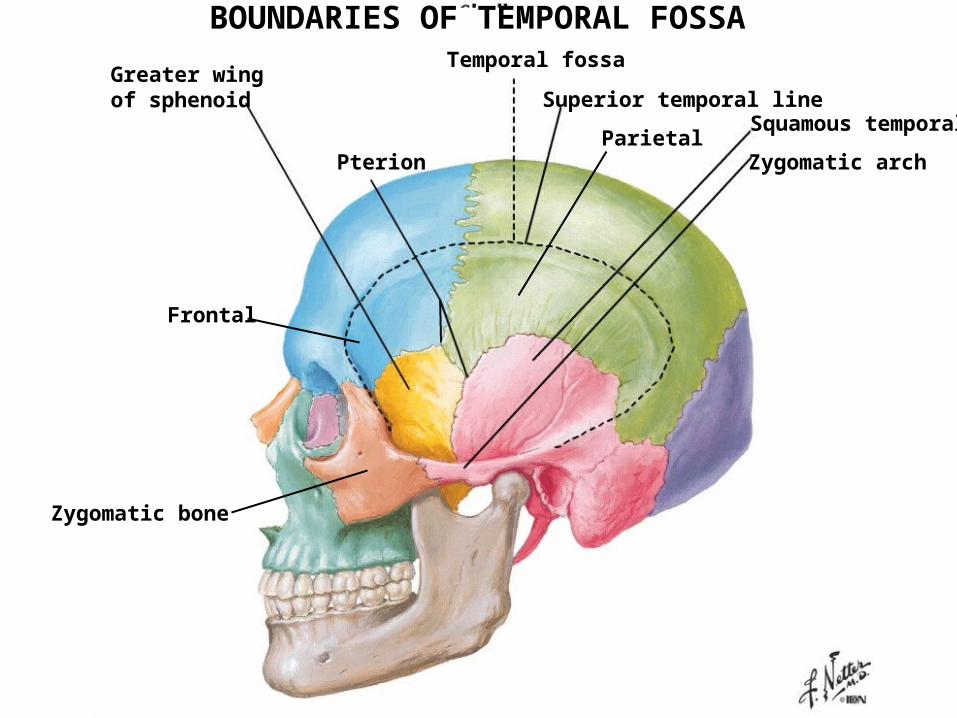
BOUNDARIES OF TEMPORAL FOSSATemporal fossa
Superior temporal line
Zygomatic arch
Squamous temporalParietal
Pterion
Greater wingof sphenoid
Frontal
Zygomatic bone
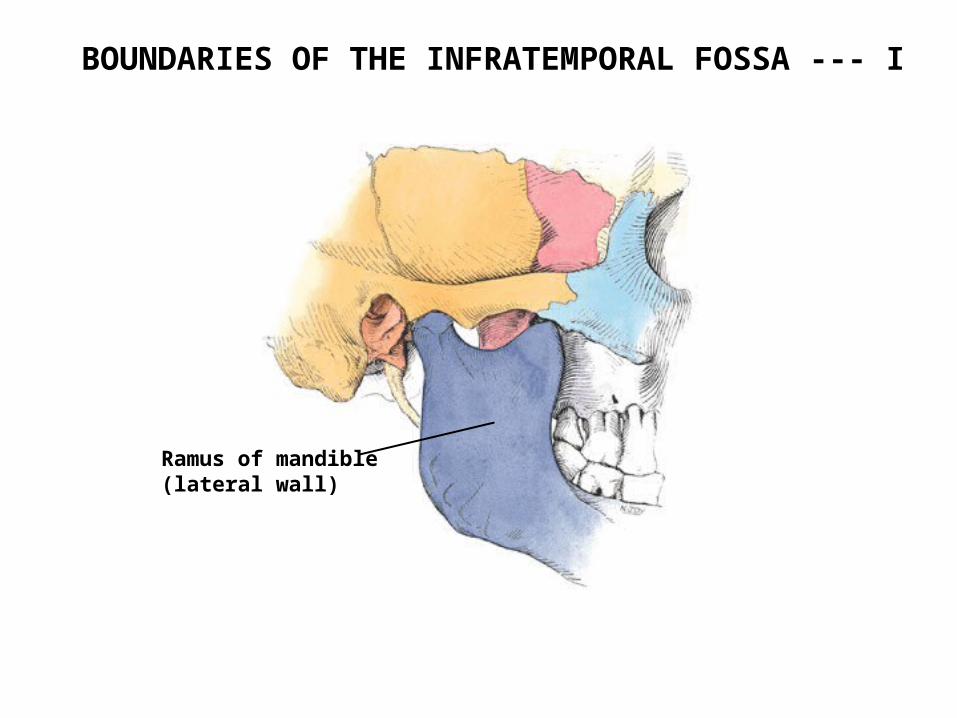
BOUNDARIES OF THE INFRATEMPORAL FOSSA --- I
Ramus of mandible(lateral wall)
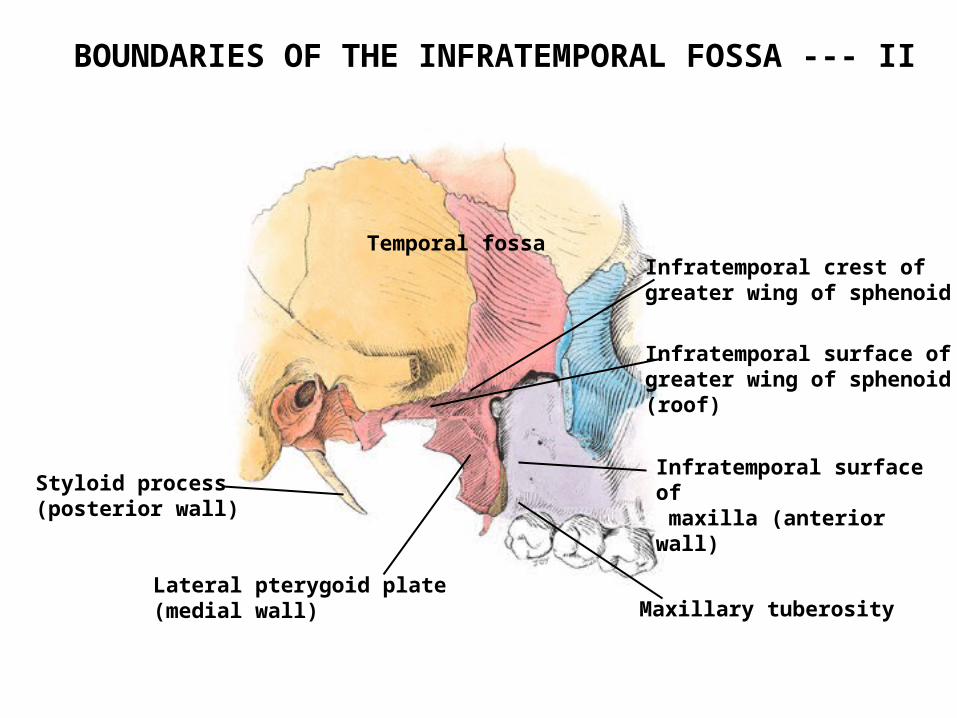
Infratemporal surface of maxilla (anterior wall)Styloid process
(posterior wall)
Lateral pterygoid plate(medial wall)
Infratemporal surface ofgreater wing of sphenoid(roof)
Infratemporal crest ofgreater wing of sphenoid
Temporal fossa
BOUNDARIES OF THE INFRATEMPORAL FOSSA --- II
Maxillary tuberosity
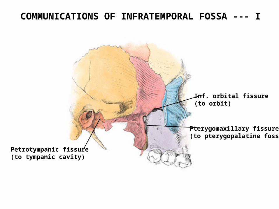
COMMUNICATIONS OF INFRATEMPORAL FOSSA --- I
Inf. orbital fissure(to orbit)
Pterygomaxillary fissure(to pterygopalatine fossa)
Petrotympanic fissure(to tympanic cavity)
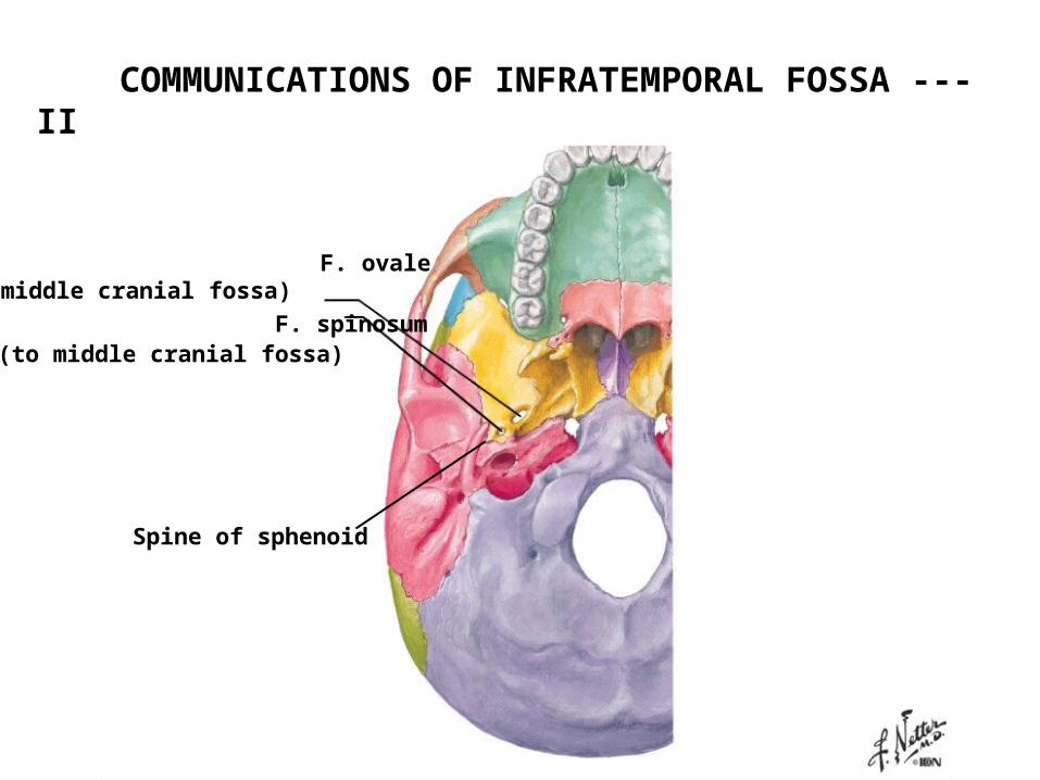
COMMUNICATIONS OF INFRATEMPORAL FOSSA --- II
F. ovale (to middle cranial fossa)
F. spinosum (to middle cranial fossa)
Spine of sphenoid
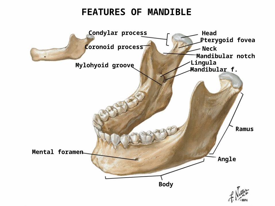
Coronoid process
Condylar process Head
Neck
Pterygoid fovea
Mandibular notchLingulaMandibular f.
Mylohyoid groove
Mental foramen
Body
Ramus
Angle
FEATURES OF MANDIBLE
MUSCLES OFMASTICATION
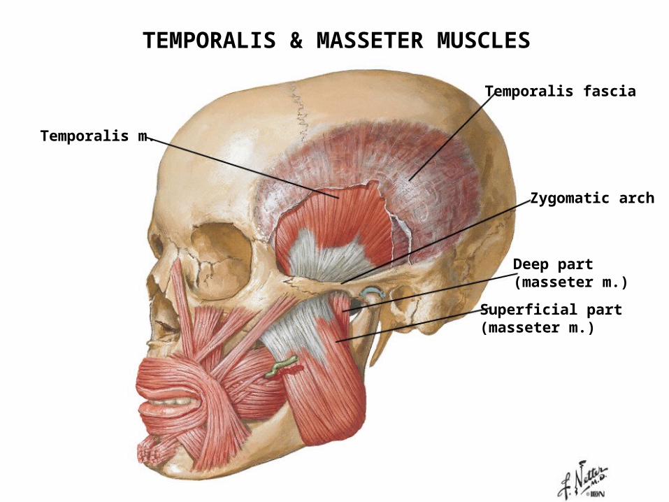
Temporalis m.
Temporalis fascia
Zygomatic arch
Deep part (masseter m.)
Superficial part(masseter m.)
TEMPORALIS & MASSETER MUSCLES
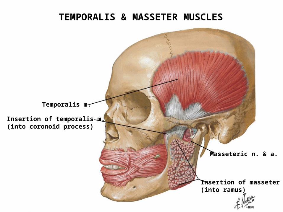
Temporalis m.
Insertion of temporalis m.(into coronoid process)
Insertion of masseter m.(into ramus)
Masseteric n. & a.
TEMPORALIS & MASSETER MUSCLES
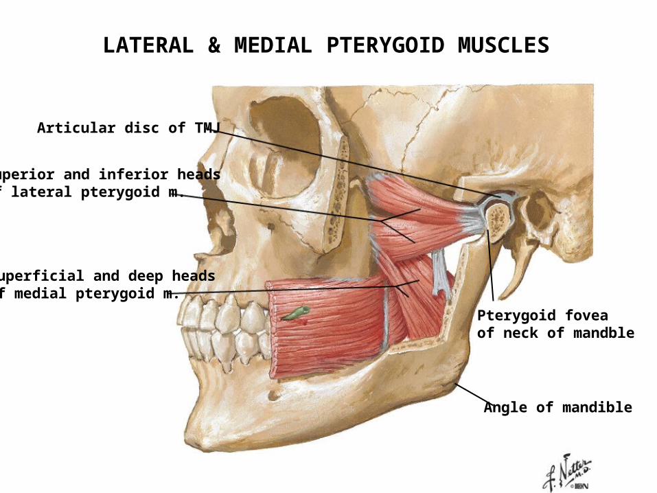
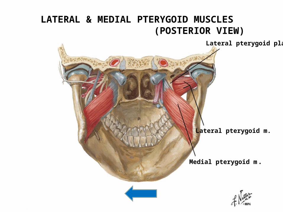
Superior and inferior headsof lateral pterygoid m.
Superficial and deep headsof medial pterygoid m.
Articular disc of TMJ
Pterygoid foveaof neck of mandble
Angle of mandible
LATERAL & MEDIAL PTERYGOID MUSCLES
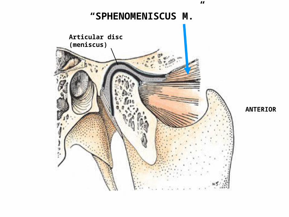
“SPHENOMENISCUS M.”
ANTERIOR
Articular disc(meniscus)
Lateral pterygoid plate
Lateral pterygoid m.
Medial pterygoid m.
LATERAL & MEDIAL PTERYGOID MUSCLES (POSTERIOR VIEW)
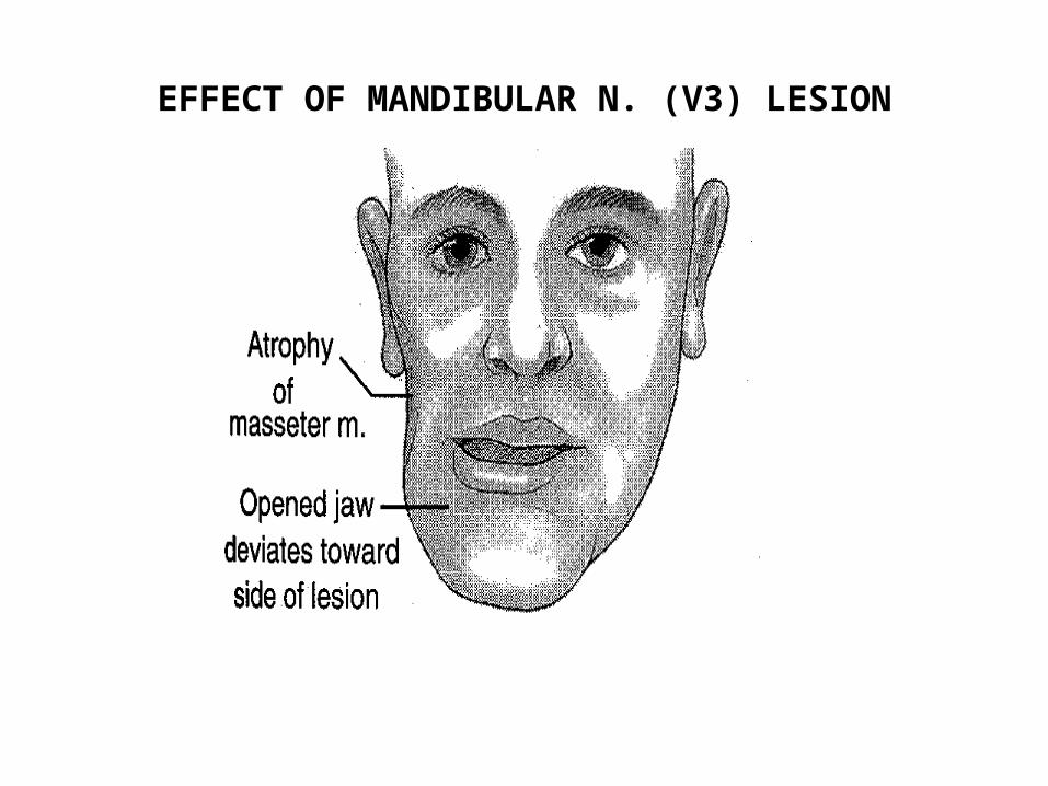
EFFECT OF MANDIBULAR N. (V3) LESION

NERVES
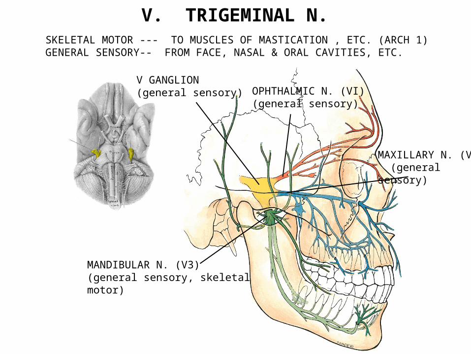
V. TRIGEMINAL N.SKELETAL MOTOR --- TO MUSCLES OF MASTICATION , ETC. (ARCH 1) GENERAL SENSORY-- FROM FACE, NASAL & ORAL CAVITIES, ETC.
OPHTHALMIC N. (VI)(general sensory)
MAXILLARY N. (V2) (general sensory)
MANDIBULAR N. (V3)(general sensory, skeletal motor)
V GANGLION(general sensory)
A
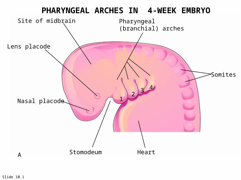
Site of midbrain
Lens placode
Nasal placode
Stomodeum Heart
Somites
Pharyngeal(branchial) arches
12
3 4
Slide 10.1
PHARYNGEAL ARCHES IN 4-WEEK EMBRYO
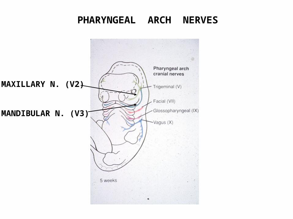
PHARYNGEAL ARCH NERVES
MAXILLARY N. (V2)
MANDIBULAR N. (V3)

Temporalis fascia and m.
Ant. division (V3) (mostly motor)
Post. division (V3) (mostly sensory)
Foramen ovale
Auriculotemporal n.
Inferior alveolar n. (cut)
Inferior alveolar n. (cut)
Lingual n.
Chorda tympani n.(br. of VII)
Posterior andanterior deep temporal nn.
Masseteric n.
Lateral pterygoid n.and m.
Buccal n.
Mylohyoid m. (cut)
Digastric m. (anterior belly)
Mylohyoid n.
Mental n.Submandibular ganglion & gland
Sublingual gland
MANDIBULAR NERVE (V3)
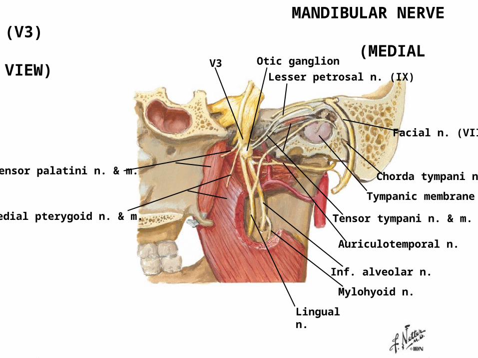
V3
Medial pterygoid n. & m.
Tensor palatini n. & m.
Otic ganglion
Lesser petrosal n. (IX)
Facial n. (VII)
Chorda tympani n.
Tympanic membrane
Auriculotemporal n.
Lingual n.
Inf. alveolar n.
Mylohyoid n.
Tensor tympani n. & m.
MANDIBULAR NERVE (V3) (MEDIAL VIEW)
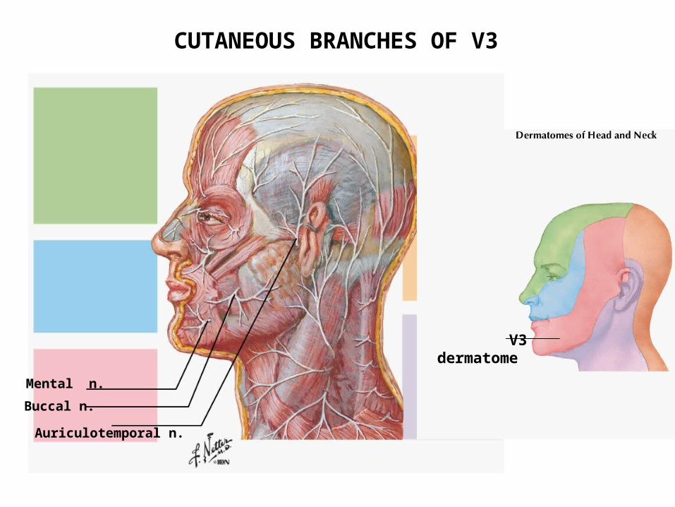
Mental n.
Buccal n.
Auriculotemporal n.
V3 dermatome
cCUTANEOUS BRANCHES OF V3
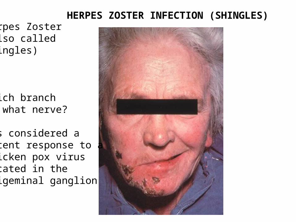
Herpes Zoster(also called shingles)
Which branchof what nerve?
-is considered alatent response to a chicken pox virus located in thetrigeminal ganglion
HERPES ZOSTER INFECTION (SHINGLES)
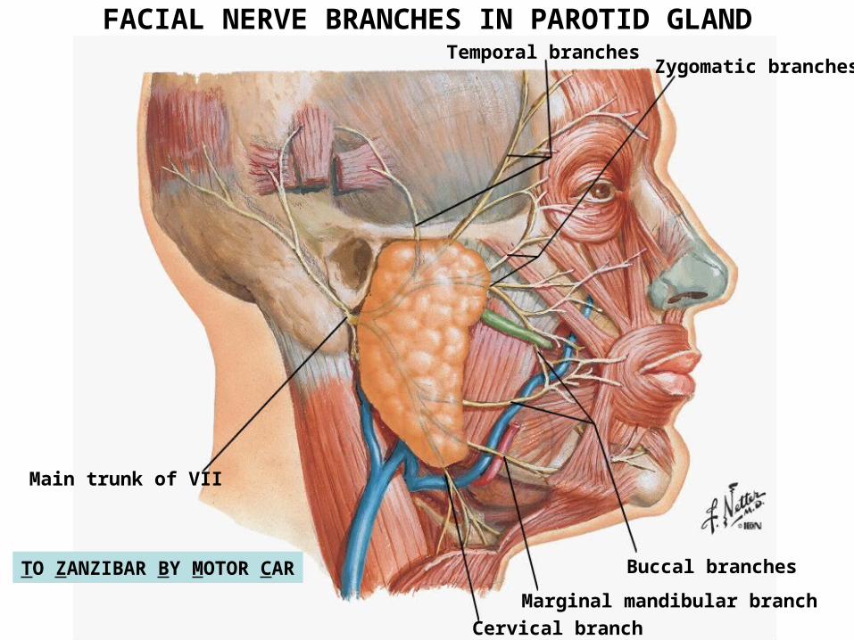
Temporal branchesZygomatic branches
Buccal branches
Marginal mandibular branch
Cervical branch
Main trunk of VII
TO ZANZIBAR BY MOTOR CAR
FACIAL NERVE BRANCHES IN PAROTID GLAND

VESSELS
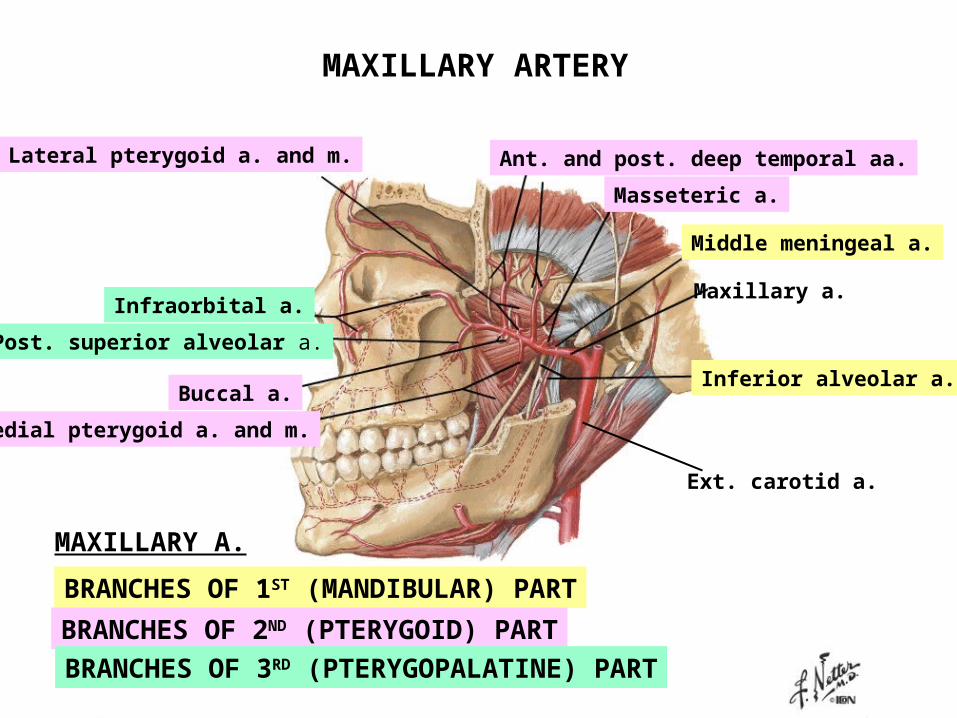
Maxillary a.
Inferior alveolar a.
Middle meningeal a.
Ant. and post. deep temporal aa.
Medial pterygoid a. and m.
Buccal a.
Lateral pterygoid a. and m.
Masseteric a.
Post. superior alveolar a.
Infraorbital a.
MAXILLARY A.
BRANCHES OF 1ST (MANDIBULAR) PART
BRANCHES OF 2ND (PTERYGOID) PART
BRANCHES OF 3RD (PTERYGOPALATINE) PART
Ext. carotid a.
MAXILLARY ARTERY
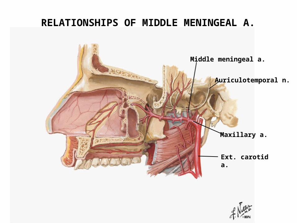
Middle meningeal a.
Auriculotemporal n.
Maxillary a.
Ext. carotid a.
RRELATIONSHIPS OF MIDDLE MENINGEAL A.
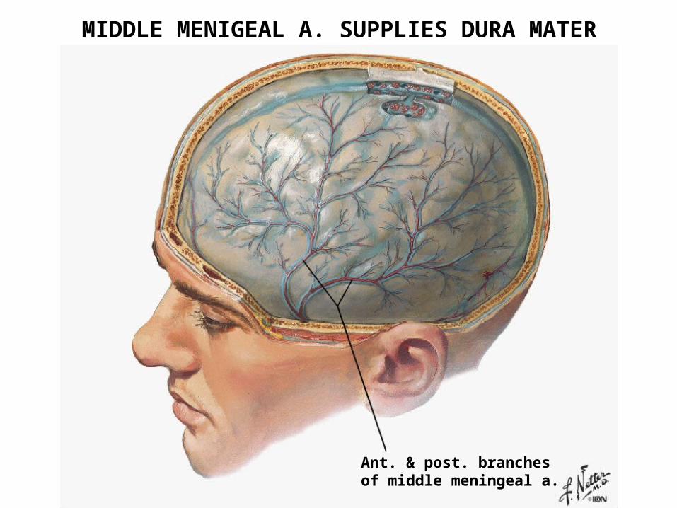
Ant. & post. branchesof middle meningeal a.
MIDDLE MENIGEAL A. SUPPLIES DURA MATER
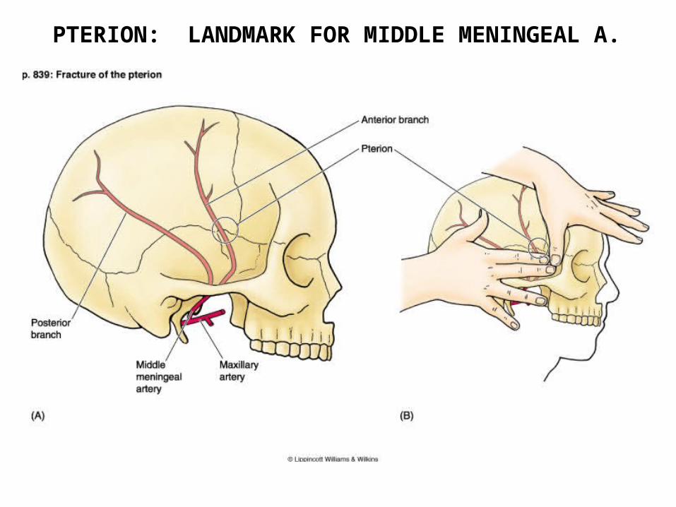
PTERION: LANDMARK FOR MIDDLE MENINGEAL A.
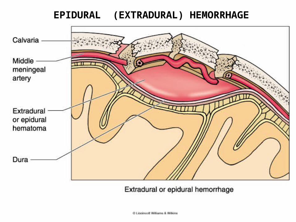
EPIDURAL (EXTRADURAL) HEMORRHAGE
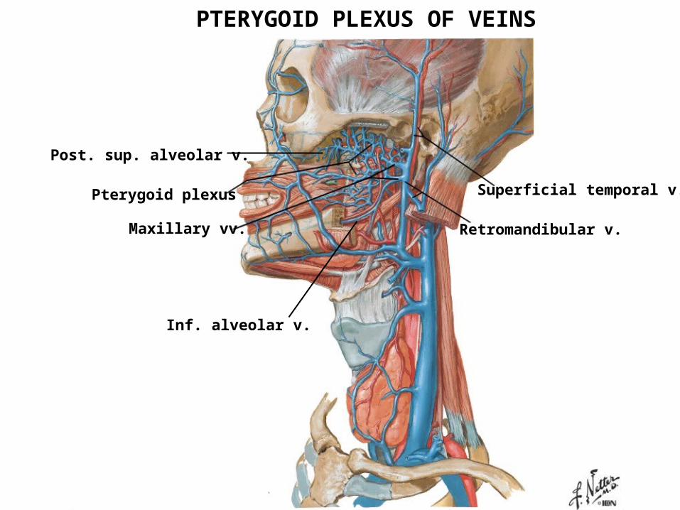
Pterygoid plexus
Maxillary vv.
Superficial temporal v.
Retromandibular v.
PTERYGOID PLEXUS OF VEINS
Inf. alveolar v.
Post. sup. alveolar v.
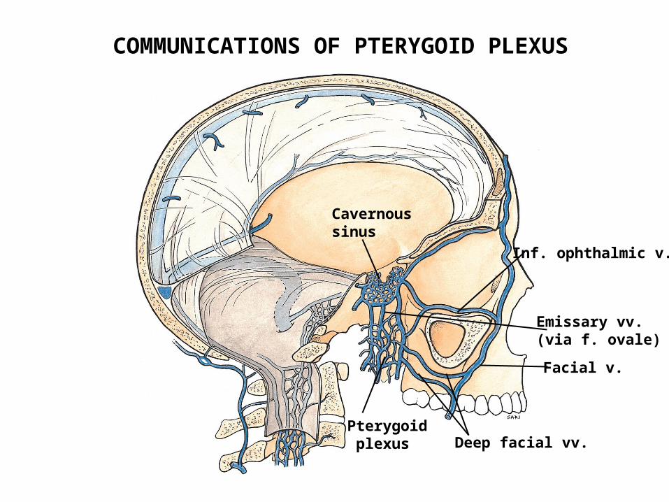
Cavernous sinus
Pterygoid plexus
Facial v.
Inf. ophthalmic v.
Deep facial vv.
Emissary vv.(via f. ovale)
COMMUNICATIONS OF PTERYGOID PLEXUS
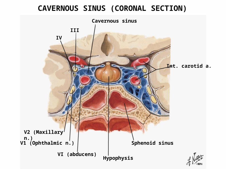
Int. carotid a.
Sphenoid sinus
HypophysisVI (abducens)
Cavernous sinus
III
IV
V2 (Maxillary n.)
V1 (Ophthalmic n.)
CAVERNOUS SINUS (CORONAL SECTION)
TEMPOROMANDIBULAR JOINT
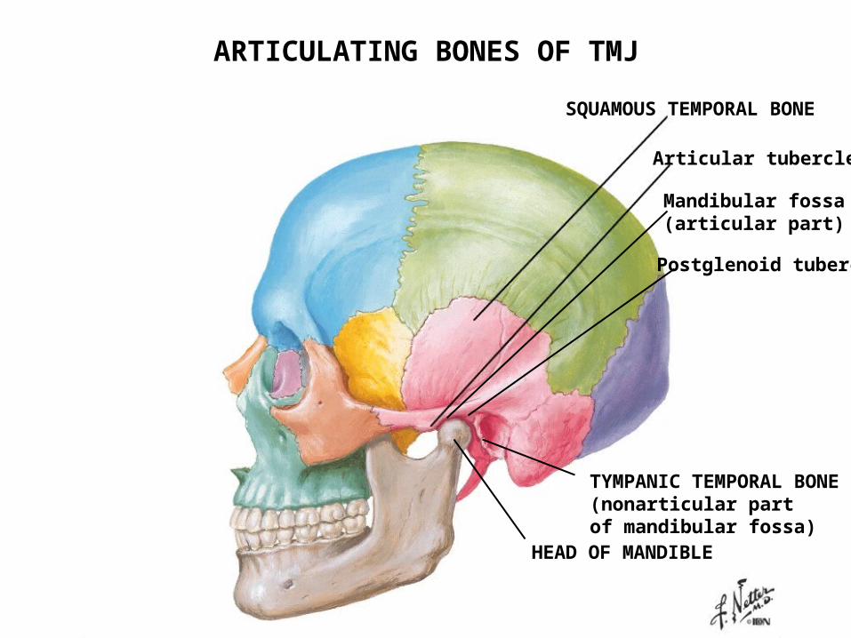
SQUAMOUS TEMPORAL BONE
Articular tubercle
Mandibular fossa(articular part)
Postglenoid tubercle
HEAD OF MANDIBLE
TYMPANIC TEMPORAL BONE(nonarticular part of mandibular fossa)
ARTICULATING BONES OF TMJ
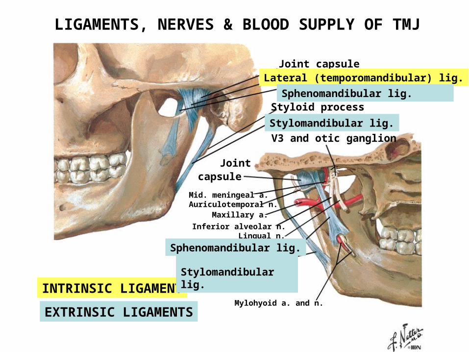
Joint capsuleLateral (temporomandibular) lig.
Sphenomandibular lig.Styloid process
Stylomandibular lig.
V3 and otic ganglion
Joint capsule
Mid. meningeal a. Auriculotemporal n.
Maxillary a.
Inferior alveolar n.Lingual n.
Mylohyoid a. and n.
INTRINSIC LIGAMENT
EXTRINSIC LIGAMENTS
Stylomandibular lig.Sphenomandibular lig.
LIGAMENTS, NERVES & BLOOD SUPPLY OF TMJ
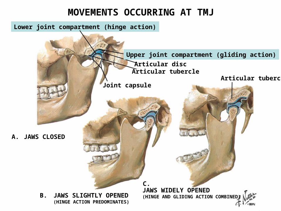
Articular discArticular tubercle
Joint capsule
JAWS CLOSED
JAWS SLIGHTLY OPENED(HINGE ACTION PREDOMINATES)
JAWS WIDELY OPENED(HINGE AND GLIDING ACTION COMBINED)
Upper joint compartment (gliding action)
Lower joint compartment (hinge action)
MOVEMENTS OCCURRING AT TMJ
A.
B.
C.
Articular tubercle
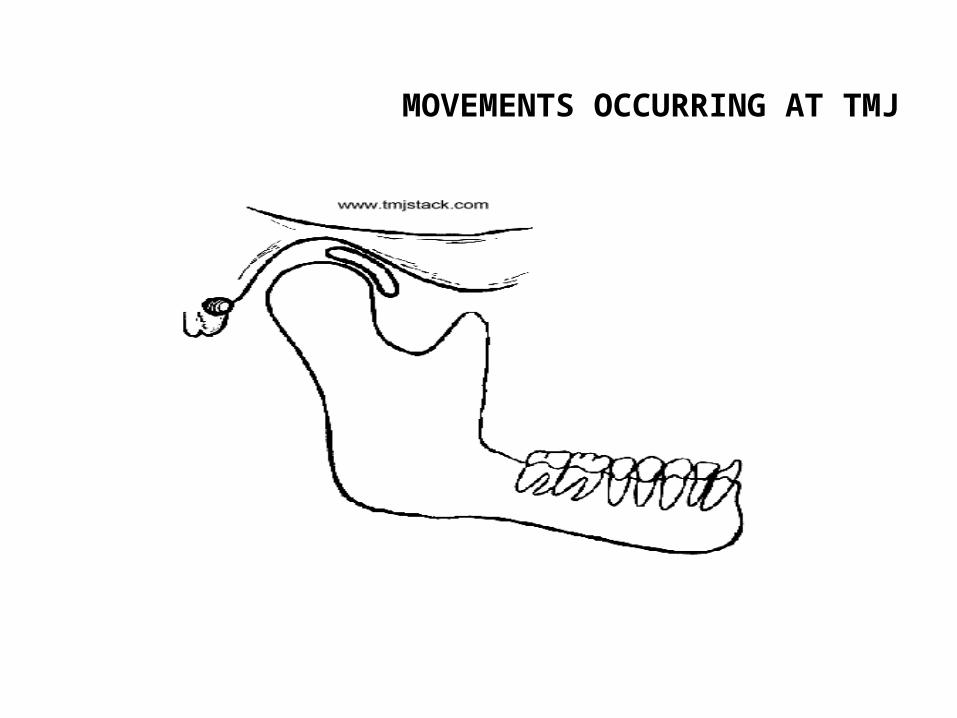
MOVEMENTS OCCURRING AT TMJ
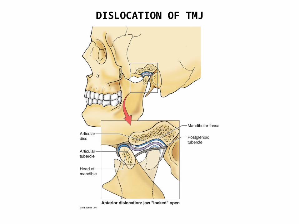
DISLOCATION OF TMJ
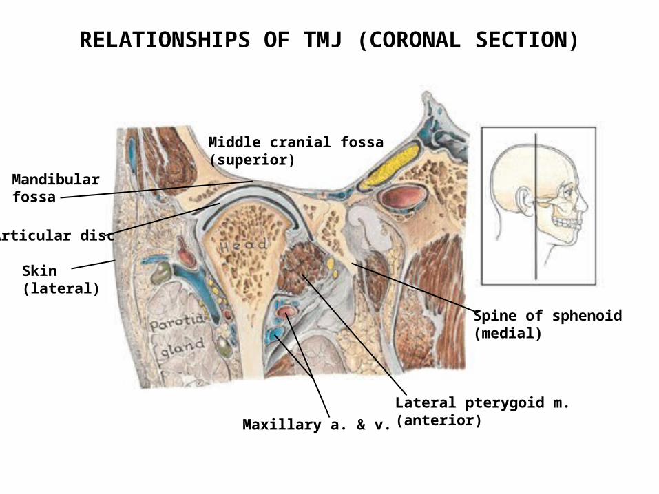
Middle cranial fossa(superior)
Articular disc
Spine of sphenoid(medial)
Lateral pterygoid m.(anterior)Maxillary a. & v.
Skin (lateral)
Mandibular fossa
RELATIONSHIPS OF TMJ (CORONAL SECTION)

END OF LECTURE