Embed Size (px)
Citation preview

7/31/2019 Strees Tolerance of Baker Yeast Cells, Stress Protective Molecules and Genes Involved in Stress Tolerance
http://slidepdf.com/reader/full/strees-tolerance-of-baker-yeast-cells-stress-protective-molecules-and-genes 1/10
Biotechnol. Appl. Biochem. (2009) 53, 155–164 (Printed in Great Britain) doi:10.1042/BA20090029 155
MINIREVIEW
Stress-tolerance of baker’s-yeast (Saccharomyces cerevisiae)cells: stress-protective molecules and genes involvedin stress tolerance
Jun Shima*1 and Hiroshi Takagi†
*National Food Research Institute, 2-1-12 Kannondai, Tsukuba, Ibaraki 305-8642, Japan, and †Graduate School of Biological Sciences, Nara Institute of Science and Technology, 8916-5 Takayama, Ikoma, Nara 630-0192, Japan
During the fermentation of dough and the production
of baker’s yeast (Saccharomyces cerevisiae), cells are
exposed to numerous environmental stresses (baking-
associated stresses) such as freeze–thaw, high sugarconcentrations, air-drying and oxidative stresses.
Cellular macromolecules, including proteins, nucleic
acids and membranes, are seriously damaged under
stress conditions, leading to the inhibition of cell
growth, cell viability and fermentation. To avoid lethal
damage, yeast cells need to acquire a variety of stress-
tolerant mechanisms, for example the induction of
stress proteins, the accumulation of stress protectants,
changes in membrane composition and repression of
translation, and by regulating the corresponding gene
expression via stress-triggered signal-transduction
pathways. Trehalose and proline are considered to
be critical stress protectants, as is glycerol. It is
known that these molecules are effective for providing
protection against various types of environmental
stresses. Modifications of the metabolic pathways of
trehalose and proline by self-cloning methods have
significantly increased tolerance to baking-associated
stresses. To clarify which genes are required for stress
tolerance, both a comprehensive phenomics analysis
and a functional genomics analysis were carried out
under stress conditions that simulated those occuring
during the commercial baking process. These analyses
indicated that many genes are involved in stress
tolerance in yeast. In particular, it was suggested thatvacuolar H+-ATPase plays important roles in yeast cells
under stress conditions.
Introduction
Baker’s yeast (mostly strains of Saccharomyces cerevisiae)
is an essential ingredient in bakery products produced by
fermentation [1,2]. Around the world, about 2 million tons
of baker’s yeast are produced (based on 30% dry weight) per
year [3,4]. The function of baker’s yeast in bread making can
be summarized as follows: (i) to increase dough volume by
gas generation during fermentation; (ii) to develop structure
and texture in the dough; and (iii) to add a distinctive flavour
to the dough [5]. Baker’s yeast is produced in the form of
cream yeast (an aqueous suspension containing approx. 20%dry weight of cells), compressed yeast or dried yeast. The
compressed yeast is manufactured by partial dehydration
and contains approximately 30 % dry weight of cells. In Japan,
most baker’s yeasts are produced as cream or compressed
yeasts. However, dried yeast, which contains less than 5%
water, is imported from other countries and used in home
baking and bakery shops owing to the convenience of its
storage and delivery to Japan.
During the fermentation of dough and the production
of baker’s yeast, yeast cells are exposed to numerous
environmental stresses, including freeze–thaw, high sugar
concentrations and air-drying (baking-associated stresses)
[3]. In addition, the yeast cells encounter such stresses in a
multiple and sequential manner (for example, freeze–thaw
plus high sugar concentrations) [3]. It is believed that, by
undergoing freeze–thaw and air-drying treatments, yeast
cells are exposed to oxidative stress [3,6,7]. In general,
micro-organisms show some ability to adapt to environ-
mental stresses. Yeast cells also need to acquire a variety
of stress-adaptation mechanisms, for example, induction of
stress proteins, accumulation of stress protectants, changes
in membrane composition and repression of translation,
and by regulating the corresponding gene expression via
stress-triggered signal transduction pathways. Under se-
vere stress conditions, however, the fermentation ability of yeast is rather limited. To develop the commercial ferment-
ation and growth of baker’s yeast, it is necessary to construct
yeast strains with a higher tolerance to various stresses. The
Key words: baker’s yeast (Saccharomyces cerevisiae), baking-associated stress,
functional genomics, phenomics, stress protectant, stress tolerance.
Abbreviations used: AZC, azetidine-2-carboxylate; GK, γ -glutamyl kinase;
GPR, γ -glutamyl phosphate reductase; HS, high sugar; IC 50 , 50 % inhibitory
concentration; LS, low sugar; ORF, open reading frame; OTA, ornithine
transaminase; P5C , 1-pyrroline-5-carboxylate; PO, proline oxidase; ROS,
reactive oxygen species; SOD, superoxide dismutase; V-ATPase, vacuolar
H+-ATPase.1 To whom correspondence should be addressed (email [email protected]).
C 2009 Portland Press Ltd
www.babonline.org
B i
t
h
l
d A
l i d
B i
h
i t

7/31/2019 Strees Tolerance of Baker Yeast Cells, Stress Protective Molecules and Genes Involved in Stress Tolerance
http://slidepdf.com/reader/full/strees-tolerance-of-baker-yeast-cells-stress-protective-molecules-and-genes 2/10
156 J. Shima and H. Takagi
Scheme 1 Process of baker’s-yeast production and bread making and the
environmental stresses associated with such processes
present minireview focuses on the mechanisms of tolerance
to environmental stresses to which baker’s-yeast cells are
exposed during industrial processes and the construction
of stress-tolerant baker’s-yeast strains. We describe stress-
tolerance mechanisms based on stress protectants, which
include trehalose and proline, and clarify, by post-genomic
analysis, which genes are required for stress tolerance.
Baking-associated environmental
stresses
During the bread-making process, baker’s yeast cells are
exposed to various types of environmental stresses in
a multiple and sequential manner [3]. We pay particular
attention to the baking-associated stresses freeze–thaw, high
sugar concentrations stress and air-drying, because yeast
cells are considered to be susceptible to damage from such
stresses during the commercial processes that baker’s yeast
undergoes (Scheme 1).
Frozen-dough baking is one of the key technologiesof bread making because it improves labour conditions
for bakers and allows for the supply of oven-fresh bakery
products to consumers. Because baker’s-yeast cells suffer
from freeze–thaw injuries in frozen-dough baking processes,
the fermentation ability of yeast cells after freeze–thaw
dramatically decreases [8,9]. Freeze–thaw injuries to yeast
cells may depend on many factors, including the genetic
background of the yeast strains, the physiological condition
of the yeast cells and the freezing conditions, such as the
length of the freezing period and the speed of freezing.
During frozen-dough baking, yeast cells are frozen in a
process that subjects them to low temperature, ice- crystal
formation in the cells and dehydration [10,11]. Freezing
can cause not only deleterious damage to the cell wall
and membrane but also denaturation of functional pro-
teins and DNA, thus decreasing cell viability. Changes in cellphysiology due to the onset of fermentation might weaken
freeze–thaw tolerance [12–14], possibly due to activation
of the cyclic AMP pathway [15]. A decrease in freeze–thaw
tolerance is strongly related to trehalose degradation, and
the levels of intracellular trehalose affect the tolerance of
baker’s yeast to freeze–thaw stress (for further details, see
below) [12,16]. In commercial frozen-dough processes, pre-
fermentation before freezing is desirable, because bread
made from pre-fermented frozen dough has the proper
texture and taste [8,17]. It is known that the freeze–
thaw tolerance of yeast is also determined by the genetic
characteristics of the strains. Some yeast strains with higherfreeze–thaw tolerance have been isolated from natural
sources and constructed by gene manipulation [16,18–20].
The freezing period is a critical parameter for freeze–thaw
injury; prolonged storage of the frozen dough damages yeast
cells greatly, owing to the growth of ice crystals [21,22].
Recently, the involvement of oxidative stress in freeze–
thaw injury to yeast cells has been analysed using mutants
defective in antioxidant functions [23–25]. Park et al. [26]
reported that superoxide anions and free radicals were
generated in yeast (S. cerevisiae) cells during aerobic freeze–
thaw and that cytoplasmic Cu,Zn-SOD (Cu,Zn-superoxide
dismutase) is required for tolerance to freeze–thaw stress.
Also, an oxidative burst during freeze–thaw has been
considered to lead to oxidative damage to many cellular
molecules, including proteins, lipids and DNA through the
generation of ROS (reactive oxygen species) [27]. Oxidative
stress is considered to cause serious injury to yeast cells
during freeze–thaw in addition to the physical damage caused
by ice nucleation and dehydration.
Yeasts used in breadmaking are exposed to different
sugar conditions during the dough-fermentation processes
[3,28]. Dough can be classified into lean or sweet dough
based on the sugar concentrations contained in the
dough. Lean dough contains no sugar (non-sugar dough) or
small amounts of sugar [below 5 % (w/w) of flour]. In general,sweet dough (high-sugar dough) contains up to 40 % sucrose
(w/w of flour). Such high sucrose concentrations exert
severe osmotic stress on yeast cells [29]. Yeast strains that
show higher tolerance to high concentrations of sucrose
are desirable for sweet-dough fermentation [3,28,30]. By
contrast, because there is little sucrose in non-sugar dough,
the yeast must use maltose derived from the flour [28,30].
Yeast strains that have a higher maltose-utilization ability
are required for non-sugar dough fermentation [30]. In
the modern baking industry, HS (high sugar; high-sucrose-
tolerant) and LS (low suger; maltose-utilizing) yeasts have
C 2009 Portland Press Ltd
7/31/2019 Strees Tolerance of Baker Yeast Cells, Stress Protective Molecules and Genes Involved in Stress Tolerance
http://slidepdf.com/reader/full/strees-tolerance-of-baker-yeast-cells-stress-protective-molecules-and-genes 3/10
Stress tolerance of baker’s-yeast cells 157
been developed by using breeding techniques, and are
currently used commercially [28,30]. Baker’s-yeast cells
may change their level of metabolic activity, such as their
carbon metabolism and nitrogen metabolism, as well as
their stress response to environmental conditions [30,31].An understanding of the molecular basis behind the meta-
bolic adaptation that occurs under different sugar conditions
is needed to develop yeast strains that have tolerance to
high-sucrose stress and have maltose-utilization ability.
During the production of dried yeast, yeast cells are
also exposed to air-drying stress [32,33]. In breadmaking,
the distinct advantages of using dried yeast, such as active
dried yeast (dry matter 92–94%) and instant dried yeast
(dry matter 94–96 %), include their greater stability and
lower moisture content, both of which reduce both storage
and transport costs [34]. To gain greater advantages from
dried yeast, it must retain a higher fermentation abilityand flavour formation after the air-drying process. During
the drying process using cell dryers such as fluidized beds
or Roto-Louver® dryers [33], yeast cells are exposed
to air-drying stress. In general, owing to the flow of
hot air during these processes, the temperature of the
yeast cells is relatively high, namely about 37 ◦C. Air-drying
stress is considered to be a complex environmental stress
composed of dehydration and heat stresses. Dehydration
generated by drying processes, such as air-drying and
evaporation, causes severe damage to yeast cells, particularly
to the cellular membrane and proteins [35]. Dehydration
promotes the generation of ROS, which induce lipid
peroxidation, protein denaturation and nucleic acid damage
[36–38]. Therefore the ROS-scavenging systems mediated
by glutathione, catalase and SOD, for example, are believed
to confer tolerance to dehydration [39,40].
Trehalose as a stress protectant
Many research groups have analysed key factors and their
roles in the stress tolerance of S. cerevisiae. For instance,
heat-shock proteins function in the disaggregation and disas-
sembly of proteins denatured by environmental stress [41].
A variety of antioxidant enzymes, including SOD, catalase,and glutathione peroxidase, detoxify ROS by the direct
disproportion, decomposition or reduction of ROS under
oxidative-stress conditions [3]. In S. cerevisiae, it is known
that glycerol [42] and trehalose function as major stress
protectants and that the synthesis of glycerol or trehalose is
induced by many stress conditions at the transcriptional level
[43]. Owing to the ability of these compounds to prevent
the influx of excess salts into the cell or irreversible cell
dehydration, the osmotic imbalance after hyper- or hypo-
osmotic shock can be rapidly restored. Panadero et al.
[44] suggested that the heterologus expression of type I
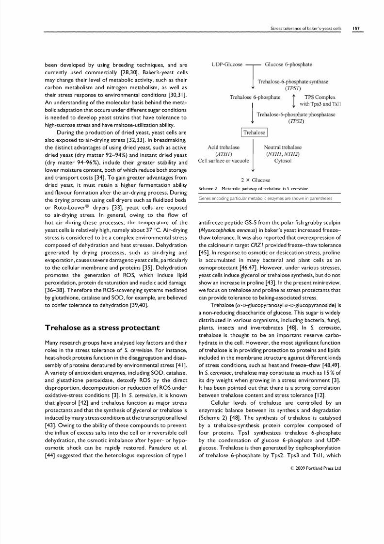
Scheme 2 Metabolic pathway of trehalose in S. cerevisiae
Genes encoding particular metabolic enzymes are shown in parentheses
antifreeze peptide GS-5 from the polar fish grubby sculpin
( Myoxocephalus aenaeus) in baker’s yeast increased freeze–
thaw tolerance. It was also reported that overexpression of
the calcineurin target CRZ1 provided freeze–thaw tolerance
[45]. In response to osmotic or desiccation stress, proline
is accumulated in many bacterial and plant cells as an
osmoprotectant [46,47]. However, under various stresses,
yeast cells induce glycerol or trehalose synthesis, but do not
show an increase in proline [43]. In the present minireview,
we focus on trehalose and proline as stress protectants that
can provide tolerance to baking-associated stress.
Trehalose (α-D-glucopyranosyl α-D-glucopyranoside) is
a non-reducing disaccharide of glucose. This sugar is widely
distributed in various organisms, including bacteria, fungi,
plants, insects and invertebrates [48]. In S. cerevisiae,
trehalose is thought to be an important reserve carbo-
hydrate in the cell. However, the most significant function
of trehalose is in providing protection to proteins and lipids
included in the membrane structure against different kinds
of stress conditions, such as heat and freeze–thaw [48,49].
In S. cerevisiae, trehalose may constitute as much as 15 % of its dry weight when growing in a stress environment [3].
It has been pointed out that there is a strong correlation
between trehalose content and stress tolerance [12].
Cellular levels of trehalose are controlled by an
enzymatic balance between its synthesis and degradation
(Scheme 2) [48]. The synthesis of trehalose is catalysed
by a trehalose-synthesis protein complex composed of
four proteins. Tps1 synthesizes trehalose 6-phosphate
by the condensation of glucose 6-phosphate and UDP-
glucose. Trehalose is then generated by dephosphorylation
of trehalose 6-phosphate by Tps2. Tps3 and Tsl1, which
C 2009 Portland Press Ltd

7/31/2019 Strees Tolerance of Baker Yeast Cells, Stress Protective Molecules and Genes Involved in Stress Tolerance
http://slidepdf.com/reader/full/strees-tolerance-of-baker-yeast-cells-stress-protective-molecules-and-genes 4/10

7/31/2019 Strees Tolerance of Baker Yeast Cells, Stress Protective Molecules and Genes Involved in Stress Tolerance
http://slidepdf.com/reader/full/strees-tolerance-of-baker-yeast-cells-stress-protective-molecules-and-genes 5/10
Stress tolerance of baker’s-yeast cells 159
Scheme 3 Metabolic pathway of proline in S. cerevisiae
Genes encoding particular metabolic enzymes are shown in parentheses
to various environmental changes, including temperature,
ethanol, oxidation, pH and osmolarity [79,80]. Recently,
the correlation between gene-expression profiles and
intracellular contents of glycerol, trehalose, and proline were
determined under various stress conditions [43]. When
yeast cells were exposed to osmotic stress, the expression
of GPD1, encoding glycerol-3-phosphate dehydrogenase,
was induced, leading to glycerol accumulation. In the
presence of ethanol, the rapid induction of TPS2 resulted
in trehalose accumulation. By contrast, the expression of
genes involved in proline metabolism and the cellular level
of proline did not change during exposure to these stresses
[43]. These results suggest that yeast cells do not accumulate
proline in response to osmotic or ethanol stress.
It was shown that a put1-disruptant yeast in minimal
medium supplemented with external proline accumulated
higher levels of proline in the cells and conferred a
higher tolerance to freeze–thaw and dehydration stresses
[73]. Enhancement of the biosynthetic activity is also
important for the intracellular accumulation of proline. AZC
(azetidine-2-carboxylate), which is a toxic four-membered
ring analogue of proline, is transported into yeast cells
through proline transporters [81]. Once inside a cell, AZCcompetes with proline for incorporation into nascent
proteins, resulting in protein misfolding, which inhibits cell
growth [82]. Overproduction of proline dilutes the effect
of AZC. To increase cellular proline accumulation, AZC-
resistant mutants have been isolated from the put1-deficient
strain [69]. This mutant was recently found to carry an
allele of PRO1 encoding GK and to have a single amino
acid replacement at position 154 (aspartic acid replaced
by asparagine) [72]. Yeast cells expressing the mutant
GK accumulated proline and showed a drastic increase
in cell viability after freezing at −20 ◦C compared with
cells harbouring wild-type PRO1. The D154N mutant of
GK [IC50 (50 % inhibitory concentration)= 32 mM] showed
lower sensitivity to feedback inhibition than did the wild-type
enzyme (0.5 mM) and thermostability. These characteristics
lead to a higher level of proline accumulation [83,84].A high level of freeze–thaw tolerance clearly correlated
with the accumulation of proline in yeast cells. Such
a proline-accumulating laboratory strain was found to
be more tolerant to various stresses, including freezing,
dehydration, hydrogen peroxide and ethanol, than the wild-
type strain [69,71,72,74,84,85]. To improve the enzymatic
properties of GK, PCR random mutagenesis in PRO1
was employed [83]. Several mutant GKs that, owing to
extreme desensitization to inhibition, enhanced the ability
to synthesize proline, were successfully isolated. Further-
more, yeast cells expressing I150T and N142D/I166V
mutant GKs were found to be more tolerant to freezingstress than cells expressing the D154N mutant.
Recently we constructed self-cloning diploid baker’s-
yeast strains by disrupting PUT1 and replacing the wild-type
PRO1 with the pro1 (D154N) or pro1 (I150T ) allele [43]. The
resultant baker’s-yeast strains accumulated higher levels of
intracellular proline (3–6 % dry weight) compared with that
of the wild-type strain (0.20 %). As expected, the proline-
accumulating strains retained their higher fermentation
abilities in the frozen doughs at the same levels as those
observed before freezing, although that of the parent strain
fell to approx. 80 % of the prefreezing level. Therefore,
proline-accumulating baker’s yeast is considered to be
suitable for frozen-dough baking. It is noteworthy that
the combination of proline and trehalose might further
contribute to the enhancement of tolerance to baking-
associated stresses.
Phenomics approach to the
identification of genes required for
stress tolerance to baking-associated
stresses
In addition to analyses of cell-protective molecules, theidentification of novel genes which determine the tolerance
to baking-associated stress were attempted. Our group at-
tempted to find the genes for stress tolerance by two differ-
ent approaches, namely phenomics and functional genomics,
under freeze–thaw, high-sugar-concentration and air-drying
stress conditions [6,7,31,86–89]. The data for phenomics
and functional genomics under baking-associated stress
conditions are available at our web site (http://nfri.naro.
affrc.go.jp/english/Useful/yeast/index.pdf). A yeast deletion-
mutant collection should be a powerful tool for determining
gene function by analysing the phenotype of mutants lacking
C 2009 Portland Press Ltd

7/31/2019 Strees Tolerance of Baker Yeast Cells, Stress Protective Molecules and Genes Involved in Stress Tolerance
http://slidepdf.com/reader/full/strees-tolerance-of-baker-yeast-cells-stress-protective-molecules-and-genes 6/10
160 J. Shima and H. Takagi
Figure 1 Schematic view of overlap in genes required for each baking-
associated stress tolerance
The numbers in parentheses are the gene numbers determined by phenomics
analyses. The genes encoding the components of V-ATPase are included in thearea where the three circles overlap.
the gene (phenomics) [90–92]. An international consortium
has carried out the systematic deletion of all of the ORFs
(open reading frames) of S. cerevisiae by using a PCR-
mediated gene-deletion strategy [91]. Analysis using this
deletion-mutant collection relies on the number of genes
whose mutations that affect components of an important
pathway show a phenotype of sensitivity or tolerance
[90,92,93]. Identification of the genes required to cope
with high-sugar-concentrations, freeze–thaw and air-drying
stresses was carried out using phenomics [6,7,88]. The
complete deletion-strain collection of diploid S. cerevisiae
was used in these analyses because commercial baker’s
yeasts are generally diploid.
The screening identified 273 strains with high sucrose-
sensitivity, 58 strains with freeze–thaw-sensitivity and 278
strains with air-drying-sensitivity [6,7,88] (Figure 1). These
deleted genes were classified on the basis of their cellular
function and the localization of their gene products. A
total of 30 strains were sensitive to all the environmental
stresses tested. As described below, the deletion strains
of the genes encoding V-ATPase (vacuolar H+-ATPase) and
proline metabolism were included in the 30 strains. Otherthan such genes, the deletion strains of genes encoding
heat-shock response and vacuolar protein sorting were
included. The deletion strains of genes encoding trehalose
metabolism were sensitive to high-sucrose stress and air-
drying stress, but not to freeze–thaw stress, in this assay.
The deletion strains of the genes involved in trehalose
did not show sensitivity to freeze–thaw stress, because
we used exponential-phase cells (no-accumulation condition
for trehalose) in this assay. Interestingly, we found that
V-ATPase function is required for tolerance to all of the
baking-associated stresses tested. In yeast, V-ATPases are
responsible for the acidification of vacuoles, endosomes
and the late Golgi apparatus and contribute to cellular
pH homoeostasis [94,95]. The deletion mutant of STV1,
encoding a Golgi-type subunit, did not show sensitivity to
baking-associated stresses, suggesting that acidification of vacuoles may be important for stress tolerance (A. Ando,
H. Takagi and J. Shima, unpublished work). This V-ATPase−
(vma) phenotype is characterized by a distinct pattern of
pH- and calcium-sensitive growth, metal-ion-sensitivity and
an inability to grow on non-fermentable carbon sources
[95]. The phenomics approach using the yeast deletion-
mutant collection has indicated that the loss of V-ATPase
activity has unexpectedly diverse consequences [94]. As
Kane [94] summarized, the deletion strains of the gene
encoding V-ATPase showed multiple phenotypes. These
previous findings, together with our own results, suggest
that V-ATPase has highly critical roles in stress tolerance toenvironmental stresses.
Cross-sensitivity of the high-sucrose-sensitive mutants
to high concentrations of NaCl and sorbitol wasdetermined.
Among the 273 sucrose-sensitive deletion mutants, 269
showed cross-sensitivities to sorbitol or NaCl [88].
However, four mutants involved in purine metabolism
(ade5,7 , ade6, ade8 and pde2) were specifically sensitive to
high sucrose concentrations. In the presence of high-sucrose
stress, the intracellular contents of ATP in the ade mutants
were at least two-fold lower than that of the wild-type cells,
suggesting that depletion of ATP is a factor in sensitivity to
high-sucrose stress [88]. The genes involved in freeze–thaw-
stress-sensitivity were then classified on the basis of their
cellular function and on the localization of their products [6].
The results showed that the genes required for tolerance
to freeze–thaw stress were frequently involved in vacuole
functions and cell-wall biogenesis. The cross-sensitivity of
the freeze–thaw-sensitive mutants to oxidative stress and
to cell-wall stress was analysed, because these stresses are
considered to be environmental stresses closely related
to freeze–thaw stress. The results showed that defects in
the functions of V-ATPase conferred sensitivity to oxidative
stress and to cell-wall stress. However, defects in gene
products involved in cell-wall assembly conferred sensitivity
to cell-wall stress, but not to oxidative stress. These resultsshowed the presence of at least two different mechanisms
of freeze–thaw injury: oxidative stress generated during the
freeze–thaw process and defects in cell-wall assembly [6].
The genes involved in air-drying-sensitivity were classified
on the basis of their cellular function and on the localization
of their gene products [7]. The results showed that the genes
required for air-drying tolerance were frequently involved in
mitochondrial functions and V-ATPase. Intracellular pH was
monitored to determine the role of vacuolar acidification
in air-drying-stress tolerance. The results showed that
intracellular acidification was induced during air-drying and
C 2009 Portland Press Ltd
7/31/2019 Strees Tolerance of Baker Yeast Cells, Stress Protective Molecules and Genes Involved in Stress Tolerance
http://slidepdf.com/reader/full/strees-tolerance-of-baker-yeast-cells-stress-protective-molecules-and-genes 7/10
Stress tolerance of baker’s-yeast cells 161
that this acidification was amplified in a deletion mutant
of the VMA2 gene encoding a component of V-ATPase,
suggesting that V-ATPase helps to maintain intracellular pH
homoeostasis, which is affected by air-drying stress [7].
Mitochondrial membrane potential under air-drying stressconditions was assessed using MitoTracker® to determine
the effects of air-drying stress on mitochondria, and the
results showed that mitochondria were extremely sensitive
to air-drying stress. These data strongly suggested that a
mitochondrial function is required for tolerance to air-
drying stress. We also analysed the correlation between
oxidative-stress- and air-drying-stress sensitivities. It has
been suggested that oxidative stress is a critical determinant
of sensitivity to air-drying stress, although ROS-scavenging
systems are not necessary for air-drying stress tolerance
[7].
The genes identified by phenomics analyses might beimportant for tolerance to baking-associated stresses, and,
as such, should be target genes in future research into
molecular modification for the breeding of yeast strains
tolerant to such stresses.
Functional-genomics approach to the
identification of genes required for
baking-associated stress tolerance
Global studies of yeast cells via functional-genomics
approaches using DNA microarray profiling are a promising
tool for the analysis of metabolic adaptation [96–99].
Because laboratory yeast has been the model organism in
the development of DNA microarray techniques, metabolic
adaptation and stress tolerance under laboratory conditions
have been extensively studied [100–103]. Global gene-
expression analyses of laboratory yeast have frequently
been performed, but only a few studies have analysed
gene-expression profiles during fermentation of commercial
strains such as brewer’s [104,105] and wine [97,106–109]
yeasts. Because the fermentation of beer and wine takes
longer than that of bread dough, ranging from several days
to weeks, the gene-expression profile of brewer’s yeast maybe different from that of baker’s yeast.
Because information on gene expression during ferm-
entation simulating dough fermentation had not been previ-
ously obtained, our group first attempted to determine the
gene-expression pattern to obtain insights at the molecular
level into the rapid adaptation mechanisms of baker’s yeast
using a liquid fermentation medium, which simulates actual
dough baking [31]. The onset of fermentation caused drastic
changes in gene-expression profiles within 15 min. Genes
involved in the tricarboxylic acid cycle were down-regulated
and genes involved in glycolysis were up-regulated, indicating
a metabolic shift from respiration to fermentation. Genes
involved in ethanol production (the PDC genes and ADH1),
glycerol synthesis (GPD1 and HOR2) and low-affinity hexose
transport (HXT1 and HXT3) were up-regulated at the
beginning of fermentation. The genes involved in amino acid(e.g. arginine) metabolism and vitamin (e.g. riboflavin and
thiamin) biosynthesis were subsequently up-regulated after
30 min. Interestingly, the genes involved in the unfolded-
protein-response pathway were also subsequently up-
regulated. These results will provide the scientific basis for
genomic responses to various stresses during commercial
fermentation processes.
As described above, HS and LS yeasts have recently
been used in the baking industry. It is known that sugar
utilization and high-sucrose tolerance differ significantly
between HS and LS yeasts [3]. The gene-expression profiles
of HS and LS yeasts were analysed under different sucroseconditions to determine their basic physiology [87]. The
clustering analysis clearly showed that the gene-expression
patterns of LS yeast differed from those of HS yeast.
Quality threshold clustering was used to identify the gene
clusters containing up-regulated genes (cluster 1) and down-
regulated genes (cluster 2) under high-sucrose conditions.
Clusters 1 and 2 contained numerous genes involved
in carbon and nitrogen metabolism respectively. The
expression level of the genes involved in the metabolism of
glycerol and trehalose, which are known to be osmoprotec-
tants, was higher in LS yeast than in HS yeast under sucrose
concentrations of 5–40 %. No clear correlation between the
expression level of the genes involved in the biosynthesis of
such osmoprotectants and the intracellular contents of the
osmoprotectants was found [87].
Singh et al. analysed the transcriptional response of a
laboratory yeast strain during dehydration and rehydration
processes under minimal-glucose conditions [110]. To
gain insight into the physiology of yeast cells during the
commercial production of dried yeast, we obtained gene-
expression profiles under air-drying stress [86]. In a different
way from Singh et al. [110], we analysed gene expression
in commercial baker’s yeast under conditions that simulate
conditions used in dried-yeast production. To simulate the
dried-yeast production process, a compressed yeast productwas used as a model. The compressed yeast was
propagated by fed-batch cultivation using molasses as the
medium, and the cells in the compressed yeast were in
the quiescent condition. Therefore, the transcriptional res-
ponse of cells in compressed yeast products might signifi-
cantly differ from that in laboratory-yeast cells in culture
medium. Up- or down-regulated genes in yeast cells exposed
to air-drying were clarified by using gene clustering and func-
tional categorization. K-means clustering suggested that the
genes involved in protein folding, such as heat-shock proteins
and in the proteasome, were transiently up-regulated at
C 2009 Portland Press Ltd

7/31/2019 Strees Tolerance of Baker Yeast Cells, Stress Protective Molecules and Genes Involved in Stress Tolerance
http://slidepdf.com/reader/full/strees-tolerance-of-baker-yeast-cells-stress-protective-molecules-and-genes 8/10
162 J. Shima and H. Takagi
early stages. It was clarified that the genes involved in fatty
acid metabolism were continuously up-regulated.
The data obtained by functional genomics under
baking-associated stresses are important as probes for
determining cell physiology during commercial processes.The information on gene expression under baking-asso-
ciated stress conditions should be useful for the design of a
stress-tolerant baker’s yeast. We are currently attempting
to determine the effects of freeze–thaw stress on gene
expression by DNA microarray analysis.
Conclusions and future perspectives
Commercial baker’s yeasts are exposed to various
environmental stresses, including high sugar concentrations,
freeze–thaw and air-drying stress. Yeast cells have a widevariety of strategies for adapting to these environmental
changes. If stress levels are higher than the level to which
yeast cells can adapt, however, the cells’ fermentation
abilities are greatly restricted. In terms of applied aspects,
stress tolerance is the key for yeast cells. To improve
the fermentation or production process of yeast products,
baker’s yeast with higher stress tolerance should be
developed. Trehalose and proline are important molecules
in the stress tolerance of baker’s-yeast cells. In fact,
as described in the present minireview, the engineering
of trehalose and proline metabolism is a promising
approach to the development of stress-tolerant yeast strains.
However, the detailed molecular mechanisms of the pro-
tective functions of such molecules are poorly understood.
Stress tolerance mechanisms have been widely studied
by many research groups. Several important genes and mo-
lecules involved in tolerance have been identified. However,
there is a strong possibility that unknown mechanisms still
exist in yeast cells. Our group attempted to find novel
genes involved in stress tolerance using both phenomic
and functional-genomic approaches. In these screening
methods, we found many genes that make important
contributions to stress tolerance. Next, we plan to analyse
the functions of the genes identified by the screening
and attempt to construct novel stress-tolerant strains bygene modification. Other commercial yeasts, such as those
used to produce grape wine and sake (Japanese rice wine)
were exposed to environmental stress. During wine and
sake fermentation, high-sugar-stress tolerance may be an
important characteristic for yeasts. Our data involved in
high-sugar-stress tolerance may be useful for breeding of
yeasts for the fermentation of alcoholic drinks. Deletion
strains for the genes encoding V-ATPase are highly sensitive
to all of the baking-associated stresses, which suggests that
V-ATPase should be a key part of the machinery involved in
the construction of stress-tolerant strains. Improvement
in V-ATPase functions would be an interesting subject for
further research. Although the integration of data on pheno-
mics and functional genomics is difficult at present, data from
both types of analysis provide an important basis for the
construction of novel stress-tolerant baker’s-yeast strains.
Acknowledgements
We thank Dr Akira Ando and Dr Toshihide Nakamura
(National Food Research Institute, Tsukuba, Ibaraki, Japan)
for critical comments on the manuscript before its
submission.
Funding
Part of the work described was supported by the Program
for Promotion of Basic Research Activities for Innovative
Biosciences (PROBRAIN).
References
1 Linko, Y., Javanainen, P. and Linko, S. (1997) Trends. Food Sci.
Technol. 8, 339–344
2 Randez-Gil, F., Sanz, P. and Prieto, J. A. (1999) Trends
Biotechnol. 17, 237–244
3 Attfield, P. V. (1997) Nat. Biotechnol. 15, 1351–1357
4 Evans, I. H. (1990) in Yeast Technology (Spencer, J. F. T. and
Spencer, D. M., eds.), pp. 13–45, Springer-Verlag, Berlin
5 Burrows, S. (1970) in The Yeasts, Volume 3: Yeast Technology
(Rose, A. H. and Harrison, J. S., eds), pp. 349–420, Academic
Press, London
6 Ando, A., Nakamura, T., Murata, Y., Takagi, H. and Shima, J.
(2007) FEMS Yeast Res. 7, 244–253
7 Shima, J., Ando, A. and Takagi, H. (2008) Yeast 25, 179–190
8 Hsu, K. H., Hoseney, R. C. and Seib, P. A. (1979) Cereal Chem.
56, 424–426
9 Kline, L. and Sugihara, T. (1968) Baker’s Dig. 42, 44–69
10 Muldrew, K. and McGann, L. E. (1990) Biophys. J. 57, 525–532
11 Morris, G. J., Coulson, G. E. and Clarke, K. J. (1988) Cryobiology
25, 471–48212 Van Dijck, P., Colavizza, D., Smet, P. and Thevelein, J . M. (1995)
Appl. Environ. Microbiol. 61, 109–115
13 Versele, M., Thevelein, J. M. and Van Dijck, P. (2004) Yeast 21,
75–86
14 Lewis, J. G., Learmonth, R. P. and Watson, K. (1993) Appl.
Environ. Microbiol. 59, 1065–1071
15 Park, J. I., Grant, C. M., Attfield, P. V. and Dawes, I. W. (1997)
Appl. Environ. Microbiol. 63, 3818–3824
16 Shima, J., Hino, A., Yamada-Iyo, C., Suzuki, Y., Nakajima, R.,
Watanabe, H., Mori, K. and Takano, H. (1999) Appl. Environ.
Microbiol. 65, 2841–2846
C 2009 Portland Press Ltd
7/31/2019 Strees Tolerance of Baker Yeast Cells, Stress Protective Molecules and Genes Involved in Stress Tolerance
http://slidepdf.com/reader/full/strees-tolerance-of-baker-yeast-cells-stress-protective-molecules-and-genes 9/10
Stress tolerance of baker’s-yeast cells 163
17 Teunissen, A., Dumortier, F., Gorwa, M. F., Bauer, J., Tanghe,
A., Loiez, A., Smet, P., Van Dijck, P. and Thevelein, J. M. (2002)
Appl. Environ. Microbiol. 68, 4780–4787
18 Oda, Y., Uno, K. and Ohta, S. (1986) Appl. Environ. Microbiol.
52, 941–943
19 Hino, A., Takano, H. and Tanaka, Y. (1987) Cereal Chem. 64,
269–275
20 Nishida, O., Kuwazaki, S., Suzuki, C. and Shima, J. (2004) Biosci.
Biotechnol. Biochem. 68, 1442–1448
21 Mazur, P. (1970) Science 168, 939–949
22 Toner, M., Cravalho, E. G. and Karel, M. (1993) J. Biomech. Eng.
115, 169–174
23 Lewis, J. G., Learmonth, R. P., Attfield, P. V. and Watson, K.
(1997) J. Ind. Microbiol. Biotechnol. 18, 30–36
24 Du, X. and Takagi, H. (2005) J. Biochem. (Tokyo) 138, 391–397
25 Hermes-Lima, M. and Storey, K. B. (1993) Am. J. Physiol. 265,
R646–65226 Park, J. I., Grant, C. M., Davies, M. J. and Dawes, I. W. (1998) J.
Biol. Chem. 273, 22921–22928
27 Takagi, H. (2008) Appl. Microbiol. Biotechnol. 81, 211–223
28 Nagodawithana, T. J. and Trivedi, N. B. (1990) in Yeast Strain
Selection (Panchal, C. J., ed.), pp. 139–184, Marcel Dekker,
New York
29 Verstrepen, K. J., Iserentant, D., Malcorps, P., Derdelinckx, G.,
Van Dijck, P., Winderickx, J., Pretorius, I. S., Thevelein, J. M. and
Delvaux, F. R. (2004) Trends Biotechnol. 22, 531–537
30 Higgins, V. J., Bell, P. J., Dawes, I. W. and Attfield, P. V. (2001)
Appl. Environ. Microbiol. 67, 4346–4348
31 Tanaka, F., Ando, A., Nakamura, T., Takagi, H. and Shima, J.
(2006) Food Microbiol. 23, 717–728
32 Evans, L. H. (1990) in Yeast Technology (Spencer, J. F. T.
and Spencer, D.M., eds.), pp. 13–54, Springer-Verlag,
Berlin and Heidelberg
33 Beudeker, R. F., Van Dam, H. W., Van der Plaat, J. B. and
Vellenga, K. (1990) in Yeast Biotechnology and Biocatalysis
(Verachtert, H. and De Mot, R., eds), pp. 103–146, Marcel
Dekker, New York
34 Burrows, S. (1970) in The Yeasts (Rose, A. H. and Harrison, J.
S., eds.), pp. 349–420, Academic Press, New York
35 Franca, M. B., Panek, A. D. and Eleutherio, E. C. (2006) Comp.
Biochem. Physiol. A Mol. Integr. Physiol. 146, 621–631
36 Berlett, B. S. and Stadtman, E. R. (1997) J. Biol. Chem. 272,20313–20316
37 Wolff, S. P., Garner, A. and Dean, R. T. (1986) Trends Biochem.
Sci. 11, 27–31
38 Storz, G., Christman, M. F., Sies, H. and Ames, B. N. (1987)
Proc. Natl. Acad. Sci. U.S.A. 84, 8917–8921
39 Jamieson, D. J. (1998) Yeast 14, 1511–1527
40 Oliver, A. E., Leprince, O., Wolkers, W. F., Hincha, D. K., Heyer,
A. G. and Crowe, J. H. (2001) Cryobiology 43, 151–167
41 Sanchez, Y. and Lindquist, S. L. (1990) Science 248, 1112–1115
42 Albertyn, J., Hohmann, S., Thevelein, J. M. and Prior, B. A.
(1994) Mol. Cell. Biol. 14, 4135–4144
43 Kaino, T. and Takagi, H. (2008) Appl. Microbiol. Biotechnol. 79,
273–283
44 Panadero, J., Randez-Gil, F. and Prieto, J. A. (2005) J. Agric.
Food Chem. 53, 9966–9970
45 Panadero, J., Hernandez-Lopez, M. J., Prieto, J. A. and
Randez-Gil, F. (2007) Appl. Environ. Microbiol. 73, 4824–4831
46 Csonka, L. N. and Hanson, A. D. (1991) Annu. Rev. Microbiol.
45, 569–606
47 Delauney, A. J. and Verma, D. P. S. (1993) Plant J. 4, 215–223
48 Nwaka, S. and Holzer, H. (1998) Prog. Nucleic Acid Res. Mol.
Biol. 58, 197–237
49 Francois, J. and Parrou, J. L. (2001) FEMS Microbiol. Rev. 25,
125–145
50 Thevelein, J. M. and Hohmann, S. (1995) Trends Biochem. Sci.
20, 3–10
51 App, H. and Holzer, H. (1989) J. Biol. Chem. 264,
17583–1758852 Destruelle, M., Holzer, H. and Klionsky, D. J. (1995) Yeast 11,
1015–1025
53 Kopp, M., Muller, H. and Holzer, H. (1993) J. Biol. Chem. 268,
4766–4774
54 Londesborough, J. and Varimo, K. (1984) Biochem. J. 219,
511–518
55 Nwaka, S., Mechler, B., Destruelle, M. and Holzer, H. (1995)
FEBS Lett. 360, 286–290
56 Jules, M., Beltran, G., Francois, J. and Parrou, J. L. (2008) Appl.
Environ. Microbiol. 74, 605–614
57 Mittenbuhler, K. and Holzer, H. (1988) J. Biol. Chem. 263,
8537–8543
58 Nwaka, S., Mechler, B. and Holzer, H. (1996) FEBS Lett. 386,
235–238
59 Keller, F., Schellenberg, M. and Wiemken, A. (1982) Arch.
Microbiol. 131, 298–301
60 Jules, M., Guillou, V., Francois, J. and Parrou, J. L. (2004) Appl.
Environ. Microbiol. 70, 2771–2778
61 Hino, A., Mihara, K., Nakashima, K. and Takano, H. (1990) Appl.
Environ. Microbiol. 56, 1386–1391
62 Nakamura, T., Takagi, H. and Shima, J. (2008) Cryobiology 58,
170–174
63 Pereira Ede, J., Panek, A. D. and Eleutherio, E. C. (2003) Cell
Stress Chaperones 8, 120–124
64 Yancey, P. H., Clark, M. E., Hand, S. C., Bowlus, R. D. andSomero, G. N. (1982) Science 217, 1214–1222
65 Gadd, G. M., Chalmers, K. and Reed, R. H. (1987) FEMS
Microbiol. Lett. 48, 249–254
66 Buitink, J. and Leprince, O. (2004) Cryobiology 48,
215–228
67 Crowe, J. H., Hoekstra, F. A. and Crowe, L. M. (1992) Annu.
Rev. Physiol. 54, 579–599
68 Kim, J., Alizadeh, P., Harding, T., Hefner-Gravink, A. and Klionsky,
D. J. (1996) Appl. Environ. Microbiol. 62, 1563–1569
69 Takagi, H., Iwamoto, F. and Nakamori, S. (1997) Appl.
Microbiol. Biotechnol. 47, 405–411
C 2009 Portland Press Ltd

7/31/2019 Strees Tolerance of Baker Yeast Cells, Stress Protective Molecules and Genes Involved in Stress Tolerance
http://slidepdf.com/reader/full/strees-tolerance-of-baker-yeast-cells-stress-protective-molecules-and-genes 10/10
164 J. Shima and H. Takagi
70 Matsuura, K. and Takagi, H. (2005) J. Biosci. Bioeng. 100,
538–544
71 Morita, Y., Nakamori, S. and Takagi, H. (2002) J. Biosci. Bioeng.
94, 390–394
72 Morita, Y., Nakamori, S. and Takagi, H. (2003) Appl. Environ.
Microbiol. 69, 212–219
73 Takagi, H., Shichiri, M., Takemura, M., Mohri, M. and Nakamori,
S. (2000) J. Bacteriol. 182, 4249–4256
74 Takagi, H., Takaoka, M., Kawaguchi, A. and Kubo, Y. (2005)
Appl. Environ. Microbiol. 71, 8656–8662
75 Rudolph, A. S. and Crowe, J. H. (1985) Cryobiology 22,
367–377
76 Carpenter, J. F. and Crowe, J. H. (1988) Cryobiology 25,
459–470
77 Samuel, D., Kumar, T. K., Ganesh, G. , Jayaraman, G. , Yang, P. W.,
Chang, M. M., Trivedi, V. D., Wang, S. L., Hwang, K. C., Chang,
D. K. and Yu, C. (2000) Protein Sci. 9, 344–35278 Hong, Z., Lakkineni, K., Zhang, Z. and Verma, D. P. (2000) Plant
Physiol. 122, 1129–1136
79 Alexandre, H., Ansanay-Galeote, V., Dequin, S. and Blondin, B.
(2001) FEBS Lett. 498, 98–103
80 Causton, H. C ., Ren, B., Koh, S. S., Harbison, C . T., Kanin, E.,
Jennings, E. G., Lee, T. I., True, H. L., Lander, E. S. and Young,
R. A. (2001) Mol. Biol. Cell 12, 323–337
81 Helliwell, S. B., Losko, S. and Kaiser, C . A. (2001) J. Cell Biol.
153, 649–662
82 Trotter, E. W., Kao, C . M., Berenfeld, L., Botstein, D., Petsko,
G. A. and Gray, J. V. (2002) J. Biol. Chem. 277, 44817–44825
83 Sekine, T., Kawaguchi, A., Hamano, Y. and Takagi, H. (2007)Appl. Environ. Microbiol. 73, 4011–4019
84 Terao, Y., Nakamori, S. and Takagi, H. (2003) Appl. Environ.
Microbiol. 69, 6527–6532
85 Takagi, H., Sakai, K., Morida, K. and Nakamori, S. (2000) FEMS
Microbiol. Lett. 184, 103–108
86 Nakamura, T., Mizukami-Murata, S., Ando, A., Murata, Y.,
Takagi, H. and Shima, J. (2008) J. Biosci. Bioeng. 106, 405–408
87 Tanaka-Tsuno, F., Mizukami-Murata, S., Murata, Y., Nakamura,
T., Ando, A., Takagi, H. and Shima, J. (2007) Yeast 24, 901–911
88 Ando, A., Tanaka, F., Murata, Y., Takagi, H. and Shima, J. (2006)
FEMS Yeast Res. 6, 249–267
89 Shima, J., Kuwazaki, S., Tanaka, F., Watanabe, H., Yamamoto, H.,Nakajima, R., Tokashiki, T. and Tamura, H. (2005) Int. J. Food
Microbiol. 102, 63–71
90 Fernandez-Ricaud, L., Warringer, J., Ericson, E., Pylvanainen, I.,
Kemp, G. J., Nerman, O. and Blomberg, A. (2005) Nucleic
Acids Res. 33, D369–D373
91 Giaever, G., Chu, A. M., Ni, L., Connelly, C., Riles, L.,
Veronneau, S., Dow, S., Lucau-Danila, A., Anderson, K.,
Andre, B. et al. (2002) Nature 418, 387–391
92 Warringer, J., Ericson, E., Fernandez, L., Nerman, O. and
Blomberg, A. (2003) Proc. Natl. Acad. Sci. U.S.A. 100,
15724–15729
93 Birrell, G. W., Giaever, G., Chu, A. M., Davis, R. W. and Brown,
J. M. (2001) Proc. Natl. Acad. Sci. U.S.A. 98, 12608–12613
94 Kane, P. M. (2007) J. Bioenerg. Biomembr. 39, 415–421
95 Kane, P. M. (2006) Microbiol. Mol. Biol. Rev. 70, 177–191
96 Perez-Ortin, J. E., Garcia-Martinez, J. and Alberola, T. M. (2002)
J. Biotechnol. 98, 227–241
97 Erasmus, D. J., van der Merwe, G. K. and van Vuuren, H. J.
(2003) FEMS Yeast Res. 3, 375–399
98 Varela, C., Cardenas, J., Melo, F. and Agosin, E. (2005) Yeast
22, 369–383
99 Hirasawa, T., Nakakura, Y., Yoshikawa, K., Ashitani, K.,Nagahisa, K., Furusawa, C., Katakura, Y., Shimizu, H. and Shioya,
S. (2006) Appl. Microbiol. Biotechnol. 70, 346–357
100 Fernandes, P. M., Domitrovic, T., Kao, C. M. and Kurtenbach,
E. (2004) FEBS Lett. 556, 153–160
101 Sahara, T., Goda, T. and Ohgiya, S. (2002) J. Biol. Chem. 277,
50015–50021
102 Lucau-Danila, A., Lelandais, G., Kozovska, Z., Tanty, V.,
Delaveau, T., Devaux, F. and Jacq, C. (2005) Mol. Cell Biol.
25, 1860–1868
103 Odani, M., Komatsu, Y., Oka, S. and Iwahashi, H. (2003)
Cryobiology 47, 155–164
104 James, T. C., Campbell, S., Donnelly, D. and Bond, U. (2003) J.
Appl. Microbiol. 94, 432–448
105 Olesen, K., Felding, T., Gjermansen, C. and Hansen, J. (2002)
FEMS Yeast Res. 2, 563–573
106 Backhus, L. E., DeRisi, J. and Bisson, L. F. (2001) FEMS Yeast
Res. 1, 111–125
107 Zuzuarregui, A. and del Olmo, M. L. (2004) FEMS Yeast Res.
4, 699–710
108 Marks, V. D., van der Merwe, G. K. and van Vuuren, H. J. (2003)
FEMS Yeast Res. 3, 269–287
109 Rossignol, T., Dulau, L., Julien, A. and Blondin, B. (2003) Yeast
20, 1369–1385
110 Singh,J., Kumar, D., Ramakrishnan, N.,Singhal, V., Jervis, J., Garst,
J. F., Slaughter, S. M., DeSantis, A. M., Potts, M. and Helm, R. F.(2005) Appl. Environ. Microbiol. 71, 8752–8763
Received 22 January 2009/4 March 2009; accepted 9 March 2009
Published on the Internet 29 May 2009, doi:10.1042/BA20090029
C 2009 Portland Press Ltd

![Thermodynamics of Abiotic Stress and Stress Tolerance of ... · duced [1]. Since plants are sessile organisms, mechanisms of tolerance (i.e., stress avoidance and stress adaptation)](https://img.pdfslide.net/doc/110x75/5e864cf4d2610b3dcb2ed849/thermodynamics-of-abiotic-stress-and-stress-tolerance-of-duced-1-since-plants.jpg)