Embed Size (px)
Citation preview
THE JOURNAL OP‘ BIOLOGKXL CHEMLBTRY VoI 252, No. 23, Issue of December 10, pp. 8615-3623, 1917
Printed in U.S.A.
Systematic Purification of Five Glycosidases from Streptococcus (Diplococcus) pneumoniae”
(Received for publication, July 25, 1977)
LOWRIE R. GLASGOW,$ JAMES C. PAULSON, AND ROBERT L. HILL
From the Department of Biochemistry, Duke University Medical Center, Durham, North Carolina 27710
Five of the six known glycosidases in the culture medium of Streptococcus pneumoniae have been purified from 600- to 55,000-fold by a systematic procedure of ion exchange and affinity chromatography. Following partial separation of the glycosidases on DEAE-Sephadex, the neuraminidase, the endo-cu-N-acetylgalactosaminidase, the /3-galactosidase, and the P-iV-acetylglucosaminidase were further purified on agarose affinity adsorbents with ligands derived, respec- tively, from ovine submaxillary mucin glycopeptides, anti- freeze glycoprotein, p-aminophenyl-1-thio-p-o-galactoside, or p-aminophenyl-1-thio-/?-o-N-acetylglucosaminide. The purified enzymes had specific activities from 25 to 48 pmoll minlmg. The endo-P-N-acetylglucosaminidase was also pur- ified further by gel filtration and ion exchange chromatog- raphy and a persistent contaminant of P-N-acetylglucosa- minidase was removed by adsorption on p-aminophenyl-l- thio-P-u-N-acetylglucosaminide-agarose. Each glycosidase preparation was substantially free of contaminating glyco- sidic, hemolytic, and proteolytic activities. Sodium dodecyl sulfate polyacrylamide gel electrophoresis showed a single polypeptide species for the P-galactosidase, the endo-cu-N- acetylgalactosaminidase, and for the @V-acetylglucosamini- dase, corresponding to apparent molecular weights of 350,000, 190,000, and 180,000, respectively.
Rapid assay procedures for three of the glycosidases were developed. Substrates for neuraminidase and endo-/3-galac- tosidase were synthesized by treatment of asialo-q-acid glycoprotein with specific glycosyltransferases to produce either [‘“C]NeuAccu2 + 6Ga1, or [W]GalNAccYl + 3 (Fuccul --f 2)Gal at the nonreducing termini of the oligosac- charide chains. The only ovalbumin glycopeptide, (Asn(GlcNAc),(Man),), that served as a substrate for the S. pneumoniae endo-/3-N-acetylglucosaminidase was labeled in the terminal mannose residues by reduction with NaB3H, after mild periodate treatment.
Glycosidases are useful tools for the structural and func- tional analysis of oligosaccharides associated with glycopro-
* These studies were supported by Research Grant HL-06400 from the National Heart and Lung Institute, National Institutes of Health, and Research Grant GB-29334 from the National Science Foundation. The costs of publication of this article were defrayed in part by the payment of page charges. This article must therefore be hereby marked “‘uduertisement” in accordance with 18 U.S.C. Section 1734 solely to indicate this fact.
$ Research Fellow, National Institutes of Arthritis, Metabolism and Digestive Diseases, National Institutes of Health, 1975 to 19’77.
teins and cell membranes. However, many glycosidases that have been described are of limited value because of low pH optima (l), activity only with low molecular weight substrates (2), or contaminating activities (3).
Streptococcus (Diplococcus) pneumoniae type I’ is a rich source for six extracellular glycosidases which are active at neutral pH on both low and high molecular weight substrates. Neuraminidase (5), endo-P-galactosidase (61, /%galactosidase (5), endo-a-N-acetylgalactosaminidase (7, 81, /Z-N-acetylglu- cosaminidase (9), and endo-/3-iV-acetylglucosaminidase (101, have been partially purified and characterized from this source although none has been purified to homogeneity.
The neuraminidase, the /3-galactosidase, and the P-N-ace- tylglucosaminidase are exoglycosidases which hydrolyze gly- cosidic bonds formed by sialic acid, galactose, and N-acetylglu- cosamine, respectively, when these unsubstituted monosac- charides are at the nonreducing end of oligosaccharides (5, 9). The endo-cY-N-acetylgalactosaminidase hydrolyzes glycosidic bonds formed by N-acetylgalactosamine and the hydroxyl group of either serine or threonine when in ‘the sequence GalPl + 3GalNAc-cr-0-Ser/Thr (7, 8). The endo-p-N-acetyl- glucosaminidase hydrolyzes the glycosidic bond between 2 N- acetylglucosamine residues in the oligosaccharide with the following structure (11, 12).
Man(u1 + 6) \
Man/31 + 4GlcNAcPl+ 4GlcNAc-Asn /
Mancal ---* 3)
Other sugars may be substituted on the mannose linked (~1 --) 6 to mannose but the mannose linked al -+ 3 to mannose cannot be substituted (11, 12). Moreover, the N-acetylglucos- amine linked to asparagine may be substituted in (~1 + 6 linkage with fucose. The endo-/3-galactosidases hydrolyze oli- gosaccharides and A and B blood group types to release trisaccharides with the structures GalNAcal + 3 (Fucc~l + 2)Gal (A type) and Gala1 + 3 (Fuccul ---f 2)Gal (B type) when the galactose is in a /31 + 4 linkage but not in a /31 --, 3
linkage with N-acetylglucosamine or N-acetylgalactosamine (6).
Following published procedures it has been difficult to purify each enzyme free of residual contamination of the
‘Streptococcus pneumoniae type I is often designated in the literature as Diplococcus pneumoniae. Although the organism was earlier classified by the latter name, S. pneumoniae is now preferred (4).
8615
by guest on April 12, 2019
http://ww
w.jbc.org/
Dow
nloaded from
8616 Glycosidases of Streptococcus pneumoniae
other glycosidases (3). This paper presents a systematic method of purification for five of these glycosidases free of contaminating glycosidic, proteolytic, or hemolytic activities. Four of the enzymes have specific activities from 75 to 2000 times those reported previously. The P-galactosidase, the endo-a-IV-acetylgalactosaminidase, and the P-N-acetylglucos- aminidase appear homogeneous as judged by polyacrylamide gel electrophoresis in sodium dodecyl sulfate. A preliminary report of this work has been presented (13).
EXPERIMENTAL PROCEDURES
Materials
p-Nitrophenyl P-n-gala&side andp-nitrophenyl P-D-N-aC&ylglU- cosaminide were obtained from Sigma Chemical Co. p-Aminophenyl- I-thio-P-n-galactoside was obtained from Vega Fox (Tucson, Ariz.) and p-aminophenyl-1-thio-P-n-N-acetylglucosaminide from Bachem Inc. (Marina Del Ray, Calif.). CMP-[4-‘“ClNeuAc, UDP-[l- ‘C]GalNAc GDP-[U-‘ClFuc, and NaB”H, were obtained from New England Nuclear. Ovalbumin was purchased from Sigma Chemical Co. GDP-fucose (14) and ovine submaxillary mucin (15) were pre- pared as previously described. Porcine submaxillary mucin was prepared from mixed glands by the methods of Plantner (16). Antifreeze glycoprotein was prepared from Dissostichus mawsoni serum kindly supplied by Dr. A. L. DeVries (University of Illinois) (171. Dr. K. Schmid (Boston University School of Medicine) supplied a generous gift of a,-acid glycoprotein. Streptococcus pneumoniae type I was a gift of Dr. G. Ashwell (National Institutes of Health).
Analytical Methods
Free sialic acid was estimated by the thiobarbituric acid procedure (18) and total sialic acid by the Svennerholm (19) or the periodate resorcinol methods (20). N-Acetylgalactosamine and Galpl + 3GalNAc were assayed by the Morgan-Elson reaction (21). The galactose content in acid hydrolysates of oligosaccharides was deter- mined with galactose dehydrogenase (22-24). Protein was deter- mined by the Lowry method (25) using bovine serum albumin as standard. Polyacrylamide gel electrophoresis in sodium dodecyl sulfate was performed as previously described (26) and molecular weights were estimated by this means employing a cross-linked protein with known molecular weights (53,000 to 318,000) obtained from Gallard-Schlesinger Chemical Mfg. Corp. Hemolytic activity was determined by incubation of the sample (20 ~1) with 0.5 ml of human erythrocytes (50% v/v) in 25 mM sodium barbitol, pH 7.1, and 0.9% sodium chloride at 37” for 30 min. Cells were removed by centrifugation and the absorbance of the diluted supernatants deter- mined at 560 nm. Each sample was compared to a control in which the cells were lysed with deionized water. Proteolytic activity was assayed by the method of Lin (27).
Preparation of Enzymes
a-N-Acetylgalactosaminidase was obtained from Clostridium per- fringens (15). Partially purified a-fucosidase from C. perfringens was prepared by a procedure employing chromatography of the cell- free growth medium on Bio-Gel P-100 and DEAE-cellulose as de- scribed for the a-N-acetylgalactosaminidase. C. perfringens neura- minidase from Worthington Biochemical Co. was freed of protease activity as described previously (28). /3-n-Galactoside a2 + 6 sialyl- transferase (29) and p-o-(fucosyl al + 2)galactoside al + 3 N- acetylgalactosaminyltransferase (30) were prepared as previously described. p-Galactoside al + 2 fucosyltransferase was prepared from a Triton X-100 extract of porcine submaxillary glands by chromatography on SP-Sephadex and GDP-agarose.%
Preparation of 0-P-D-Galactopyranosyl-(1 + 3)-N-acetylgalactosa- mine -The disaccharide GalPl + 3GalNAc was obtained by diges- tion of antifreeze glycoproteins with pure endo-a-N-acetylgalactosa- minidase and isolated by gel filtration on a column (1.5 x 23 cm) of Sephadex G-25 (superfine) equilibrated with water. The disaccharide contained only galactose and N-acetylgalactosamine in a 1:l molar ratio and gave a single reducing sugar spot (31) on descending paper chromatography (Whatman No. 3MM, ethyl acetatelpyridinel water, 2:1:2).
2 T. A. Beyer and R. L. Hill, unpublished observations.
Preparatzon of (Gal/~?1 + SGalNAci-Porcine Submaxillary Mucin”-Porcine submaxillary mucin (174 mg) in 20 ml of 0.01 M cacodylate, pH 6, was incubated with 0.2 unit of a-N-acetylgalacto- saminidase, 1 unit of neuraminidase, and 0.1 unit of fucosidase for 3 days at 37”; the same amount of each glycosidase was then added and the mixture incubated for an additional 24 h. The solution was dialyzed extensively against water and lyophilized. This procedure removed >95% of the sialic acid, fucose, and nonreducing terminal N-acetylgalactosamine to leave Gal@ --f 3GalNAc as the only carbohydrate prosthetic group.
Preparation of ([‘4CINeuAc)-cu,-acid Glycoprotein -The reaction mixture (0.65 ml) contained 50 +mol of sodium phosphate, pH 6.8, 8.5 mg of asialo-a,-acid glycoprotein, bovine serum albumin (0.5 mg), 2 &i of CMP-[‘“ClNeuAc (200 $i/pmol), and 25 milliunits of P-galactoside a2 + 6 sialyltransferase. The reaction was allowed to proceed for 16 h at 37” and the ([‘4ClNeuAc)-a,-acid glycoprotein was isolated by gel filtration on a column (1.5 x 23 cm) of Sephadex G-25 (superfine) equilibrated with 50 mM sodium cacodylate, pH 6.0. Over 90% of the labeled sialic acid was transferred to the asialo-a, acid glycoprotein.
Preparation of ([‘~IGalNAcal -+ S(Fucal + 2)Gal)-al-acid Gly- coproteuz-A mixture (1.85 ml) containing 50 pmol of sodium cacodylate, pH 6.0, 1 mg of bovine serum albumin, 13 mg of asialo- a,-acid glycoprotein, 1 pmol of GDP-[“Clfucose (250 cpm/nmol), and 15 milliunits of p-n-galactoside al + 2 fucosyltransferase was incubated at 37” for 13.5 h and the fucosylated product isolated by gel filtration on a column (1.5 x 23 cm) of Sephadex G-25 (superfine) equilibrated with 50 rnM sodium cacodylate, pH 6.0. The (Fucal + aGal)-a,-acid glycoproteit? was collected in -5.0 ml and concen- trated to 1 ml by ultrafiltration (Minicon B-15, Amicon Corpl. The product, which contained 330 nmol of Fuccul --) 2Gal . sites, was dissolved in a solution (final volume, 1.2 ml) containing 50 pmol of sodium cacodylate, pH 6.0, 10 mg of Triton X-100, 20 pmol of MnCl,, 40 milliunits of p-n-(fucosyl al + 2)galactoside al + 3 N- acetylgalactosaminyltransferase, and 40 nmol of UDP-[‘*C]GalNAc (1 x lo5 cpm/nmol). The solution was incubated for 4 h at 37” and the ‘C-labeled product was isolated by gel filtration as described in the previous step. The transfer of [“ClGalNAc to (Fucal + 2Gal)- a,-acid glycoprotein was virtually quantitative and yielded about 40 nmol of the sequence [i4ClGalNAcal + 3(Fucal + 2)Gal at the nonreducing termini of the oligosaccharides of the asialo-a,-acid glycoprotein.
Preparation of Dansyl-[3HIAsn(GlcNAc),(Manj5,3 - Glycopeptides were obtained from a pronase digest of ovalbumin by gel filtration as previously reported (32), and further fractionated with Dowex 50 as described by Huang et al. (33). The glycopeptides with the composition Asn(GlcNAc),(Man), were labeled with tritium as de- scribed earlier (34) after periodate oxidation of the terminal mannose residues followed by reduction with NaB3H,. Asn(GlcNAc),(Man), (2 ymol) in 2 ml of 10 m&f acetic acid was treated with 1.6 pmol of periodic acid for 2.5 h at 4” in the dark, and 200 ~1 of 0.7 M potassium borate and 1.9 mg of NaB3H, (300 mCi/mmol) were added before further incubation at 4” overnight. Excess NaBH, was de- stroyed with several drops of glacial acetic acid, and the 13HlAsn(GlcNAc),(Man), was isolated on a column (1.5 x 23 cm) of Sephadex G-25 (superfine) equilibrated in 0.1 M acetic acid. The 3H- glycopeptides were dansylated as described previously (34) to give the final product, dansyl-[3H]Asn(GlcNAc)z(Man),.
3 All glycoses are of the D configuration except for fucose which is of the L configuration. Abbreviations for glycoprotein derivatives prepared as described under “Experimental Procedures” are as follows: (Gal/31 + 3 GalNAc) - porcine submaxillary mucin is used to designate the product of porcine submaxillary mucin which had been treated with neuraminidase, a-fucosidase, and a-N-acetylga- lactosaminidase to leave Gal@ + 3 GalNAc as the only oligosaccha- ride prosthetic group. Asialo-a,-acid glycoprotein after treatment with CMP-[“‘ClNeuAc and P-galactoside a2 + 6 sialyltransferase is referred to as ([“C]NeuAc)-a, acid glycoprotein, and after treatment with P-galactoside 01 + 2 fucosyltransferase, GDP-fucose, p-(fuco- syl al + 2)galactoside al + 3-N-acetylgalactosaminyltransferase, and UDP-[‘ClGalNAc is referred to as ([i4ClGalNAcal + 3 (Fucal + 2)Gal)-a, acid glycoprotein. Dansyl-13HlAsn (GlcNAc),(Man), is used to indicate the derivative of the ovalbumin glycopeptide of composition Asn(GlcNAc),(Man), which had been periodate treated, reduced with NaB3H, and dansylated.
by guest on April 12, 2019
http://ww
w.jbc.org/
Dow
nloaded from
Glycosidases of Streptococcus pneumoniae 8617
Glycosidase Assays
Neuraminidase-Assay mixtures (75 ~1) contained 2 ,~mol of sodium cacodylate, pH 6.0, 1 mg of cu,-acid glycoprotein, and 7.5 pg of bovine serum albumin. Reactions were initiated by the addition of enzyme and were incubated at 37” for 10 min. The sialic acid released was estimated by the thiobarbituric acid procedure (18). One unit of activity will release 1 pmol of sialic acid/min under these assay conditions.
An alternate assay used to locate neuraminidase in column eluates employed ([“ClNeuAc)-u,-acid glycoprotein. Incubation mixtures (55 ~1) contained ([‘ClNeuAc)-a,-acid glycoprotein (12,000 cpml, 0.55 pmol of sodium cacodylate, pH 6.0, and 7.5 pg of bovine serum albumin. Enzyme (2 ~1) was added and the reaction incubated for 6 min at 37” and stopped with 1 ml of 50 mM sodium citrate, pH 3.0. The mixture was applied to a column (1 ml) of Dowex 50-X8 (H’, 200 to 400 mesh) in a Pasteur pipette followed by 1.5 ml of 50 mM sodium citrate, pH 3.0. The eluate, which contained the [“Clsialic acid, was collected directly into scintillation vials and counted. The assay was linear with time and enzyme concentration when less than 25% of the total counts were released.
Endo- P-galactosidase -Assay mixtures (50 ~11 contained ([“‘ClGalNAccul + 3(Fucol + Z)Gall-a,-acid glycoprotein (13,200 cpml, 0.5 pmol of sodium cacodylate, pH 6.0, and 7.5 /Ig of bovine serum albumin. Enzyme was added to initiate the reaction and the mixtures were incubated at 37” for 20 min. Reaction was stopped with 1.0 ml of sodium citrate, pH 3.0, and the free [“ClGalNAcal -+ B(Fucal + 2)Gal was separated from the “C-protein substrate on a column (1 ml) of Dowex 50-X8 (H+, 200 to 400 mesh) as described above for the neuraminidase assay. Activity is expressed as counts per min released under the assay condition.
PGaZactosidose -Assay mixtures (50 $1 contained 0.2 pmol ofp- aminophenyl-P-n-galactoside, 2.5 pmol of sodium cacodylate, pH 6.0, and 25 +g of bovine serum albumin. Enzyme (2 to 10 ~1) was added to initiate the reaction, and after incubation at 37” for 5 to 15 min, the reaction was stopped by adding 1 ml of 0.5 M sodium carbonate and the absorbance at 400 nm was measured (emM 400nm = 17.7). One unit of activity will hydrolyze 1 pmol/min under these assay conditions.
Endo-ol-N-acetylgalactosaminidase -Either (Galfll + 3GalNAc)- porcine submaxillary mucin or antifreeze glycoprotein was used as substrate for the endo-a-N-acetylgalactosaminidase. Enzyme (2 ~1) was added to 100 ~1 of substrate (0.2 pmol in Gal61 -+ 3GalNAc sites) in 20 rnM sodium cacodylate, pH 6.0, and incubated at 37” for 20 min. The reaction was stopped with the addition of 50 ~1 of 0.8 M potassium borate, pH 9.1, and the released disaccharide measured with the Morgan-Elson method forN-acetylamino sugars (21). Galpl + 3GalNAc gives 110% of the color yield of free GalNAc. Units of activity of the enzyme are defined as micromoles of Gal@ + 3GalNAc released per min under these assay conditions. The assay with antifreeze glycoprotein was linear with time and enzyme concentration up to a final absorbance at 585 nm of 0.25.
P-N-Acetylglucosaminidase - P-N-Acetylglucosaminidase was as- sayed as described for the P-galactosidase with p-nitrophenyl-p-n- N-acetylglucosaminidase as substrate.
Endo-PN-acetylglucosaminidase -The enzyme was assayed by a modification of the method of Tarentino and Maley (34). Enzyme (2 ~1) was added to 50 ~1 of 20 mM sodium cacodylate, pH 6.0, containing 3H-labeled dansyl-Asn(GlcNAc&.(Man), (20,000 cpm) and 25 pg of bovine serum albumin and the mixture was incubated at 37” for 5 to 10 min. The reaction was terminated by addition of 30 rnM ammonium hydroxide (1 ml) and the released 3H-oligosaccharide was separated from the 3H-glycopeptides on a column (1 ml) of Dowex l-X2 (acetate cycle, 200 to 400 mesh) in a Pasteur pipette followed by an. additional 1.5 ml of 30 mM ammonium hydroxide. Eluates were collected directly into scintillation vials and counted. The assay was linear with up to 20% of the total counts released and about 50% of the total counts in the assay were susceptible to enzymatic cleavage. One unit of activity represents the amount of enzyme that will release 1 pmol of oligosaccharide/min at saturating substrate concentration. Since the final substrate concentration (15 pM) was below the K, (0.25 mM) of the enzyme (101, the value obtained under these assay conditions was multiplied by 17.5.
Preparation of Affinity Adsorbents
All adsorbents were prepared by reacting cyanogen bromide- activated Sepharose 4B (29, 35) with the appropriate ligand. Adsorb- ents were stored in 0.1% sodium azide.
Ovine Submaxillary Mucin-Sepharose 4B - Glycopeptides of ovine
submaxillary mucin were prepared by digestion with thermolysin. Mucin (10 mg/ml) dissolved in 0.1 M Tris/HCl, pH 8.0, containing 10 mM calcium chloride, was incubated with thermolysin (30 pg/ml) overnight at 37”, dialyzed against deionized water, and lyophilized. Cyanogen bromide-activated Sepharose-4B was washed with 10 volumes of 0.2 M sodium pyrophosphate, pH 8.5 at 4”, and then added to an equal volume of the same buffer containing ovine submaxillary mucin glycopeptides (10 mg/ml). After 2 h at room temperature, the gel was washed in a sintered glass funnel, and the filtrate retained for quantitation of uncoupled protein (E#;L = 3.6). Under these conditions about 60 to 80% of the total ligand was coupled to give - 7pmol of sialic acid/ml of settled gel.
Antifreeze Glycoprotein-Sepharose 4B -Cyanogen bromide-acti- vated gel was washed at 4” with 0.2 M sodium pyrophosphate, pH 8.5, and added to an equal volume of the same buffer containing antifreeze glycoprotein (14 mg/ml). The reaction was allowed to proceed at 4” for 5 min and then for an additional 2 h at room temperature. The gel was washed with 1.0 M NaCl in a sintered glass funnel and the filtrate was retained for quantitation of uncou- pled protein (#%A = 1.3). About 50% of the antifreeze glycoprotein was coupled giving 12 pmol of Gal01 + 3GalNAciml of settled gel.
p-Aminophenyl-l-thio-@n-galactoside-Sepharose 4B-Cyanogen bromide-activated gel was added to 2 volumes of 0.1 M sodium bicarbonate, pH 10.0, containing approximately 6.7 rnM p-amino- phenyl-1-thio-P-n-galactoside. Coupling was allowed to proceed over- night at 4” with gentle shaking. The gel was washed with deionized water in a sintered glass funnel with filtrate retained for quantita- tion of uncoupled ligand (E!#“., = 2.8). About 30% of the total ligand was coupled to give 4 +mollml of settled gel.
p-Aminophenyl-l-thio-po-N-acetylglucosaminide-Sepharose 4B - The ligand was coupled as for thep-aminophenyl-l-thio-~-n-galacto- side to yield 3 pmol of p-aminophenyl-1-thio-P-n-N-acetylglucosa- minide/ml of settled gel. Before use the gel was treated with an equal volume of 1 M ethanolamine, pH 8.5, at room temperature for 2 h and washed exhaustively with deionized water.
Purification of S. pneumoniae Glycosidases
Step 1: Culture Supernatant -A strain of S. pneumoniae, type I, was grown for 72 h at 37” in a 15-liter culture medium as described earlier (51. The cells were collected by centrifugation at 7200 x g for 20 min and discarded. Further ,procedures were performed at 4” unless otherwise noted.
Step 2: First Ammonium Sulfate Precipitation -Ammonium sul- fate (510 g/liter) was added with stirring to the cell-free growth medium, the solution stirred overnight, and then centrifuged at 7200 x g for 30 min. The resulting sticky brown precipitate was dissolved in a minimum volume of water and dialyzed overnight against distilled water.
Step 3: Second Ammonwm Sulfate Precipitation-Ammonium sulfate (175 g/liter) was added to the dialyzed fraction from Step 2, stirred for 2 h, and the precipitate removed by centrifugation at 7200 x g for 40 min and discarded. Additional ammonium sulfate (310 g/liter) was added to the supernatant and after 1 h, the precipitate containing the glycosidases was collected by centrifuga- tion (7200 x g for 40 min), dissolved in a minimum amount of water, and dialyzed against 10 rnM Tris. HCl, pH 7.5.
Step 4: Chromatography on DEAE-Sephadex A-25 -The dialyzed fraction was applied to a column of DEAE-Sephadex A-25 (5.0 x 120 cm) equilibrated in 10 mM Tris. HCl, pH 7.5 (10). The column was washed with 3 liters of equilibration buffer followed by a linear gradient of sodium chloride formed with 12.5 liters of 10 mM Tris. HCl and 12.5 liters of 10 rnM Tris. HCl containing 0.35 M sodium chloride. Fractions (15 ml) were collected at a flow rate of 5.5 ml/min. Peaks of glycosidase activity were pooled as they eluted from the column to give fractions enriched in neuraminidase and endo-P-galactosidase (A), P-galactosidase (B), endo-a-N-acetylga- lactosaminidase (Cl, p-N-acetylglucosaminidase CD), and endo-p-N- acetylglucosaminidase (E).
Step 5 (A to E): Concentration of Glycosidase Fractions-Each fraction (A to E) was stirred with ammonium sulfate (510 g/liter) for 1 h, centrifuged at 7200 x g for 30 min, and the precipitate containing the glycosidases was dissolved in a minimum amount of water and dialyzed against 0.01 M cacodylate, pH 6. Fraction A (60 ml) was further concentrated to -10 ml by ultrafiltration over a PM-30 membrane (Amicon Carp).
Step 6A: Chromatography of Neuraminidase on Ovine Submaxil- lary Mucin-Sepharose-4B -Concentrated neuraminidase from Step 5 (Fraction A, 10 ml) was applied to a column (9 x 4 cm) of ovine
by guest on April 12, 2019
http://ww
w.jbc.org/
Dow
nloaded from
8618 Glycosidases of Streptococcus pneumoniae
submaxillary mucin-Sepharose 4B equilibrated with 0.1 M sodium cacodylate, pH 6.0 at 4”, and the column was then washed with 400 ml of 20 rnM sodium cacodylate, pH 6.0, containing 2.0 M sodium chloride. The column uias then immersed in a constant temperature bath (37”) and neuraminidase was eluted by the immediate applica- tion of 50 rnM sodium borate, pH 9.0, containing 1.0 M sodium chloride. Fractions were collected in an ice bath, adjusted to pH - 7.5 with 1 M cacodylic acid, and those containing neuraminidase were pooled. A flow rate of 12 ml/min was maintained throughout the column development.
Step 6B: Chromatography of pGalactosidase on p-Aminophenyl- thio-6-n-galactoside-Sepharose 4B - The concentrated galactosidase solution from Step 5 (Fraction B, 61 ml in 10 m&r sodium cacodylate, pH 6.0) was made 0.15 M in sodium chloride by addition of the solid salt and applied to a column (1.5 x 4 cm) ofp-aminophenylthio-p-n- galactoside-agarose equilibrated with 0.1 M sodium cacodylate, pH 6.0. A flow rate of about 1.5 mUmin was maintained throughout the column development. The column was washed with 60 ml of 1 M
sodium chloride in 0.1 M sodium cacodylate, pH 6.0. Elution of the p-galactosidase was achieved with 15 mMp-aminophenyl-l-thio-b-n- galactoside in 50 rnM sodium cacodylate, pH 6.0, in the following manner. One bed volume (8 ml) was applied at 4”, the column was immersed in a 37” constant temperature bath and elution immedi- ately resumed. Fractions were moved to an ice bath soon after collection. Active fractions were pooled and dialyzed exhaustively against 25 rnM sodium cacodylate, pH 6.0. Elution of the P-galacto- sidase could also be achieved by using 20% ethylene glycol (v/v) instead of the 15 rnM p-aminophenyl-1-thio-P-o-gala&side in the elution buffer.
Step 6C: Chromatography of Endo-a-N-acetylgalactosaminidase on Antifreeze Glycopeptide-Sepharose 4B -The concentrated solution of endo-cr-N-acetylgalactosaminidase from Step 5 (Fraction C, 85 ml) in 0.01 M sodium cacodylate, pH 6.0, was made 300 mM in NaCl by adding the solid salt, and applied at 4” to a column (0.6 x 3 cm) of antifreeze glycoprotein-agarose at a flow rate of 1 mUmin. The column was washed with 60 ml of 2 M NaCl in 20 mM sodium cacodylate, pH 6.0. The column was brought to 37” and elution continued with the same buffer. Fractions of 2 ml were collected and those containing the endo-a-N-acetylgalactosaminidase were pooled.
Step 6D: Chromatography of BN-Acetylglucosaminidase on p- Aminophenylthio-B-~-N-acetylglucosaminide-Sepharose 4B -The concentrated N-acetylglucosaminidase from Step 5 (Fraction D, 60 ml) was mixed with 6 ml of 1 M sodium chloride containing 0.1 M
sodium cacodylate, pH 6.0, and applied to a column (1.5 x 8.5 cm) of p-aminophenylthio-P-n-N-acetylglucosaminide-agarose equilibrated with 0.15 M NaCl in 0.1 M sodium cacodylate, pH 6.0. The column was washed with 100 ml of 0.5 M NaCl, 0.05 M sodium cacodylate, pH 6.0, and the p-N-acetylglucosaminidase then eluted with 1 M NaCl, 50 rnM Tris. HCl, pH 8.6, at a flow rate of 1 ml/min. Pooled active fractions were adjusted to pH 6.5 to 7.0 with 1 M cacodylic acid and dialyzed overnight against 50 rnM sodium cacodylate, pH 6.0.
Step 6E: Chromatography of Endo-/3-N-acetylglucosaminidase on Sephacryl G-200 -A column (5 x 88 cm) of Sephacryl G-200 was equilibrated with 0.01 M Tris.HCl, pH 7.5, containing 0.1 M NaCl. Endo-/3-N-acetylglucosaminidase concentrated to 85 ml in Step 5E was applied to the column. The exclusion volume of the column containing the enzyme activity was pooled.
Step 7E: Chromatography of Endo-/?-N-acetylglucosaminidase on DEAE-Sephadez G-25 and p-Aminophenyl-1-thio-EN-acetylglucosa- mmrde-agorose -The endo-/3-N-acetylglucosaminidase was further purified on DEAE-Sephadex G-25 as previously described (10). To remove the substantial P-N-acetylglucosaminidase activity that remained, the enzyme was adjusted to pH 6.0 with 1 M cacodylic acid and applied to an 8-ml column of p-aminophenyl-l-thio-fi-n- acetylglucosaminide-agarose. The unretarded endo-a-N-acetylglu- cosaminidase activity was pooled and stored at -20”.
RESULTS
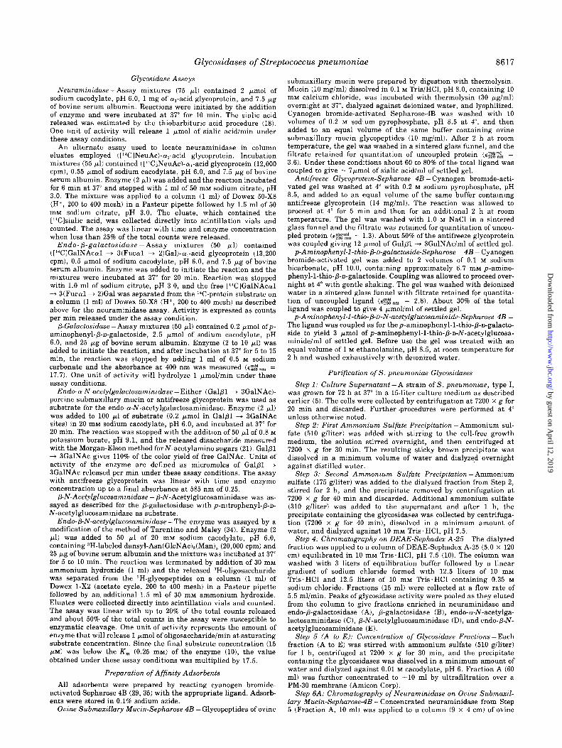
Purification of Streptococcus pneumoniae Glycosidases - Glycosidases were prepared from 15 liters of S. pneumoniae culture filtrate. The enzymes were initially fractionated on DEAE-Sephadex A-25 as shown in Fig. 1, and each partially pure glycosidase was purified further to remove contaminat- ing glycosidases. A summary of the purification is given in
Fraction Number (22.5ml)
FIG. 1. Chromatography of the partially purified glycosidases on DEAE-Sephadex A-25. Glycosidases from Step 3 (578 ml) were applied to a column of DEAE-Sephadex A-25 (5.0 x 120 cm) in 10 rnM Tris.HCl, pH 7.5. Fractions were collected as the column was developed with the same buffer followed by a linear gradient of sodium chloride (0 to 0.35 M). Details of the column conditions are given in Step 4 of “Experimental Procedures.” Symbols refer to protein (O), NaCl concentration (-), neuraminidase by the thio- barbiturate assay (a, 1 = 0.05 unit/ml), P-galactosidase (0, 1 = 0.005 unit/ml), endo-P-N-acetylgalactosaminidase by the (Galpl + SGalNAc)-porcine submaxillary mucin assay (A, 1 = 0.01 unit/ml), and p-N-acetylglucosaminidase (0, 1 = 0.1 unit/ml). Endo-p-galac- tosidase elution at 0.05 M NaCl (6) and the endo-@N-acetylglucosa- minidase elution at 0.25 M NaCl (10) are not shown. The major peaks of activity (A to E) were pooled as indicated.
Table I, which lists the steps common for all enzymes through chromatography on DEAE-Sephadex A-25, and the further purification of each enzyme separately.
Purification of Neuraminidase - The neuraminidase from Step 5 was contaminated with endo-P-galactosidase and /3- galactosidase. Two affinity adsorbents that have been used in the purification of other neuraminidases, N-(p-aminophenylj- oxamic acid-agarose (36) and ol,-acid glycoprotein-agarose (28), were found unsuitable for further purification since the other enzymatic activities also bound these adsorbents and eluted with the neuraminidase. One problem with the cy,-acid glycoprotein-agarose is that degradation during chromatogra- phy may produce terminal gala&se residues that serve as potential binding sites for the P-galactosidase. For this reason, ovine submaxillary mucin-agarose containing the disaccha- ride NeuAccu2 + 6GalNAc was examined and found to be effective.
An example of the purification of 145 units of neuraminidase obtained on ovine submaxillary mucin-agarose is shown in Fig. 2. The neuraminidase was eluted substantially free of other glycosidases with variable yields of 27 to 80%. The final specific activity (45 units/mg of protein) is about 400 times higher than reported previously for this enzyme (51. The purified enzyme was found to be unstable, but with the addition of 0.25 mg of bovine serum albumin/ml, no loss of activity was observed over 2 months when stored at -20”.
The column size and the time spent on the affinity adsorbent were critical factors for optimal purification. Loading of excess enzyme or prolonging the time of development of the column allowed for enzymatic cleavage of the sialic acid and prema- ture elution of the enzyme. Several test columns showed that a minimum of 2 pmol of sialic acid bound to the adsorbent was necessary for application of 1 unit of neuraminidase when the total chromatography time was 30 min or less.
Purification of Po-Galactosidase - The purification of the p-galactosidase is summarized in Table I. An example of the purification of P-galactosidase obtained on the affinity adsorb-
by guest on April 12, 2019
http://ww
w.jbc.org/
Dow
nloaded from
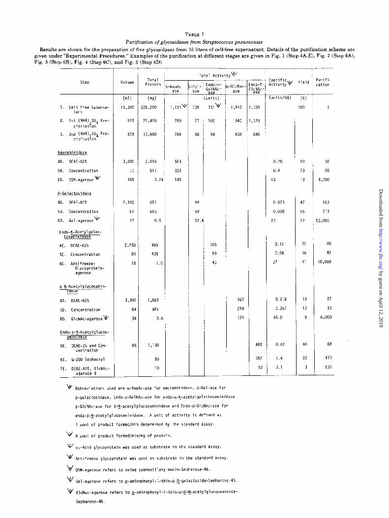
TABLE I
Purification of glycosidases from Streptococcus pneumonine
Results are shown for the preparation of five glycosidases from 15 liters of cell-free supernatant. Details of the purification scheme are given under “Experimental Procedures.” Examples of the purification at different stages are given in Fig. 1 (Step 4A-El, Fig. 2 (Step 6A), Fig. 3 (Step 6B), Fig. 4 (Step 6C), and Fig. 5 (Step 6D).
Total Protein
NeuAc- ase
Total Activityw 'urifi- :ation
Volume Gal- ase
(ml)
15,300
(m9)
!28,200 1,134v
915 22,420 789 77
578 13,900 789 68
3,200 2,016 563
10 811 328
185 3.24 145
2,100 651 49
61 603 59
17 0.5 12.
2,250 945
85 935
18 1.5
3,3oc
61
31
8:
1,683
975
3.6
1,138
80
10
ndo-a- $;",Ac-
nits)
351v
-GlcNac- ase
(units/mg)
1. ,100 i 1 1
940 1, ,120
838 846
0.28 50 56
0.4 29 80
45 13 9,000
0.075 47 163
0.098 56 213
25 12 i5,ooo
0.12 30 80
0.06 16 42
27 11 18,000
363 0.216 19 27
254 0.261 13 33
174 48.0 9 6,000
482 0.42 44 88
352 4.4
3.1
32 913
30 3 633
1. Cell Free Superna- tant
Neuraminidase
4A. DEAE-AZ5
5A. Concentration
6A. OSM-agarose w
B-Galactosidase
48. DEAE-A25
58. Concentration
68. Gal-agarose \f/
Endo-y-Acetylgalac- tosamlnldase
4c. DEAE-A25
5c. Concentration
6C. Antifreeze- Glycoprotein- agarose
d
B-l-Acetylglucosamin- ldase
40. DEAE-A25
5D. Concentration
6D. GlcNAc-agarosev
Endo-B-tj-Acetylglucos- amlnldase
5E. DEAE-25 and Con- centration
6E. G-ZOO Sephacryl
7E. DEAE-A25, GlcNAc- agarose~&
v Abbreviations used are a-NeuAc-ase for neuraminidase, B-Gal-ase for
B-galactosidase, Endo-a-GalNAc-ase for endo-a-I+acetylgalactosaminidase
B-GlcNAc-ase for B-lj-acetylglucosaminidase and Endo+GlcNAc-ase for
endo-B-F-acetylglucosaminidase. A unit of activity is defined as
1 umol of product formedfmin determined by the standard assay.
\br' A pm01 of product formed/min/mg of protein.
w _ a, Acid glycoprotein was used as substrate in the standard assay.
\e/ Antifreeze glycoprotein was used as substrate in the standard assay.
ki - OSM agarose refers to ovine submaxillary mucin-Sepharose-4B.
V Gal-agarose refers to ~-aminophenyl-l-thio-B-~-galactoside-Sepharose-4B.
\4/ GlcNac-agarose refers to e-aminophenyl-1-thio-B-g-y-acetylglucosaminide-
Seoharose-4B.
by guest on April 12, 2019
http://ww
w.jbc.org/
Dow
nloaded from
8620 Glycosidases of Streptococcus pneumoniae
FIG. 2. Purification of neuraminidase on ovine submaxillary mu- tin-agarose. The concentrated neuraminidase fraction (10 ml in 10 mM sodium cacodylate) from Step 5A was applied to a column of ovine submaxillary mucin-agarose (9 x 4 cm). The column was washed at 4” with 2.0 M sodium chloride in 0.02 M sodium cacodylate, pH 6.0 (A) and elution of the neuraminidase was initiated by bringing the column to 37” and washing with 1.0 M sodium chloride in 0.05 M sodium borate, pH 9.0 (B). Details of the column conditions are given in Step 6A of “Experimental Procedures.” Symbols refer to protein (O), neuraminidase (a, 1 = 200 cpm in standard ([14ClNeuAc)-cu, acid glycoprotein assay), endo-P-galactosidase (a, 1 = 200 cpm in standard ([14C]GalNAc~l + S(Fucoll + B)Gal)-alp- acid glycoprotein assay) and /3-galactosidase (0, 1 = 0.001 unit/ml).
I ” ” ‘I
‘ii 0.5 IO c
s E z 0.4 6 .- ,”
.- .? f L ;
; 0.3 6a J ; 5 2
” 0.2 .;
41;
z L a 0.1 2
20 40 60 SO Fraction Number (1.51111)
100
FIG. 3. Purification of /3-galactosidase on p-aminophenyl-l-thio- /3-n-galactoside-agarose. The concentrated P-galactosidase fraction obtained from Step 5B (61 ml in 10 mM sodium cacodylate, pH 6.0, 0.15 M sodium chloride) was applied to a column (1.5 x 4 cm) ofp- aminophenyl-1-thio-/3-n-galactoside-agarose. The column was washed with 0.1 M sodium cacodylate, pH 6.0, 1.0 M sodium chloride (A) and the enzyme eluted with 15 mM p-aminophenyl-1-thio-p-D-galac- toside in 50 mM sodium cacodylate, pH 6.0 @?I. Details of column conditions are given in Step 6B of “Experimental Procedures.” Symbols denote protein concentration (0, Lowry assay), P-galacto- sidase (0, 1 = 0.25 unit/ml), neuraminidase (a, 1 = 360 cpm in standard [L4C1NeuAc-o,-acid glycoprotein assay), endo-cy-l\r-acetyl- galactosaminidase (A, 1 = 0.025 unit/ml), and endo-p-galactosidase (@, 1 = 40 cpm in standard assay).
ent, p-aminophenyl-1-thio+n-galactoside-agarose, is shown in Fig. 3. The majority of the protein and the contaminating glycosidases present in the galactosidase (Fraction B) from the DEAE-step are unretarded on the column to yield a highly purified P-galactosidase substantially free of other glycosidases. The specific activity (25 units/mg of protein) was about 75 times that reported previously (5). Variable yields of 20 to 80% were obtained at this step. Although the
Fraction Number (2.0ml)
FIG. 4. Purification of endo-a-N-acetylgalactosaminidase on an- tifreeze glycoprotein-agarose. The concentrated endo-a-N-acetylga- lactosaminidase (85 ml in 10 mM sodium cacodylate, pH 6.0, 0.15 M sodium chloride) obtained from Step 5C was applied (1 mUmin) at 4” to a column of antifreeze glycoprotein-agarose (0.6 x 3 cm) equili- brated in the same buffer. The column was washed with 2.0 M sodium chloride in 20 rnM sodium cacodylate, pH 6.0 (A), and the enzyme eluted by bringing the column to 37” and continuing the wash (B). Details of the column conditions are given in Step 6C of “Experimental Procedures.” Symbols refer to protein (O), P-galac- tosidase (0, 1 = 0.01 unit/ml), endo-o-N-acetylgalactosaminidase (A, 1 = 1 unit/ml by (Galfil + 3 GalNAc)-porcine submaxillary mucin assay), P-N-acetylglucosaminidase (0, 1 = 0.01 unit/ml).
basis for the occasional low yields has not been systematically examined, better yields were obtained when 20% ethylene glycol was used to elute the enzyme instead ofp-aminophehyl- l-thio-/3-n-galactoside. The enzyme lost 10 to 15% activity in 30 days when stored at either 4” or -20” at a concentration of 30 pglml in 50 mM sodium cacodylate, pH 6.0. No loss of activity was observed when the enzyme was stored at 4” or -20” in bovine serum albumin (0.5 mg/ml).
Purification of Endo-a-N-acetylgalactosaminidase -The purification of the endo-cu-N-acetylgalactosaminidase on anti- freeze glycoprotein-agarose is shown in Fig. 4. The enzyme was eluted free of other enzymatic activities with a 68% step yield to give a specific activity (27 units/mg of protein) over 2000 times that obtained by conventional purification proce- dures (7). The /3-galactosidase does not bind presumably because of its strict substrate specificity for GalPl + 4R linkage.4 The enzyme can be stored for at least 2 months at -20” with no loss of activity. Limitations on column capacity and chromatography time have not been observed but signifi- cant changes in the method used could cause hydrolysis of the affmity ligand from the agarose.
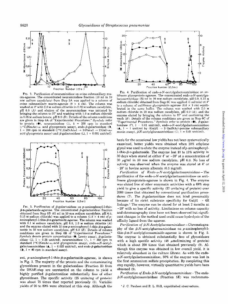
Purification of P-N-Acetylglucosaminidase - Chromatogra- phy of the j3-N-acetylglucosaminidase on p-aminophenyl-l- thio-/3-n-N-acetylglucosaminide-agarose is shown in Fig. 5. The enzyme is obtained substantially free of glycosidases with a high specific activity (48 pmol/min/mg of protein) which is about 200 times that obtained previously (9). Al- though this enzyme was obtained in low overall yield, it is relatively abundant in the culture filtrate. As with the endo- c-u-N-acetylgalactosaminidase, 50% of the enzyme was lost in the first ammonium sulfate precipitation. By completing this step rapidly, however, virtually quantitative yields have been obtained (9).
Purification of Endo-/3-N-acetylglucosaminidase -The endo- P-N-acetylglucosaminidase (Fraction 5E) was rechromato-
4 J. C. Paulson and R. L. Hill, unpublished observations.
by guest on April 12, 2019
http://ww
w.jbc.org/
Dow
nloaded from
Glycosidases of Streptococcus pneumoniae 8621
Fraction Number (3.5ml)
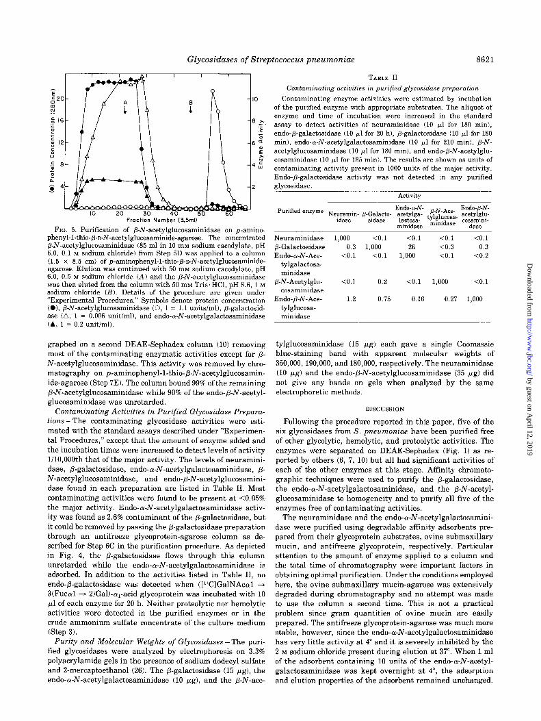
FIG. 5. Purification of P-N-acetylglucosaminidase on p-amino- phenyl-1-thio-p-n-N-acetylglucosaminide-agarose. The concentrated P-N-acetylglucosaminidase (85 ml in 10 mM sodium cacodylate, pH 6.0, 0.1 M sodium chloride) from Step 5D was applied to a column (1.5 x 8.5 cm) of p-aminophenyl-1-thio-p-D-N-acetylglucosaminide- agarose. Elution was continued with 50 mM sodium cacodylate, pH 6.0, 0.5 M sodium chloride (A) and the /3-N-acetylglucosaminidase was then eluted from the column with 50 mivr Tris’HCl, pH 8.6, 1 M sodium chloride (B). Details of the procedure are given under “Experimental Procedures.” Symbols denote protein concentration (01, P-N-acetylglucosaminidase (0, 1 = 1.1 units/ml), P-galactosid- ase (a, 1 = 0.006 unit/ml), and endo-Lu-N-acetylgalactosaminidase (A, 1 = 0.2 unit/ml).
graphed on a second DEAE-Sephadex column (10) removing most of the contaminating enzymatic activities except for p- N-acetylglucosaminidase. This activity was removed by chro- matography on p-aminophenyl-1-thio-P-N-acetylglucosamin- ide-agarose (Step 7E). The column bound 99% of the remaining P-N-acetylglucosaminidase while 90% of the endo-b-hr-acetyl- glucosaminidase was unretarded.
Contaminating Activities in Purified Glycosidase Prepara- tions -The contaminating glycosidase activities were esti- mated with the standard assays described under “Experimen- tal Procedures,” except that the amount of enzyme added and the incubation times were increased to detect levels of activity l/lO,OOOth that of the major activity. The levels of neuramini- dase, P-galactosidase, endo-a-N-acetylgalactosaminidase, p- N-acetylglucosaminidase, and endo-p-N-acetylglucosamini- dase found in each preparation are listed in Table II. Most contaminating activities were found to be present at ~0.05% the major activity. Endo-cY-N-acetylgalactosaminidase activ- ity was found as 2.6% contaminant of the P-galactosidase, but it could be removed by passing the P-galactosidase preparation through an antifreeze glycoprotein-agarose column as de- scribed for Step 6C in the purification procedure. As depicted in Fig. 4, the P-galactosidase flows through this column unretarded while the endo-a-N-acetylgalactosaminidase is adsorbed. In addition to the activities listed in Table II, no endo-P-galactosidase was detected when ([‘ClGalNAcal + 3(Fuccul + 2)Gal)-a,-acid glycoprotein was incubated with 10 ~1 of each enzyme for 20 h. Neither proteolytic nor hemolytic activities were detected in the purified enzymes or in the crude ammonium sulfate concentrate of the culture medium (Step 3).
Purity and Molecular Weights of Glycosidases-The puri- fied glycosidases were analyzed by electrophoresis on 3.3% polyacrylamide gels in the presence of sodium dodecyl sulfate and 2-mercaptoethanol (26). The P-galactosidase (15 pg), the endo-a-N-acetylgalactosaminidase (10 pg), and the p-N-ace-
TABLE II
Contaminating activities in purified glycosidase preparation
Contaminating enzyme activities were estimated by incubation of the purified enzyme with appropriate substrates. The aliquot of enzyme and time of incubation were increased in the standard assay to detect activities of neuraminidase (10 ~1 for 180 min), endo-p-galactosidase (10 ~1 for 20 h), P-galactosidase (10 /Al for 180 min), endo-a-N-acetylgalactosaminidase (10 yl for 210 min), P-N- acetylglucosaminidase (10 ~1 for 180 min), and endo-P-N-acetylglu- cosaminidase (10 ~1 for 185 min). The results are shown as units of contaminating activity present in 1000 units of the major activity. Endo-p-galactosidase activity was not detected in any purified glycosidase.
Activity
Neuraminidase 1,000 co.1 CO.1 co.1 co.1 /3-Galactosidase 0.3 1,000 26 co.3 0.3 Endo-a-N-Ace- co.1 co.1 1,000 co.1 co.2
tylgalactosa- minidase
/3-N-Acetylglu- co.1 0.2 10.1 1,000 co.1 cosaminidase
Endo-p-N-Ace- 1.2 0.78 0.16 0.27 1,000 tylglucosa- minidase
tylglucosaminidase (15 pg) each gave a single Coomassie blue-staining band with apparent molecular weights of 350,000,190,000, and 180,000, respectively. The neuraminidase (10 pg) and the endo-/3-N-acetylglucosaminidase (35 pg) did not give any bands on gels when analyzed by the same electrophoretic methods.
DISCUSSION
Following the procedure reported in this paper, five of the six glycosidases from S. pneumoniae have been purified free of other glycolytic, hemolytic, and proteolytic activities. The enzymes were separated on DEAE-Sephadex (Fig. 1) as re- ported by others (6, 7, 10) but all had significant activities of each of the other enzymes at this stage. Affinity chromato- graphic techniques were used to purify the p-galactosidase, the endo-a-N-acetylgalactosaminidase, and the /3-N-acetyl- glucosaminidase to homogeneity and to purify all five of the enzymes free of contaminating activities.
The neuraminidase and the endo-a-N-acetylgalactosamini- dase were purified using degradable affinity adsorbents pre- pared from their glycoprotein substrates, ovine submaxillary mucin, and antifreeze glycoprotein, respectively. Particular attention to the amount of enzyme applied to a column and the total time of chromatography were important factors in obtaining optimal purification. Under the conditions employed here, the ovine submaxillary mucin-agarose was extensively degraded during chromatography and no attempt was made to use the column a second time. This is not a practical problem since gram quantities of ovine mucin are easily prepared. The antifreeze glycoprotein-agarose was much more stable, however, since the endo-a-N-acetylgalactosaminidase has very little activity at 4” and it is severely inhibited by the 2 M sodium chloride present during elution at 37”. When 1 ml of the adsorbent containing 10 units of the endo-a-N-acetyl- galactosaminidase was kept overnight at 4”, the adsorption and elution properties of the adsorbent remained unchanged.
by guest on April 12, 2019
http://ww
w.jbc.org/
Dow
nloaded from
8622 Glycosiduses of Streptococcus pneumoniae
The S. pneumoniae P-galactosidase and P-N-acetylglucosa- minidase have been purified to homogeneity on affinity ad- sorbents substituted with p-aminophenyl-1-thio-@n-galacto- side or p-aminophenyl-1-thio-p-n-N-acetylglucosaminide, re- spectively. The fact that these two enzymes can be purified free of each other on these adsorbents attests to the speci- ficity with which the glycosidases bind the appropriate sub- strate analog. p-Aminophenyl-1-thioglycoside substituted ad- sorbents have been widely used as potential affinity adsorbents for glycosidases with variable results (37-43). In most studies a spacer arm between the ligand and the gel matrix was em- ployed and frequently, an adsorbent was found to adsorb several glycosidases in addition to the one which is expected to bind (39-41). In one case, Escherichia coli P-galactosidase has been shown to adsorb equally well to the p-aminophenyl-l- thio-/3-n-galactoside adsorbent and to a column containing only the spacer arm (44). In the present report, the p-amino- phenyl-1-thioglycosides were coupled directly to cyanogen bro- mide-activated Sepharose 4B. Since spacer arms provide po- tential sites for nonspecific adsorption of inert protein or unwanted enzyme activities, it is noteworthy that in this case they were not required. Thus spacer arms need not be consid- ered a necessary component for the design of ligands for all glycosidase affinity adsorbents.
Specific glycosyltransferases have been utilized to synthe- size radiolabeled substrates for neuraminidase and endo-P- galactosidase. For the neuraminidase, resialylation of asialo- a,-acid glycoprotein by incubation with pure P-gala&side (~2 + 6 sialyltransferase and CMP-[14C]NeuAc produces oligosac- charides with the terminal sequence [i4C]NeuAc(u2 + 6Galpl * 4GlcNAc (24). The endo-P-galactosidase is known to act on substrates that contain the blood group A positive sequence GalNAccul + 3(Fucal + 2)GalPl + 4GlcNAc . . . (6). While the oligosaccharides of a,-acid glycoprotein do not normally carry this structure, it can be synthesized with the aid of two glycosyltransferases. The P-gala&side al + 2 fucosyltrans- ferase incubated with GDP-fucose and asialo-a,-acid glycopro- tein yields a product with the terminal sequence Fuccul -+ 2GalPl + 4GlcNAc. This product is an acceptor for the fi- (fucosyl (~1 + 2) gala&side (~1 + 3 N-acetylgalactosaminyl- transferase (45) which on incubation with UDP-[i4C]GalNAc gives the final product [i4C]GalNAc~1 + 3(Fuccul + 2)GalPl + 4GlcNAc. In addition to the trisaccharide [‘*C]GalNAcal + 3(Fuccul + 2)Gal released by the endo-p-galactosidase, a- N-acetylgalactosaminidase would also release a radiolabeled product as free [‘C]GalNAc. This activity, however, is not found in the culture medium of S. pneumoniae when incu- bated with p-nitrophenyl-cY-N-acetylgalactosaminide.5
A third radiolabeled substrate was prepared from the oval- bumin glycopeptide with composition Asn(GlcNAc),(Man),. Tai and Kobata (11) have shown that of the five ovalbumin glycopeptides hydrolyzed by the endo-P-N-acetylglucosamini- dase from Streptomyces griseus, only this one is susceptible to cleavage by the S. pneumonias enzyme. The procedure used to label Asn(GlcNAc),(Man), introduces tritium by NaB3H, reduction of periodate-oxidized mannose residues (1 mol of periodate/mol of glycopeptide (34)). At least 50% of the labeled glycopeptides were susceptible to cleavage by the S. pneumoniae endo-/3-N-acetylglucosaminidase. Thus, of the 3 terminal mannose residues susceptible to periodate oxidation at least one of these may be modified without destroying the ability of the glycopeptide to serve as a substrate.
5 L. R. Glasgow and R. L. Hill, unpublished observations.
The substrate specificities for the S. pneumoniae endo-/3- galactosidase (61, the endo-a-N-acetylgalactosaminidase (7, 81, and the endo-P-N-acetylglucosaminide (11, 12) are well documented. For the neuraminidase, the p-galactosidase and the /3-N-acetylglucosaminidase, however, little has been re- ported about their specificity toward the penultimate sugar or the glycosidic linkage. The P-galactosidase appears to be quite specific toward galactose in a /31 + 4 linkage since GalPl -+ 4GlcNAc is a good substrate and GalPl -+ 4Glc is also a substrate, but no hydrolysis of Galpl + 3GlcNAc, Gal/31 + GGlcNAc, or methyl P-galactoside can be detected.4 Thus, until the substrate specificities of these exoglycosidases are better defined, it should not be assumed that the appropri- ate unsubstituted monosaccharide will be hydrolyzed when found at the nonreducing end of oligosaccharides.
The glycosidases of S. pneumoniue are active at neutral pH values and act on oligosaccharides of intact glycoproteins. These features make them particularly suitable for investigat- ing the role of oligosaccharides on glycoproteins with a labile function of biological interest. Since these enzymes can be obtained virtually free of contaminating activities, they can be used in large excess to ensure complete digestion of the desired moiety without producing partial digestions from unexpected activities. In principle, the enzymes can remove most of the carbohydrate from a wide variety of carbohydrate groups linked to the polypeptide chain through asparagine or threonine/serine residues. The neuraminidase, P-galactosid- ase, /3-N-acetylglucosaminidase, and endo-p-N-acetylglucosa- minidase have been used together to remove >85% of the asparagine linked carbohydrate from rabbit IgG (46) and (Ye- acid glycoprotein.6 The endo-a-N-acetylgalactosaminidase re- moved about 90% of the Gal@ + 3GalNAc units linked to the threonine residues of antifreeze glycoprotein’ and in conjunc- tion with a neuraminidase, a-fucosidase, and a-N-acetylgalac- tosaminidase from Clostridium perfrirzgens virtually all of the carbohydrate from porcine submaxillary mucin.8
Acknowledgments -We wish to thank J. I. Rearick and T. A. Beyer (Department of Biochemistry, Duke University Medical Center) for the preparation and characterization of the Gal@ + 3GalNAc from antifreeze glycoprotein and p-n- galactoside (~1 -+ 2 fucosyltransferase, respectively.
5.
6.
7. 8.
REFERENCES
Elbein, A. D., Sureka, A., and Lee, Y. C. (1977) J. Bid. Chem. 252, 2026-2031
Opheim, D. J., and Touster, 0. (1977). J. Biol. Chem. 252, 739- 743
Kawasaki, T., and Ashwell, G. (1976) J. Bud. Chem 251, 5292- 5299
Buchanan, R. E., and Gibbons, N. E. (1974) Bergey’s Manual of Determinative Bacteriology, 8th Ed, Williams and Wilkins Co., Baltimore, Md.
Hughes, R. C., and Jeanloz, R. W. (1964) Biochemistry 3, 1535 1543
Takasaki, S., and Kobata, A. (1976) J. Biol. Chem. 251, 3603- 3609
Endo, Y., and Kobata, A. (1976) J. Biochem. (Tokyo) 80, l-8 Bhavanandan, V. P., Umemoto, J., and Davidson, E. A. (1976)
Biochem. Biophys. Res. Commun. 70, 738-745
6 J. C. Paulson, C. S. Lowman, and R. L. Hill, unpublished observations.
‘J. I. Rearick, L. R. Glasgow, and R. L. Hill, unpublished observations.
B L. R. Glasgow, M. J. Holroyde, and R. L. Hill, unpublished observations.
by guest on April 12, 2019
http://ww
w.jbc.org/
Dow
nloaded from
Glycosidases of Streptococcus pneumoniae 8623
9. Hughes, R. C., and Jeanloz, R. W. (1964) Biochemistry 3, 1543- 28. Geisow, M. J. (1975)Bzochem. J. 151, 181-183 1548 29. Paulson, J., Beranek, W., and Hill, R. L. (1977) J. Biol. Chem.
10. Koide, N., and Muramatsu, T. (1973) J. Biol. Chem. 249, 4897- 252, 2356-2362 4904 30. Schwyser, M., and Hill, R. L. (1977) J. Biol. Chem. 252, 2338-
11. Tai, T., Yamashita, K., Ogata-Arakawa, M., Koide, N., Mura- 2345 matsu, T., Iwashita, S., Inoue, Y., and Kobata, A. (1975) J. 31. Trevelan, W. E., Procter, D. P., and Harrison, J. S. (1950) Biol. Chem. 250, 8569-8575 Nature 166, 444-446
12. Ito, S., Muramatsu, T., and Kobata, A. (1972)Biochem. Biophys. 32. Fletcher, A. P., Marks, G. S., Marshall, R. P., and Neuberger, Res. Commun. 63. 938-944 A. (1963) Blochem. J. 87. 265-272
13. Paulson, J. C., Glasgow, L. R., Lowman, C., Holroyde, M. J., and Hill, R. L. (1977) Fourth International Symposium on Glycoconjugates, Woods Hole, Mass.
14. Schacter, H., Hanako, I., and Heath, E. C. (1972) Methods Enzymol. 28, 285-287
15. Hill, H., Reynolds, J. A., and Hill, R. L. (1977) J. Biol. Chem. 252, 3791-3798
16. Plantner, J. (1974) Doctoral thesis, Case Western Reserve Uni- versity
17. DeVries, A. L., Komatsu, S. K., and Feeney, R. E. (1970) J. Biol. Chem. 245, 2901-2908
18. Warren, L. (1959) J. Biol. Chem. 234, 1971-1975 19. Svennerholm, L. (1959) Biochim. Biophys. Acta 24, 604-611 20. Jourdian, G. W., Dean, L., and Roseman, S. (1971) J. Biol.
Chem. 246, 430-435 21. Reissig, J. L., Strominger, J. L., and Leloir, L. F. (1955) J.
Biol. Chem. 217, 959-966 22. Doudoroff, M. (1962) Methods Enzymol. 5, 339-341
33. Huang, C., Mayer, H. E.,’ and Montgomery, R. (1970) Carbo- hydr. Res. 13, 127-137
34. Tarentino, A., and Maley, F. (1977) Anal. Bzochem. 77, 185-194 35. Porath, J. (1974) Methods Enzymol. 34, 13-29 36. Cuatrecasas, P., and Illiano, G. (1971) Biochem. Biophys. Res.
Commun. 44, 178-184 37. Steers, E., Jr., Cuatrecasas, P., and Pollard, H. B. (1971) J.
Biol. Chem. 246, 196-200 38. Grebner, E. E., and Parikh, I. (1974) Biochim. Biophys. Acta
350, 437-441 39. Junowicz, E., and Parris, J. E. (1973) Bzochim. Biophys. Acta
321, 234-245 40. Kiohara, R., Terao, T., Shioiri-Nakano, K., and Osawa, T.
(1976) J. Biochem (Tokyo) 80, 9-17 41. Mega, T., and Matsushima, Y. (1977) J. Biochem. (Tokyo) 81,
571-578 42. Mapes, C. A., and Sweeley, C. C. (1973) J. Biol. Chem. 248,
2461-2470 23. Wallenfels, K., and Kurz, G. (1966) Methods Enzymol. 9, 112- 43. Distler, J. J., and Jourdian, G. W. (1973) J. Biol. Chem. 248,
116 6772-6780 24. Paulson, J. C., Rearick, J. I., and Hill, R. L. (1976) J. Biol. 44. Hirotoshi, N., and Bailon, P. (1975) Arch. Biochem. Biophys.
Chem. 252, 2363-2371 168, 576-584 25. Lowry, 0. H., Rosebrough, N. J., Farr, A. L., and Randall, R. 45. Schwyzer, M., and Hill, R. L. (1977) J. Biol. Chem. 252, 2346-
J. (1951) J. Biol. Chem. 193, 265-275 2355 26. Weber, K., and Osborn, M. (1969) J. Biol. Chem. 244, 4406-4412 46. Koide, N., Masuto, N., and Muramatsu, T. (1977) Biochem. 27. Williams, H. R., and Lin, T. (1971) Biochim. Biophys. Acta 250, Biophys. Res. Commun. 75, 838-844
603-607
by guest on April 12, 2019
http://ww
w.jbc.org/
Dow
nloaded from
L R Glasgow, J C Paulson and R L Hillpneumoniae.
Systematic purification of five glycosidases from Streptococcus (Diplococcus)
1977, 252:8615-8623.J. Biol. Chem.
http://www.jbc.org/content/252/23/8615.citation
Access the most updated version of this article at
Alerts:
When a correction for this article is posted•
When this article is cited•
to choose from all of JBC's e-mail alertsClick here
http://www.jbc.org/content/252/23/8615.citation.full.html#ref-list-1
This article cites 0 references, 0 of which can be accessed free at
by guest on April 12, 2019
http://ww
w.jbc.org/
Dow
nloaded from