Embed Size (px)
Citation preview

Published: April 19, 2011
r 2011 American Chemical Society 4615 dx.doi.org/10.1021/bi200321c | Biochemistry 2011, 50, 4615–4622
ARTICLE
pubs.acs.org/biochemistry
Streptomyces clavuligerusHlmI Is an Intramolecular Disulfide-FormingDithiol Oxidase in Holomycin BiosynthesisBo Li and Christopher T. Walsh*
Department of Biological Chemistry and Molecular Pharmacology, Harvard Medical School, 240 Longwood Avenue, Boston,Massachusetts 02115, United States
bS Supporting Information
Natural product antibiotics holomycin and its variants, such asthiolutin, aureothricin, xenorhabdin, and thiomarinol, share
a common N-acylated bicyclic dithiolopyrrolone scaffold (alsoknown as pyrrothine) (Figure 1A).1�4 These dithiolopyrroloneantibiotics have been reported to interfere with bacterial RNAsynthesis;5�7 however, the mode of action and the relevance ofthe disulfide to activity remain unclear as direct inhibition ofRNA polymerases has yet to be reconstituted in vitro.7
Disulfides are present in a wide variety of natural products, inlarger ribosomally encoded constructs, e.g., conotoxins and cyclo-tides, as well as smaller, non-ribosomal peptides such as gliotoxinand the antitumorigen FK228. In all of these natural products,the disulfide is essential for the activity of the molecule. In thecyclotides, cystine disulfides impart increased thermal stability,conformational rigidity, and cell permeability.8 Similarly, inFK228, the disulfide has been shown to promote cell permeabilitywhile masking the active form containing free thiol of theepigenetically active prodrug.9 The mycotoxin gliotoxin employsthe disulfide in two complementary and damaging activities: (1)redox cycling to produce destructive superoxide free radicals and(2) cross-linking and inhibition of cellular proteins via formationof mixed disulfides with the reduced form.10 Given the importanceof the disulfide linkage to stability and mobility of these molecules,it is of considerable interest as to when and how the disulfides areinstalled in the mature natural products. Formation of disulfides inFK228 and gliotoxin is catalyzed by thioredoxin reductase-likeenzymes DepH and GliT, both of which are encoded in their
respective biosynthetic gene clusters (Figure 1B).11,12 Prior tothe work reported herein, it was unclear which enzyme may beresponsible for generation of the unusual disulfide present inholomycin.
We recently identified the holomycin biosynthetic genecluster through genome mining of the producing bacteriumS. clavuligerus.13 In addition to a non-ribosomal peptide synthe-tase (NRPS) module which activates and loads the essentialbuilding block L-Cys and an acetyl transferase for introduction ofthe N-acetyl moiety, four predicted flavin-dependent oxidore-ductases are encoded in the gene cluster (Figure 2A). Thepresence of four such oxidoreductases is consistent with apathway where L-Cys-L-Cys undergoes oxidations removing anet total of eight electrons in four two-electron oxidation steps,each catalyzed by one of the flavoenzymes (Figure 2B). One ofthe flavoenzymes, HlmI, displays homologies to the large familyof thioredoxin reductases, involved in dithiol�disulfide redoxinterconversions in protein substrates. We demonstrate thatHlmI is a dithiol oxidase, converting the reduced dithiol formof holomycin (red-holomycin), 2, to the disulfide form, 1(Figure 1B). Heterologous expression of the holomycin genecluster in S. albus was recently reported by Deng and co-workers,
Received: March 2, 2011Revised: April 17, 2011
ABSTRACT: Holomycin and related dithiolopyrrolone anti-biotics display broad-spectrum antimicrobial activities andcontain a unique 5,5-bicyclic ring structure with an N-acylatedaminopyrrolone fused to a cyclic ene�disulfide. Here we showthat the intramolecular disulfide bridge is constructed from theacyclic ene�dithiol at a late stage in the pathway by a thior-edoxin oxidoreductase-like enzyme HlmI from the holomycinproducer Streptomyces clavuligerus. Recombinant HlmI waspurified from E. coli with bound flavin adenine dinucleotide(FAD) and converts reduced holomycin to holomycin utilizing O2 as cosubstrate. As a dithiol oxidase, HlmI is functionallyhomologous to GliT and DepH, which perform a similar dithiol to disulfide oxidation in the biosynthesis of fungal natural productgliotoxin and epigenetic regulator compound FK228, respectively. Deletion of the hlmI gene in the wild type S. clavuligerus and in aholomycin-overproducing mutant resulted in decreased level of holomycin production and increased sensitivity toward holomycin,suggesting a self-protection role of HlmI in the holomycin biosynthetic pathway. HlmI belongs to a new clade of uncharacterizedthioredoxin oxidoreductase-like enzymes, distinctive from theGliT-like enzymes and the DepH-like enzymes, and represents a thirdexample of oxidoreductases that catalyzes disulfide formation in the biosynthesis of small molecules.

4616 dx.doi.org/10.1021/bi200321c |Biochemistry 2011, 50, 4615–4622
Biochemistry ARTICLE
and deletion of the hlmI gene abolished holomycin production,suggesting a role for HlmI in holomycin biosynthesis.14
’MATERIALS AND METHODS
Molecular Cloning of hlmI and Overexpression and Pur-ification of the HlmI Protein. The hlmI gene was clonedinto pET30-Ek/LIC vector (Novagen) using primers listed inTable S1, and the gene sequence was verified by DNA sequen-cing. This vector was introduced into BL21-CodonPlus (DE3)-RIPL chemical competent cells (Stratagene). A 2 L LB broth wasinoculated with 20 mL of overnight culture that was started froma single colony, and protein overexpression was induced at anOD600 nm of 0.6 with 0.2 mM isopropyl-β-D-thiogalactopyrano-side (IPTG) at 18 �C for 18 h. The cells were harvested bycentrifugation, resuspended in 20 mL of column buffer (50 mMHEPES, 300 mM NaCl, 10% glycerol, pH 7.5), and lysed with a
cell disruptor. The lysis mixture was clarified by ultracentrifuga-tion, and the supernatant was incubated with 2 mL of nickel-NTA agarose resin (Qiagen) at 4 �C with gentle mixing for 1 h.The resin with bound protein was loaded on to a column to drainthe flowthrough, washed with 50 mL of column buffer and 20 mLof column buffer containing 20 mM imidazole, and eluted with10 mL of elution buffer (300 mM imidazole in column buffer).The elution fractions were analyzed by SDS-PAGE, and thefractions containing HlmI were pooled and concentrated to5 mL with a 15 mL Amicon (Millipore) (10 kDa MW cutoff).Concentrated protein was desalted with a PD-10 desalting column(GE Healthcare) twice to remove excess imidazole. The resultingprotein was flash frozen with liquid N2 and stored at �80 �C.Molecular Cloning of gliT, Overexpression, and Purifica-
tion of theGliT Protein.Aspergillus fumigatusAf293 cDNAwas agift from Robert A. Cramer Jr. (Durham, NC).15 Using thiscDNA as template, gliT was amplified by PCR and cloned intopET24b betweenNdeI and XhoI sites. The sequence was verifiedby DNA sequencing. BL21 (DE3) cells containing the con-structed vector were grown overnight at 37 �C. 6 L of LBcontaining 40 μg/mL kanamycin and 5 mM MgCl2 was inocu-lated with 1% of the overnight culture. When OD600 nm of theculture reached 0.7, protein expression was induced with 0.1 mMIPTG and the culture was grown for an additional 12 h at 15 �C.Cells were harvested and resuspended in 28 mL of lysis buffer(25 mM Tris, pH 8.0, 500 mM NaCl), lysed with two passes onan Emulsiflex-C5 cell disruptor (Avestin). Cell lysate was clearedat 35 000 rpm (95 000g) for 35 min and incubated with 1.1 mL ofNi-NTA resin (2.2 mL 50% slurry) that was prewashed with lysisbuffer for 1.5 h at 4 �C. Resin was then transferred to column, anda gradient imidazole elution was performed: 25 mL for 0 and5mM imidazole, 15mL for 25mM, 10mL for 200mM, and 5mLfor 500 mM imidazole. GliT was eluted in the 25, 200, and500 mM imidazole elution fractions, which were combined andconcentrated to less than 2 mL and injected for gel filtration in20 mM Tris, pH 8, 50 mM NaCl. Fractions containing GliTwere pooled and concentrated, and 10% glycerol was added. Atotal of 4.5 mL of GliT was obtained, and its concentration wasdetermined by Bradford as 294 μM.Determination of HlmI Oligomeric State and FAD Content
in HlmI and GliT. Purified HlmI was analyzed by size exclusionchromatography using a 10/300 Superdex 200 gel-filtrationcolumn and running buffer containing 50 mM HEPES, 150 mMNaCl, and 5% glycerol at pH 7.5. The calculated molecular weightfor HlmI, including the His tag, is 39 163 Da, and the apparentmolecular weight for HlmI is ∼80 kDa based on the standardcurve on the gel filtration column, suggesting that HlmI exists as adimer. BothHlmI andGliTwere denatured by boiling for 5min torelease the flavin cofactor. The supernatant was injected onAccurate-Mass Q-Tof LC/MS instrument (Agilent Technologies6520), and the flavin cofactor was identified as FAD. To measurethe FAD content, UV scans between 200 and 600 nm (CaryUV�vis) were taken for HlmI and GliT and also the FAD-containing supernatant from the denatured samples. Absorbanceat 450 nm for the latter samples was used to determine the con-centration of FAD using extinction coefficient 11 300 M�1 cm�1,and the background absorbance at 280 nm from FAD wassubtracted from the A280 nm values of the nondenatured HlmIand GliT samples. The adjusted A280 nm values were used forcalculation of protein concentrations using theoretical extinctioncoefficients 26 595M�1 cm�1 for HlmI and 30 160M�1 cm�1 forGliT. Duplicate experiments were performed, and the average
Figure 1. (A) Structures of holomycin and related dithiolopyrroloneantibiotics. (B) Conversion of the reduced form of holomycin, gliotoxin,and FK228 into the disulfide form by thioredoxin oxidoreductase-likeenzymes.

4617 dx.doi.org/10.1021/bi200321c |Biochemistry 2011, 50, 4615–4622
Biochemistry ARTICLE
FAD content was 20% forHlmI and 35% for GliT. To reconstitutethe binding of FAD in HlmI and GliT, both proteins wereincubated with excess FAD on ice for 30 min. Excess FAD wasremoved by passing the proteins through Micro Bio-Spin 6chromatography columns pre-equilibrated in buffer containing20mMTris, 10mMNaCl, 5% glycerol (pH8.5). HlmIwas furtherpurified using a MonoQ 10/100 GL anion exchange column (GEHealthcare). A linear gradient from 10mMNaCl to 1MNaCl wasrun over seven column volumes at 1mL/min. Fractions containingHlmI were combined and concentrated using Amicon Ultra cen-trigual filters (0.5 mL, 3 kDa molecular weight cutoff). The resultingprotein was shown to be over 95% pure by SDS-PAGE. The FADcontent was determined using the same method as described aboveand increased to 80% and 76% in GliT and HlmI, respectively.Enzymatic Assays To Measure Steady-State Kinetic Para-
meters. Holomycin was isolated from overproducing strainS. clavuligerus ΔORF15 and purified by HPLC as previouslydescribed.13 The concentration of holomycin was calculatedbased on UV absorbance at 388 nm using a reported extinctioncoefficient of 11 220 M�1 cm�1.16 Reduced holomycin wasgenerated by reduction of holomycin with 1 mol equiv of TCEPin the presence of 100 mM phosphate buffer (pH 6.5), and asample of 3 μL of 2 μM HlmI (50 nM final total concentration,10 nM effective concentration) or 3 μL of 1 μM GliT (25 nMfinal total concentration, 8.75 nM effective concentration) wasadded to initiate the reaction (total reaction volume 120 μL).Because of the detectable nonenzymatic oxidation by O2, multi-ple concentrations for HlmI and GliT were tested to findoxidation rates that are well above background yet still in thelinear range, and the optimal concentrations for HlmI and GliTare 50 and 25 nM, respectively. Initial velocity was measuredin a continuous assay by taking a UV scan (Cary UV�vis) from200 to 600 nm every 0.2 min for a total of 10 min at reducedholomycin concentrations of 5, 10, 25, 50, and 100 μM. Initial
velocity was calculated from the decrease in absorbance ofreduced holomycin at 340 nm with a extinction coefficient at15 900 M�1 cm�1. This extinction coefficient was obtained byfully converting a solution of holomycin of known concentrationto reduced holomycin with excess TCEP and measuring theabsorbance at 340 nm. Background oxidation by O2 was takenfor each substrate concentration without enzyme. The enzymecatalyzed rate was obtained by subtracting the nonenzymaticrate from the overall rate. These experiments were carried outin duplicates for calculation of error. Data were analyzed andplotted using GraphPad Prism. Kinetic measurement was alsoperformed with the FAD-reconstituted HlmI and GliT, whichcontained a much higher percentage of FAD, at a concentrationof 25 nM (76% FAD, 19 nM effective concentration) and12.5 nM (80% FAD, 10 nM effective concentration), respectively.Determination of Half-Life of Reduced Holomycin and
Reduced Holothin. Holothin was generated by microwave-assisted deacetylation of holomycin dissolved in 1,3-dioxane andheated at 100 �C for 10min in the presence of 2NHCl. The waterlayer of product mixture was vacuum-dried and resuspendedin water and further purified by HPLC. A sample of 50 μMholomycin or holothin was mixed with 1 equiv of TCEP in thepresence of 100 mM phosphate buffer (pH 6.5) to generatereduced holomycin or reduced holothin. The conversion from thereduced form to the disulfide form was monitored continuouslyby UV. Absorbance at 340 and 335 nm was used respectively tocalculate the half-life of reduced holomycin and reduced holothin.Data were analyzed and plotted using GraphPad Prism.Genetic Deletion of hlmIGene in S. clavuligerus.Deletion of
hlmI gene was achieved using ReDirect PCR-targeting strategy.17
The hlmI gene with a 2 kb extension of the chromosomal seque-nce on each end was cloned into a PCR-Blunt vector (KanR,Invitrogen). This vector was introduced into E. coli BW25113/pIJ790 strain allowing for λ red-mediated recombination. The
Figure 2. (A) Holomycin (hlm) gene cluster with genes colored by function. hlmA, acyltranferase; hlmB, acyl-CoA dehydrogenase; hlmC, thioesterase;hlmD, glucose�methanol�choline oxidoreductase; hlmE, NRPS (cyclization�adenylation�thiolation, Cy-A-T); hlmF, lantibiotic decarboxylase;hlmG, globin; hlmH, major facilitator family transporter; hlmI, thioredoxin�disulfide oxidoreductase; hlmJ, hlmM, transcriptional regulators; hlmK,inactive thioesterase domain; hlmL, condensation domain. (B) Proposed holomycin biosynthetic pathway. HlmI, the focus of this study, is shown in ahighlighted box and is proposed to be involved in the late stages of holomycin biosynthesis.

4618 dx.doi.org/10.1021/bi200321c |Biochemistry 2011, 50, 4615–4622
Biochemistry ARTICLE
resulting strain was transformed with PCR fragments containingthe desired antibiotic resistance cassette including a 36-nt exten-sion homologous to the target region directly upstream anddownstream of the hlmI gene. Correct recombined vectors con-taining the desired disruption cassettes were selected based onantibiotic resistance. The apramycin resistance cassette was con-structed using pIJ773 (ApraR, acc(3)IV-oriT) vector as templatefor disruption in S. clavuligerus wild type. The vector pIJ10700(HygR, hyg-oriT) was used as template for generating the hygro-mycin B resistance cassette to target the ORF15::apr holomycin-overproducing mutant, which already contains an apramycinresistance marker. Both pIJ vectors were obtained from JohnInnesCentre, Norwich,UK.TheE. coliWM6062 strain containingthe disruption vectors was conjugated with the wild type orORF15::apr S. clavuligerus strains following stardard protocols.18
Correct exoconjugants resulting from homologous recombinationwere selected based on antibiotic resistance and verified by PCRusing an internal primer for the disruption cassette (pIJ773-RP603: 50-GAG TTG TCT CTG ACA CAT TCT GGC G-30;or pIJ10700-FP1000: 50-GGG AAC ACC GTG CTC ACC-30)and an external primer that anneals to hlmF gene (Scl-HlmF-LICFP: 50-GGT ATT GAG GGT CGC ATG GAA AAT CCGATC CCC TCT CCT TC-30). The PCR products were alsopurified and verified by DNA sequencing.Analysis of Holomycin Production by hlmI Deletion Mu-
tants. The correct ΔhlmI deletion mutants were grown in TSBmedia for 24�48 h. A sample of 10 mL of liquid GSPG(glycerol�sucrose�proline�glutamate) medium19 was inocu-lated with 10 μL of the starter culture and grown with shakingat 30 �C for 3�5 days. The resulting culture supernatant wasfiltered with Spin-X centrifuge tube filters (Corning), and a sample
of 10 μL was injected on the Accurate-Mass Q-Tof LC/MSinstrument (Agilent Technologies 6520) for detection of holo-mycin production via electrospray ionization (ESI).Agar Diffusion Assays To Test Holomycin Sensitivity of
S. clavuligerus Strains. Spore stocks were prepared for wildtype, ΔhlmI, ΔORF15, and ΔORF15/ΔhlmI strains. The sporeconcentrations were determined by serial dilutions of the stockand counting of colony forming units. Approximately 5 � 105
spores were plated for each strain, and a sample of 5 μL of7 mg/mL holomycin was added to the center of the MYM plate(4.0 g of yeast extract, 10.0 g of malt extract, 2.0 g of dextrose,and 20.0 g of agar per liter of media). The plates were incubatedat 30 �C for 4�7 days, and the diameter of the inhibition zonewas determined as a measurement for the sensitivity of thesestrains toward holomycin.Phylogenetic Analysis of HlmI. HlmI was aligned with a
number of bacterial and fungal thioredoxin�oxidoreductase likeenzymes from the protein database using ClustalX. Alignmentwas edited using MacClade and imported into RAxML forgeneration of dendrogram by maximum likelihood.20 The result-ing dendrogram was further colored by Adobe Illustrator.
’RESULTS AND DISCUSSION
Holomycin Biosynthetic Gene Cluster Nomenclature. Pre-viously, we demonstrated that ORFs 3483-3496 from theS. clavuligerus genome (locus tag SSCG_03483 throughSSCG_03496) constitute a likely holomycin biosynthetic genecluster by both genetic manipulation and biochemical character-ization of ORFs 3483 and 3488.13 In this work we renamedORFs3483-3496 as hlmA-hlmM (Figure 2A), where hlm stands forholomycin and is a three-letter acronym not used elsewhere inbacterial gene assignments. ORF3492 as the focus of this work isrenamed as hlmI.Preparation and Characterization of Reduced Holomycin.
The disulfide formation is proposed to occur at late stages ofholomycin assembly via oxidation of the dithiol groups in reducedholothin (red-holothin) or red-holomycin, 2 (Figure 2B). Becausea large quantity of holomycin was readily available from apreviously described overproducing strain of S. clavuligerus,21 wefirst reduced holomycin with tris(2-carboxyethyl)phosphine(TCEP) to generate red-holomycin, 2, as a substrate for enzymaticanalysis. LC-MS analysis of the TCEP reduction mixture showedchromatographic separation of 1 ([Mþ H]þ expected 214.9943,observed 214.9954) from 2 ([M þ H]þ expected 217.0100,observed 217.0111), which was eluted earlier than holomycin onthe C18 column and showed two mass unit increase in molecularweight (Figure 3A) as anticipated. The conversion of the disulfideform to the dithiol form of holomycin by TCEP resulted in asignificant blue shift of the λmax of UV absorbance from 385 nm in2 to 340 nm in 1with an∼50% increase in extinction coefficient to15 900 M�1 cm�1 (Figure 3B). The reduction of holomycin tored-holomycin is consistent with prior findings fromHertweck andcolleagues that TCEP generates the reduced form of gliotoxin.11
The change in UV absorbance affords an analytical method formonitoring the reduction�oxidation reaction described below.S. clavuligerus HlmI Is an FAD-Dependent Oxidase for
Reduced Holomycin. Among the four predicted flavoenzymes inthe S. clavuligerus cluster, HlmI shares sequence homology withthioredoxin oxidoreductase superfamily (Figure S1), in particularthe recently characterized gliotoxin dithiol oxidase GliT.11 Toevaluate a comparable function ofHlmI as a red-holomycin oxidase,
Figure 3. (A) LC-MS analysis of holomycin treated with 2 equiv ofTCEP. UV trace at 390 nm is shown with a clear separation of red-holomycin (5.0 min) and holomycin (7.2 min). The presence ofholomycin in the UV trace is due to the reoxidation of red-holomycinby O2 during the sample injection and column separation. (B) UVspectra of red-holomycin and holomycin.

4619 dx.doi.org/10.1021/bi200321c |Biochemistry 2011, 50, 4615–4622
Biochemistry ARTICLE
HlmIwas expressed heterologously inE.coli and purified as a yellowprotein at a yield of 20 mg/L (Figure 4A and Figure S2a). Thebound cofactor was determined as FAD by LC/MS after heatdenaturation of HlmI (Figure S2b,c) and was present in 0.2 molequiv to the 40 kDa protein. In vitro reconstitution by incubationwith excess FAD resulted in an increase in FAD content to 0.76equiv. The apparent oligomeric state of HlmI was determined bygel filtration elution profile as dimeric.To assay the activity of purifiedHlmI, holomycin,1, was reduced
in situ in buffer solutions with 1 equiv of TCEP to generate red-holomycin, 2. The reduction process appeared to be complete as
addition of excess TCEP did not further increase the UV absor-bance at 340 nm, indicating that nearly all holomycin in solutionhad been reduced. Subsequent addition of HlmI led to rapid loss ofthe A340 nm peak as the dithiol substrate was oxidized to intramo-lecular disulfide in holomycin. A family of progression curves couldbe recorded to monitor reaction progress and enabled determina-tion of the catalytic parameters of HlmI as a dithiol oxidoreductasefor red-holomycin (Figure 4B).Concentration of 2 (from quantitative TCEP-mediated re-
duction of 1 in situ) was varied at a fixed concentration of HlmI(50 nM, effective concentration 10 nM based on 20% activeenzyme by FAD content) to determine the steady-state kineticparameters. A Michaelis�Menten curve was generated yieldinga kcat of 333 ( 28 min�1 and a KM of 4.6 ( 1.9 μM for 2(Figure 4C). Kinetic measurements using FAD recontitutedHlmI with 0.76 equiv of FAD generated similar results (Figure S3).In control experiments under the same incubation conditionswithout addition of enzyme, the nonenzymatic rate of reoxida-tion of 2 to 1was monitored and a half-life of 7 min was obtainedfor 2, corresponding to a kuncat of about 0.1 min�1 (Figure S4).The kcat/kuncat ratio for HlmI is 3330/1, suggesting a 3 orders ofmagnitude rate enhancement in accelerating the disulfide for-mation. The half-life of reduced holomycin was not altered inthe presence of EDTA, suggesting metal-mediated thiol oxida-tion was not responsible for the nonenzymatic formation of theintramolecular disulfide.No electron receptorswere added in theHlmI oxidation reaction,
suggesting that molecular oxygen is the terminal electron acceptor.Addition of nicotinamide cofactors (NADþ or NADPþ) did not
Figure 4. (A)UV�vis absorbance spectrum ofHlmI containing 20% of FAD. (B) Time course of oxidation of 100μM red-holomycin by 50 nMHlmI in10 min (UV spectra scan was taken every 0.4 min). Arrow indicates the direction of UV spectra progression. (C) Michaelis�Menten kinetics curve ofHlmI (20% FAD) and GliT (35% FAD) with varying concentrations of red-holomycin substrate. Standard deviation is shown as error bars. (D)Anaerobic assay of 50 μM of red-holomycin and 50 nM HlmI in the presence of 1 mM NADþ (NADþ, no O2). HlmI was unable to convert 2 to 1without oxygen. When the assay was opened to the atmosphere (NADþ, O2), facile oxidation of 2 to 1 by HlmI was observed. Substitution of NADþ
with NADPþ yielded the very similar results and therefore is not shown in the graph.
Figure 5. Proposed mechanism of catalysis by HlmI to convert red-holomycin to holomycin.
4620 dx.doi.org/10.1021/bi200321c |Biochemistry 2011, 50, 4615–4622
Biochemistry ARTICLE
increase the oxidation rate. Furthermore,HlmI failed to convert 2 to1 under anaerobic conditions in the presence of NADþ or NADPþ
but readily catalyzed the disulfide formation once the reaction isopened to the atmosphere (Figure 4D). These results indicate thatHlmI acts as a dithiol oxidase, similar to GliT. GliT has beenproposed to use its active site disulfide as an initial redox port ofentry for electrons from the dithiol substrate into FAD.11 TheresultedFADH2�enzyme intermediatewould be rapidly reoxidizedby O2, presumably through the canonical FAD-C4a-OOH inter-mediate. Such a pathway for electron flow is also likely for HlmI,which contains a conserved CXXC motif (Figure S1). We proposethat the FAD reductive half-reaction is initiated by attack of the thiolgroup or the ene�thiol group of 2 on the active site disulfide inHlmI to yield a covalent disulfide adduct with the enzyme.Intramolecular attack of the second thiol upon proton abstractionby an active site base would lead to formation of the cyclic disulfidein 1 with generation of the reduced form of the enzyme (it is notknown which thiol of 2 would initiate attack on the disulfide in theactive site of HlmI; the enethiol, as a tautomer of a thioaldehyde, islikely to be less nucleophilic, but nothing is known about theorientation of 2 in the enzyme active site) (Figure 5). FADox thenregenerates the active site disulfide in the enzyme to form FADH2.Oxidation of FADH2 by O2 yields a transient FAD-C4a-OOH onthe way to release of hydrogen peroxide and regeneration of FADox,presumably constituting the oxidative half-reaction. The utilizationof O2 differentiates HlmI andGliT fromDepH, whichmakes use ofNADþ or NADPþ as electron receptors.12
GliT Has Reduced Holomycin Oxidase Activity. Recombi-nant GliT was purified from E. coli (Dr. Carl Balibar thesis)a andcontains 0.35 equiv of bound FAD. We assayed GliT at 25 nM(effective concentration 8.75 nM with bound FAD) to determinewhether it can oxidize 2 in addition to its normal substrate reducedgliotoxin (red-gliotoxin). This fungal dithiol oxidase can indeedoxidize 2 back to 1 (Figure 4C) with a KM for 2 at 198( 64 μM,20�130-fold higher than HlmI. Interestingly, the measured kcatwas about 10-fold higher thanHlmI, at 3166( 742min�1. Similar
KM and kcat values were obtained for GliT reconstituted with 0.8equiv of FAD (Figure S3). The kcat/KM catalytic efficiency ratiosare 72min�1 μM�1 for HlmI and 16min�1 μM�1 for GliT for theoxidation of red-holomycin, respectively. Therefore, HlmI is over-all more efficient than GliT to make the cyclic disulfide inholomycin, but GliT is substantially promiscuous.red-Holothin vs red-Holomycin as HlmI Substrates.During
the initial characterization of the holomycin biosynthetic path-way, we demonstrated that an acetyltransferase HlmA encodedin the gene cluster catalyzes N-acetylation of holothin(deacetylated holomycin, see Figure 2B for structure) as a latestep in the pathway.13 Acetylation of the exocyclic amino groupof the dithiolopyrrolone was proposed to be involved in stabiliz-ing the heterocyclic scaffold and/or be a self-protection strategy.We attempted to evaluate whether the reduced dithiol form ofholothin could also be a substrate for HlmI. Holothin wasgenerated by microwave-mediated deacetylation of holomycinand isolated in its oxidized form.13 Subsequent reduction ofholothin by TCEP resulted in a blue shift in the UV spectrumconsistent with the maximum absorbance around 340 nm ob-served in red-holomycin (Figure S5a). The dithiol group in red-holothin was much more prone to nonenzymatic oxidation todisulfide than red-holomycin. The half-life of red-holothin underaerobic conditions was very short, about 0.3 min (Figure S5b).HlmI qualitatively accelerated the reoxidation of red-holothin toholothin, but the high nonenzymatic background rate precludedkinetic measurements. The significantly faster nonenzymaticoxidation of red-holothin compared to red-holomycin indicatesthat acetylation of the exocyclic amine of the aminopyrrolonering confers a substantial electronic effect on the dithiol/disulfideredox properties. In both theN-acetylated red-holomycin and thefree amine-containing red-holothin, the intramolecular dithiolgroups are set up geometrically for facile closure to cyclicdisulfide.Phenotype of an HlmI deletion in S. clavuligerus.To assess
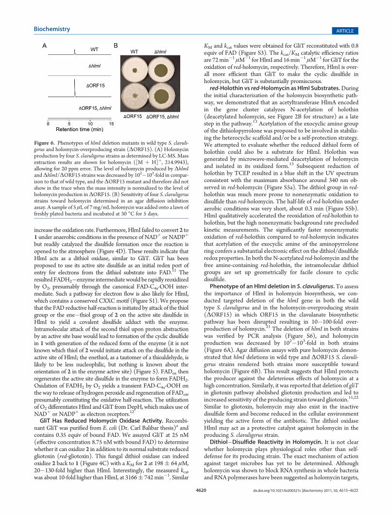
the importance of HlmI in holomycin biosynthesis, we con-ducted targeted deletion of the hlmI gene in both the wildtype S. clavuligerus and in the holomycin-overproducing strain(ΔORF15) in which ORF15 in the clavulanate biosyntheticpathway has been disrupted resulting in 10�100-fold over-production of holomycin.21 The deletion of hlmI in both strainswas verified by PCR analysis (Figure S6), and holomycinproduction was decreased by 102�103-fold in both strains(Figure 6A). Agar diffusion assays with pure holomycin demon-strated that hlmI deletions in wild type and ΔORF15 S. clavuli-gerus strains rendered both strains more susceptible towardholomycin (Figure 6B). This result suggests that HlmI protectsthe producer against the deleterious effects of holomycin at ahigh concentration. Similarly, it was reported that deletion of gliTin gliotoxin pathway abolished gliotoxin production and led toincreased sensitivity of the producing strain toward gliotoxin.11,22
Similar to gliotoxin, holomycin may also exist in the inactivedisulfide form and become reduced in the cellular environmentyielding the active form of the antibiotic. The dithiol oxidaseHlmI may act as a protective catalyst against holomycin in theproducing S. clavuligerus strain.Dithiol�Disulfide Reactivity in Holomycin. It is not clear
whether holomycin plays physiological roles other than self-defense for its producing strain. The exact mechanism of actionagainst target microbes has yet to be determined. Althoughholomycin was shown to block RNA synthesis in whole bacteriaand RNA polymerases have been suggested as holomycin targets,
Figure 6. Phenotypes of hlmI deletion mutants in wild type S. clavuli-gerus and holomycin-overproducing strain (ΔORF15). (A) Holomycinproduction by four S. clavuligerus strains as determined by LC-MS. Massextraction results are shown for holomycin ([M þ H]þ, 214.9943),allowing for 20 ppm error. The level of holomycin produced by ΔhlmIandΔhlmI/ΔORF15 strains was decreased by 102�103-fold in compar-ison to that of wild type, and theΔORF15 mutant and therefore did notshow in the trace when the mass intensity is normalized to the level ofholomycin production inΔORF15. (B) Sensitivity of four S. clavuligerusstrains toward holomycin determined in an agar diffusion inhibitionassay. A sample of 5 μL of 7mg/mL holomycin was added onto a lawn offreshly plated bacteria and incubated at 30 �C for 5 days.

4621 dx.doi.org/10.1021/bi200321c |Biochemistry 2011, 50, 4615–4622
Biochemistry ARTICLE
E. coliRNA polymerases are not susceptible toward holomycin inin vitro assays.7 The disulfide embedded in a conjugated bicyclicframework is likely to engage in dithiol�disulfide redox chem-istry with holomycin targets. We have conducted an initialexamination of holomycin reactivity with the most abundantcellular thiol, glutathione, as a prototypic marker of redoxbehavior. We first incubated the disulfide-containing holomycinwith the reduced form of glutathione (GSH) and monitored forreduction of holomycin by gain of UV absorbance at 340 nm. Nochanges were detected over a wide range of GSH concentrationsand molar excess over 1. When we assayed in the oppositedirection, for the ability of 2 to reduce oxidized glutathione(GS-SG), a rapid decrease in UV absorbance at 340 nm wasobserved, corresponding to the oxidation of 2 by GS-SG. Therate of oxidation of 2 as a function of oxidized GS-SG is shown inFigure S7.Subsequent studies will be necessary to determine redox
equilibria and ultimately reduction potentials of holomycin underconditions where competing oxidation by molecular O2 is sup-pressed. It is clear that red-holomycin is kinetically and thermo-dynamically competent to reduce the intermolecular disulfide inoxidized glutathione. Thus, the reduced form of holomycin andpresumably holothin (perhaps even more so) may be strongthiol reductants in both microbial producers and target cells.The combination of oxidation by HlmI and acetylation by HlmA
may ensure accumulation of the natural product in the oxidizedand acetylated state with low adventitious redox reactivity. Futurestudies will involve the examination of the formation of any mixeddisulfides between holomycin and intracellular proteins as part ofthe antibacterial effect of holomycin and related dithiolopyrrolonescaffolds.An alternative mode of reactivity is also possible involving
conjugated addition of cellular nucleophiles to the dienonechromophore for holomycin. Amine nucleophiles such asN-acetylcysteamine to a certain degree perturbed the UV spec-trum of holomycin by changing its λmax at 385 nm in bufferedsolutions at pH 9 (Figure S8), although no adduct formation hasbeen observed by LC-MS, suggesting that the conjugatedMichael addition of amines with holomycin may be transient.Phylogeny Analysis of HlmI. Alignment of HlmI with a
number of bacterial and fungal proteins that are likely to catalyzedithiol�disulfide redox chemistry revealed four distinctive phy-logeny groups: the thioredoxin oxidoreductase (TrxB) groupand three small molecule oxidoreductase groups (Figure 7 andFigure S8). The TrxB group catalyzes the reduction of disulfidesin thioredoxins, which are small proteins involved in a wide rangeof cellular processes such as redox-sensitive signal transduction,stress response, and detoxification.23 This group includes bothbacterial and fungal thioredoxin oxidoreductases. Interestingly,among the small molecule oxidoreductases, HlmI, GliT, andDepH belong to three different groups. The GliT group consistsof fungal proteins from gene clusters for biosyntheses of mol-ecules closely related to gliotoxin and is likely differentiated fromthe HlmI and DepH groups because of its fungal origin. TheDepH group contains a number of bacterial putative oxidore-ductases including TdpH encoded in a FK228 homologous genecluster from Burkholderia thailandensis E264. The products ofthis gene cluster have recently been identified as burkholdac Aand B, which are new HDAC inhibitors containing an intramo-lecular disulfide.24Many hypothetical proteins in the HlmI groupare predicted dithiol�disulfide oxidoreductases, and they maybe involved in similar cellular processes such as small moleculebiosynthesis or detoxification. Further interrogation of thegenome context of these hypothetical proteins may lead to thediscovery of novel natural product biosynthetic gene clusters.
’SUMMARY
HlmI joins GliT as a member of FAD-dependent dithioloxidases, using O2 as cosubstrate to accelerate the formation ofintramolecular disulfide bridges in the late steps of small mole-cule biosynthesis. These two enzymes differ from the FK228dithiol oxidase DepH, which utilizes NADPþ as the electronacceptor. Both HlmI and GliT were shown to play a role inprotecting the producer organisms from the action of their ownnatural products. Similar to gliotoxin and FK228, the reduceddithiol form of holomycin may be the active form of theantibiotic, and the redox properties of the thiol/enethiol pair inreduced holomycin remain to be examined. HlmI and relatedproteins constitute a new phylogenetic group of oxidoreductasesthat catalyze disulfide formation in small molecules and aredifferent from the GliT and DepH dithiol oxidase groups. Thesesmall molecule dithiol oxidases are phylogenetically separatedfrom the peptide oxidoreductases, suggesting they have evolvedfor specific and efficient catalysis of small molecule substrates.
Figure 7. Phylogenetic analysis of HlmI and HlmI homologues (HlmIgroup), DepH and homologues (DepH group), GliT and homologuesfrom other ETP fungal clusters (GliT group), and thioredoxin reduc-tase-like proteins from bacteria and fungi (TrxB group). This dendro-gram is generated by RAxML and bootstrap support values and thedistance scale are displayed.

4622 dx.doi.org/10.1021/bi200321c |Biochemistry 2011, 50, 4615–4622
Biochemistry ARTICLE
’ASSOCIATED CONTENT
bS Supporting Information. Figures S1�S8 and Table S1.This material is available free of charge via the Internet at http://pubs.acs.org.
’AUTHOR INFORMATION
Corresponding Author*E-mail: [email protected]. Phone: 617.432.1715. Fax: (þ1) 617.432.0483.
Funding SourcesThis research was financially supported byNIHGrant GM49338(C.T.W.).
’ACKNOWLEDGMENT
We thank Dr. Carl Balibar for the generous gift of purifiedGliT, Dr. Kapil Tahlan for providing S. clavuligerus ORF15::aprmutant, Dr. Emily Balskus and Dr. Rebecca Case for assistance inconstructing the phylogeny tree, and Dr. Albert Bowers forsuggestions regarding the manuscript.
’ABBREVIATIONS
red-holomycin, reduced holomycin; red-holothin, reduced holothin;red-gliotoxin, reduced gliotoxin; red-FK228, reducedFK228;HDAC,histone deacetylase; FAD, flavin adenine dinucleotide;NRPS, non-ribosomal peptide synthetase; IPTG, isopropyl-β-D-thiogalactopyr-anoside;TCEP, tris(2-carboxyethyl)phosphine;GSH, glutathione;GS-SG, oxidized glutathione;NADþ, nicotinamide adenine dinu-cleotide;NADPþ, nicotinamide adenine dinucleotide phosphate;SDS-PAGE, sodium dodecyl sulfate�polyacrylamide gel electro-phoresis;HPLC, high-performance liquid chromatography; LC-MS,liquid chromatography�mass spectrometry.
’ADDITIONAL NOTEa Purified GliT was generously provided by Dr. Carl Balibar,a previous graduate student in the Walsh group.
’REFERENCES
(1) Celmer, W. D., and Solomons, I. A. (1955) The structures ofthiolutin and aureothricin, antibiotics containing a unique pyrrolinono-dithiole nucleus. J. Am. Chem. Soc. 77, 2861–2865.(2) Kenig, M., and Reading, C. (1979) Holomycin and an antibiotic
(MM 19290) related to tunicamycin, metabolites of Streptomycesclavuligerus. J. Antibiot. (Tokyo) 32, 549–554.(3) McInerney, B. V., Gregson, R. P., Lacey, M. J., Akhurst, R. J.,
Lyons, G. R., Rhodes, S. H., Smith, D. R., Engelhardt, L. M., and White,A. H. (1991) Biologically active metabolites from Xenorhabdus spp.,Part 1. Dithiolopyrrolone derivatives with antibiotic activity. J. Nat. Prod.54, 774–784.(4) Shiozawa, H., Kagasaki, T., Kinoshita, T., Haruyama, H., Domon,
H., Utsui, Y., Kodama, K., and Takahashi, S. (1993) Thiomarinol, anew hybrid antimicrobial antibiotic produced by a marine bacterium.Fermentation, isolation, structure, and antimicrobial activity. J. Antibiot.(Tokyo) 46, 1834–1842.(5) Jimenez, A., Tipper, D. J., andDavies, J. (1973)Mode of action of
thiolutin, an inhibitor of macromolecular synthesis in Saccharomycescerevisiae. Antimicrob. Agents Chemother. 3, 729–738.(6) Tipper, D. J. (1973) Inhibition of yeast ribonucleic acid poly-
merases by thiolutin. J. Bacteriol. 116, 245–256.
(7) Oliva, B., O’Neill, A., Wilson, J. M., O’Hanlon, P. J., and Chopra,I. (2001) Antimicrobial properties and mode of action of the pyrrothineholomycin. Antimicrob. Agents Chemother. 45, 532–539.
(8) Ireland, D. C., Clark, R. J., Daly, N. L., and Craik, D. J. (2010)Isolation, sequencing, and structure-activity relationships of cyclotides.J. Nat. Prod. 73, 1610–1622.
(9) Newkirk, T. L., Bowers, A. A., and Williams, R. M. (2009)Discovery, biological activity, synthesis and potential therapeutic utilityof naturally occurring histone deacetylase inhibitors. Nat. Prod. Rep26, 1293–1320.
(10) Gardiner, D. M., Waring, P., and Howlett, B. J. (2005) Theepipolythiodioxopiperazine (ETP) class of fungal toxins: distribution,mode of action, functions and biosynthesis.Microbiology 151, 1021–1032.
(11) Scharf, D. H., Remme, N., Heinekamp, T., Hortschansky, P.,Brakhage, A. A., and Hertweck, C. (2010) Transannular disulfideformation in gliotoxin biosynthesis and its role in self-resistance of thehuman pathogen Aspergillus fumigatus. J. Am. Chem. Soc.132, 10136–10141.
(12) Wang, C., Wesener, S. R., Zhang, H., and Cheng, Y. Q. (2009)An FAD-dependent pyridine nucleotide-disulfide oxidoreductase isinvolved in disulfide bond formation in FK228 anticancer depsipeptide.Chem. Biol. 16, 585–593.
(13) Li, B., andWalsh, C. T. (2010) Identification of the gene clusterfor the dithiolopyrrolone antibiotic holomycin in Streptomyces clavu-ligerus. Proc. Natl. Acad. Sci. U.S.A. 107, 19731–19735.
(14) Huang, S., Zhao, Y., Qin, Z., Wang, X., Onega, M., Chen, L., He,J., Yu, Y., and Deng, H. (2011) Identification and heterologous expressionof the biosynthetic gene cluster for holomycin produced by Streptomycesclavuligerus. Process Biochem.10.1016/j.procbio.2010.1011.1024.
(15) Cramer, R. A., Jr., Gamcsik, M. P., Brooking, R. M., Najvar,L. K., Kirkpatrick, W. R., Patterson, T. F., Balibar, C. J., Graybill, J. R.,Perfect, J. R., Abraham, S. N., and Steinbach,W. J. (2006) Disruption of anon-ribosomal peptide synthetase in Aspergillus fumigatus eliminatesgliotoxin production. Eukaryot. Cell 5, 972–980.
(16) Okamura, K., Soga, K., Shimauchi, Y., Ishikura, T., and Lein, J.(1977) Holomycin and N-propionylholothin, antibiotics produced by acephamycin C producer. J. Antibiot. (Tokyo) 30, 334–336.
(17) Gust, B., Challis, G. L., Fowler, K., Kieser, T., and Chater, K. F.(2003) PCR-targeted Streptomyces gene replacement identifies aprotein domain needed for biosynthesis of the sesquiterpene soil odorgeosmin. Proc. Natl. Acad. Sci. U.S.A. 100, 1541–1546.
(18) Kieser, T., Bibb, M. J., Buttner, M. J., Chater, K. F., andHopwood, D. A. (2000) Practical Streptomyces Genetics, John InnesFoundation, Norwich, UK.
(19) Romero, J., Liras, P., and Martin, J. F. (1986) Utilization ofornithine and arginine as specific precursors of clavulanic acid. Appl.Environ. Microbiol. 52, 892–897.
(20) Stamatakis, A. (2006) RAxML-VI-HPC: maximum likelihood-based phylogenetic analyses with thousands of taxa and mixed models.Bioinformatics 22, 2688–2690.
(21) de la Fuente, A., Lorenzana, L. M., Martin, J. F., and Liras, P.(2002) Mutants of Streptomyces clavuligerus with disruptions in differentgenes for clavulanic acid biosynthesis produce large amounts of holo-mycin: possible cross-regulation of two unrelated secondary metabolicpathways. J. Bacteriol. 184, 6559–6565.
(22) Schrettl, M., Carberry, S., Kavanagh, K., Haas, H., Jones, G. W.,O’Brien, J., Nolan, A., Stephens, J., Fenelon, O., and Doyle, S. (2010)Self-protection against gliotoxin--a component of the gliotoxin biosyn-thetic cluster, GliT, completely protects Aspergillus fumigatus againstexogenous gliotoxin. PLoS Pathog. 6, e1000952.
(23) Meyer, Y., Buchanan, B. B., Vignols, F., and Reichheld, J. P.(2009) Thioredoxins and glutaredoxins: unifying elements in redoxbiology. Annu. Rev. Genet. 43, 335–367.
(24) Biggins, J. B., Gleber, C. D., and Brady, S. F. (2011) Acyldepsi-peptide HDAC Inhibitor Production Induced in Burkholderia thailan-densis. Org. Lett.10.1021/ol200225v.