Embed Size (px)
Citation preview

Strongyloides
Morning ReportDec 14th, 2009Nicole Cullen
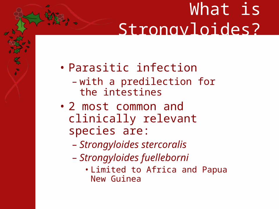
What is Strongyloides?
• Parasitic infection – with a predilection for the
intestines
• 2 most common and clinically relevant species are:– Strongyloides stercoralis– Strongyloides fuelleborni
• Limited to Africa and Papua New Guinea
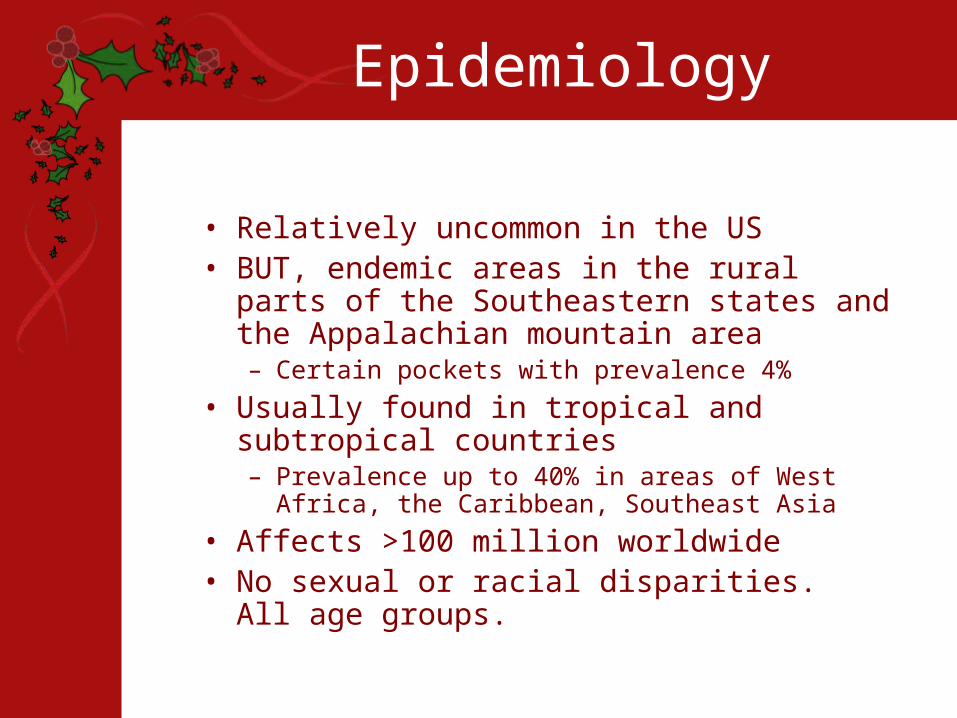
Epidemiology
• Relatively uncommon in the US• BUT, endemic areas in the rural parts of the
Southeastern states and the Appalachian mountain area– Certain pockets with prevalence 4%
• Usually found in tropical and subtropical countries– Prevalence up to 40% in areas of West Africa, the
Caribbean, Southeast Asia
• Affects >100 million worldwide• No sexual or racial disparities. All age groups.
How Do You Get It?
• Penetration of intact skin by filiariform larvae in the soil, or ingestion through contaminated food or water
• Larvae enter the circulation– Lungs alveoli ascension up
tracheobronchial tree swallowed molt in the small bowel and mature into adult female
• Females enter the intestinal mucosa and produce several eggs daily through parthenogenesis (hatch during transit through the gut)

Clinical Presentation
• Acute infection:– Lower extremity itching (mild
erythematous maculopapular rash at the site of skin penetration)
– Cough, dyspnea, wheezing– Low-grade fevers– Epigastric discomfort, n/v/d

Clinical Presentation
• Chronic Infection– Can be completely asymptomatic– Abdominal pain that can be very vague, crampy,
burning • Often worse after eating
– Intermittent diarrhea• Can alternate with constipation
– Occasional n/v– Weight loss (if heavy infestation)– Larva currens (“racing larva” – a recurrent
maculopapular or serpiginous rash)• Usually begins perianally and extends up the buttocks,
upper thighs, abdomen– Chronic urticaria

Larva Currens
Clinical Presentation• Severe infection
– Can be abrupt or insidious in onset– N/v/d, severe abdominal pain, distention– Cough, hemoptysis, dyspnea, wheezing, crackles– Stiff neck, headache, MS changes
• If CNS involved– Fever/chills– Hematemesis, hematochezia– Rash (petechiae, purpura) over the trunk and proximal extremities
• Caused by dermal blood vessel disruption brought on by massive migration of larvae within the skin
• Risk factors for severe infection– Immunosuppressant meds (steroids, chemo, TNF modulators,
tacro, etc – all BUT cyclosporine)– Malignancy– Malabsorptive state– ESRD– DM– Advanced age– HIV– HTLV1– Etoh

Clinical Presentation
• Can replicate in the host for decades with minimal or no sx
• High morbidity and mortality when progresses to hyperinfection syndrome or disseminated strongyloidiasis– Usually in immunocompromised
hosts (pregnancy?)
Dangerous Complications
• Hyperinfection Syndrome– Acceleration of the normal life cycle, causing
excessive worm burden– Autoinfection (turn into infective filariform larva within
the lumen– Spread of larvae outside the usual migration pattern
of GI tract and lungs • Disseminated strongyloidiasis
– Widespread dissemination of larvae to extraintestinal organs
• CNS (meningitis), heart, urinary tract, bacteremia, etc– Can be complicated by translocation of enteric
bacteria• Travel on the larvae themselves or via intestinal ulcers
– Mortality rate close to 80%• Due to delayed diagnosis, immunocompromised state
of the host at this point
Laboratory Findings
• CBC– WBC usually wnl for acute and
chronic cases, can be elevated in severe cases
– Eosinophilia common during acute infection, +/- in chronic infection (75%), usually absent in severe infection
Diagnostic Testing
• Stool O&P– Microscopic ID of S. sterocoralis larvae is
the definitive diagnosis– Ova usually not seen (only helminth to
secrete larva in the feces)• Stool wet mount (direct exam)
– In chronic infection, sensitivity only 30%, can increase to 75% if 3 consecutive stool exams
– Can enhance larvae recovery with more obscure methods (Baermann funnel, agar plate, Harada-Mori filter paper)
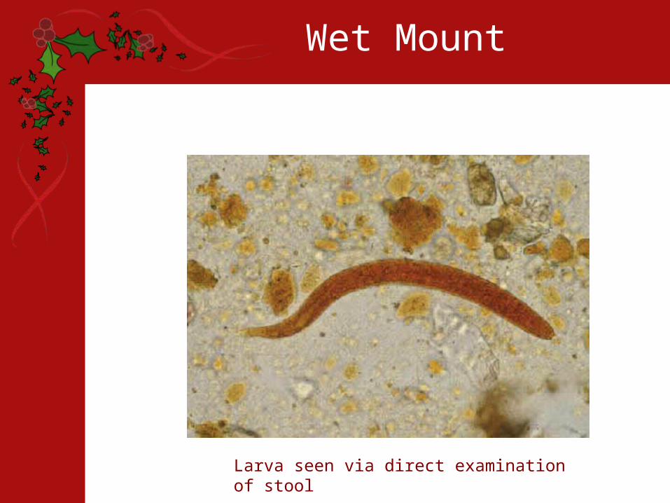
Wet Mount
Larva seen via direct examination of stool

Serology
• ELISA• Most sensitive method (88-95%)
– May be lower in immunocompromised patients
• Cannot distinguish between past and present infections
• Can cross-react with other nematode infections
• If results are positive, can move on to try and establish a microscopic dx
Imaging
• CXR – patchy alveolar infiltrates, diffuse interstitial infiltrates, pleural effusions
• AXR – Loops of dilated small bowel, ileus
• Barium swallow – stenosis, ulceration, bowel dilitation
• Small bowel follow-through – worms in the instestine
• CT abdomen/pelvis – nonspecific thickening of the bowel wall
Procedures
• EGD – duodenitis, edematous mucosa, white villi, erythema
• Colonoscopy – colitis• Duodenal aspiration – examine for larvae• Sputum sample, bronchial washings, BAL –
show larvae• Sputum cx
– Nl respiratory flora organisms pushed to the outside in groups as a result of migrating larvae
– Characteristic pattern can be diagnostic of S.Stercoralis infection
• If CNS involved, LP – gram stain, cell count/diff ( protein, ↓ glu, poly predominance), wet mount prep
Histology
• Larvae typically found in proximal portion of small intestine– Embedded in lamina propria
• Cause edema, cellular infiltration, villous atrophy, ulcerations
• In-long standing infections, may see fibrosis
Treatment
• Antihelminitic therapy– Ivermectin– Albendazole– Thiabendazole
• Abx directed toward enteric pathogens if bacteremia or meningitis (2-4wks)
• Minimize immunosuppression as possible• Directed supportive tx
– Transfusions if GI bleed, antihistamines for itching, surgery if bowel perf, etc
• Repeat course of antihelminitic therapy if immunocompromised, as relapse common

Follow-Up
• Repeat stool exams or duodenal aspirations in 2-3 mos to document cure
• Repeat serologies 4-8 mos after therapy – Ab titer should be low or undetectable 6-18
mos after successful tx• If titer not falling, additional
antihelminitic tx• Precautions for travelers to endemic
areas, but no prophylaxis or vaccine available

References
• Arch EL, Schaefer JT and Dahiya A. Cutaneous manifestation of disseminated strongyloidiasis in a patient coinfected with HTLV-1. Dermatology Online Journal. 2008;14(12):6.
• Chadrasekar PH, Bharadwaj RA, Polenakovik H, Polenakovik S. Emedicine: Strongyloidiasis. April 3, 2009.
• Concha R, Harrington W and Rogers A. Intestinal Strongyloidiasis. Recognition, Management and Determinants of Outcome. Journal of Clinical Gastroengerology. 2005;39(3):203-211.
• Greiner K, Bettencourt J, and Semolic C. Strongyloidiasis: A Review and Update by Case Example. Clinical Laboratory Science. 2008;21(2):82-8.
• Siddiqui AA, Berk SL. Diagnosis of Strongyloides stercoralis infection. Clin Infect Dis. October 1, 2001;33:1040-7.
• Zeph, Bill. Strongyloides stercoralis Infection Can Be Fatal. American Family Physician. March 15, 2002.