Embed Size (px)
Citation preview
Structural and Biophysical Characterization of Bacillusthuringiensis Insecticidal Proteins Cry34Ab1 andCry35Ab1Matthew S. Kelker1*, Colin Berry2, Steven L. Evans1, Reetal Pai1, David G. McCaskill1, Nick X. Wang1,
Joshua C. Russell1¤a, Matthew D. Baker2, Cheng Yang3, J. W. Pflugrath3, Matthew Wade4, Tim J. Wess4¤b,
Kenneth E. Narva1
1 Dow AgroSciences, LLC, Indianapolis, Indiana, United States of America, 2 Cardiff School of Biosciences, Cardiff University, Cardiff, Wales, United Kingdom, 3 Rigaku
Americas Corporation, The Woodlands, Texas, United States of America, 4 School of Optometry & Vision Sciences, Cardiff University, Cardiff, Wales, United Kingdom
Abstract
Bacillus thuringiensis strains are well known for the production of insecticidal proteins upon sporulation and these proteinsare deposited in parasporal crystalline inclusions. The majority of these insect-specific toxins exhibit three domains in themature toxin sequence. However, other Cry toxins are structurally and evolutionarily unrelated to this three-domain familyand little is known of their three dimensional structures, limiting our understanding of their mechanisms of action and ourability to engineer the proteins to enhance their function. Among the non-three domain Cry toxins, the Cry34Ab1 andCry35Ab1 proteins from B. thuringiensis strain PS149B1 are required to act together to produce toxicity to the western cornrootworm (WCR) Diabrotica virgifera virgifera Le Conte via a pore forming mechanism of action. Cry34Ab1 is a protein of,14 kDa with features of the aegerolysin family (Pfam06355) of proteins that have known membrane disrupting activity,while Cry35Ab1 is a ,44 kDa member of the toxin_10 family (Pfam05431) that includes other insecticidal proteins such asthe binary toxin BinA/BinB. The Cry34Ab1/Cry35Ab1 proteins represent an important seed trait technology having beendeveloped as insect resistance traits in commercialized corn hybrids for control of WCR. The structures of Cry34Ab1 andCry35Ab1 have been elucidated to 2.15 A and 1.80 A resolution, respectively. The solution structures of the toxins werefurther studied by small angle X-ray scattering and native electrospray ion mobility mass spectrometry. We present here thefirst published structure from the aegerolysin protein domain family and the structural comparisons of Cry34Ab1 andCry35Ab1 with other pore forming toxins.
Citation: Kelker MS, Berry C, Evans SL, Pai R, McCaskill DG, et al. (2014) Structural and Biophysical Characterization of Bacillus thuringiensis Insecticidal ProteinsCry34Ab1 and Cry35Ab1. PLoS ONE 9(11): e112555. doi:10.1371/journal.pone.0112555
Editor: Juan Luis Jurat-Fuentes, University of Tennessee, United States of America
Received June 13, 2014; Accepted October 7, 2014; Published November 12, 2014
Copyright: � 2014 Kelker et al. This is an open-access article distributed under the terms of the Creative Commons Attribution License, which permitsunrestricted use, distribution, and reproduction in any medium, provided the original author and source are credited.
Data Availability: The authors confirm that all data underlying the findings are fully available without restriction. All crystal structure files are available from theProtein Data Bank (accession numbers 4JOX and 4JP0). All relevant data are within the paper and its Supporting Information files.
Funding: Portions of this work were performed at the DuPont-Northwestern-Dow Collaborative Access Team (DND-CAT) located at Sector 5 of the AdvancedPhoton Source (APS). DND-CAT is supported by E.I. DuPont de Nemours & Co., The Dow Chemical Company and Northwestern University. Use of the APS, anOffice of Science User Facility operated for the U.S. Department of Energy (DOE) Office of Science by Argonne National Laboratory, was supported by the U.S. DOEunder Contract No. DE-AC02-06CH11357. Molecular graphics and analyses were performed with the UCSF Chimera package. Chimera was developed by theResource for Biocomputing, Visualization, and Informatics at the University of California, San Francisco (supported by NIGMS P41-GM103311). The funders had norole in study design, data collection and analysis, decision to publish, or preparation of the manuscript.
Competing Interests: The authors have read the journal’s policy and have the following competing interests: MSK, SLE, RP, DM, NXW and KEN are currentemployees of Dow AgroSciences. JCR was formerly employed by Dow AgroSciences. CY and JWP are employed by Rigaku Americas Corporation. A MaterialTransfer Agreement was in place between Dow AgroSciences and Cardiff University. Cry34Ab1 and Cry35Ab1 are protected under US patents 7,309,785 andfamily members and 7,524,810 and family members and are part of Dow AgroSciences’ product, Herculex RW. This does not alter the authors’ adherence to PLOSONE policies on sharing data and materials.
* Email: [email protected]
¤a Current address: Department of Biochemistry, University of Washington, Seattle, Washington, United States of America¤b Current address: Office of the Dean of Science, Charles Sturt University, New South Wales, Victoria, Australia
Introduction
Bacillus thuringiensis strains are well-known for the production
of insecticidal toxins on sporulation and these proteins are
deposited in parasporal crystalline inclusions, closely associated
with the spore. To date, many crystal toxins (Cry) have been
discovered and these are currently divided into 73 major classes
(see http://www.lifesci.susx.ac.uk/home/Neil_Crickmore/Bt/for
an updated list). The great majority of these toxins belong to a
single structural class of proteins, exhibiting 3 domains in the
mature toxin sequence. However, an increasing number of other
Cry toxins are structurally and evolutionarily unrelated to this
three-domain family. Unfortunately, little is known of their three
dimensional structures, limiting our understanding of their
mechanisms of action and our ability to engineer the proteins to
enhance their function. Amongst the non-three domain Cry
toxins, the Cry34Ab1 and Cry35Ab1 proteins from B. thur-ingiensis strain PS149B1 are required to act together to produce
toxicity to the western corn rootworm (WCR) Diabrotica virgiferavirgifera via a pore forming mechanism of action [1,2,3]. Very few
PLOS ONE | www.plosone.org 1 November 2014 | Volume 9 | Issue 11 | e112555
Cry proteins have been described with activity against WCR [4]
and among them the binary mode of action of Cry34Ab1/
Cry35Ab1 and related Cry family members is unique. The
Cry34Ab1/Cry35Ab1 proteins are important for WCR resistance
trait technology, having been introduced to corn hybrids through
genetic transformation event DAS-59122-7 to provide protection
from WCR feeding in commercialized corn hybrids [5].
Cry34Ab1 is a protein of ,14 kDa with features of the
aegerolysin family (Pfam06355) of proteins that have known ability
to interact with membranes to form pores [6] while Cry35Ab1
appears to contain b-trefoil sequences reminiscent of the
carbohydrate-binding domain of ricin B subunit (Pfam00652)
and is a member of the toxin_10 family (Pfam05431) that includes
the binary toxin BinA/BinB, the Cry49Aa1 component of a
second binary toxin (Cry48Aa1/Cry49Aa1) from Lysinibacillussphaericus, the Cry36Aa1 protein of B. thuringiensis and a
hypothetical protein from Chlorobium phaeobacteroides [7,8,9]. In
this study we have elucidated the structures of both Cry34Ab1 and
Cry35Ab1 and further probed the consistency of the crystal
structure data with their structures in solution using small angle X-
ray scattering (SAXS) and native electrospray ion mobility mass
spectrometry. We present here the first published structure from
the aegerolysin protein family and compare the Cry34Ab1 and
Cry35Ab1 protein structures with other pore forming toxins.
Materials and Methods
Expression and purification of full length Cry34Ab1 andCry35Ab1
Full length Cry35Ab1 and Cry34Ab1 toxins were over-
expressed in the inclusion body fraction of recombinant Pseudo-monas fluorescens (Pf) [10] and were purified as follows. Whole
broth, including Pf cells, was frozen at 220uC. To isolate and
wash the inclusion bodies, the broth was thawed at 37uC and
200 mL lysis buffer (50 mM Tris-HCl, pH 7.5, 0.2 M NaCl, 5%
glycerol, 1 mM DTT 20 mM EDTA and 0.5% Triton X-100) was
added for every 60 grams of frozen broth, mixed and centrifuged
at 10,000 g for 20 minutes at 4uC. The cell pellet was resuspended
at 200 mg cell pellet/mL cold lysis buffer and the cells disrupted
by micro-fluidization using a pressure difference of 16,000 psi.
Lysozyme was then added to 0.6 mg/mL and the mixture briefly
incubated at 37uC, then placed on ice for one hour with stirring.
Cell lysis was confirmed by microscopy. Magnesium sulfate was
added to 60 mM and DNase I added to 0.25 mg/mL and the
mixture incubated overnight at 4uC with stirring. The mixture was
gently homogenized using a hand held homogenizer to shear any
undigested genomic DNA and centrifuged at 10,000 g for
20 minutes at 4uC. The pellet was washed in lysis buffer and
centrifuged three more times.
Purification and crystallization of Cry35Ab1Freshly prepared Cry35Ab1 inclusions (100 mg) were solubi-
lized in 100 mL of 50 mM sodium citrate, pH 3.5, precipitated by
the addition of 80% ammonium sulfate then isolated by
centrifugation at 15,000 g for 15 minutes at 4uC. The pellet was
resuspended in 4.0 mL of 15 mM sodium citrate, pH 3.5 and
dialyzed against 6 L of the same buffer overnight using a 10,000
MWCO dialysis membrane. The pellet was completely dissolved.
This method reliably yielded 60 mg of highly pure Cry35Ab1.
The final concentration of Cry35Ab1 used in crystallization
experiments was 10–15 mg/mL in 15 mM sodium citrate,
pH 3.5. The results from SDS-PAGE analysis and dynamic light
scattering scan indicated Cry35Ab1 had reached.90% purity and
was monodisperse in solution.
Microbatch crystallization experiments using the Hampton
Index screen were set up with the Cry35Ab1 protein. Small
crystals appeared within several conditions after two days. After
optimization, the best crystals were produced in 0.72 M
NaH2PO4, pH 4.5, 80 mM K2HPO4 and 0.2 M NaCl with
,15 mg/mL Cry35Ab1 protein at 16uC.
Purification and crystallization of Cry34Ab1In order to achieve a highly concentrated protein solution for
crystallization, washed Cry34Ab1 inclusions were dissolved in 7 M
urea then refolded using a specially designed dialyzer. This
dialyzer has an open-end tube with a 10 K cutoff membrane at the
bottom end, which was hung in the center of a micro-centrifuge
tube. The membrane allows the solution outside of the dialysis
tube to equilibrate slowly into the tube. The small membrane
surface limits the rate of diffusion and may also create gradients of
components of dialysate in the tube.
About 8.9 mg of Cry34Ab1 powder was initially dissolved in
0.5 mL of 7 M urea, 20 mM potassium phosphate buffer pH 7.8.
A 40 mL sample of this protein solution was transferred into the
microdialysis apparatus described above and dialyzed against a
3.0 mL solution of 25% (v/v) PEG 400 and 50 mM sodium
acetate, pH 4.4, at 16uC. The results from SDS-PAGE analysis
and dynamic light scattering scan indicated Cry34Ab1 had
reached.90% purity and were monodisperse in solution.
After two days, small hexagonally-shaped crystals
(0.1560.1560.03 mm3) were observed on the membrane.
Data collection and phasing of Cry34Ab1Data collection on both Cry34Ab1 (Table 1) and full length
Cry35Ab1 (Table 2) crystals were carried out at 2180uC on a
home X-ray system (Rigaku MicroMax-007 X-ray generator and
R-AXIS IV++ detector). Two different wavelengths of X-ray
radiation (Cu Ka, 1.54 A and Cr Ka, 2.29 A) were used to collect
data sets for structure refinement and enhanced anomalous signal
to phase the protein diffraction data. All data sets were processed
using the d*TREK data processing package [11].
As a novel structure, the multiple isomophous replacement
(MIR) method was employed to phase Cry34Ab1 diffraction data.
After numerous trials, a Pb-derivatized crystal was prepared by
soaking a native Cry34Ab1 crystal in a crystallization solution
containing 10 mM lead acetylacetonate for 24 hours. The
diffraction data were collected at 2180uC (Table 1). One major
lead site was found on three Harker sections of an isomorphous
difference Patterson map. Two more minor lead sites were found
in the isomorphous difference Fourier map of FPH – FP using the
phases calculated from the major lead site. Three lead sites were
initially used in phase calculation up to 3.0 A resolution with the
program MLPHARE in CCP4 [12]. The figure of merit (FOM) of
the single isomorphous replacement with anomalous scattering
(SIRAS) phase set was 0.41, while an electron density map
calculated with these phases showed clear protein-solvent bound-
aries but with many broken regions. The anomalous differences of
the native data with the initial SIRAS phases were used to
generate an anomalous difference Fourier map. A peak with a
height of ,5 sigma above the average value was found in this
anomalous Fourier map. It was considered as a sulfur site arising
from one of the two methionine residues (the other methionine
residue at the N-terminus is disordered and not visible in these
maps). The sulfur position and the anomalous difference in the
native data were used in further phase calculation. The FOM of
the MIR phase set was improved to 0.45. Furthermore, since the
anomalous differences of the native data were included as a new
independent data set in the phase calculation, it greatly enhanced
Implications of the Cry34Ab1 and Cry35Ab1 Structures
PLOS ONE | www.plosone.org 2 November 2014 | Volume 9 | Issue 11 | e112555
the power to resolve the phase ambiguity of the initial SIRAS
phases from the Pb derivative. The electron density map
calculated with these new phases was improved and revealed
clear and recognizable regions, such as several b-strands and some
large side chain electron density. A solvent-flattening procedure
was employed to improve the quality of this electron density map
by using program DM [12].
Data collection and phasing of Cry35Ab1A platinum derivative of Cry35Ab1 was prepared by an
overnight soaking of a native crystal in 20 mM platinum
diammine dichloride and the crystallization condition. Crystals
were cryoprotected by addition of a final concentration of 20% (v/
v) of glycerol to the well condition. Diffraction data sets from both
native and heavy-atom derivatives were collected at 100 K with
home source X-ray equipment. All the data sets were processed
using the d*TREK data processing package [11]. The statistics of
data collection are listed in Table 2.
Structure determination and refinement of Cry34Ab1The electron density map was used to build an initial model
with the program O [13]. The value of the Matthews number
indicated that Cry34Ab1 crystals have one molecule per asym-
metric unit. The chain tracing and sequence match started at the
position of Met54 that was recognized in the anomalous difference
Fourier map of the native data set and were extended from there
in both directions. Initially, 78 amino acids out of 123 residues
were fitted into their densities. The model was refined using the
program REFMAC5 [12] and improved by rebuilding after
recalculation of the electron density map using weighted
combinations of model and MIR phases. Some regions missing
in the MIR electron density map gradually appeared in the partial
model combined electron density maps. The rigid-body, overall
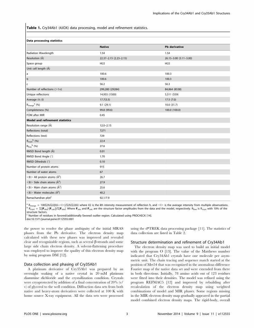
Table 1. Cry34Ab1 (4JOX) data processing, model and refinement statistics.
Data processing statistics
Native Pb derivative
Radiation Wavelength 1.54 1.54
Resolution (A) 22.37–2.15 (2.23–2.15) 26.15–3.00 (3.11–3.00)
Space group I422 I422
Unit cell length (A)
a 100.6 100.3
b 100.6 100.3
c 56.2 56.3
Number of reflections (.1s) 299,280 (29284) 84,864 (8538)
Unique reflections 14,955 (1500) 5,511 (559)
Average I/s (I) 17.7(3.5) 17.5 (7.0)
Rmergea (%) 9.1 (29.1) 10.0 (31.7)
Completeness (%) 99.8 (99.6) 100.0 (100.0)
FOM after MIR 0.45
Model and refinement statistics
Resolution range (A) 12.0–2.15
Reflections (total) 7271
Reflections (test) 729
Rcrystb (%) 22.4
Rfreeb (%) 27.6
RMSD Bond length (A) 0.01
RMSD Bond Angle (u) 1.70
RMSD Dihedrals (u) 0.10
Number of protein atoms 915
Number of water atoms 67
,B. All protein atoms (A2) 26.7
,B. Side chain atoms (A2) 27.9
,B. Main chain atoms (A2) 25.6
,B. Water molecules (A2) 40.2
Ramachandran plotc 92.1/7.9
a Rmerge = 100S(h)S(i)|I(i)-,I.|/S(h)S(i)I(i) where I(i) is the ith intensity measurement of reflection h, and ,I. is the average intensity from multiple observations.b Rcryst = S||Fobs|-|Fcalc||/S|Fobs|. Where Fobs and Fcalc are the structure factor amplitudes from the data and the model, respectively. Rfree is Rcryst with 10% of thestructure factors.c Number of residues in favored/additionally favored outlier region. Calculated using PROCHECK [14].doi:10.1371/journal.pone.0112555.t001
Implications of the Cry34Ab1 and Cry35Ab1 Structures
PLOS ONE | www.plosone.org 3 November 2014 | Volume 9 | Issue 11 | e112555
B-factor, individual B-factor and TLS refinement procedure were
iterated a number of times to refine the model. The final model
was refined to 2.15 A and contains 117 (from Ala3 to Tyr119) out
of 123 amino acids and 67 water molecules. Rcryst and Rfree factors
for the final model were 22.4% and 27.6%, respectively. Analysis
of the model by the program PROCHECK [14] indicated that
92.1% of the residues fell into the most favored regions of a
Ramachandran plot while the remaining 7.9% occurred in
additionally allowed regions. The refinement statistics and
structure analysis are listed in Table 1. Coordinates and reflection
files were assigned the PDB accession code, 4JOX.
Structure determination and refinement of Cry35Ab1The initial model was built using the program O [13]. One
Cry35Ab1 molecule was determined to be in an asymmetric unit
based on calculated Matthews number. The chain tracing and
sequence match of Cry35Ab1 was started simultaneously at the
position of Cys183 and Met185. They were recognized through
their unique densities in the anomalous difference Fourier map of
Cr Ka derived native data set. The sequence match was further
confirmed by the unique motif of electron densities of Met176,
Gly177 and Trp178 and anomalous peak of the sulfur of Met176.
About 200 amino acids were fitted into their densities in the first
round of map fitting. The model was refined using the program
REFMAC5 [12] which includes the procedures of idealization,
rigid-body, overall B-factor, TLS and individual B-factor. The
electron density map was recalculated using weighted combina-
tions of model and MIR phases. During the refinement process,
the electron density was improved in each new map, especially in
some uninterpretable regions. The final model contains 378 (from
Leu2 to His381) out of 385 amino acids and 295 water molecules.
Pro163 and Thr164 were excluded from final model. Some of
their electron density was observed in the maps of later cycles, but
these two residues cannot be refined into a conformation with both
good geometry and density coverage. It may result from their
structural location at a loop region with high thermomobility. The
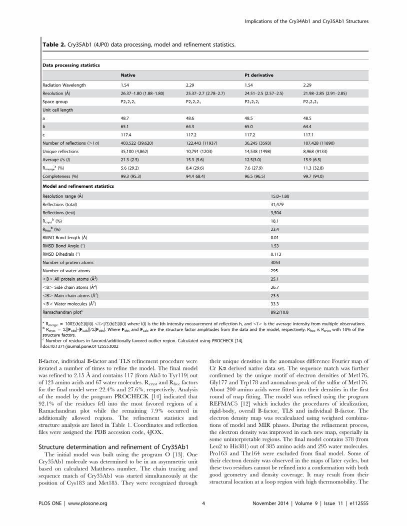
Table 2. Cry35Ab1 (4JP0) data processing, model and refinement statistics.
Data processing statistics
Native Pt derivative
Radiation Wavelength 1.54 2.29 1.54 2.29
Resolution (A) 26.37–1.80 (1.88–1.80) 25.37–2.7 (2.78–2.7) 24.51–2.5 (2.57–2.5) 21.98–2.85 (2.91–2.85)
Space group P212121 P212121 P212121 P212121
Unit cell length
a 48.7 48.6 48.5 48.5
b 65.1 64.3 65.0 64.4
c 117.4 117.2 117.2 117.1
Number of reflections (.1s) 403,522 (39,620) 122,443 (11937) 36,245 (3593) 107,428 (11890)
Unique reflections 35,100 (4,862) 10,791 (1203) 14,538 (1498) 8,968 (9133)
Average I/s (I) 21.3 (2.5) 15.3 (5.6) 12.5(3.0) 15.9 (6.5)
Rmergea (%) 5.6 (29.2) 8.4 (29.6) 7.6 (27.9) 11.3 (32.8)
Completeness (%) 99.3 (95.3) 94.4 68.4) 96.5 (96.5) 99.7 (94.0)
Model and refinement statistics
Resolution range (A) 15.0–1.80
Reflections (total) 31,479
Reflections (test) 3,504
Rcrystb (%) 18.1
Rfreeb (%) 23.4
RMSD Bond length (A) 0.01
RMSD Bond Angle (u) 1.53
RMSD Dihedrals (u) 0.113
Number of protein atoms 3053
Number of water atoms 295
,B. All protein atoms (A2) 25.1
,B. Side chain atoms (A2) 26.7
,B. Main chain atoms (A2) 23.5
,B. Water molecules (A2) 33.3
Ramachandran plotc 89.2/10.8
a Rmerge = 100S(h)S(i)|I(i)-,I.|/S(h)S(i)I(i) where I(i) is the ith intensity measurement of reflection h, and ,I. is the average intensity from multiple observations.b Rcryst = S||Fobs|-|Fcalc||/S|Fobs|. Where Fobs and Fcalc are the structure factor amplitudes from the data and the model, respectively. Rfree is Rcryst with 10% of thestructure factors.c Number of residues in favored/additionally favored outlier region. Calculated using PROCHECK [14].doi:10.1371/journal.pone.0112555.t002
Implications of the Cry34Ab1 and Cry35Ab1 Structures
PLOS ONE | www.plosone.org 4 November 2014 | Volume 9 | Issue 11 | e112555
Ta
ble
3.
Me
asu
red
and
the
ore
tica
lva
lue
sfo
rC
CS
for
Cry
34
Ab
1an
dtr
Cry
35
Ab
1.
Th
eo
reti
cal
Pro
tein
com
po
ne
nt
cha
rge
(z)
DT
(ms,
ob
s.)
CC
SV
(A2
)P
A(A
2)
EH
HS
(A2
)T
M(A
2)
Cry
34
Ab
11
72
7.5
71
40
4.7
13
68
.61
72
0.2
17
06
.6
27
29
.86
14
77
.5
38
27
.39
16
33
.9
49
25
.06
16
75
.9
59
27
.22
18
19
.1
69
31
.04
20
92
.9
79
36
.62
49
7.0
81
02
5.3
21
84
1.1
91
02
7.3
42
04
7.1
10
10
32
.62
41
5.3
11
10
34
.84
25
97
.5
12
10
36
.79
27
71
.9
13
11
23
.91
19
22
.0
14
11
26
.55
22
53
.2
15
11
30
.35
24
06
.4
16
11
32
.52
28
37
.2
17
11
34
.72
87
2.5
trC
ry3
5A
b1
11
43
2.6
63
46
6.4
32
01
.04
14
6.0
42
11
.5
21
43
4.3
43
48
7.8
31
53
2.4
73
67
3.3
41
53
2.5
63
71
7.9
51
63
1.0
93
70
3.6
61
73
0.0
33
76
2.0
71
73
6.7
64
80
8.8
81
82
9.1
43
81
9.5
91
83
5.6
35
14
3.1
Th
eo
reti
cal
valu
es
we
reca
lcu
late
du
sin
gth
ep
roje
ctio
nap
pro
xim
atio
n(P
A),
exa
cth
ard
sph
ere
scat
teri
ng
(EH
SS)
and
traj
ect
ory
me
tho
d(T
M)
wit
hh
eliu
mas
the
colli
sio
ng
as.
Exp
eri
me
nta
llym
eas
ure
dC
CS
use
dn
itro
ge
nas
the
colli
sio
ng
as.
do
i:10
.13
71
/jo
urn
al.p
on
e.0
11
25
55
.t0
03
Implications of the Cry34Ab1 and Cry35Ab1 Structures
PLOS ONE | www.plosone.org 5 November 2014 | Volume 9 | Issue 11 | e112555
final model was refined to 1.80 A. Rcryst and Rfree factors for the
final model were 18.1% and 23.4%, respectively. The analysis of
the model by the program PROCHECK [14] indicated 89.2% of
the residues fell into the most favored regions of a Ramachandran
plot while the remaining 10.8% occurred in additionally allowed
regions. The refinement statistics and structure analysis are listed
in Table 2. Coordinates and reflection files were assigned the PDB
accession code, 4JP0.
Expression and purification of soluble, truncatedCry35Ab1
A transgenic corn line encoding full length versions of both
Cry34Ab1and Cry35Ab1 was jointly developed by Dow Agro-
Sciences and Pioneer Hi-Bred International [15,16] and sold
under the brand name HERCULEX RW. Full length Cry35Ab1
is 44 kDa; however, during characterization of the proteins
expressed in transgenic corn, a 40 kDa C-terminal truncation of
Cry35Ab1 (trCry35Ab1) was isolated. Interestingly, this 40 kDa
form retains both the insecticidal activity and immunoreactivity of
the full length Cry35Ab1 [17]. In this study we wished to use this
construct to examine the solution state of the truncated molecule
in comparison to the crystal structure. In addition, trCry35Ab1 is
highly soluble and stable over the time course of experimentation.
A plasmid encoding residues 1–354 of Cry35Ab1 (trCry35Ab1;
lacking 31 residues at the C-terminus) was transformed into a Dow
AgroSciences P. fluorescens expression strain [10]. Seed cultures
were grown overnight. Production cultures were inoculated with
2% volume of the overnight culture and grown in production
media with trace elements and fermented in 2 L controlled
bioreactors. Twenty-four hours post inoculation, the cultures were
induced with 0.3 mM IPTG. The cells were harvested at 48 hours
post-induction by centrifugation. The pellets were stored at 2
80uC until purification. Routine expression levels are ,30 grams
of soluble trCry35Ab1 per liter of cell culture.
The trCry35Ab1 is expressed in the soluble fraction of the cell
lysate. Frozen cell pellets were resuspended in 0.1 M Na acetate,
pH 3.3, 1 mM EDTA and 1 mM TCEP (tris(2-carboxyethyl)pho-
sphine). The suspension was sonicated for 30 seconds, followed by
a 1 minute rest on ice, three times. After lysis, the lysate was
centrifuged at 19,000 rpm for 20 minutes at 4 uC. The
supernatant was filtered through a 0.22 mm filter. Purified protein
was obtained by using cation exchange chromatography with a
Source 30S 16/20 column pre-equilibrated in 0.1 M sodium
acetate pH 3.3, and gradient elution with 0.1 M sodium acetate
pH 3.3 and 1 M NaCl. The fractions containing trCry35Ab1 were
concentrated with 10,000 MWCO 15 mL, Amicon concentrators,
centrifuged at 5000 g for 10 minutes. Final samples were filtered
through a 0.22 mm filter and applied to a Superdex 75 26/90
column pre-equilibrated in 20 mM sodium citrate, pH 3.3.
Small angle X-ray scatteringFull length Cry34Ab1 and trCry35Ab1 samples, prepared as
described above, were diluted to various concentrations between
2.07 to 6.86 mg/mL in 20 mM sodium citrate pH 3.3. Immedi-
ately prior to the data collection, both samples were centrifuged at
14,000 rpm in a tabletop centrifuge for one hour, then filtered
through a 0.22 mm syringe filter. Synchrotron scattering data were
collected and processed at beamline 5-ID-D at the Advanced
Photon Source at Argonne National Laboratories, Illinois, USA.
Data were analyzed using the ATSAS package [18]. Buffer
scattering intensities were subtracted from the sample image to
remove background scattering using PRIMUS [19]. For the SAXS
data, the radius of gyration and the particle distance distribution
function, p(r) were evaluated with the GNOM program [20].
Particle shapes were generated using the ab-initio software
program DAMMIN [21]. Multiple DAMMIN runs were per-
formed (,25) to check the ‘uniqueness’ of the solution and to
generate 25 similar shapes that were combined and filtered to
produce an averaged model using the DAMAVER and DAM-
FILT programs [22].
Cry34Ab1 and Cry35Ab1 crystal structures were docked with
the SAXS calculated envelopes using the Chimera program [23].
The C-terminal residues 355–381 of the full length Cry35Ab1
crystal structure were removed for SAXS docking purposes.
Native electrospray ion mobility mass spectrometryThe behavior of Cry34Ab1 and trCry35Ab1 in solution were
probed using native electrospray ion mobility mass spectrometry.
Stock solutions of Cry34Ab1 (3.5 mg/mL stored in 20 mM
sodium citrate buffer, pH 3.5) and a trCry35Ab1 (3.2 mg/mL
stored in 20 mM sodium citrate buffer, pH 3.5) were used for
direct infusion under non-denaturing nano-electrospray condi-
tions.
In the case of the Cry34Ab1 sample, the stock solution was
diluted 4 – fold with 0.1% formic acid and buffer exchanged into
0.1% formic acid (pH 3.0) using a Zeba spin desalting column
(ThermoFisher Scientific) pre-equilibrated with 0.1% formic acid.
The buffer exchanged sample was subsequently diluted 5 – fold to
give a stock solution of approximately 12.9 mM.
The trCry35Ab1 sample was buffer exchanged without initial
dilution into 0.1% formic acid using a Zeba spin desalting column
pre-equilibrated with 0.1% formic acid. The resulting buffer
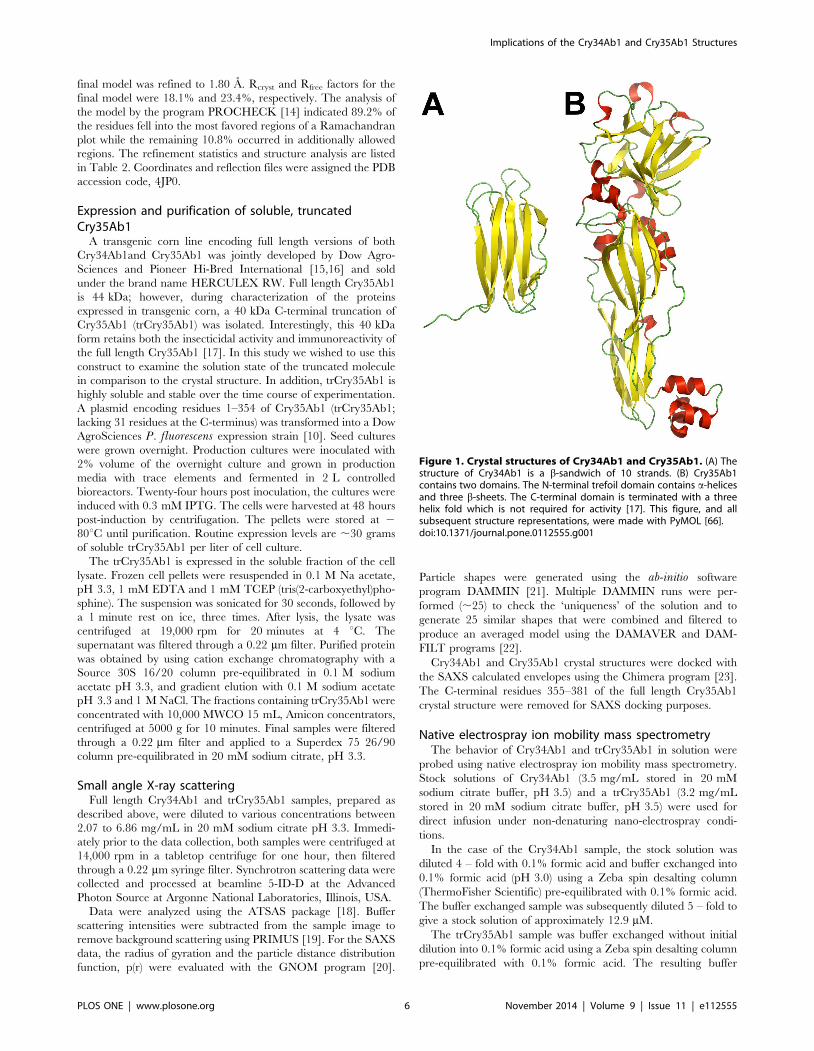
Figure 1. Crystal structures of Cry34Ab1 and Cry35Ab1. (A) Thestructure of Cry34Ab1 is a b-sandwich of 10 strands. (B) Cry35Ab1contains two domains. The N-terminal trefoil domain contains a-helicesand three b-sheets. The C-terminal domain is terminated with a threehelix fold which is not required for activity [17]. This figure, and allsubsequent structure representations, were made with PyMOL [66].doi:10.1371/journal.pone.0112555.g001
Implications of the Cry34Ab1 and Cry35Ab1 Structures
PLOS ONE | www.plosone.org 6 November 2014 | Volume 9 | Issue 11 | e112555
exchanged material was diluted 5 – fold to give a stock solution of
approximately 15.9 mM.
Electrospray mass spectrometry of the Cry34Ab1 and
trCry35Ab1 stock solutions were carried out by directly infusing
the proteins with a syringe pump at 500 nL/min with an unheated
nanospray inlet. Detection and ion mobility measurements of the
proteins was carried out using a prototype ion mobility quadrupole
time-of-flight (model 6560 IM-QTOF) mass spectrometer at
Agilent Technologies (Santa Clara, CA). This instrument utilizes
a drift tube configuration with nitrogen collision gas for ion
mobility measurements. The drift tube was operated at 27uC, with
4 Torr of nitrogen collision gas.
For calculation of the measured collisional cross sectional areas
(CCS) of the analyzed proteins, the drift tube was calibrated
according to the manufacturer’s directions using infusion of a
colchicine standard (400 m/z, literature value for CCS = 196.2 A),
and the calibration was confirmed using infusion of a standard of
ondansetrone (m/z 294, measured CCS value = 172.5 A, literature
value = 172.7 A) [24]. Measured CCS values for Cry34Ab1 and
trCry35Ab1 were calculated using these calibration values with
software provided by Agilent.
Determining collision cross sectional areas by MOBCALTheoretical collision cross sectional areas (CCS) of Cry34Ab1
and trCry35Ab1 were calculated using the open source software
program MOBCAL [25,26]. MOBCAL source code was down-
loaded from the website of Professor M.F. Jarrold’s group at
Indiana University (http://www.indiana.edu/,nano/software.
html) and compiled with Fortran 95 in an in-house Linux work
station. The MOBCAL program was further modified to process
protein systems up to 15,000 atoms. PDB files of Cry34Ab1 and
Cry35Ab1 were used as input files. The calculations were carried
out with a uniform charge distribution. A scaling factor of 1.0 was
applied throughout the calculations. MOBCAL implements three
different types of calculations to derive the CCS area between a
protein and helium buffer gas: the projection approximation (PA),
the exact hard sphere scattering (EHSS) and the trajectory method
(TM). In this study, the PA values are consistently in better
agreement with experimental IM-MS measurements. All calcu-
lated CCS values from three the methods were included in
Table 3.
Protein structure alignment by combinatorial extensionStructures of Cry34Ab1 and Cry35Ab1 were aligned against all
the 3D structures in the Protein Data Bank (http://www.rcsb.org/
pdb/home/home.do). These alignments were performed using the
Combinatorial Extension (CE) algorithm [27]. This method is a
fast and accurate way to perform structural alignment against
large protein databases. It identifies the optimal alignment
between any two structures by defining an alignment path
between aligned fragment pairs in the two structures. Similarity
between fragment pairs is calculated on the basis of inter-residue
distances between the fragments after the superposition. Other
structural features like secondary structure, solvent exposure,
dihedral angles, etc. are also included to increase the accuracy of
the alignment between the fragment pairs. The algorithm provides
the sequence identity, r.m.s. of superposition and a Z-score for
each alignment.
Modeling of related proteinsUsing the coordinates of Cry35Ab1 and BinB as a template for
Cry49Aa1 and Cry34Ab1 as a template for Pam, the possible
structures of the related proteins were modeled using Modeller
9.11 [28,29]. Briefly, for Cry49Aa1 modelling, structure-based
sequence alignments were performed using the amino acid
sequences of Cry49Aa1, BinA and BinB as template sequences
along with the structure of Cry35Ab1 (PDB ID 4JPO) and BinB
(PDB ID 3WA1), followed by automated model building and
minimization. Manual inspection of clashes and rebuilding of
surface loops was performed using Chimera [23]. Final model
selection was based on the GA341 score of Modeller [30] and
Ramachandran plots.
Results and Discussion
Cry34Ab1 and Cry35Ab1 crystal structuresThe crystal structure of the Bacillus thuringiensis Cry34Ab1
protein was refined to 2.15 A resolution (Table 1). The Cry34Ab1
structure has one distinct structural domain containing 117 amino
acids (Figure 1A). The protein folds in a typical b-sandwich
conformation, which has two b-sheets packed against each other.
b-sheet I containing the N- and C-termini is composed of four b-
strands while b-sheet II has five b-strands. All b-strands, except the
adjacent N- and C-terminal strands, are antiparallel. N- and C-
terminal strands are located at the center of sheet I and parallel to
each other. The peptide fragment comprising residues Thr115 to
Tyr119 extends beyond its b-sheet toward a symmetry-related
molecule within the crystal lattice. The entire b-sandwich has a
relatively flat layer-like conformation and two slightly twisted b-
sheets. When the side chains are excluded, the distance between
the two b-sheets is between ,7–10 A. The molecule is ,45 A in
length and ,20 A in width.
As expected, the Cry34Ab1 structure has a very hydrophobic
core between the two b-sheets including Val6, Ile8, Val10, Leu18,
Trp31, Ile61, Tyr63, Ile71, Leu73, Phe75, Ile96, Val108, Tyr110
and Ile112. The phenol groups of Tyr63 and Tyr110 hydrogen
bond with the side chain of Ser106 and the carbonyl oxygen of
Thr36, respectively. Residue Trp31 is located at the loop region
between b-strand 2 and 3 and its indole group is inserted directly
into the core.
Nearly every residue in the final model of the Cry34Ab1
structure has well-defined electron density except for residues in
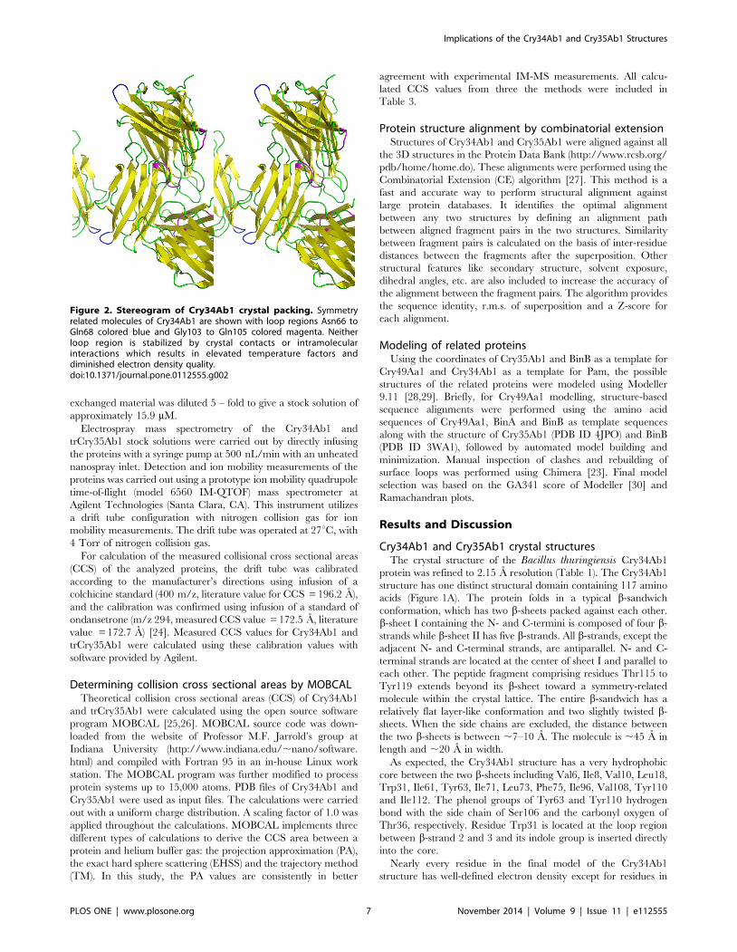
Figure 2. Stereogram of Cry34Ab1 crystal packing. Symmetryrelated molecules of Cry34Ab1 are shown with loop regions Asn66 toGln68 colored blue and Gly103 to Gln105 colored magenta. Neitherloop region is stabilized by crystal contacts or intramolecularinteractions which results in elevated temperature factors anddiminished electron density quality.doi:10.1371/journal.pone.0112555.g002
Implications of the Cry34Ab1 and Cry35Ab1 Structures
PLOS ONE | www.plosone.org 7 November 2014 | Volume 9 | Issue 11 | e112555
two short loop regions (Asn66 to Gln68 and Gly103 to Gln105),
which have relatively poorly defined electron density. Both these
regions have higher average temperature factors for their main
chain and side chain atoms than that of residues calculated over
the entire structure. This indicates these loops might have multiple
conformations in the crystal and reside in flexible regions due to
the lack of crystal contacts to stabilize them (Figure 2).
A total of 67 water molecules were included in the final model of
Cry34Ab1. Among these water molecules, ,60% of them have
high temperature factors (.40A2). indicating these positions were
partially occupied or otherwise disordered in the entire crystal.
One water molecule (HOH1) is located at a special position of a
two-fold crystallography axis and participates in hydrogen bonds
with the side chains of two symmetry-related His107 residues.
The crystal structure of the Cry35Ab1 protein was refined to
1.80 A resolution (Table 2). The Cry35Ab1 structure has an
elongated rectangular shape with dimensions 426456105 A and
is composed of two distinct domains (Figure 1B). Residues Pro163
and Thr164, had no interpretable electron density. The N-
terminal domain is a b-trefoil fold, which contains a very
hydrophobic core including Ala19, Val30, Leu32, Trp47, Ile59
Trp70, Val72, Ile77, Val79, Trp91, Ile93, Leu123, Trp135,
Tyr100, Ile102, Leu110, Ile121, and Leu137.
The next domain contains six helices and three antiparallel b-
sheets. A four antiparallel strand b-sheet sits below the N-terminal
domain and another two b-strands form a b-sandwich. Within this
fold, a p-p stacking interaction is formed between the phenol rings
of Tyr231 and Tyr341. Additionally, Tyr341 is also hydrogen-
bonded with Tyr229. The hydrophobic side chains of Val219,
Leu221 and Met307 cluster around Ile299.
The N-terminal domain and C-terminal domain pack tightly
against each other with more than 400 A2 area buried at the
interface. The buried region includes hydrophobic residues Ile184,
Ile197, Phe50, Phe48, Pro182, Met182, Ile58, Ile52, Ile271. In
addition, a hydrogen bonding network exists between the side
chains of residues Tyr82 and Glu270, the side chains and main
chains of Tyr202 and Thr4 and Asp53 and Thr273, respectively,
and the main chains of residues Gly270 and Asp53. These
interactions appear to keep domain packing very strong and the
conformation of the entire molecule very rigid. The two cysteine
residues (Cys67 and Cys187) are present in the interface but their
sulfur atoms are 6.1 A apart, which is too distant to form a
disulfide bond. Cys187 is conserved in all the toxins within this
family, except Cry36. It is interesting to note that replacement of
the Cys187-equivalent residue in BinA (Cys195) drastically
reduces its activity [31] while substitution of the equivalent in
BinB (Cys241) has no effect [32].
The Cry35Ab1 structure is ended with a C-terminal cluster of
three a-helices. The first two helices form a typical helix-loop-
helix. The third helix is perpendicular to this helix-loop-helix and
the group is held together through a hydrophobic core, consisting
of Leu378, Leu353, Leu356, Ala352, Leu375, Val364. Leu353
and Leu356 are the first and last residues of a distinct sequence
pattern of four tandem leucines (353-LLLL-356). Due to these
structural characteristics, this C-terminal domain is very stable and
tightly packed.
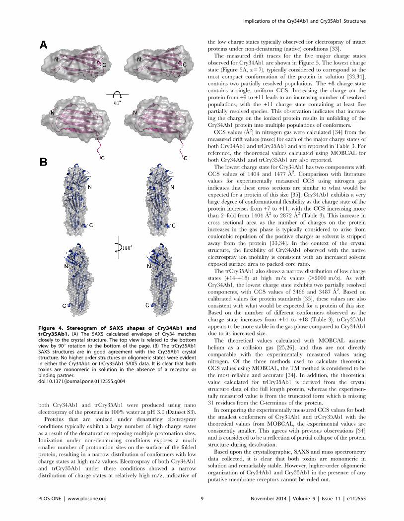
Cry34Ab1 and Cry35Ab1 solution structuresAnalysis of the individual crystal structures suggested that both
toxins are monomeric in solution, with no obvious higher order
associations based upon content of the asymmetric unit or
symmetry related molecules. To confirm the monomeric state in
solution and under native conditions, we calculated the solution
structures of both Cry34Ab1 and trCry35Ab1 by small angle X-
ray scattering. The molecular envelope was generated using the
ATSAS software package [18] and superimposed with the crystal
structures (Dataset S1 and Dataset S2). The SAXS data indicate
that both proteins exist as monomers in these conditions with the
predicted radii of gyration of 14.6 and 26.7 A calculated from the
crystal structures of Cry34Ab1 and Cry35Ab1 (with the C-
terminal three helix domain removed, consistent with the
trCry35Ab1 sequence) respectively, matching closely with those
of the SAXS models (14.9 and 25.9 A, respectively). The overlap
of the SAXS envelope and crystal structures (Figure 3) correlates
well for both Cry34Ab1 (Figure 4A) and trCry35Ab1 (Figure 4B).
It also suggests that the structures remain stable over a range of pH
values since this match is seen despite differences in the pH of the
crystallization and SAXS conditions (SAXS carried out at pH 3.3
compared to Cry35Ab1 crystallization at pH 4.5 and Cry34Ab1
crystallization at pH 7.8). Removal of the C-terminal 31 amino
acids of Cry35Ab1 in the trCry35Ab1 SAXS structure does not
appear to alter the core structure of the toxin in solution. When
taken together, these finding support a monomeric solution state.
To expand upon the calculated SAXS solution structures,
Cry34Ab1 and trCry35Ab1 were further assessed by native
electrospray ion mobility mass spectrometry. Experimentally
measured values for the collisional cross sectional (CCS) area of
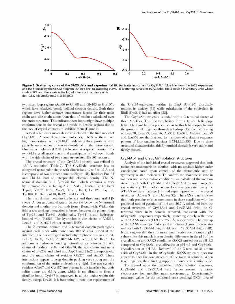
Figure 3. Scattering curve of the SAXS data and experimental fit. (A) Scattering curves for Cry34Ab1 (blue line) from the SAXS experimentand the fit made by the GNOM program [20] (red line) to scattering curve. (B) Scattering curves for trCry35Ab1. The X axis is s in arbitrary units wheres = 4psinh/l and the Y axis is the log of intensity in arbitrary units.doi:10.1371/journal.pone.0112555.g003
Implications of the Cry34Ab1 and Cry35Ab1 Structures
PLOS ONE | www.plosone.org 8 November 2014 | Volume 9 | Issue 11 | e112555
both Cry34Ab1 and trCry35Ab1 were produced using nano
electrospray of the proteins in 100% water at pH 3.0 (Dataset S3).
Proteins that are ionized under denaturing electrospray
conditions typically exhibit a large number of high charge states
as a result of the denaturation exposing multiple protonation sites.
Ionization under non-denaturing conditions exposes a much
smaller number of protonation sites on the surface of the folded
protein, resulting in a narrow distribution of conformers with low
charge states at high m/z values. Electrospray of both Cry34Ab1
and trCry35Ab1 under these conditions showed a narrow
distribution of charge states at relatively high m/z, indicative of
the low charge states typically observed for electrospray of intact
proteins under non-denaturing (native) conditions [33].
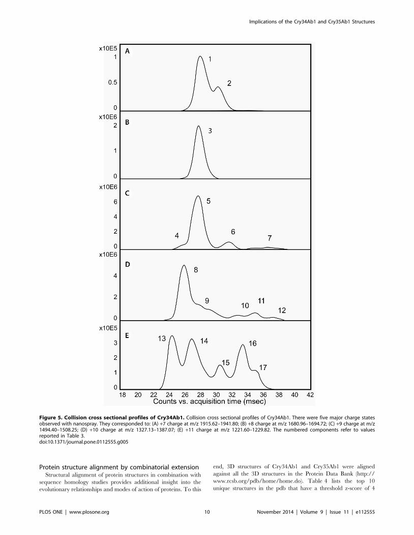
The measured drift traces for the five major charge states
observed for Cry34Ab1 are shown in Figure 5. The lowest charge
state (Figure 5A, z = 7), typically considered to correspond to the
most compact conformation of the protein in solution [33,34],
contains two partially resolved populations. The +8 charge state
contains a single, uniform CCS. Increasing the charge on the
protein from +9 to +11 leads to an increasing number of resolved
populations, with the +11 charge state containing at least five
partially resolved species. This observation indicates that increas-
ing the charge on the ionized protein results in unfolding of the
Cry34Ab1 protein into multiple populations of conformers.
CCS values (A2) in nitrogen gas were calculated [34] from the
measured drift values (msec) for each of the major charge states of
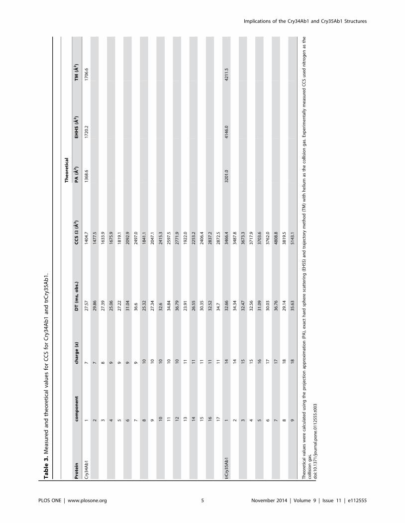
both Cry34Ab1 and trCry35Ab1 and are reported in Table 3. For
reference, the theoretical values calculated using MOBCAL for
both Cry34Ab1 and trCry35Ab1 are also reported.
The lowest charge state for Cry34Ab1 has two components with
CCS values of 1404 and 1477 A2. Comparison with literature
values for experimentally measured CCS using nitrogen gas
indicates that these cross sections are similar to what would be
expected for a protein of this size [35]. Cry34Ab1 exhibits a very
large degree of conformational flexibility as the charge state of the
protein increases from +7 to +11, with the CCS increasing more
than 2–fold from 1404 A2 to 2872 A2 (Table 3). This increase in
cross sectional area as the number of charges on the protein
increases in the gas phase is typically considered to arise from
coulombic repulsion of the positive charges as solvent is stripped
away from the protein [33,34]. In the context of the crystal
structure, the flexibility of Cry34Ab1 observed with the native
electrospray ion mobility is consistent with an increased solvent
exposed surface area to packed core ratio.
The trCry35Ab1 also shows a narrow distribution of low charge
states (+14–+18) at high m/z values (.2000 m/z). As with
Cry34Ab1, the lowest charge state exhibits two partially resolved
components, with CCS values of 3466 and 3487 A2. Based on
calibrated values for protein standards [35], these values are also
consistent with what would be expected for a protein of this size.
Based on the number of different conformers observed as the
charge state increases from +14 to +18 (Table 3), trCry35Ab1
appears to be more stable in the gas phase compared to Cry34Ab1
due to its increased size.
The theoretical values calculated with MOBCAL assume
helium as a collision gas [25,26], and thus are not directly
comparable with the experimentally measured values using
nitrogen. Of the three methods used to calculate theoretical
CCS values using MOBCAL, the TM method is considered to be
the most reliable and accurate [34]. In addition, the theoretical
value calculated for trCry35Ab1 is derived from the crystal
structure data of the full length protein, whereas the experimen-
tally measured value is from the truncated form which is missing
31 residues from the C-terminus of the protein.
In comparing the experimentally measured CCS values for both
the smallest conformers of Cry34Ab1 and trCry35Ab1 with the
theoretical values from MOBCAL, the experimental values are
consistently smaller. This agrees with previous observations [34]
and is considered to be a reflection of partial collapse of the protein
structure during desolvation.
Based upon the crystallographic, SAXS and mass spectrometry
data collected, it is clear that both toxins are monomeric in
solution and remarkably stable. However, higher-order oligomeric
organization of Cry34Ab1 and Cry35Ab1 in the presence of any
putative membrane receptors cannot be ruled out.
Figure 4. Stereogram of SAXS shapes of Cry34Ab1 andtrCry35Ab1. (A) The SAXS calculated envelope of Cry34 matchesclosely to the crystal structure. The top view is related to the bottomview by 90u rotation to the bottom of the page. (B) The trCry35Ab1SAXS structures are in good agreement with the Cry35Ab1 crystalstructure. No higher order structures or oligomeric states were evidentin either the Cry34Ab1 or trCry35Ab1 SAXS data. It is clear that bothtoxins are monomeric in solution in the absence of a receptor orbinding partner.doi:10.1371/journal.pone.0112555.g004
Implications of the Cry34Ab1 and Cry35Ab1 Structures
PLOS ONE | www.plosone.org 9 November 2014 | Volume 9 | Issue 11 | e112555
Protein structure alignment by combinatorial extensionStructural alignment of protein structures in combination with
sequence homology studies provides additional insight into the
evolutionary relationships and modes of action of proteins. To this
end, 3D structures of Cry34Ab1 and Cry35Ab1 were aligned
against all the 3D structures in the Protein Data Bank (http://
www.rcsb.org/pdb/home/home.do). Table 4 lists the top 10
unique structures in the pdb that have a threshold z-score of 4
Figure 5. Collision cross sectional profiles of Cry34Ab1. Collision cross sectional profiles of Cry34Ab1. There were five major charge statesobserved with nanospray. They corresponded to: (A) +7 charge at m/z 1915.62–1941.80; (B) +8 charge at m/z 1680.96–1694.72; (C) +9 charge at m/z1494.40–1508.25; (D) +10 charge at m/z 1327.13–1387.07; (E) +11 charge at m/z 1221.60–1229.82. The numbered components refer to valuesreported in Table 3.doi:10.1371/journal.pone.0112555.g005
Implications of the Cry34Ab1 and Cry35Ab1 Structures
PLOS ONE | www.plosone.org 10 November 2014 | Volume 9 | Issue 11 | e112555
or higher with the Cry34Ab1 structure. It also includes the CATH
description for the protein where available. Table 5 provides a
similar analysis for the Cry35Ab1 structure.
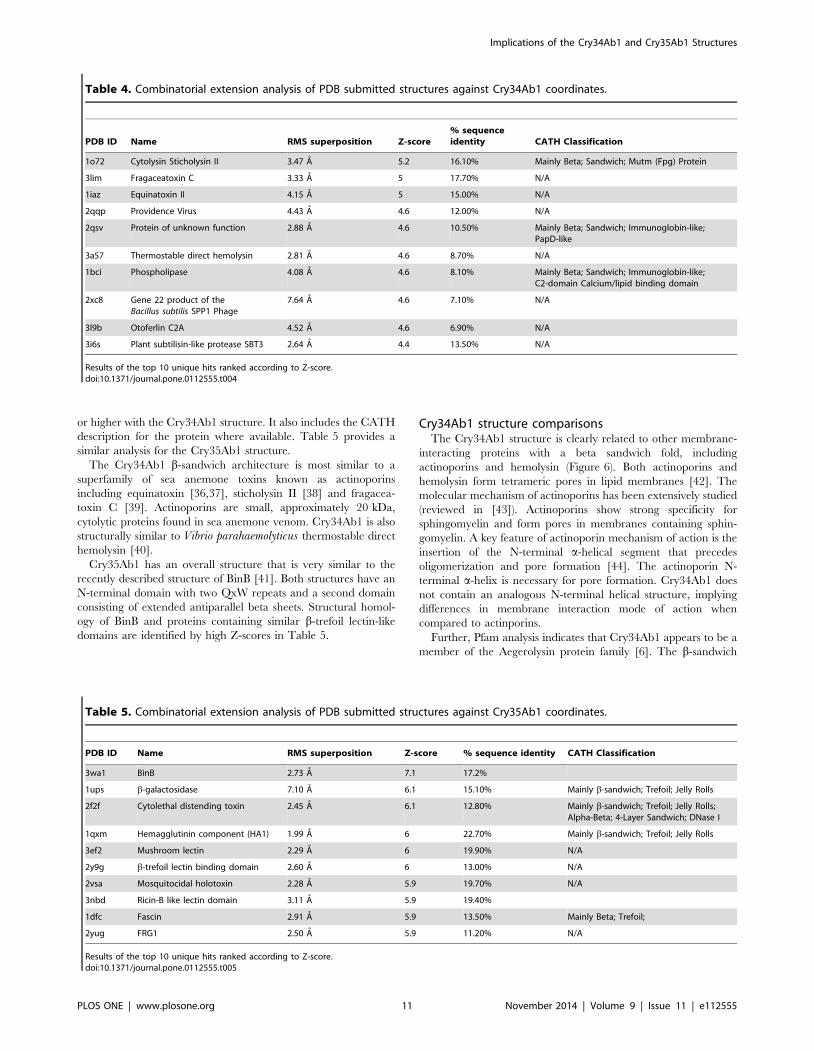
The Cry34Ab1 b-sandwich architecture is most similar to a
superfamily of sea anemone toxins known as actinoporins
including equinatoxin [36,37], sticholysin II [38] and fragacea-
toxin C [39]. Actinoporins are small, approximately 20 kDa,
cytolytic proteins found in sea anemone venom. Cry34Ab1 is also
structurally similar to Vibrio parahaemolyticus thermostable direct
hemolysin [40].
Cry35Ab1 has an overall structure that is very similar to the
recently described structure of BinB [41]. Both structures have an
N-terminal domain with two QxW repeats and a second domain
consisting of extended antiparallel beta sheets. Structural homol-
ogy of BinB and proteins containing similar b-trefoil lectin-like
domains are identified by high Z-scores in Table 5.
Cry34Ab1 structure comparisonsThe Cry34Ab1 structure is clearly related to other membrane-
interacting proteins with a beta sandwich fold, including
actinoporins and hemolysin (Figure 6). Both actinoporins and
hemolysin form tetrameric pores in lipid membranes [42]. The
molecular mechanism of actinoporins has been extensively studied
(reviewed in [43]). Actinoporins show strong specificity for
sphingomyelin and form pores in membranes containing sphin-
gomyelin. A key feature of actinoporin mechanism of action is the
insertion of the N-terminal a-helical segment that precedes
oligomerization and pore formation [44]. The actinoporin N-
terminal a-helix is necessary for pore formation. Cry34Ab1 does
not contain an analogous N-terminal helical structure, implying
differences in membrane interaction mode of action when
compared to actinporins.
Further, Pfam analysis indicates that Cry34Ab1 appears to be a
member of the Aegerolysin protein family [6]. The b-sandwich
Table 4. Combinatorial extension analysis of PDB submitted structures against Cry34Ab1 coordinates.
PDB ID Name RMS superposition Z-score% sequenceidentity CATH Classification
1o72 Cytolysin Sticholysin II 3.47 A 5.2 16.10% Mainly Beta; Sandwich; Mutm (Fpg) Protein
3lim Fragaceatoxin C 3.33 A 5 17.70% N/A
1iaz Equinatoxin II 4.15 A 5 15.00% N/A
2qqp Providence Virus 4.43 A 4.6 12.00% N/A
2qsv Protein of unknown function 2.88 A 4.6 10.50% Mainly Beta; Sandwich; Immunoglobin-like;PapD-like
3a57 Thermostable direct hemolysin 2.81 A 4.6 8.70% N/A
1bci Phospholipase 4.08 A 4.6 8.10% Mainly Beta; Sandwich; Immunoglobin-like;C2-domain Calcium/lipid binding domain
2xc8 Gene 22 product of theBacillus subtilis SPP1 Phage
7.64 A 4.6 7.10% N/A
3l9b Otoferlin C2A 4.52 A 4.6 6.90% N/A
3i6s Plant subtilisin-like protease SBT3 2.64 A 4.4 13.50% N/A
Results of the top 10 unique hits ranked according to Z-score.doi:10.1371/journal.pone.0112555.t004
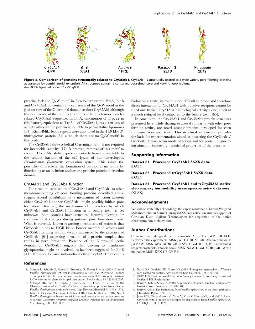
Table 5. Combinatorial extension analysis of PDB submitted structures against Cry35Ab1 coordinates.
PDB ID Name RMS superposition Z-score % sequence identity CATH Classification
3wa1 BinB 2.73 A 7.1 17.2%
1ups b-galactosidase 7.10 A 6.1 15.10% Mainly b-sandwich; Trefoil; Jelly Rolls
2f2f Cytolethal distending toxin 2.45 A 6.1 12.80% Mainly b-sandwich; Trefoil; Jelly Rolls;Alpha-Beta; 4-Layer Sandwich; DNase I
1qxm Hemagglutinin component (HA1) 1.99 A 6 22.70% Mainly b-sandwich; Trefoil; Jelly Rolls
3ef2 Mushroom lectin 2.29 A 6 19.90% N/A
2y9g b-trefoil lectin binding domain 2.60 A 6 13.00% N/A
2vsa Mosquitocidal holotoxin 2.28 A 5.9 19.70% N/A
3nbd Ricin-B like lectin domain 3.11 A 5.9 19.40%
1dfc Fascin 2.91 A 5.9 13.50% Mainly Beta; Trefoil;
2yug FRG1 2.50 A 5.9 11.20% N/A
Results of the top 10 unique hits ranked according to Z-score.doi:10.1371/journal.pone.0112555.t005
Implications of the Cry34Ab1 and Cry35Ab1 Structures
PLOS ONE | www.plosone.org 11 November 2014 | Volume 9 | Issue 11 | e112555
fold exemplified in Cry34Ab1 is common among other cytolytic
proteins found in nature including necrosis and ethylene-inducing
peptide 1 (Nep1)-like proteins (NLPs) from microbial plant
pathogens [45] and fungal fruit lectins [46]. Cry34Ab1 protein
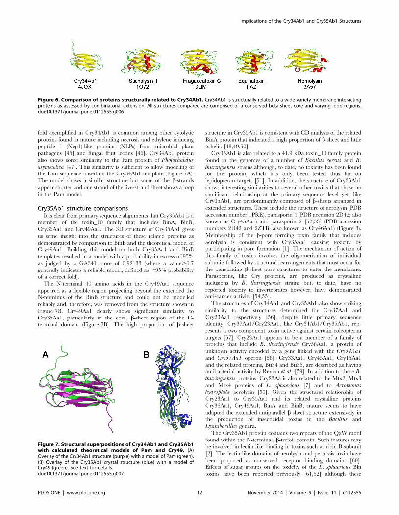
also shows some similarity to the Pam protein of Photorhabdusasymbiotica [47]. This similarity is sufficient to allow modeling of
the Pam sequence based on the Cry34Ab1 template (Figure 7A).
The model shows a similar structure but some of the b-strands
appear shorter and one strand of the five-strand sheet shows a loop
in the Pam model.
Cry35Ab1 structure comparisonsIt is clear from primary sequence alignments that Cry35Ab1 is a
member of the toxin_10 family that includes BinA, BinB,
Cry36Aa1 and Cry49Aa1. The 3D structure of Cry35Ab1 gives
us some insight into the structures of these related proteins as
demonstrated by comparison to BinB and the theoretical model of
Cry49Aa1. Building this model on both Cry35Aa1 and BinB
templates resulted in a model with a probability in excess of 95%
as judged by a GA341 score of 0.92133 (where a value.0.7
generally indicates a reliable model, defined as $95% probability
of a correct fold).
The N-terminal 40 amino acids in the Cry49Aa1 sequence
appeared as a flexible region projecting beyond the extended the
N-terminus of the BinB structure and could not be modelled
reliably and, therefore, was removed from the structure shown in
Figure 7B. Cry49Aa1 clearly shows significant similarity to
Cry35Aa1, particularly in the core, b-sheet region of the C-
terminal domain (Figure 7B). The high proportion of b-sheet
structure in Cry35Ab1 is consistent with CD analysis of the related
BinA protein that indicated a high proportion of b-sheet and little
a-helix [48,49,50].
Cry35Ab1 is also related to a 41.9 kDa toxin_10 family protein
found in the genomes of a number of Bacillus cereus and B.thuringiensis strains although, to date, no toxicity has been found
for this protein, which has only been tested thus far on
lepidopteran targets [51]. In addition, the structure of Cry35Ab1
shows interesting similarities to several other toxins that show no
significant relationship at the primary sequence level yet, like
Cry35Ab1, are predominantly composed of b-sheets arranged in
extended structures. These include the structure of aerolysin (PDB
accession number 1PRE), parasporin 4 (PDB accession 2D42; also
known as Cry45Aa1) and parasporin 2 [52,53] (PDB accession
numbers 2D42 and 2ZTB; also known as Cry46Aa1) (Figure 8).
Membership of the b-pore forming toxin family that includes
aerolysin is consistent with Cry35Aa1 causing toxicity by
participating in pore formation [1]. The mechanism of action of
this family of toxins involves the oligomerisation of individual
subunits followed by structural rearrangements that must occur for
the penetrating b-sheet pore structures to enter the membrane.
Parasporins, like Cry proteins, are produced as crystalline
inclusions by B. thuringiensis strains but, to date, have no
reported toxicity to invertebrates however, have demonstrated
anti-cancer activity [54,55].
The structures of Cry34Ab1 and Cry35Ab1 also show striking
similarity to the structures determined for Cry37Aa1 and
Cry23Aa1 respectively [56], despite little primary sequence
identity. Cry37Aa1/Cry23Aa1, like Cry34Ab1/Cry35Ab1, rep-
resents a two-component toxin active against certain coleopteran
targets [57]. Cry23Aa1 appears to be a member of a family of
proteins that include B. thuringiensis Cry38Aa1, a protein of
unknown activity encoded by a gene linked with the Cry34Aa1and Cry35Aa1 operon [58]. Cry33Aa1, Cry45Aa1, Cry15Aa1
and the related proteins, Bti34 and Bti36, are described as having
antibacterial activity by Revina et al. [59]. In addition to these B.thuringiensis proteins, Cry23Aa is also related to the Mtx2, Mtx3
and Mtx4 proteins of L. sphaericus [7] and to Aeromonashydrophila aerolysin [56]. Given the structural relationship of
Cry23Aa1 to Cry35Aa1 and its related crystalline proteins
Cry36Aa1, Cry49Aa1, BinA and BinB, nature seems to have
adapted the extended antiparallel b-sheet structure extensively in
the production of insecticidal toxins in the Bacillus and
Lysinibacillus genera.
The Cry35Ab1 protein contains two repeats of the QxW motif
found within the N-terminal, b-trefoil domain. Such features may
be involved in lectin-like binding in toxins such as ricin B subunit
[2]. The lectin-like domains of aerolysin and pertussis toxin have
been proposed as conserved receptor binding domains [60].
Effects of sugar groups on the toxicity of the L. sphaericus Bin
toxins have been reported previously [61,62] although these
Figure 6. Comparison of proteins structurally related to Cry34Ab1. Cry34Ab1 is structurally related to a wide variety membrane-interactingproteins as assessed by combinatorial extension. All structures compared are comprised of a conserved beta-sheet core and varying loop regions.doi:10.1371/journal.pone.0112555.g006
Figure 7. Structural superpositions of Cry34Ab1 and Cry35Ab1with calculated theoretical models of Pam and Cry49. (A)Overlay of the Cry34Ab1 structure (purple) with a model of Pam (green).(B) Overlay of the Cry35Ab1 crystal structure (blue) with a model ofCry49 (green). See text for details.doi:10.1371/journal.pone.0112555.g007
Implications of the Cry34Ab1 and Cry35Ab1 Structures
PLOS ONE | www.plosone.org 12 November 2014 | Volume 9 | Issue 11 | e112555
proteins lack the QxW motif in b-trefoil structures. BinA, BinB
and Cry49Aa1 do contain an occurrence of the QxW motif in the
b-sheet core of the C-terminal domain as does Cry35Ab1 although
this occurrence of the motif is absent from the much more closely-
related Cry35Aa1 sequence. In BinA, substitution of Trp222 in
this feature, equivalent to Trp211 of Cry35Ab1, results in loss of
activity although the protein is still able to permeabilize liposomes
[63]. Ricin-B-like lectin repeats were also noted in the 41.9 kDa B.thuringiensis protein [51] although there are no QxW motifs in
this protein.
The Cry35Ab1 three a-helical C-terminal motif is not required
for insecticidal activity [17]. Moreover, removal of this motif to
create trCry35Ab1 shifts expression entirely from the insoluble to
the soluble fraction of the cell lysate of our heterologous
Pseudomonas fluorescens expression system. This raises the
possibility of a role in the formation of parasporal inclusions by
functioning as an inclusion anchor or a protein: protein interaction
domain.
Cry34Ab1 and Cry35Ab1 functionThe structural similarities of Cry34Ab1 and Cry35Ab1 to other
membrane-binding or pore forming proteins described above
suggest several possibilities for a mechanism of action wherein
either Cry34Ab1 and/or Cry35Ab1 might possibly initiate pore
formation. However, the mechanism of interaction by which
Cry34Ab1 and Cry35Ab1 function as a binary toxin is yet
unknown. Both proteins have structural features allowing for
conformational changes during putative pore formation event.
What is currently known regarding mechanism of action is that
Cry35Ab1 binds to WCR brush border membrane vesicles and
Cry35Ab1 binding is dramatically enhanced by the presence of
Cry34Ab1 [64] suggesting formation of a protein complex that
results in pore formation. Presence of the N-terminal lectin
domain on Cry35Ab1 suggests that binding to membrane
glycoproteins might be involved, as has been suggested for BinB
[41]. However, because iodo-radiolabelling Cry34Ab1 reduced its
biological activity, its role is more difficult to probe and therefore
direct interaction of Cry34Ab1 with putative receptors cannot be
ruled out. In fact, Cry34Ab1 has biological activity alone, albeit at
a much reduced level compared to the binary toxin [65].
In conclusion, the Cry34Ab1 and Cry35Ab1 protein structures
presented here, while sharing structural similarity with other pore
forming toxins, are novel among proteins developed for corn
rootworm resistance traits. This structural information provides
the basis for experimentation aimed at dissecting the Cry34Ab1/
Cry35Ab1 binary toxin mode of action and for protein engineer-
ing aimed at improving insecticidal properties of the proteins.
Supporting Information
Dataset S1 Processed Cry34Ab1 SAXS data.
(DAT)
Dataset S2 Processed trCry35Ab1 SAXS data.
(DAT)
Dataset S3 Processed Cry34Ab1 and trCry35Ab1 nativeelectrospray ion mobility mass spectrometry data sets.
(XLSX)
Acknowledgments
We wish to gratefully acknowledge the expert assistance of Steven Weigand
(Advanced Photon Source) during SAXS data collection and the support of
Christian Klein (Agilent Technologies) for acquisition of the native
electrospray ion mobility data.
Author Contributions
Conceived and designed the experiments: MSK CY JWP JCR SLE.
Performed the experiments: MSK JWP CY DGM JCR. Analyzed the data:
JWP CY MSK MW MDB CB NXW DGM RP TJW. Contributed
reagents/materials/analysis tools: MSK NXW DGM MDB JCR. Wrote
the paper: MSK KEN CB CY RP.
References
1. Masson L, Schwab G, Mazza A, Brousseau R, Potvin L, et al. (2004) A novelBacillus thuringiensis (PS149B1) containing a Cry34Ab1/Cry35Ab1 binary
toxin specific for the western corn rootworm Diabrotica virgifera virgiferaLeConte forms ion channels in lipid membranes. Biochemistry 43: 12349–12357.
2. Schnepf HE, Lee S, Dojillo J, Burmeister P, Fencil K, et al. (2005)
Characterization of Cry34/Cry35 binary insecticidal proteins from diverseBacillus thuringiensis strain collections. Appl Environ Microbiol 71: 1765–1774.
3. Ellis RT, Stockhoff BA, Stamp L, Schnepf HE, Schwab GE, et al. (2002) NovelBacillus thuringiensis binary insecticidal crystal proteins active on western corn
rootworm, Diabrotica virgifera virgifera LeConte. Applied and Environmental
Microbiology 68: 1137–1145.
4. Narva KE, Siegfried BD, Storer NP (2013) Transgenic approaches to Western
corn rootworm control. Adv Biochem Eng Biotechnol 136: 135–162.
5. (2012) U.S. Environmental Protection Agency Current & Previously Registered
Section 3 PIP Registrations.
6. Berne S, Lah L, Sepcic K (2009) Aegerolysins: structure, function, and putative
biological role. Protein Sci 18: 694–706.
7. Berry C (2012) The bacterium, Lysinibacillus sphaericus, as an insect pathogen.
J Invertebr Pathol 109: 1–10.
8. Jones GW, Nielsen-Leroux C, Yang Y, Yuan Z, Dumas VF, et al. (2007) A new
Cry toxin with a unique two-component dependency from Bacillus sphaericus.FASEB J 21: 4112–4120.
Figure 8. Comparison of proteins structurally related to Cry35Ab1. Cry35Ab1 is structurally related to a wide variety pore-forming proteinsas assessed by combinatorial extension. All structures contain a conserved beta-sheet core and varying loop regions.doi:10.1371/journal.pone.0112555.g008
Implications of the Cry34Ab1 and Cry35Ab1 Structures
PLOS ONE | www.plosone.org 13 November 2014 | Volume 9 | Issue 11 | e112555
9. Rupar MJ DW, Chu C-R, Pease E, Tan Y, et al. (2003) Nucleic acids encoding
coleopteran-toxic polypeptides and insect-resistant transgenic plants comprising
them. Monsanto Technology LLC (St Louis, MO).
10. Charles H. Squires DMR, Lawrence C. Chew, Tom M. Ramseier, Jane C.
Schneider, and Henry W. Talbot. (2004) Heterologous Protein Production in P.fluorescens. Bioprocess International: 54–59.
11. Pflugrath JW (1999) The finer things in X-ray diffraction data collection. Acta
Crystallographica Section D-Biological Crystallography 55: 1718–1725.
12. Dodson EJ, Winn M, Ralph A (1997) Collaborative Computational Project,
number 4: providing programs for protein crystallography. Methods Enzymol
277: 620–633.
13. Jones TA, Zou JY, Cowan SW, Kjeldgaard M (1991) Improved methods for
building protein models in electron density maps and the location of errors in
these models. Acta Crystallogr A 47 (Pt 2): 110–119.
14. Laskowski RA, MacArthur MW, Moss DS, Thornton JM (1993) PROCHECK:
a program to check the stereochemical quality of protein structures. Journal of
Applied Crystallography 26: 283–291.
15. Moellenbeck DJ, Peters ML, Bing JW, Rouse JR, Higgins LS, et al. (2001)
Insecticidal proteins from Bacillus thuringiensis protect corn from corn
rootworms. Nat Biotechnol 19: 668–672.
16. Narva KE, Storer NP, Meade T (2014) Discovery and Development of Insect-
Resistant Crops Using Genes from Bacillus thuringiensis. In: Dhadialla TS, Gill
SS, editors. Advances in Insect Physiology. Oxford: Academic Press.
17. Gao Y, Schafer BW, Collins RA, Herman RA, Xu XP, et al. (2004)
Characterization of Cry34Ab1 and Cry35Ab1 insecticidal crystal proteins
expressed in transgenic corn plants and Pseudomonas fluorescens. Journal of
Agricultural and Food Chemistry 52: 8057–8065.
18. Petoukhov MV, Franke D, Shkumatov AV, Tria G, Kikhney AG, et al. (2012)
New developments in the ATSAS program package for small-angle scattering
data analysis. Journal of Applied Crystallography 45: 342–350.
19. Konarev PV, Volkov VV, Sokolova AV, Koch MHJ, Svergun DI (2003)
PRIMUS: a Windows PC-based system for small-angle scattering data analysis.
Journal of Applied Crystallography 36: 1277–1282.
20. Semenyuk AV, Svergun DI (1991) Gnom - a Program Package for Small-Angle
Scattering Data-Processing. Journal of Applied Crystallography 24: 537–540.
21. Svergun DI (1999) Restoring low resolution structure of biological macromol-
ecules from solution scattering using simulated annealing. Biophysical Journal
76: 2879–2886.
22. Volkov VV, Svergun DI (2003) Uniqueness of ab initio shape determination in
small-angle scattering. Journal of Applied Crystallography 36: 860–864.
23. Pettersen EF, Goddard TD, Huang CC, Couch GS, Greenblatt DM, et al.
(2004) UCSF chimera - A visualization system for exploratory research and
analysis. Journal of Computational Chemistry 25: 1605–1612.
24. Campuzano I, Bush MF, Robinson CV, Beaumont C, Richardson K, et al.
(2012) Structural Characterization of Drug-like Compounds by Ion Mobility
Mass Spectrometry: Comparison of Theoretical and Experimentally Derived
Nitrogen Collision Cross Sections. Analytical Chemistry 84: 1026–1033.
25. Mesleh MF, Hunter JM, Shvartsburg AA, Schatz GC, Jarrold MF (1996)
Structural information from ion mobility measurements: Effects of the long-
range potential. Journal of Physical Chemistry 100: 16082–16086.
26. Shvartsburg AA, Jarrold MF (1996) An exact hard-spheres scattering model for
the mobilities of polyatomic ions. Chemical Physics Letters 261: 86–91.
27. Shindyalov IN, Bourne PE (1998) Protein structure alignment by incremental
combinatorial extension (CE) of the optimal path. Protein Eng 11: 739–747.
28. N. Eswar MAM-R, B. Webb, M. S . Madhusudhan, D. Eramian, M. Shen, U.
Pieper, A. Sali (2006) Comparative Protein Structure Modeling With
MODELLER.: John Wiley & Sons, Inc.
29. Sali A, Blundell TL (1993) Comparative protein modelling by satisfaction of
spatial restraints. J Mol Biol 234: 779–815.
30. Melo F, Sanchez R, Sali A (2002) Statistical potentials for fold assessment.
Protein Sci 11: 430–448.
31. Boonyos P, Soonsanga S, Boonserm P, Promdonkoy B (2010) Role of cysteine at
positions 67, 161 and 241 of a Bacillus sphaericus binary toxin BinB. Bmb
Reports 43: 23–28.
32. Promdonkoy B, Promdonkoy P, Wongtawan B, Boonserm P, Panyim S (2008)
Cys31, cys47, and cys195 in BinA are essential for toxicity of a binary toxin from
Bacillus sphaericus. Current Microbiology 56: 334–338.
33. Konijnenberg A, Butterer A, Sobott F (2013) Native ion mobility-mass
spectrometry and related methods in structural biology. Biochim Biophys Acta
1834: 1239–1256.
34. Jurneczko E, Barran PE (2011) How useful is ion mobility mass spectrometry for
structural biology? The relationship between protein crystal structures and their
collision cross sections in the gas phase. Analyst 136: 20–28.
35. Bush MF, Hall Z, Giles K, Hoyes J, Robinson CV, et al. (2010) Collision Cross
Sections of Proteins and Their Complexes: A Calibration Framework and
Database for Gas-Phase Structural Biology. Analytical Chemistry 82: 9557–
9565.
36. Athanasiadis A, Anderluh G, Macek P, Turk D (2001) Crystal structure of the
soluble form of equinatoxin II, a pore-forming toxin from the sea anemone
Actinia equina. Structure 9: 341–346.
37. Hinds MG, Zhang W, Anderluh G, Hansen PE, Norton RS (2002) Solution
structure of the eukaryotic pore-forming cytolysin equinatoxin II: Implications
for pore formation. Journal of Molecular Biology 315: 1219–1229.
38. Mancheno JM, Martin-Benito J, Martinez-Ripoll M, Gavilanes JG, Hermoso JA
(2003) Crystal and electron microscopy structures of sticholysin II actinoporin
reveal insights into the mechanism of membrane pore formation. Structure 11:
1319–1328.
39. Mechaly AE, Bellomio A, Morante K, Gonzalez-Manas JM, Guerin DMA
(2009) Crystallization and preliminary crystallographic analysis of fragaceatoxin
C, a pore-forming toxin from the sea anemone Actinia fragacea. Acta
Crystallographica Section F-Structural Biology and Crystallization Communi-
cations 65: 357–360.
40. Yanagihara I, Nakahira K, Yamane T, Kaieda S, Mayanagi K, et al. (2010)
Structure and functional characterization of Vibrio parahaemolyticus thermo-
stable direct hemolysin. J Biol Chem 285: 16267–16274.
41. Srisucharitpanit K, Yao M, Promdonkoy B, Chimnaronk S, Tanaka I, et al.
(2014) Crystal structure of BinB: A receptor binding component of the binary
toxin from Lysinibacillus sphaericus. Proteins.
42. Alvarez C, Mancheno JM, Martinez D, Tejuca M, Pazos F, et al. (2009)
Sticholysins, two pore-forming toxins produced by the Caribbean Sea anemone
Stichodactyla helianthus: Their interaction with membranes. Toxicon 54: 1135–
1147.
43. Kristan KC, Viero G, Dalla Serra M, Macek P, Anderluh G (2009) Molecular
mechanism of pore formation by actinoporins. Toxicon 54: 1125–1134.
44. Rojko N, Kristan KC, Viero G, Zerovnik E, Macek P, et al. (2013) Membrane
Damage by an alpha-Helical Pore-forming Protein, Equinatoxin II, Proceeds
through a Succession of Ordered Steps. Journal of Biological Chemistry 288:
23704–23715.
45. Ottmann C, Luberacki B, Kufner I, Koch W, Brunner F, et al. (2009) A
common toxin fold mediates microbial attack and plant defense. Proceedings of
the National Academy of Sciences of the United States of America 106: 10359–
10364.
46. Birck C, Damian L, Marty-Detraves C, Lougarre A, Schulze-Briese C, et al.
(2004) A new lectin family with structure similarity to actinoporins revealed by
the crystal structure of Xerocomus chrysenteron lectin XCL. Journal of Molecular
Biology 344: 1409–1420.
47. Jones RT, Sanchez-Contreras M, Vlisidou I, Amos MR, Yang GW, et al. (2010)
Photorhabdus adhesion modification protein (Pam) binds extracellular polysac-
charide and alters bacterial attachment. Bmc Microbiology 10.
48. Hire RS, Hadapad AB, Dongre TK, Kumar V (2009) Purification and
characterization of mosquitocidal Bacillus sphaericus BinA protein. J Invertebr
Pathol 101: 106–111.
49. Kale A, Hire RS, Hadapad AB, D’Souza SF, Kumar V (2013) Interaction
between mosquito-larvicidal Lysinibacillus sphaericus binary toxin components:
Analysis of complex formation. Insect Biochemistry and Molecular Biology 43:
1045–1054.
50. Srisucharitpanit K, Inchana P, Rungrod A, Promdonkoy B, Boonserm P (2012)
Expression and purification of the active soluble form of Bacillus sphaericusbinary toxin for structural analysis. Protein Expression and Purification 82: 368–
372.
51. Palma L, Munoz D., Berry C., Murillo J., and Caballero P. (2014) Draft genome
sequences of two Bacillus thuringiensis strains and characterization of a putative
41.9-kDa insecticidal toxin. Toxins 6: 1490–1504.
52. Akiba T, Abe Y, Kitada S, Kusaka Y, Ito A, et al. (2009) Crystal Structure of the
Parasporin-2 Bacillus thuringiensis Toxin That Recognizes Cancer Cells.
Journal of Molecular Biology 386: 121–133.
53. Akiba T, Higuchi K, Mizuki E, Ekino K, Shin T, et al. (2006) Nontoxic crystal
protein from Bacillus thuringiensis demonstrates a remarkable structural
similarity to beta-pore-forming toxins. Proteins 63: 243–248.
54. Akiba T, Abe Y, Kitada S, Kusaka Y, Ito A, et al. (2009) Crystal structure of the
parasporin-2 Bacillus thuringiensis toxin that recognizes cancer cells. J Mol Biol
386: 121–133.
55. Xu C, Wang BC, Yu Z, Sun M (2014) Structural Insights into Bacillusthuringiensis Cry, Cyt and Parasporin Toxins. Toxins (Basel) 6: 2732–2770.
56. de Maagd RA, Bravo A, Berry C, Crickmore N, Schnepf HE (2003) Structure,
diversity, and evolution of protein toxins from spore-forming entomopathogenic
bacteria. Annu Rev Genet 37: 409–433.
57. Donovan WP, J. C. Donovan, and A. C. Slaney (2000) Bacillus thuringiensiscryET33 and cryET34 compositions and uses thereof. USA: Monsanto
Company.
58. Baum JA, Chu CR, Rupar M, Brown GR, Donovan WP, et al. (2004) Binary
toxins from Bacillus thuringiensis active against the western corn rootworm,
Diabrotica virgifera virgifera LeConte. Appl Environ Microbiol 70: 4889–4898.
59. Revina LP, Kostina LI, Dronina MA, Zalunin IA, Chestukhina GG, et al. (2005)
Novel antibacterial proteins from entomocidal crystals of Bacillus thuringiensisssp. israelensis. Can J Microbiol 51: 141–148.
60. Rossjohn J, Buckley JT, Hazes B, Murzin AG, Read RJ, et al. (1997) Aerolysin
and pertussis toxin share a common receptor-binding domain. EMBO J 16:
3426–3434.
61. Broadwell AH, Baumann P (1987) Proteolysis in the gut of mosquito larvae
results in further activation of the Bacillus sphaericus toxin. Appl Environ
Microbiol 53: 1333–1337.
62. Nielsen-Leroux C, Charles JF (1992) Binding of Bacillus sphaericus binary toxin
to a specific receptor on midgut brush-border membranes from mosquito larvae.
Eur J Biochem 210: 585–590.
Implications of the Cry34Ab1 and Cry35Ab1 Structures
PLOS ONE | www.plosone.org 14 November 2014 | Volume 9 | Issue 11 | e112555
63. Kunthic T, Promdonkoy B, Srikhirin T, Boonserm P (2011) Essential role of
tryptophan residues in toxicity of binary toxin from Bacillus sphaericus. BmbReports 44: 674–679.
64. Li HR, Olson M, Lin GF, Hey T, Tan SY, et al. (2013) Bacillus thuringiensisCry34Ab1/Cry35Ab1 Interactions with Western Corn Rootworm MidgutMembrane Binding Sites. Plos One 8.
65. Herman RA, Scherer PN, Young DL, Mihaliak CA, Meade T, et al. (2002)
Binary insecticidal crystal protein from Bacillus thuringiensis, strain PS149B1:effects of individual protein components and mixtures in laboratory bioassays.
J Econ Entomol 95: 635–639.
66. DeLano WL (2006) PyMOL (v 0.99rc6). DeLano Scientific, LLC, San Carlos,CA.
Implications of the Cry34Ab1 and Cry35Ab1 Structures
PLOS ONE | www.plosone.org 15 November 2014 | Volume 9 | Issue 11 | e112555