Embed Size (px)
Citation preview
Structure and Function
of the Hip
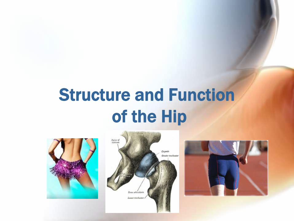
The Hip
“The San Francisco 49ers placed Frank Gore on the
injured reserve list on Tuesday after the Pro Bowl
running back suffered a season-ending hip fracture a
day earlier.” 12/1/10
•The most proximal joint of the lower extremity responsible for
motion in all 3 planes.
•This mobility makes the hip prone to injury if all of the support
structures are not working properly.

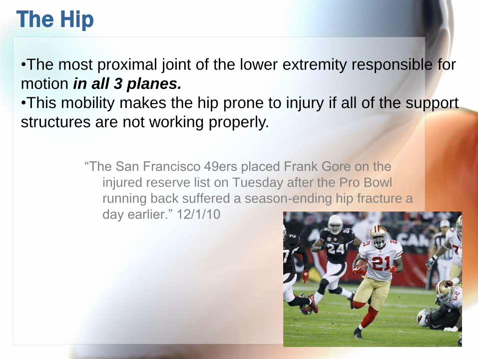
Osteology of the Hip
The “pelvis” is really the union of 3 bones:
• The ilium, the ischium & the pubis
Ilium
Pubis
Ischium

Osteology of the Hip
The “pelvis” is really the union of 3 bones:
• The ilium, the ischium & the pubis
Lateral Aspect Right Hip Posterior Aspect Left Hip

Osteology of the Hip

Osteology of the Hip
• Ilium
• Iliac fossa
• Iliac crest
• ASIS
• AIIS
• PSIS
• PIIS
• Ischium
• Body
• Ramus
• Ischial tuberosity
• spine
Lippert pg 263

Osteology of the Hip
• Pubis
• Body
• Superior ramus
• Inferior ramus
• Symphysis pubis
• Pubic tubercle
Lippert pg 264
Osteology of the Hip
The Acetabulum
• deep cup shaped structure encasing the head of the
femur formed by all 3 bones of pelvis
Permits motion in all 3 planes
Bony landmarks also
formed by a combination of
the pelvic bones
• Obturator foramen
• Greater sciatic notch
Lippert pg 264
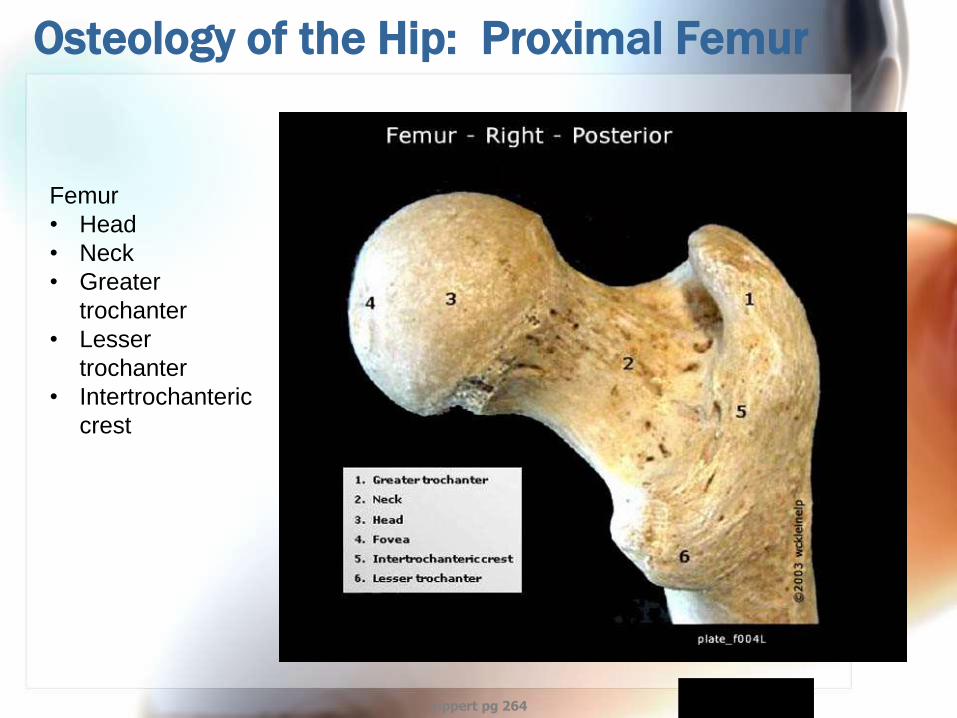
Osteology of the Hip: Proximal Femur
Femur
• Head
• Neck
• Greater
trochanter
• Lesser
trochanter
• Intertrochanteric
crest
Lippert pg 264

Osteology of the Hip: Proximal Femur

Osteology of the Hip: Proximal Femur
anatomical neck
surgical neck
Posterior Aspect Right Femur Anterior Aspect Right Femur
Femur
• Linea
aspera
• Pectineal
line
Lippert pg 265
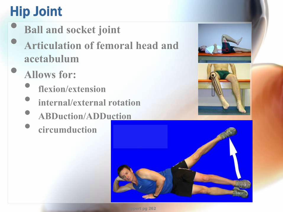
Hip Joint
• Ball and socket joint
• Articulation of femoral head and
acetabulum
• Allows for:
• flexion/extension
• internal/external rotation
• ABDuction/ADDuction
• circumduction
Lippert pg 262

The Hip Joint Structure
Lippert pg 275
Angle of Inclination
• Angle between the shaft and neck of the femur
• Normal is 125 degrees
• May be lesser or greater due to congenital
deformity, trauma, disease
• Coxa vara: angle less than 120 degrees
• Coxa valga: angle greater than 135 degrees

Ligaments in the Pelvis (anterior)

Ligaments of the Pelvis

Supporting Structures of the Hip Joint
• Joint capsule
• Ligaments
• Iliofemoral ligament
• Pubofemoral ligament
• Ischiofemoral ligament
• Inguinal ligament
• Acetabular labrum
• Fibrocartilaginous structure around rim of
acetabulum
• Increases depth of acetabulum
Lippert pg 265-266

The Ilioinguinal Ligament
This ligament runs from the
ASIS to the pubic
tubercle
It separates the anterior
abdominal wall from the
thigh

Myology of the Hip
Anterior
• Iliopsoas
• Rectus femoris
• Sartorius
Medial
• Pectineus
• Adductor magnus
• Adductor longus
• Adductor brevis
Posterior
• Gluteus maximus
• Semimembranosus
• Semitendinosus
• Biceps femoris
• Deep rotators (6)
Lateral
• Gluteus medius
• Gluteus minimus
• TFL
Lippert pg 267

Myology of the Hip
Iliopsoas
Origin Psoas Major: transverse
processes of T12-L5
Iliacus: Iliac fossa
Insertion Lesser trochanter of the
femur
Innervation Femoral n.
Action Hip flexion, trunk
flexion, anterior pelvic
tilt

Myology of the Hip
Your subtopic goes here Rectus Femoris
Origin Anterior-inferior iliac spine
Insertion Tibial tuberosity via the quadriceps
tendon
Innervation Femoral n.
Action Hip flexion, knee extension
“tidbit” One of the heads of the “quads”

Myology of the Hip
Your subtopic goes here Sartorius
Origin ASIS
Insertion Proximal-medial surface of the tibia (via
the pes anserinus)
Innervation Femoral n.
Action Hip flexion, hip ABD, Hip ER, knee
flexion
“tidbit” Longest muscle in the body

Myology of the Hip
Your subtopic goes here Tensor Fascia Latae
Origin Outer surface of the iliac crest
posterior to the ASIS
Insertion Proximal 1/3 of the ITB
Innervation Superior gluteal n.
Action Hip flexion, hip ABD, hip IR

Myology of the Hip
Your subtopic goes here Gluteus Maximus
Origin Posterior ilium, sacrum, coccyx
Insertion ITB, gluteal tuberosity of the femur
Innervation Inferior gluteal n.
Action Hip extension, hip ER

Myology of the Hip
Your subtopic goes here Semitendinosus
Origin Ischial tuberosity
Insertion Proximal-medial surface of the tibia
(pes anserinus)
Innervation Tibial portion of the sciatic n.
Action Hip extension, knee flexion,
“tidbit” One of the hamstrings
Myology of the Hip
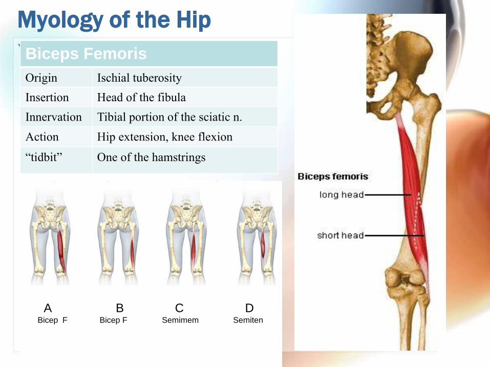
Your subtopic goes here Biceps Femoris
Origin Ischial tuberosity
Insertion Head of the fibula
Innervation Tibial portion of the sciatic n.
Action Hip extension, knee flexion
“tidbit” One of the hamstrings
A
A B C D Bicep F Bicep F Semimem Semiten

Myology of the Hip
Your subtopic goes here Semimembranosus
Origin Ischial tuberosity
Insertion Medial condyle of the tibia, posterior aspect
Innervation Tibial portion of the sciatic n.
Action Hip extension, knee flexion
“tidbit” One of the hamstrings

Myology of the Hip
Your subtopic goes here Gluteus Medius
Origin Outer surface of the ilum
Insertion Greater trochanter of the femur
Innervation Superior gluteal n.
Action Hip ABD
Gluteus
Medius
Iliac Crest
Gluteus
Maximus

Myology of the Hip
Your subtopic goes here Gluteus Minimus
Origin Outer surface of the ilium, inferior
to the gluteus medius
Insertion Greater trochanter
Innervation Superior gluteal n.
Action Hip ABD, hip IR
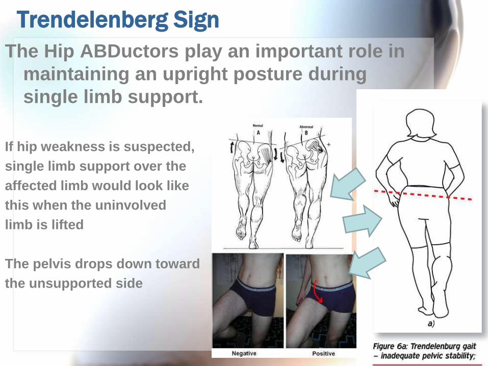
Trendelenberg Sign
The Hip ABDuctors play an important role in
maintaining an upright posture during
single limb support.
If hip weakness is suspected,
single limb support over the
affected limb would look like
this when the uninvolved
limb is lifted
The pelvis drops down toward
the unsupported side

Myology of the Hip
Your subtopic goes here Pectineus
Origin Pectineal line on superior ramus
Insertion Pectineal line on posterior surface
of the femur
Innervation Obturator n.
Action Hip ADD, hip flexion

Myology of the Hip
Your subtopic goes here ADDuctor Longus
Origin Anterior surface of the body of
the pubis
Insertion Middle 1/3 of the linea aspera of
the femur
Innervation Obturator n.
Action Hip ADD, Hip flexion
“tidbit” What’s in a name?

Myology of the Hip
Your subtopic goes here Gracillis
Origin Body and inferior ramus of the
pubis
Insertion Proximal-medial aspect of the
tibia (pes anserinus)
Innervation Obturator n.
Action Hip ADD, hip flexion, knee
flexion

Myology of the Hip
Your subtopic goes here ADDuctor Brevis
Origin Proximal Attachment: Anterior
surface of the inferior pubic ramus
Insertion Proximal 1/3 of the linea aspera of
the femur
Innervation Obturator n.
Action Hip ADD, Hip flexion
“tidbit” What’s in a name?
Myology of the Hip
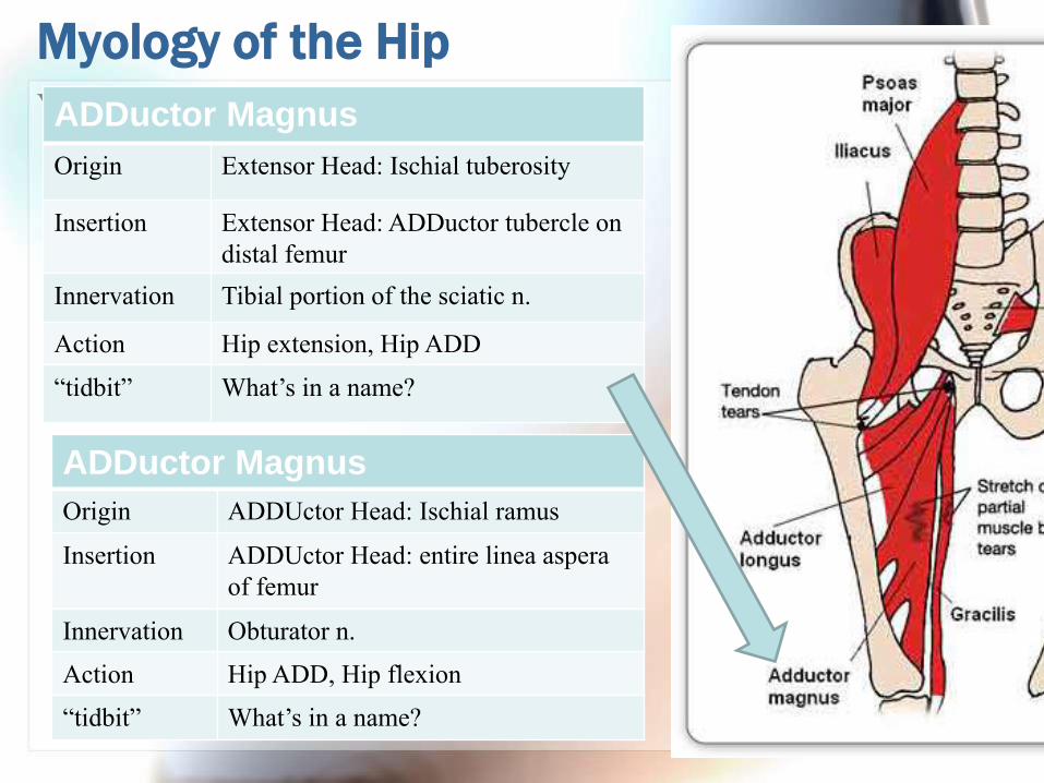
Your subtopic goes here ADDuctor Magnus
Origin Extensor Head: Ischial tuberosity
Insertion Extensor Head: ADDuctor tubercle on
distal femur
Innervation Tibial portion of the sciatic n.
Action Hip extension, Hip ADD
“tidbit” What’s in a name?
ADDuctor Magnus
Origin ADDUctor Head: Ischial ramus
Insertion ADDUctor Head: entire linea aspera
of femur
Innervation Obturator n.
Action Hip ADD, Hip flexion
“tidbit” What’s in a name?

Myology of the Hip
Intrinsic Hip ER (deep
rotators): (6 muscles)
Piriformis, Obturator Internus, Obturator
Exterunus, Gemelus Superior,
Gemelus Inferior, Quadratus Femoris
Piriformis Syndrome: The sciatic nerve passes deep to the
piriformis in most cases (approximately 85%
of people) but can in fact pierce the piriformis
itself, predisposing to piriformis syndrome
and subsequent sciatica. Even if the sciatic
nerve runs deep to the piriformis, muscle
guarding in this muscle can put direct
pressure on the nerve, causing pain and
discomfort.

Sciatic Nerve Distribution & the Piriformis

Common Hip Pathologies
Congenital Hip dysplasia
• Shallow acetabulum
• Causes femoral head to slide
upward
Legg-Calve’Perthes Disease
• Femoral head undergoes necrosis
• Children 5-10
• Head death 2-4 years then remodels
revascularization and
remodeling.
Avascular Necrosis of the
Femoral Head
Common Hip Pathologies
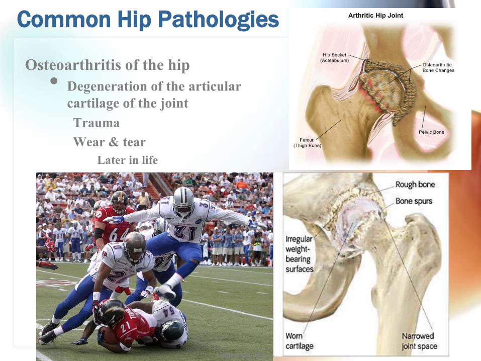
Osteoarthritis of the hip
• Degeneration of the articular
cartilage of the joint
Trauma
Wear & tear
Later in life
Lippert pg 277

Common Hip Pathologies Iliotibial Band Syndrome
Overuse injury causing lateral knee pain
Cyclists
Runners
Hamstring Strain
Overload of the muscle
Coxa vara
Coxa valga
“Hip Pointer”
Pelvic injury
Severe bruise caused by direct contact to the iliac crest of the pelvis
Lippert pg 277

Common Hip Pathologies
Lippert pg 277
Hip Fractures
intertrochanteric
femoral neck

Identify what you can! (in the hips)
Hip ADDuctors
Sartorius
ADDuctor Longus
ADDuctor Brevis
ADDuctor Magnus
Hip Flexors
Rectus femoris
TFL
Hip ABDuctors
TFL

Identify what you can! (in the hips)
Gluteus Maximus
Gluteus medius
Hamstrings
Semimembranosus
Biceps Femoris
Semitendinosus
TFL
OK go for it!
Pick out everything
else!

• How do you stretch
the hip flexors?
• How do you
strengthen the hip
extensors?
• How do you stretch
the hip adductors?
• How do you
strengthen the hip
abductors?
• How do you stretch
the hip internal
rotators?
• How do you
strengthen the hip
external rotators?