Embed Size (px)
Citation preview
Structure Of DNA & RNA
By Himanshu Dev
VMMC & SJH
DNA
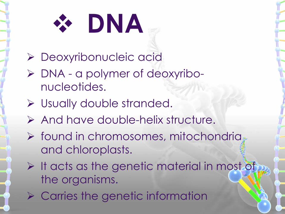
Deoxyribonucleic acid
DNA - a polymer of deoxyribo-
nucleotides.
Usually double stranded.
And have double-helix structure.
found in chromosomes, mitochondria
and chloroplasts.
It acts as the genetic material in most of
the organisms.
Carries the genetic information
DNA
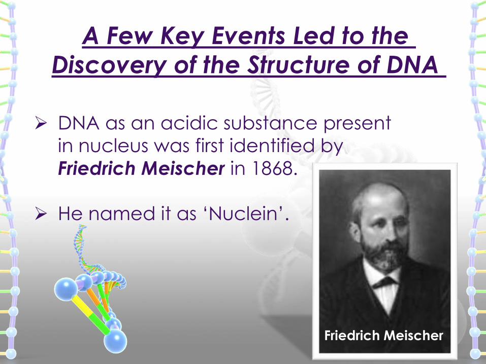
DNA as an acidic substance present
in nucleus was first identified by
Friedrich Meischer in 1868.
He named it as ‘Nuclein’.
A Few Key Events Led to the
Discovery of the Structure of DNA
Friedrich Meischer
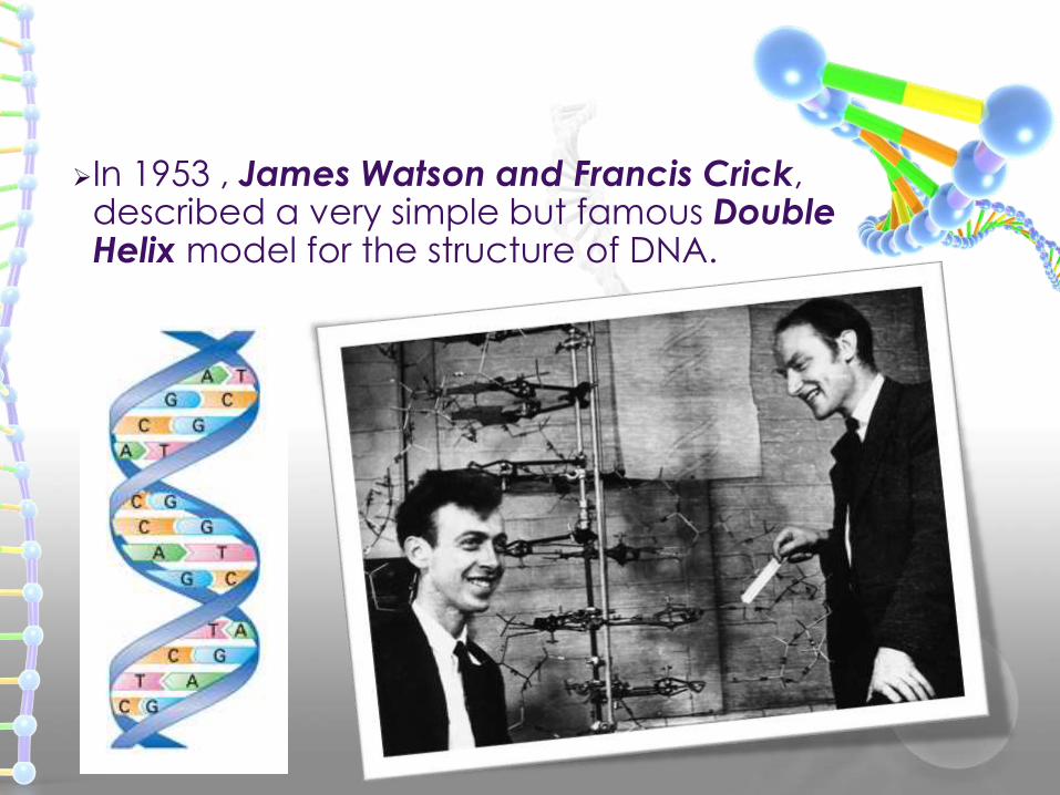
In 1953 , James Watson and Francis Crick,described a very simple but famous DoubleHelix model for the structure of DNA.
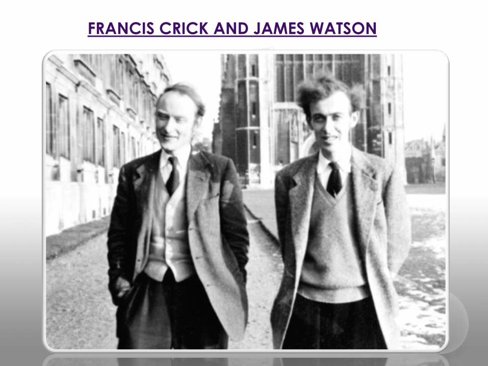
FRANCIS CRICK AND JAMES WATSON
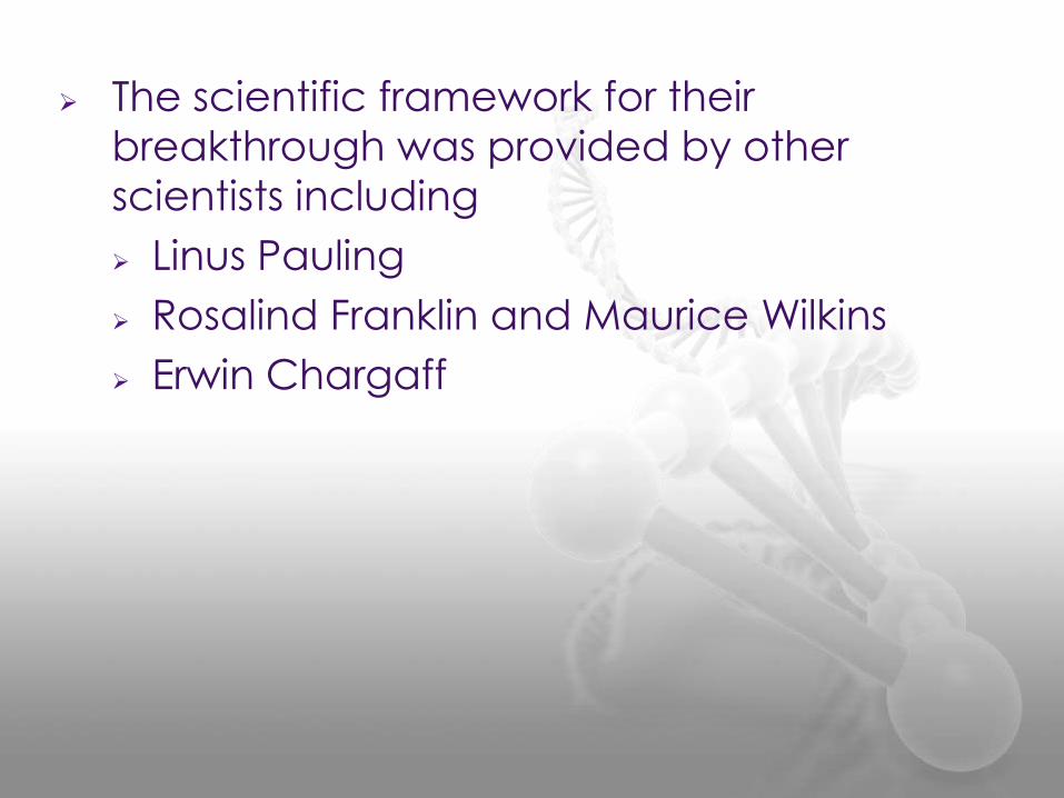
The scientific framework for their
breakthrough was provided by other
scientists including
Linus Pauling
Rosalind Franklin and Maurice Wilkins
Erwin Chargaff
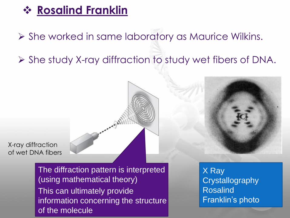
She worked in same laboratory as Maurice Wilkins.
She study X-ray diffraction to study wet fibers of DNA.
X Ray
Crystallography
Rosalind
Franklin’s photo
X-ray diffraction
of wet DNA fibers
The diffraction pattern is interpreted
(using mathematical theory)
This can ultimately provide
information concerning the structure
of the molecule
Rosalind Franklin
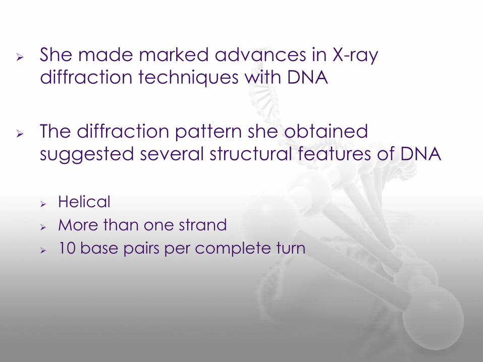
She made marked advances in X-ray
diffraction techniques with DNA
The diffraction pattern she obtained
suggested several structural features of DNA
Helical
More than one strand
10 base pairs per complete turn
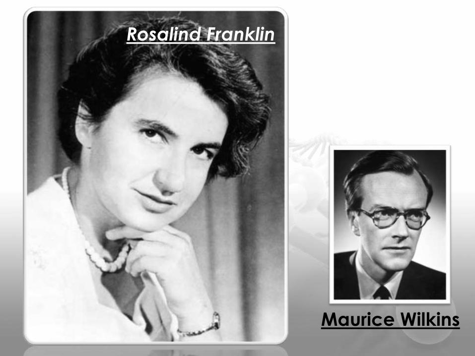
Rosalind Franklin
Maurice Wilkins

DNA Structure
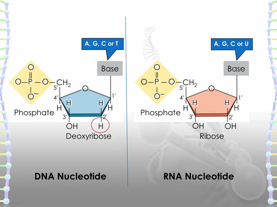
DNA has three main components
1. Deoxyribose (a pentose sugar)
2. Base (there are four different ones)
3. Phosphate
DNA structure is often divided into four different levels primary, secondary, tertiary and quaternary.
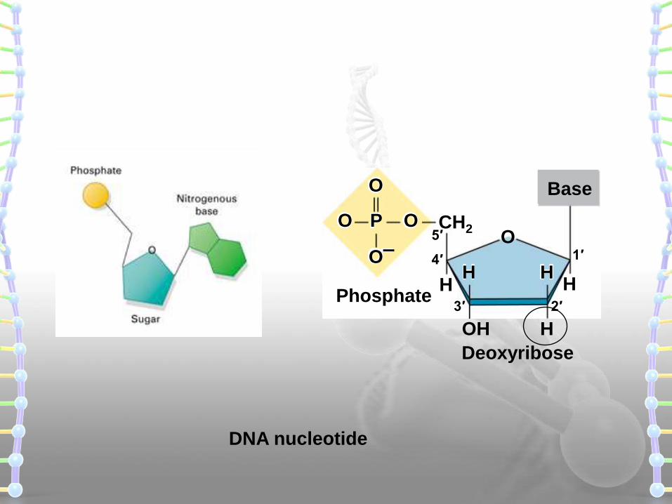
HH
OH
OCH2
Base
Phosphate
Ribose
OH
5′
4′ 1′
3′ 2′
O
O
P
O–
O
HH
A, G, C or U
HH
H
OCH2
Base
Phosphate
Deoxyribose
5′
OH
4′ 1′
3′ 2′
OO
O
P
O–HH
DNA Nucleotide RNA Nucleotide
A, G, C or T
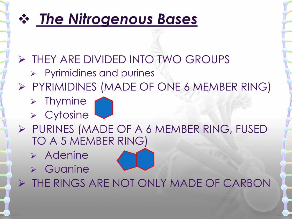
The Nitrogenous Bases
THEY ARE DIVIDED INTO TWO GROUPS
Pyrimidines and purines
PYRIMIDINES (MADE OF ONE 6 MEMBER RING)
Thymine
Cytosine
PURINES (MADE OF A 6 MEMBER RING, FUSED TO A 5 MEMBER RING)
Adenine
Guanine
THE RINGS ARE NOT ONLY MADE OF CARBON
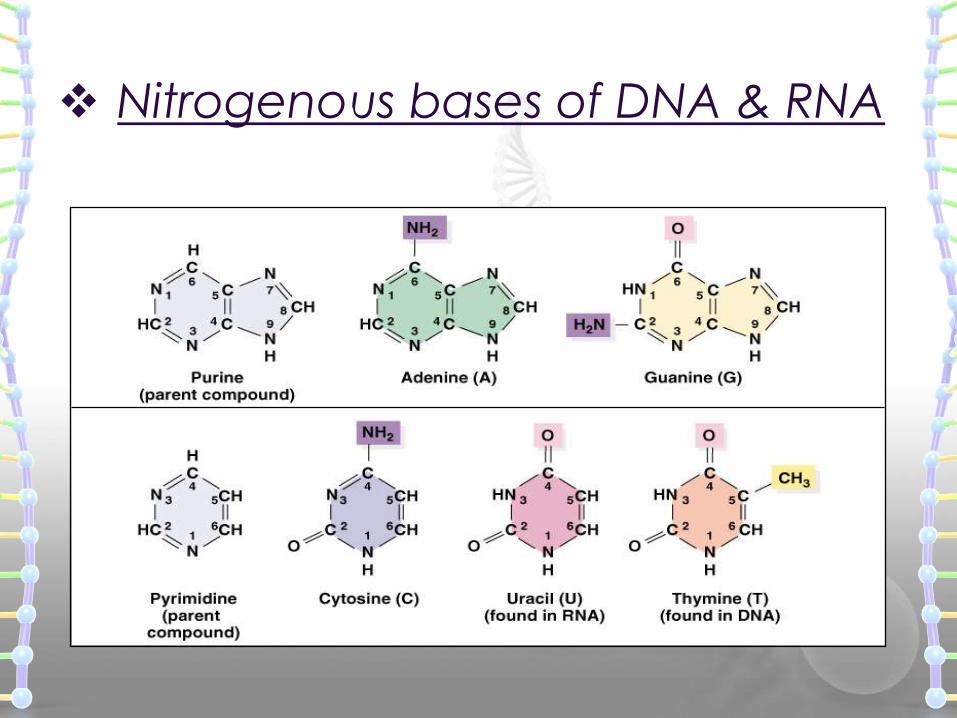
Nitrogenous bases of DNA & RNA
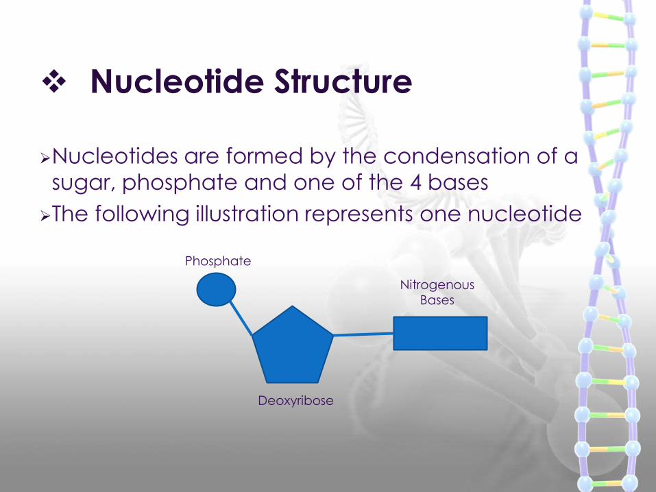
Nucleotide Structure
Nucleotides are formed by the condensation of a
sugar, phosphate and one of the 4 bases
The following illustration represents one nucleotide
Phosphate
Deoxyribose
NitrogenousBases
HH
H
OCH2
Base
DNA nucleotide
Phosphate
Deoxyribose
5′
OH
4′ 1′
3′ 2′
OO
O
P
O–HH
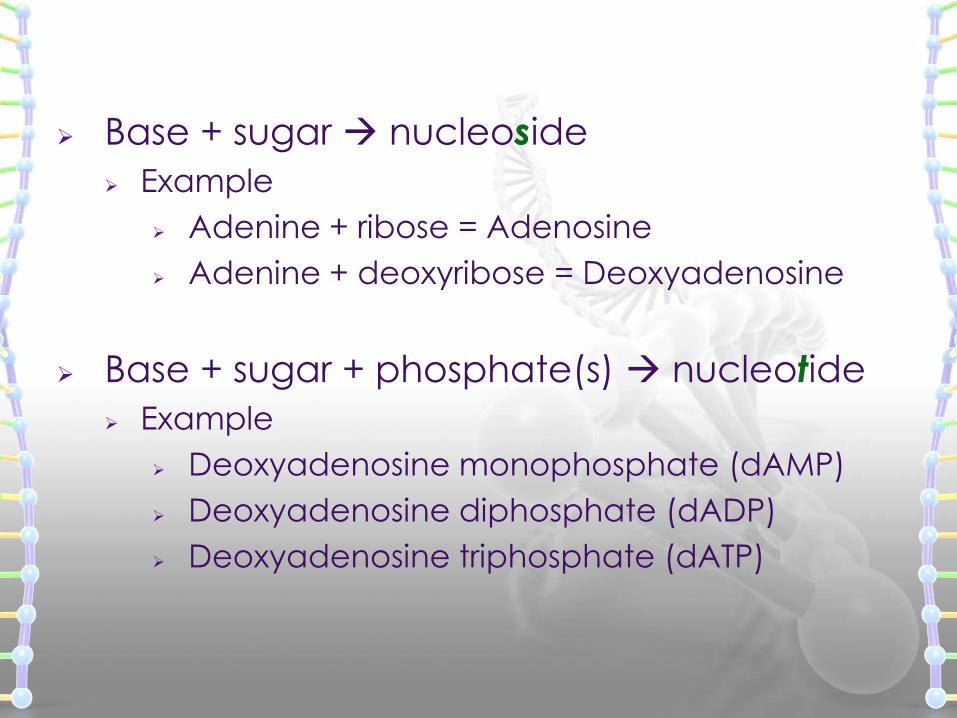
Base + sugar nucleoside
Example
Adenine + ribose = Adenosine
Adenine + deoxyribose = Deoxyadenosine
Base + sugar + phosphate(s) nucleotide
Example
Deoxyadenosine monophosphate (dAMP)
Deoxyadenosine diphosphate (dADP)
Deoxyadenosine triphosphate (dATP)
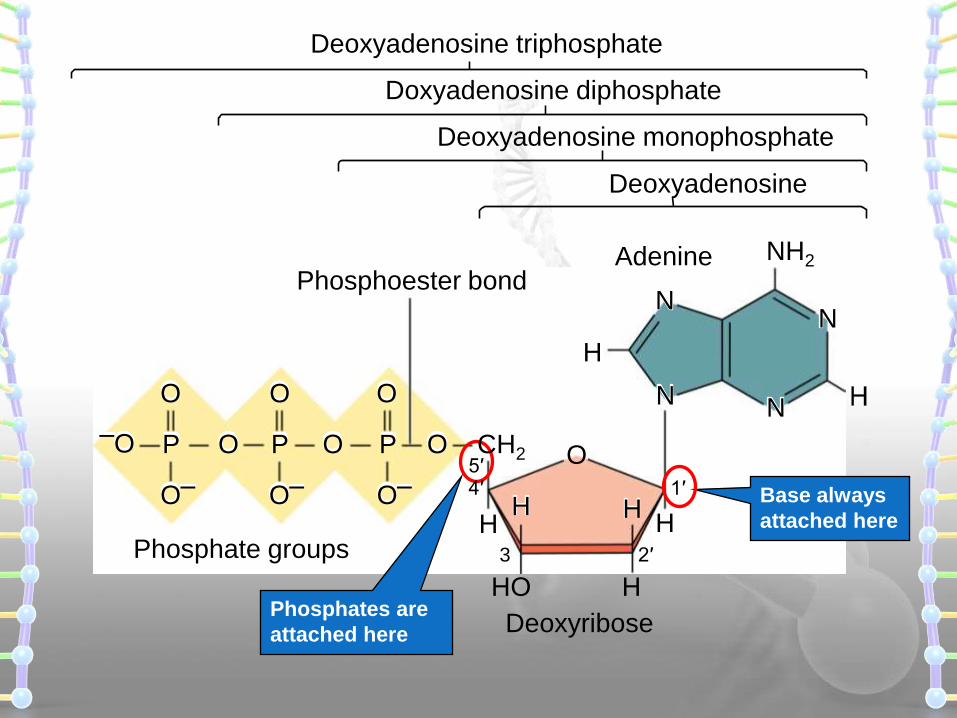
Base always
attached here
Phosphates are
attached here
Deoxyadenosine
Deoxyadenosine monophosphate
Doxyadenosine diphosphate
Adenine
Phosphate groups
Phosphoester bond
Deoxyribose
H
OP CH2
O–
OO P
O–
O O O
–O P
O–
H
HHO
O
H
2′3
1′4′5′
Deoxyadenosine triphosphate
NH2
N
H
H
N
NN
Sugar
Base
P
Sugar
Base
P
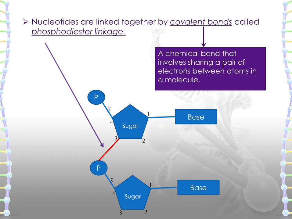
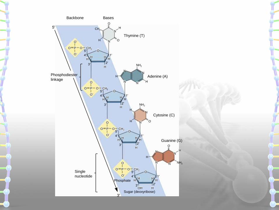
Nucleotides are linked together by covalent bonds called
phosphodiester linkage.
1
23
4
5
1
23
4
5
A chemical bond that
involves sharing a pair of
electrons between atoms in
a molecule.
NH2
N O
N
O
N O
N
Adenine (A)
Guanine (G)
Thymine (T)
BasesBackbone
Cytosine (C)
O
HH
H
H
HH
OOO
O–
P CH2
O–
HH
H
H
H
HH
OOO
O
P CH2
O–
NH2
N
N
H
N
N
HH
H
HH
OOO
O
P CH2
O–
NH2
HN
N
N
H
N
HH
HOH
HH
OOO
O
P CH2
O–
Single
nucleotide
Phosphodiester
linkage
Sugar (deoxyribose)
Phosphate
3′
5′
5′
4′ 1′
2′3′
5′
4′ 1′
2′3′
5′
4′ 1′
2′3′
5′
4′ 1′
2′3′
CH3
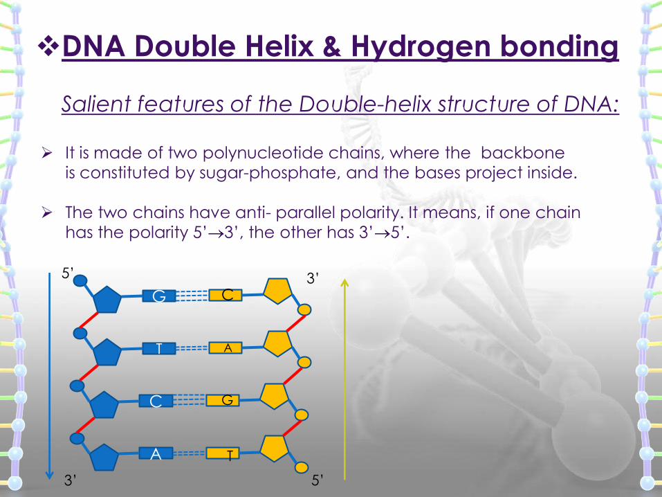
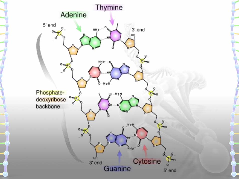
DNA Double Helix & Hydrogen bonding
Salient features of the Double-helix structure of DNA:
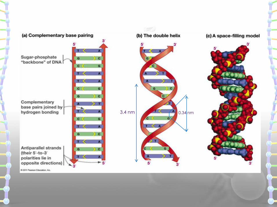
It is made of two polynucleotide chains, where the backbone
is constituted by sugar-phosphate, and the bases project inside.
The two chains have anti- parallel polarity. It means, if one chain
has the polarity 5’ 3’, the other has 3’ 5’.
G
T
C
A
5’ 3’
3’ 5’
C
A
T
G
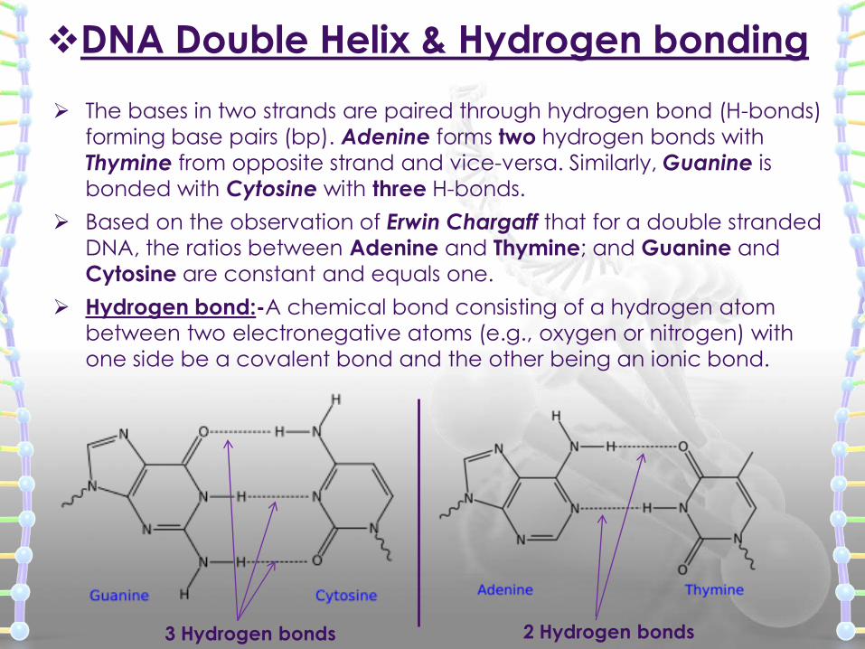
The bases in two strands are paired through hydrogen bond (H-bonds)
forming base pairs (bp). Adenine forms two hydrogen bonds with
Thymine from opposite strand and vice-versa. Similarly, Guanine is
bonded with Cytosine with three H-bonds.
Based on the observation of Erwin Chargaff that for a double stranded
DNA, the ratios between Adenine and Thymine; and Guanine and
Cytosine are constant and equals one.
Hydrogen bond:-A chemical bond consisting of a hydrogen atom
between two electronegative atoms (e.g., oxygen or nitrogen) with
one side be a covalent bond and the other being an ionic bond.
DNA Double Helix & Hydrogen bonding
3 Hydrogen bonds 2 Hydrogen bonds
Erwin Chargaff’s Experiment
Chargaff pioneered many of biochemical technique for
the isolation, purification and measurement of nucleic acids
from living cells.
It was known that DNA contained the four bases: A, G, C & T.
Chargaff analyzed the base composition DNA isolated from
many different species.
THE HYPOTHESIS
An analysis of the base composition of DNA in different
species may reveal important features about structure of DNA.
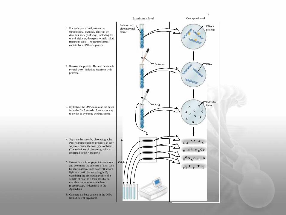
Experimental level Conceptual level
For each type of cell, extract the
chromosomal material. This can be
done in a variety of ways, including the
use of high salt, detergent, or mild alkali
treatment. Note: The chromosomes
contain both DNA and protein.
1.
Remove the protein. This can be done in
several ways, including treament with
protease.
2.
Hydrolyze the DNA to release the bases
from the DNA strands. A common way
to do this is by strong acid treatment.
3.
Separate the bases by chromatography.
Paper chromatography provides an easy
way to separate the four types of bases.
(The technique of chromatography is
described in the Appendix.)
4.
Extract bands from paper into solutions
and determine the amounts of each base
by spectroscopy. Each base will absorb
light at a particular wavelength. By
examining the absorption profile of a
sample of base, it is then possible to
calculate the amount of the base.
(Spectroscopy is described in the
Appendix.)
5.
Compare the base content in the DNA
from different organisms.
6.
Solution of
chromosomal
extract
DNA
DNA +
proteins
Individual
bases
Origin
Protease
AcidA
A
AAA A
AA
CC
C CC CC
GG G
G G GG
TT TT
TT
T
T
G
G
G
C
C
C
y
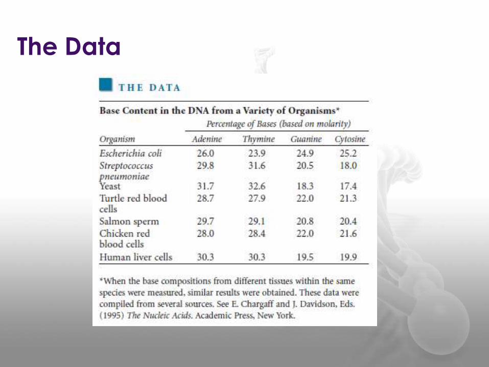
The Data
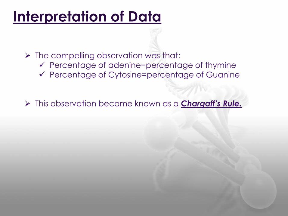
Interpretation of Data
The compelling observation was that:
Percentage of adenine=percentage of thymine
Percentage of Cytosine=percentage of Guanine
This observation became known as a Chargaff’s Rule.
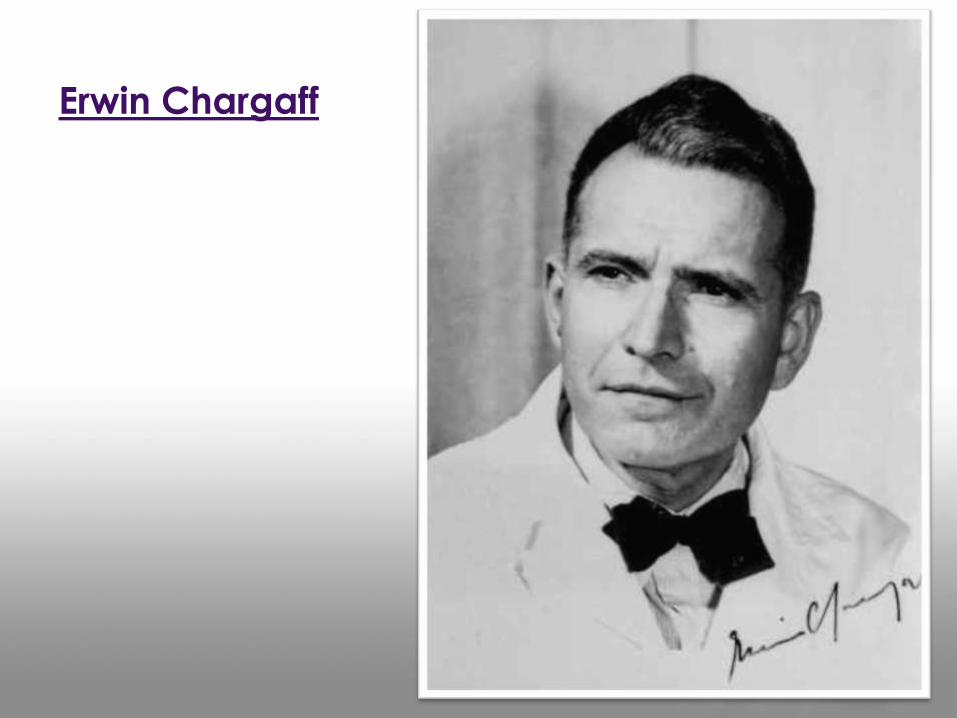
Erwin Chargaff
DNA Double Helix & Hydrogen bonding
The two strands are coiled in a right-handed fashion(Clockwise).
The pitch of the helix is 3.4 nm (a nanometer is one billionth of a
meter, that is 10-9 m) and there are roughly 10 bp in each turn.
Consequently, the distance between a bp in a helix is
approximately equal to 0.34 nm.
The plane of one base pair stacks over the other in double helix.
This, in addition to H-bonds, confers stability of the helical structure.
0.34 nm3.4 nm
DNA Double Helix & Hydrogen bonding
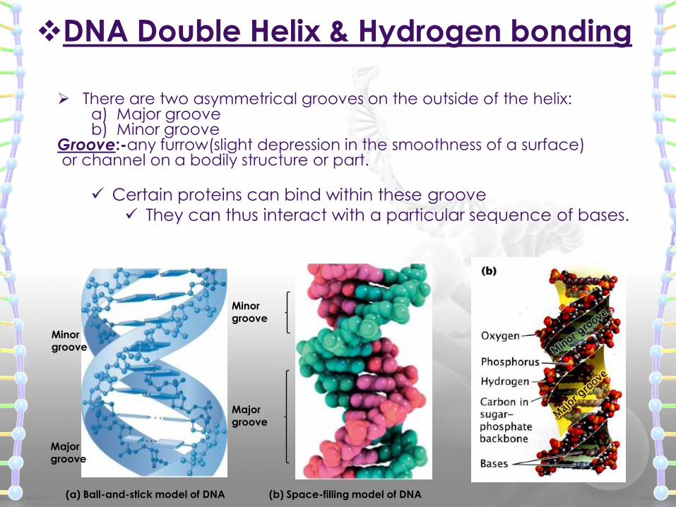
There are two asymmetrical grooves on the outside of the helix:a) Major grooveb) Minor groove
Groove:-any furrow(slight depression in the smoothness of a surface)or channel on a bodily structure or part.
Certain proteins can bind within these groove
They can thus interact with a particular sequence of bases.
(b) Space-filling model of DNA(a) Ball-and-stick model of DNA
Minor
groove
Majorgroove
Minorgroove
Major
groove
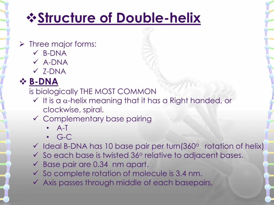
Structure of Double-helix
Three major forms:
B-DNA
A-DNA
Z-DNA
B-DNAis biologically THE MOST COMMON
It is a -helix meaning that it has a Right handed, or
clockwise, spiral.
Complementary base pairing
• A-T
• G-C
Ideal B-DNA has 10 base pair per turn(360o rotation of helix)
So each base is twisted 36o relative to adjacent bases.
Base pair are 0.34 nm apart.
So complete rotation of molecule is 3.4 nm.
Axis passes through middle of each basepairs.
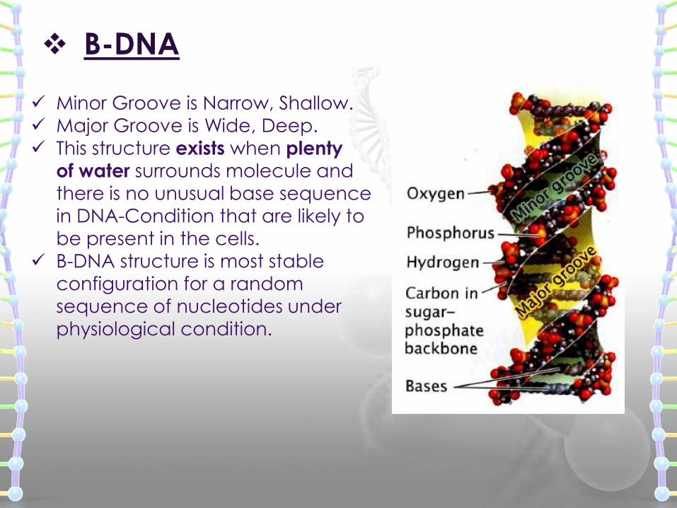
B-DNA
Minor Groove is Narrow, Shallow. Major Groove is Wide, Deep.
This structure exists when plenty
of water surrounds molecule and
there is no unusual base sequence
in DNA-Condition that are likely tobe present in the cells.
B-DNA structure is most stable
configuration for a random
sequence of nucleotides under
physiological condition.
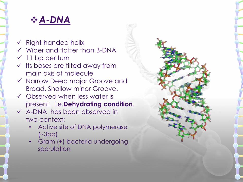
A-DNA
Right-handed helix
Wider and flatter than B-DNA
11 bp per turn
Its bases are tilted away from
main axis of molecule
Narrow Deep major Groove and
Broad, Shallow minor Groove.
Observed when less water is
present. i.e.Dehydrating condition.
A-DNA has been observed in
two context:• Active site of DNA polymerase
(~3bp)
• Gram (+) bacteria undergoing
sporulation

Z-DNA
A left-handed helix
Seen in Condition of High salt
concentration.
In this form sugar-phosphate
backbones zigzag back and
forth, giving rise to the name
Z-DNA(for zigzag).
12 base pairs per turn.
A deep Minor Groove.
No Discernible Major Groove.
Part of some active genes form
Z-DNA, suggesting that Z-DNA
may play a role in regulating
gene transcription.
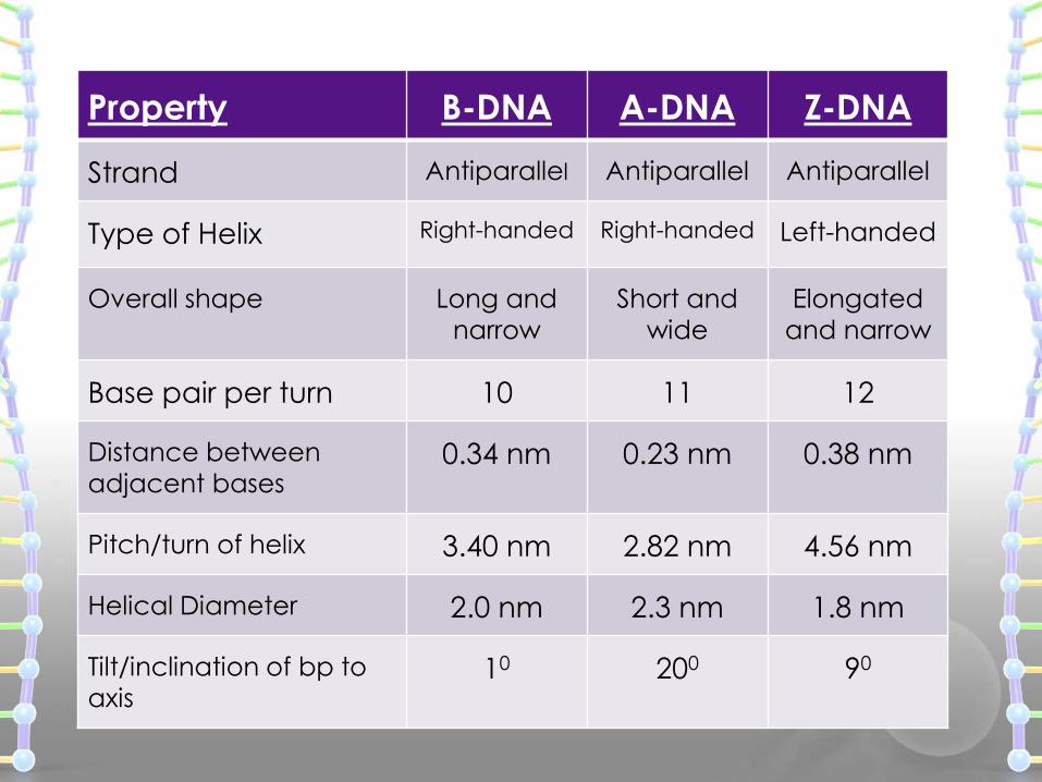
Property B-DNA A-DNA Z-DNA
Strand Antiparallel Antiparallel Antiparallel
Type of Helix Right-handed Right-handed Left-handed
Overall shape Long and
narrow
Short and
wide
Elongated
and narrow
Base pair per turn 10 11 12
Distance between
adjacent bases0.34 nm 0.23 nm 0.38 nm
Pitch/turn of helix 3.40 nm 2.82 nm 4.56 nm
Helical Diameter 2.0 nm 2.3 nm 1.8 nm
Tilt/inclination of bp to
axis10 200 90
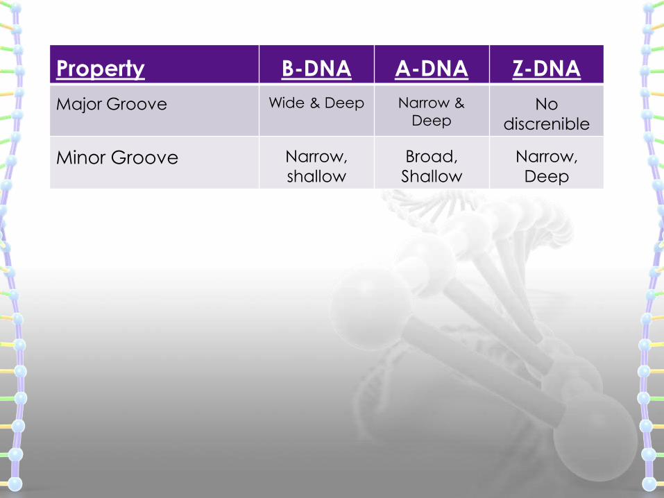
Property B-DNA A-DNA Z-DNA
Major Groove Wide & Deep Narrow &
DeepNo
discrenible
Minor Groove Narrow,
shallow
Broad,
Shallow
Narrow,
Deep
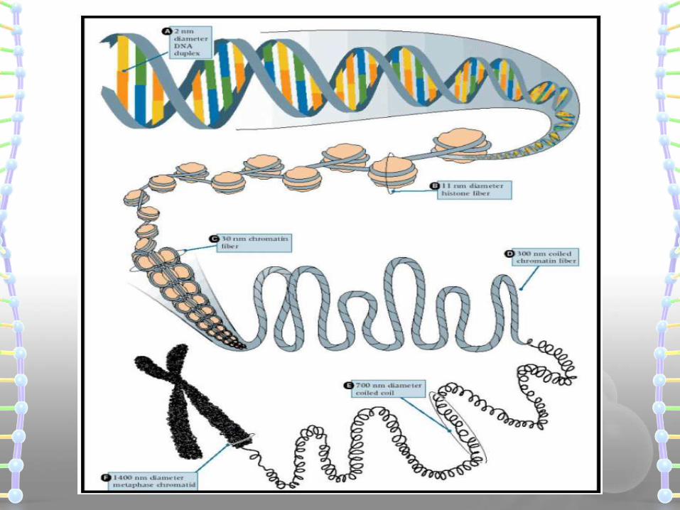
DNA Supercoiling
DNA supercoiling refers to the over or under-winding
of strands.
DNA supercoiling is important for DNA packaging
within all cells. Because the length of DNA can be
of thousands of times that of a cells, packaging this
material into the cell or nucleus (in Eukaryotes) is a
difficult feat.
Supercoiling of DNA reduces the space and allows
for much more DNA to be packaged.
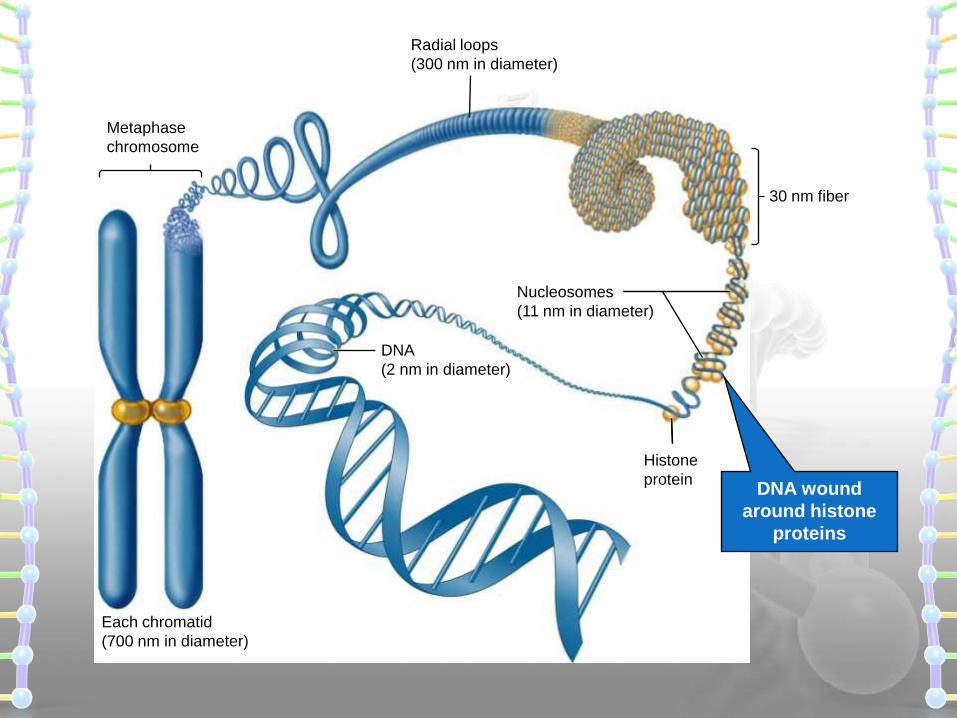
DNA wound
around histone
proteins
Radial loops
(300 nm in diameter)
Metaphase
chromosome
DNA
(2 nm in diameter)
Nucleosomes
(11 nm in diameter)
Histone
protein
Each chromatid
(700 nm in diameter)
30 nm fiber
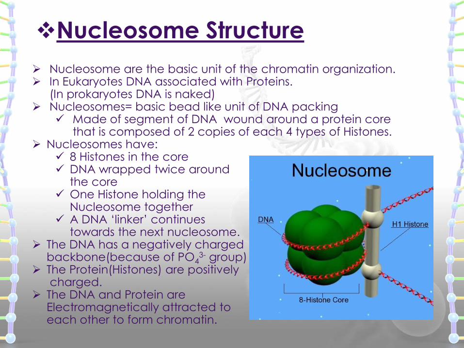
Nucleosome Structure
Nucleosome are the basic unit of the chromatin organization. In Eukaryotes DNA associated with Proteins.
(In prokaryotes DNA is naked) Nucleosomes= basic bead like unit of DNA packing
Made of segment of DNA wound around a protein corethat is composed of 2 copies of each 4 types of Histones.
Nucleosomes have: 8 Histones in the core DNA wrapped twice around
the core One Histone holding the
Nucleosome together A DNA ‘linker’ continues
towards the next nucleosome. The DNA has a negatively charged
backbone(because of PO43- group)
The Protein(Histones) are positivelycharged.
The DNA and Protein are Electromagnetically attracted to each other to form chromatin.
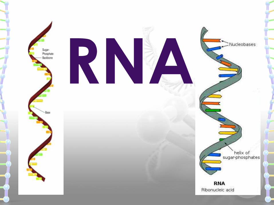
RNA
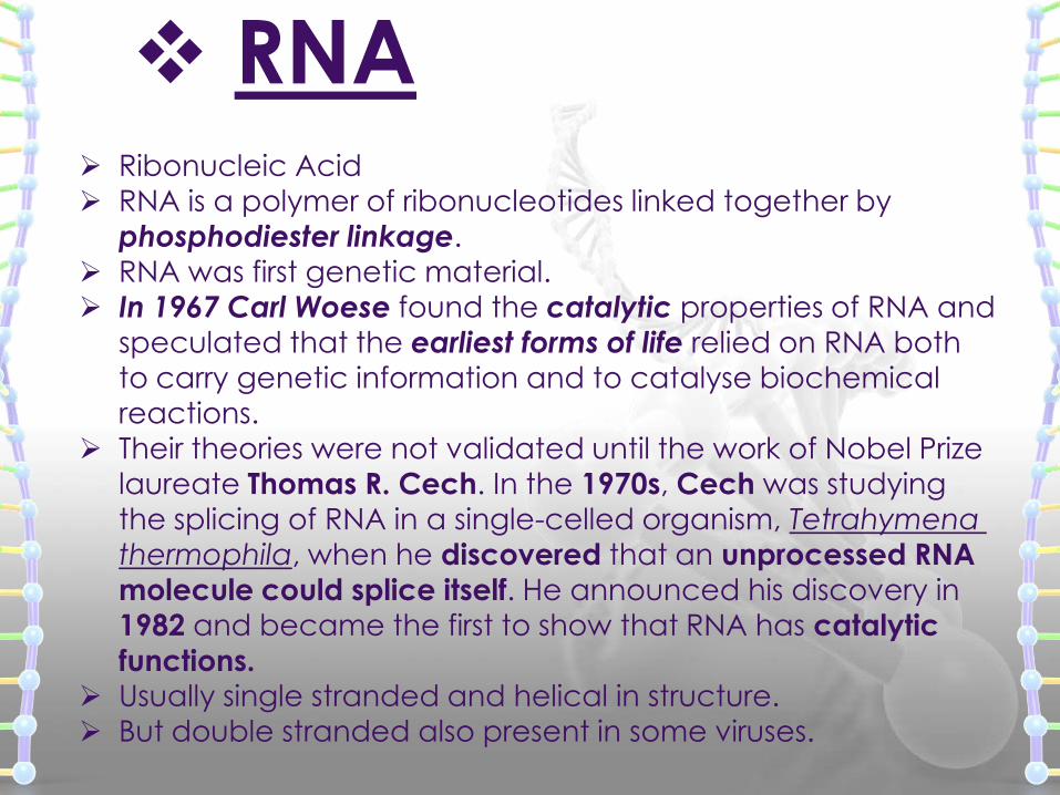
RNA Ribonucleic Acid
RNA is a polymer of ribonucleotides linked together by
phosphodiester linkage. RNA was first genetic material.
In 1967 Carl Woese found the catalytic properties of RNA and
speculated that the earliest forms of life relied on RNA both
to carry genetic information and to catalyse biochemical
reactions. Their theories were not validated until the work of Nobel Prize
laureate Thomas R. Cech. In the 1970s, Cech was studying
the splicing of RNA in a single-celled organism, Tetrahymena
thermophila, when he discovered that an unprocessed RNA
molecule could splice itself. He announced his discovery in 1982 and became the first to show that RNA has catalytic
functions. Usually single stranded and helical in structure.
But double stranded also present in some viruses.
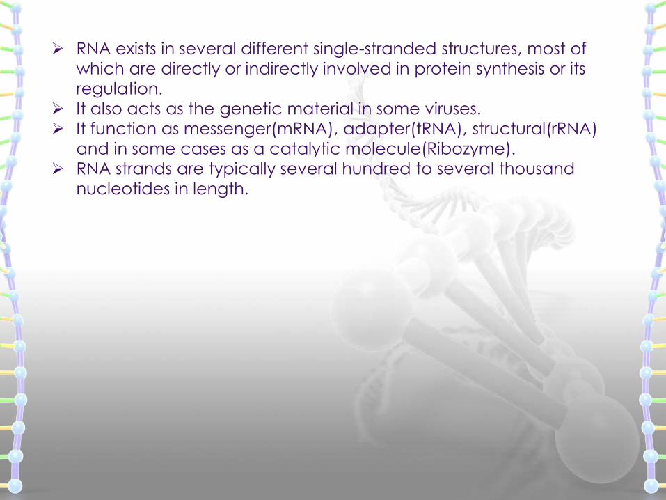
RNA exists in several different single-stranded structures, most of
which are directly or indirectly involved in protein synthesis or its
regulation.
It also acts as the genetic material in some viruses.
It function as messenger(mRNA), adapter(tRNA), structural(rRNA)
and in some cases as a catalytic molecule(Ribozyme).
RNA strands are typically several hundred to several thousand
nucleotides in length.
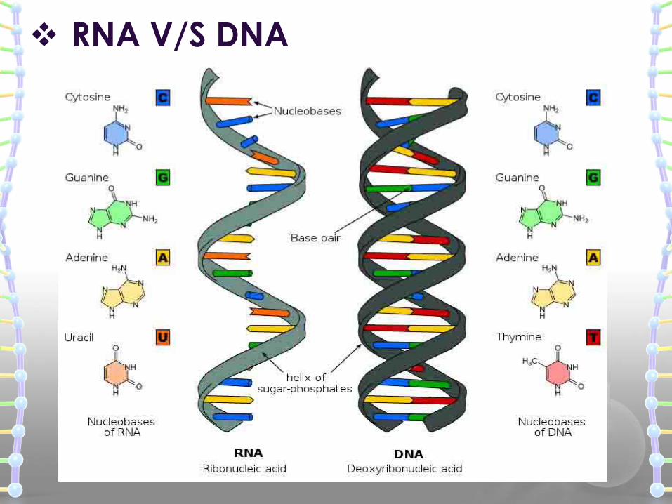
RNA V/S DNA
RNA structure
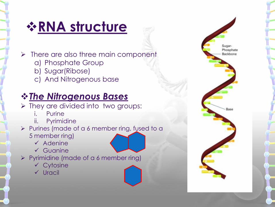
There are also three main component
a) Phosphate Group
b) Sugar(Ribose)
c) And Nitrogenous base
The Nitrogenous Bases They are divided into two groups:
i. Purine
ii. Pyrimidine
Purines (made of a 6 member ring, fused to a
5 member ring)
Adenine
Guanine
Pyrimidine (made of a 6 member ring)
Cytosine
Uracil
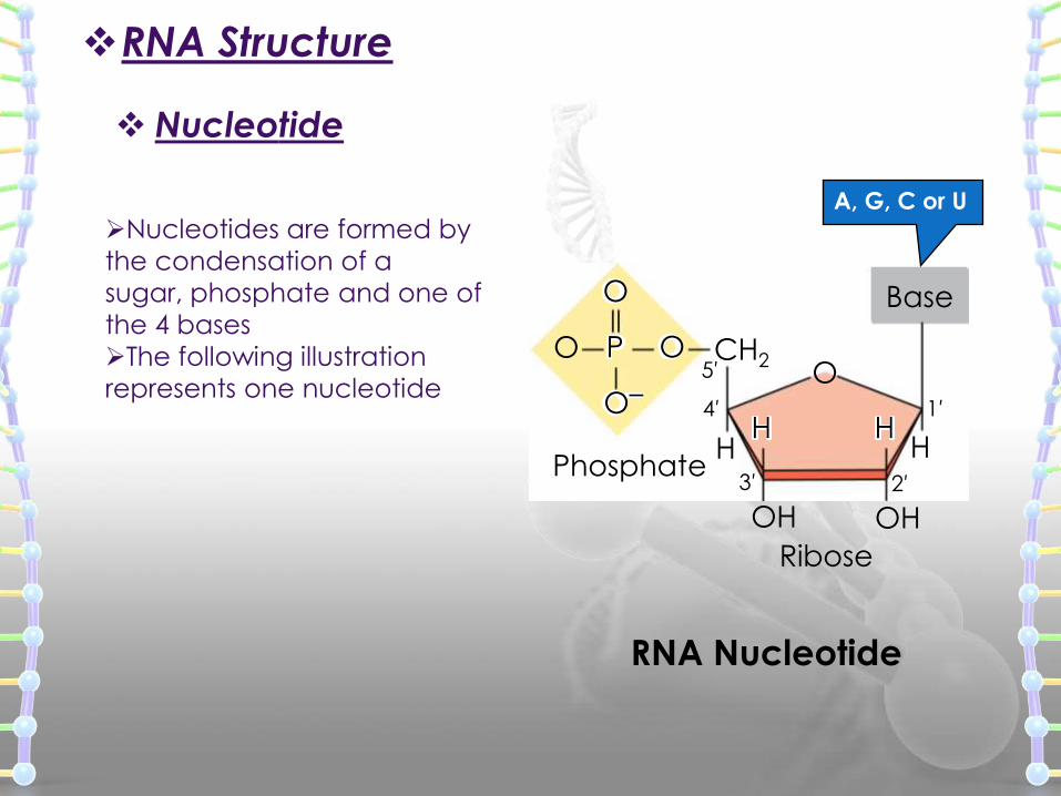
Nucleotide
HH
OH
OCH2
Base
Phosphate
Ribose
OH
5′
4′ 1′
3′ 2′
O
O
P
O–
O
HH
A, G, C or U
RNA Nucleotide
RNA Structure
Nucleotides are formed by
the condensation of a
sugar, phosphate and one of
the 4 bases
The following illustration
represents one nucleotide
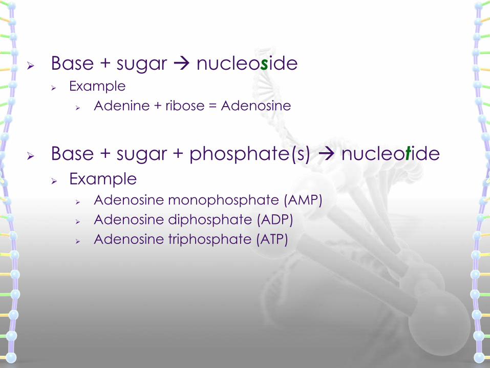
Base + sugar nucleoside Example
Adenine + ribose = Adenosine
Base + sugar + phosphate(s) nucleotide
Example
Adenosine monophosphate (AMP)
Adenosine diphosphate (ADP)
Adenosine triphosphate (ATP)
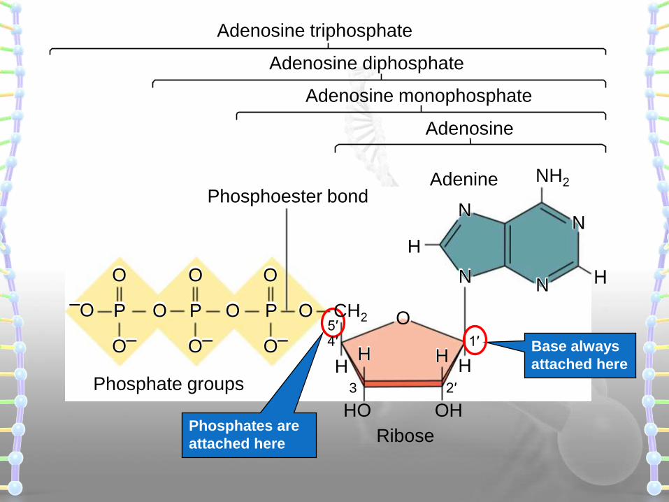
Base always
attached here
Phosphates are
attached here
Adenosine
Adenosine monophosphate
Adenosine diphosphate
Adenine
Phosphate groups
Phosphoester bond
Ribose
H
OP CH2
O–
OO P
O–
O O O
–O P
O–
H
OHHO
O
H
2′3
1′4′5′
Adenosine triphosphate
NH2
N
H
H
N
NN
Ribose
Sugar
Base
P
Ribose
Sugar
Base
P
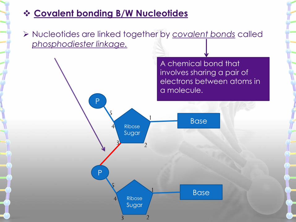
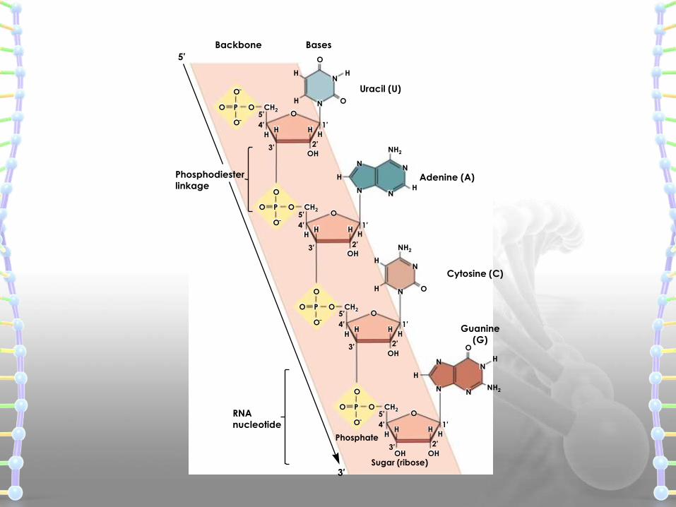
Nucleotides are linked together by covalent bonds called
phosphodiester linkage.
1
23
4
5
1
23
4
5
A chemical bond that
involves sharing a pair of
electrons between atoms in
a molecule.
Covalent bonding B/W Nucleotides
Adenine (A)
Guanine
(G)
Uracil (U)
BasesBackbone
Cytosine (C)
O
HHHH
OOO
O–
P CH2
O–
HHHH
OOO
O
P CH2
O–
NH2
H
N
HHHH
OOO
O
P CH2
O–
H
H
HH
OH
HH
OOO
O
P CH2
O–
Sugar (ribose)
Phosphate
5′
4′ 1′
2′3′
5′
4′ 1′
2′3′
5′
4′ 1′
2′3′
5′
4′ 1′
2′3′OH
OH
OH
OH
RNA
nucleotide
Phosphodiester
linkage
3′
5′
NH2
OH
H
H
O
NH O
N
NN
N
N
N
N
N N
N
NH2
H
H
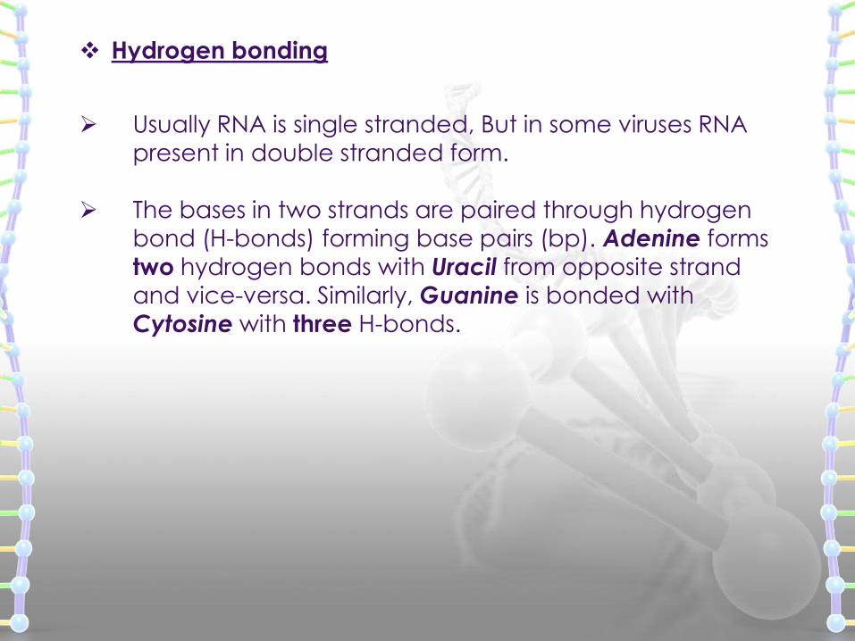
Usually RNA is single stranded, But in some viruses RNA
present in double stranded form.
The bases in two strands are paired through hydrogen bond (H-bonds) forming base pairs (bp). Adenine forms
two hydrogen bonds with Uracil from opposite strand
and vice-versa. Similarly, Guanine is bonded with
Cytosine with three H-bonds.
Hydrogen bonding
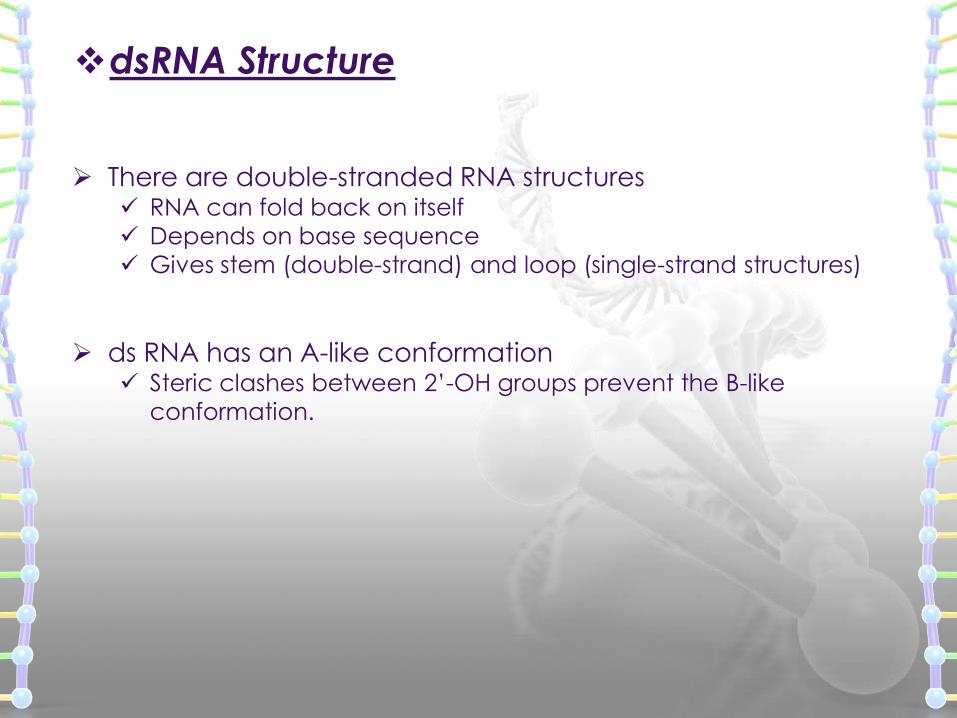
dsRNA Structure
There are double-stranded RNA structures RNA can fold back on itself
Depends on base sequence
Gives stem (double-strand) and loop (single-strand structures)
ds RNA has an A-like conformation Steric clashes between 2’-OH groups prevent the B-like
conformation.
A U
A U
U A
G C
C G
C G
A U
U A
U A
C G
C G
C G
C G
C G
A
A
U
U
G
G
C
C
C
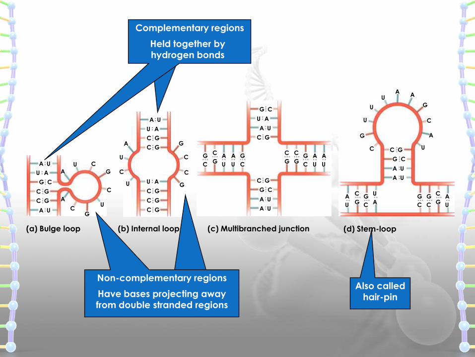
(a) Bulge loop (b) Internal loop (c) Multibranched junction (d) Stem-loop
G
C
C
G
U
AA
U
G
C
G
C
C
GA
UA U
A U
G C
A U
U A
G C
C G
C G
G
C
G
C
C
GA
U
A
U
G
C
C
G
C
GA
U
A
U
A
U
U
G
G
C
C
CA U
A U
G C
C G
A
AA
U
U
U
U
G
G
C
C
Also called hair-pin
Complementary regions
Non-complementary regions
Held together by hydrogen bonds
Have bases projecting away from double stranded regions
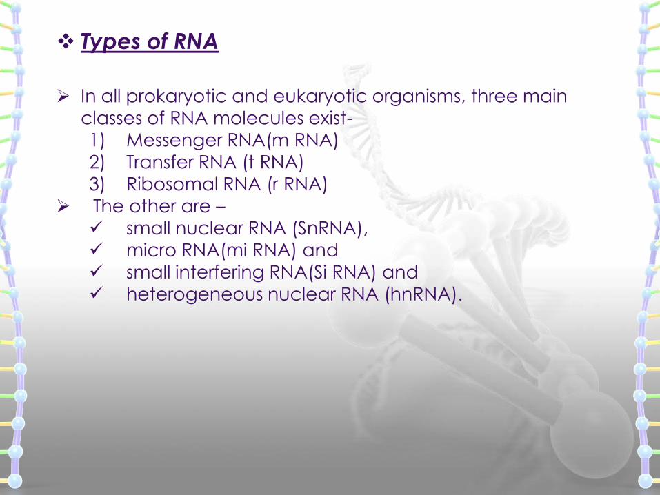
Types of RNA
In all prokaryotic and eukaryotic organisms, three main
classes of RNA molecules exist-
1) Messenger RNA(m RNA)
2) Transfer RNA (t RNA)3) Ribosomal RNA (r RNA)
The other are –
small nuclear RNA (SnRNA),
micro RNA(mi RNA) and
small interfering RNA(Si RNA) and heterogeneous nuclear RNA (hnRNA).
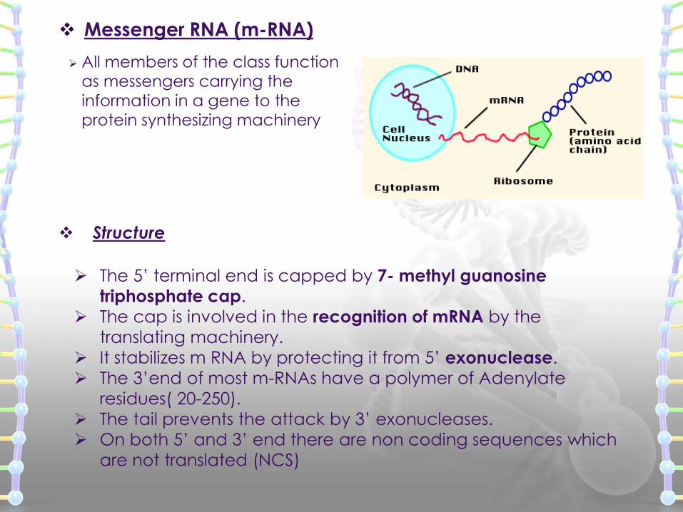
Messenger RNA (m-RNA)
All members of the class function as messengers carrying the
information in a gene to the protein synthesizing machinery
Structure
The 5’ terminal end is capped by 7- methyl guanosine
triphosphate cap.
The cap is involved in the recognition of mRNA by the
translating machinery.
It stabilizes m RNA by protecting it from 5’ exonuclease.
The 3’end of most m-RNAs have a polymer of Adenylate
residues( 20-250).
The tail prevents the attack by 3’ exonucleases.
On both 5’ and 3’ end there are non coding sequences which
are not translated (NCS)
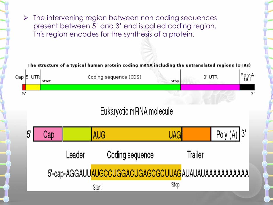
The intervening region between non coding sequences
present between 5’ and 3’ end is called coding region.
This region encodes for the synthesis of a protein.
Heterogeneous nuclear RNA (hnRNA) [Precursor mRNA]
In mammalian nuclei , hnRNA is the immediate
product of gene transcription
The nuclear product is heterogeneous in size
(Variable) and is very large.
75 % of hnRNA is degraded in the nucleus,
only 25% is processed to mature m RNA.
Mature m –RNA is formed from primary transcript
by capping, tailing, splicing and base modification.
Transfer RNA (t-RNA)
Transfer RNA are the smallest of three major speciesof RNA moleculesThey have 74-95 nucleotide residuesThey transfer the amino acids from cytoplasm to the
protein synthesizing machinery, hence the name tRNA.They are also called Adapter molecules, since they
act as adapters for the translation of the sequenceof nucleotides of the m RNA in to specific aminoacidsThere are at least 20 species of tRNA one
corresponding to each of the 20 amino acidsrequired for protein synthesis. tRNA is the only RNA species that contains the
nucleoside thymidine.
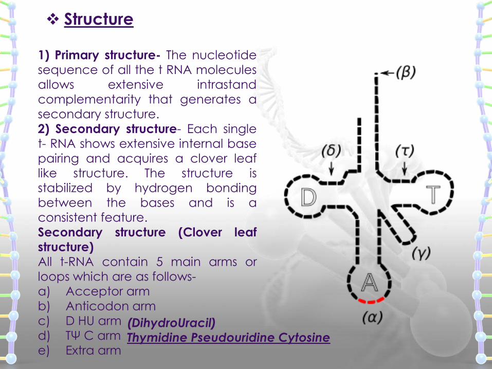
Structure
1) Primary structure- The nucleotide
sequence of all the t RNA molecules
allows extensive intrastand
complementarity that generates a
secondary structure.
2) Secondary structure- Each single
t- RNA shows extensive internal base
pairing and acquires a clover leaf
like structure. The structure is
stabilized by hydrogen bonding
between the bases and is a
consistent feature.
Secondary structure (Clover leaf
structure)
All t-RNA contain 5 main arms or
loops which are as follows-
a) Acceptor arm
b) Anticodon arm
c) D HU arm
d) TΨ C arm
e) Extra arm
(DihydroUracil)Thymidine Pseudouridine Cytosine
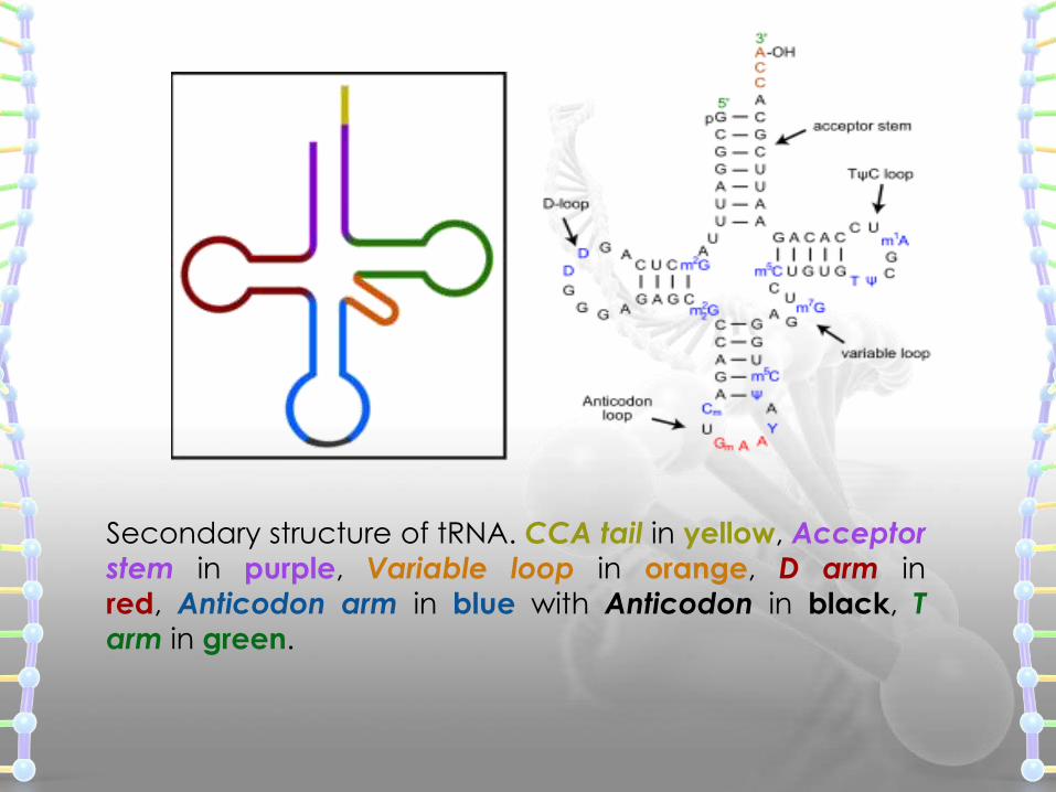
Secondary structure of tRNA. CCA tail in yellow, Acceptor
stem in purple, Variable loop in orange, D arm in
red, Anticodon arm in blue with Anticodon in black, T
arm in green.
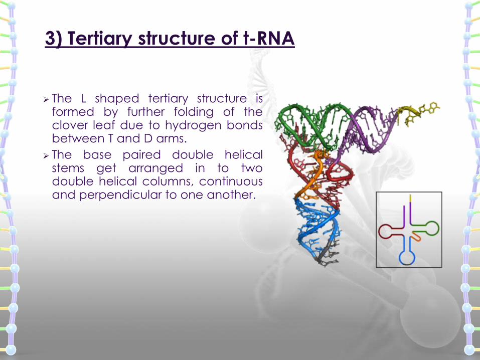
3) Tertiary structure of t-RNA
The L shaped tertiary structure isformed by further folding of theclover leaf due to hydrogen bondsbetween T and D arms.
The base paired double helicalstems get arranged in to twodouble helical columns, continuousand perpendicular to one another.
Ribosomal RNA (rRNA)
Ribosomal ribonucleic acid (rRNA) is the RNA component ofthe ribosome, and is essential for protein synthesis in all livingorganisms.
The functions of the ribosomal RNA molecules in the ribosomalparticle are not fully understood, but they are necessary forribosomal assembly and seem to play key roles in the binding ofmRNA to ribosomes and its translation
Recent studies suggest that an rRNA component performs thepeptidyl transferase activity and thus is an enzyme (a ribozyme).
It constitutes the predominant material within the ribosome, which isapproximately 60% rRNA and 40% protein by weight.
Ribosomes contain two major rRNAs and 50 or more proteins.
The ribosomal RNAs form two subunits, the large subunit (LSU) andsmall subunit (SSU). The LSU rRNA acts as a ribozyme,catalysing peptide bond formation.
Major types of small RNA molecules:
Small nuclear RNA (snRNA) - involved in mRNA splicing.
Small nucleolar RNA (snoRNA) - directs the modification ofribosomal RNAs.
Micro RNA (miRNA) and short interfering RNA (siRNA) -
regulate gene expression.
Small RNA molecules
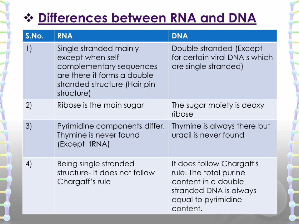
Differences between RNA and DNA
S.No. RNA DNA
1) Single stranded mainly
except when self
complementary sequences
are there it forms a double
stranded structure (Hair pin
structure)
Double stranded (Except
for certain viral DNA s which
are single stranded)
2) Ribose is the main sugar The sugar moiety is deoxy
ribose
3) Pyrimidine components differ.
Thymine is never found
(Except tRNA)
Thymine is always there but
uracil is never found
4) Being single stranded
structure- It does not follow
Chargaff’s rule
It does follow Chargaff's
rule. The total purine
content in a double
stranded DNA is always
equal to pyrimidine
content.
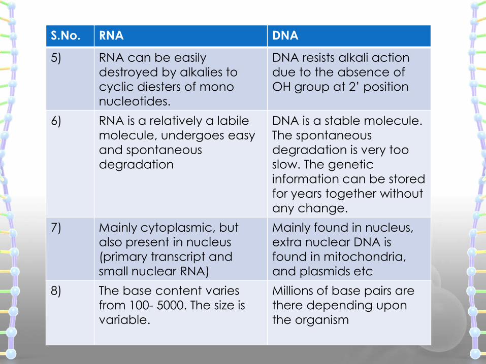
S.No. RNA DNA
5) RNA can be easily
destroyed by alkalies to
cyclic diesters of mono
nucleotides.
DNA resists alkali action
due to the absence of
OH group at 2’ position
6) RNA is a relatively a labile
molecule, undergoes easy
and spontaneous
degradation
DNA is a stable molecule.
The spontaneous
degradation is very too
slow. The genetic
information can be stored
for years together without
any change.
7) Mainly cytoplasmic, but
also present in nucleus
(primary transcript and
small nuclear RNA)
Mainly found in nucleus,
extra nuclear DNA is
found in mitochondria,
and plasmids etc
8) The base content varies
from 100- 5000. The size is
variable.
Millions of base pairs are
there depending upon
the organism
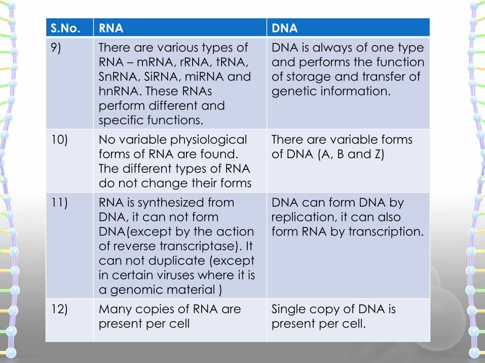
S.No. RNA DNA
9) There are various types of
RNA – mRNA, rRNA, tRNA,
SnRNA, SiRNA, miRNA and
hnRNA. These RNAs
perform different and
specific functions.
DNA is always of one type
and performs the function
of storage and transfer of
genetic information.
10) No variable physiological
forms of RNA are found.
The different types of RNA
do not change their forms
There are variable forms
of DNA (A, B and Z)
11) RNA is synthesized from
DNA, it can not form
DNA(except by the action
of reverse transcriptase). It
can not duplicate (except
in certain viruses where it is
a genomic material )
DNA can form DNA by
replication, it can also
form RNA by transcription.
12) Many copies of RNA are
present per cell
Single copy of DNA is
present per cell.