Embed Size (px)
Citation preview

Structure of the C-Terminal Domain of Lettuce Necrotic YellowsVirus Phosphoprotein
Nicolas Martinez,a,b,c,d Euripedes A. Ribeiro, Jr.,a,b,c Cédric Leyrat,e Nicolas Tarbouriech,a,b,c Rob W. H. Ruigrok,a,b,c Marc Jamina,b,c
Université Grenoble Alpes, UVHCI, Grenoble, Francea; CNRS, UVHCI, Grenoble, Franceb; Unit for Virus Host-Cell Interactions, Université Grenoble Alpes-EMBL-CNRS,Grenoble, Francec; Institut Laue Langevin, Grenoble, Franced; Division of Structural Biology, Wellcome Trust Centre for Human Genetics, University of Oxford, Oxford,United Kingdome
Lettuce necrotic yellows virus (LNYV) is a prototype of the plant-adapted cytorhabdoviruses. Through a meta-prediction of dis-order, we localized a folded C-terminal domain in the amino acid sequence of its phosphoprotein. This domain consists of anautonomous folding unit that is monomeric in solution. Its structure, solved by X-ray crystallography, reveals a lollipop-shapedstructure comprising five helices. The structure is different from that of the corresponding domains of other Rhabdoviridae, Fi-loviridae, and Paramyxovirinae; only the overall topology of the polypeptide chain seems to be conserved, suggesting that thisdomain evolved under weak selective pressure and varied in size by the acquisition or loss of functional modules.
Numerous viruses infecting plants are classified as rhabdovi-ruses most generally on the basis of electron microscopic ob-
servations of distinctive enveloped bacilliform or bullet-shapedparticles in the infected cells (1, 2). Only a few of them were con-firmed to be true rhabdoviruses by sequencing of their genomeand molecular studies (1, 2). The Rhabdoviridae belong to thenonsegmented negative-stranded RNA viruses (NNVs); togetherwith the Paramyxoviridae, the Filoviridae, and the Bornaviridae,they constitute the order Mononegavirales. Plant rhabdovirusesare classified in two genera on the basis of their site of virionmaturation; members of the genus Cytorhabdovirus assemble inthe cytoplasm, whereas members of the genus Nucleorhabdovirusassemble in the nucleus (3).
The lettuce necrotic yellows virus (LNYV), identified in 1963in Australia (4), is found only in Australia and New Zealand, but itis very similar to the lettuce yellow mottle virus that was recentlyisolated in France (5). Infection by LNYV causes fading of theleaves and necrosis of the heads, rendering the lettuce unsuitablefor consumption. LNYV is transmitted by an aphid of the genusHyperomyzus (4), and the main natural reservoir of both virus andvector is the common sowthistle (Sonchus oleraceus), which is in-fected without showing any symptoms. Disease outbreaks occurwhere the natural reservoir is densely populated and coincide withflight periods of the vector.
LNYV is the prototype member of the Cytorhabdovirus genus(3). In the late stages of LNYV replication, viral particles bud fromthe endoplasmic reticulum and accumulate in the cytoplasm (6,7). The genome of LNYV was completely sequenced, revealing anorganization conserved among other rhabdoviruses (8). It con-sists of six genes flanked by untranslated 3= leader and 5= trailersequences, and intergenic regions contain highly conserved con-sensus sequences. On the basis of sequence analysis and immuno-labeling, the genes were proposed to encode successively from the3= extremity to the 5= extremity the nucleoprotein (N), the phos-phoprotein (P), a plant-specific 4b protein assisting in cell-to-cellmovement of viral complexes through plasmodesmata and thevascular system, the matrix protein (M), the glycoprotein (G), andthe large subunit of the RNA-dependent RNA polymerase (L)(8–12).
In infected cells, the N and P proteins of LNYV interact with
each other, as in plant nucleorhabdoviruses and other rhabdovi-ruses (6), but little is known about the functions of P in LNYV orin other members of the Cytorhabdoviridae. However, the struc-ture and function of the P protein from rhabdoviruses have beenstudied, in particular, those of the P protein from vesicular stoma-titis virus (VSV), a Vesiculovirus, and from rabies virus (RAV), aLyssavirus (13). In NNVs, the phosphoprotein plays multiple rolesduring the replication process, some of which seem to be com-mon, while others are specific to a virus or a viral family. In allthese viruses, P forms a two-subunit RNA-dependent RNA poly-merase with the L protein, in which P acts as a noncatalytic cofac-tor, positioning the polymerase on its template (14). The genomicRNA of the rhabdoviruses, like that of all NNVs, is entirely coatedby the viral N, and it is the N-RNA complex rather than the nakedRNA that serves as a template for the polymerase (15). The repli-cation of the viral genome thus requires a continuous supply of N,and a second common function of P is to act as a chaperone of thenascent nucleoprotein, preventing the automatic assembly of N inthe absence of viral RNA and the encapsidation of cellular RNA(16, 17).
VSV P and RAV P form nonglobular dimers (18). Limitedproteolysis, disorder predictions, and biochemical studies of pro-tein fragments revealed that these proteins are multidomain pro-teins that consist of two structured domains and two disorderedregions (19). A central domain is responsible for the dimerizationof the protein, whereas a C-terminal domain binds to the N-RNAtemplate. The structures of both domains of VSV and RAV P, aswell as the C-terminal domain of Mokola virus (MOKV), weresolved recently (20–24). Despite a lack of sequence conservation,the structures of the three C-terminal domains are similar (24,25), suggesting that they are homologous. Conversely, the struc-tures of the central domains of VSV and RAV are so different that
Received 11 April 2013 Accepted 14 June 2013
Published ahead of print 19 June 2013
Address correspondence to Marc Jamin, [email protected].
Copyright © 2013, American Society for Microbiology. All Rights Reserved.
doi:10.1128/JVI.00999-13
September 2013 Volume 87 Number 17 Journal of Virology p. 9569–9578 jvi.asm.org 9569
Dow
nloa
ded
from
http
s://j
ourn
als.
asm
.org
/jour
nal/j
vi o
n 19
Jan
uary
202
2 by
167
.250
.166
.217
.

it is impossible to conclude that they have a common ancestry(22). The N-terminal region of the protein is globally disordered,while a flexible linker connects the central and C-terminal do-mains, making the protein highly flexible and structurally hetero-geneous. A recent integrative structural characterization of thephosphoprotein of VSV combining the atomic structures of thedomains with nuclear magnetic resonance spectroscopy andsmall-angle X-ray scattering data led to a representation of theprotein in the form of an equilibrium ensemble of interconvertingconformers rather than a unique structure (26). Disorder predic-tions and structural studies showed that a similar modular orga-nization is conserved among P proteins from the Paramyxoviridae(27, 28), Bornaviridae (29), and Filoviridae (30).
Since no structural information was available for any Cytorh-abdovirus, we used a meta-prediction of disorder to localize thedisordered and structured regions in the sequence of LNYV P. Thedisorder score (D score) provides a consensus from 16 differentweb servers that predict the localization of disordered regionsfrom the amino acid sequence of a protein (19, 24). Consequently,for a multidomain protein, this meta-prediction of disorder al-lows localization of the boundaries between ordered and disor-dered regions. On the basis of this analysis, we cloned, expressed,and purified the C-terminal domain of the LNYV P protein. Itscrystal structure revealed a fold that is different from the folds ofVSV and RAV and from the folds of paramyxo- and filoviruses butthat suggests common ancestry.
MATERIALS AND METHODSAmino acid sequence analysis, cloning, production, and purification ofthe C-terminal domain of LNYV P. The location of disordered regionswithin LNYV P was predicted by submitting its amino acid sequence to 16different algorithms accessible through web servers and by calculating aconsensus prediction, as described previously (19, 24). On the basis of thisprediction, a synthetic gene (GeneArt) optimized for expression in Esch-erichia coli and encoding residues 230 to 300 of LNYV P (71 residues) wascloned between the NcoI and BamHI restriction sites of a pET28 vector,such that the construct includes two additional N-terminal residues (Met-Ala) and a C-terminal His6 tag with a Leu-Glu linker. The constructionwas verified by DNA sequencing. The protein was expressed in E. coliBL21(DE3-RIL). Cells were grown in LB medium at 37°C until the opticaldensity at 600 nm reached a value of 0.6. Then, protein expression wasinduced by adding 1 mM IPTG (isopropyl-�-D-thiogalactopyranoside),and cells were grown for 3 h. Protein with a selenomethionine (SeMet)substitution was produced by growing the bacterial cells in minimal me-dium and by adding SeMet before induction of the heterologous proteinexpression. The cells were harvested and suspended in 20 mM Tris-HClbuffer at pH 7.5 containing 150 mM NaCl and EDTA-free protease inhib-itor cocktail (Roche) (buffer A). The cells were broken by sonication, andthe soluble fraction was loaded onto a Ni2� column equilibrated in bufferA. The column was washed with buffer A containing 20 mM imidazole,and the protein was then eluted with buffer A containing 300 mM imida-zole. The protein was further purified by gel filtration with a Superdex S75column (GE Healthcare) equilibrated in buffer A. The protein prepara-tions were checked by SDS-PAGE.
SEC combined with detection by MALLS and refractometry. Size-exclusion chromatography (SEC) was performed with a Superdex S75column (GE Healthcare) equilibrated in 20 mM Tris-HCl, pH 7.5, 150mM NaCl. Separations were performed at 20°C with a flow rate of 0.5 ml ·min�1. Fifty microliters of a protein solution at a concentration of 7 mg ·ml�1 was injected. Online multiangle laser light scattering (MALLS) de-tection was performed with a DAWN-EOS detector (Wyatt TechnologyCorp., Santa Barbara, CA) using a laser emitting at 690 nm. The proteinconcentration was measured online by the use of refractive index mea-
surements, an RI2000 detector (Schambeck SFD), and a refractive indexincrement dn/dc, the differential increment of refractive index per differ-ential increment of concentration, of 0.185 ml · g�1. Data were analyzed,and weight-average molecular weights (Mws) were calculated using thesoftware ASTRA V (Wyatt Technology Corp., Santa Barbara, CA) as de-scribed previously (18). For size determination, the column was calibratedwith proteins of known Stokes radius (RS) (31).
CD spectroscopy. Far-UV circular dichroism (CD) spectra were re-corded at 20°C on a Jasco model J-810 CD spectropolarimeter equippedwith a Peltier temperature controller. The LNYV P C-terminal domain(PCTD) was in 20 mM Tris-HCl, pH 7.5, containing 150 mM NaCl. Spec-tra were measured in a cuvette with a path length of 1 mm. The temper-ature-induced unfolding and refolding were recorded at 222 nm in a cu-vette with a path length of 10 mm by varying the temperature between 25and 60°C using a scan rate of 60°C/h.
Crystallization, data collection, and structure determination andrefinement. Crystallization conditions were screened by the hanging-drop vapor diffusion method using a PixSys4200 Cartesian robot (high-throughput crystallization laboratory at EMBL, Grenoble, France). Thescreen was performed by combining 0.1 �l of protein solution at 8 mg ·ml�1 in buffer A with 0.1 �l of Hampton crystal screen solutions.
The selenomethionine derivative of LNYV PCTD was crystallized in asolution of 20 mM sodium cacodylate buffer at pH 6.5 containing 200mM magnesium acetate and 15% 2-methyl-2,4-pentanediol (MPD).Crystals were frozen in the same solution at a final concentration of 30%MPD as the cryoprotectant.
Diffraction data were collected at Se-K edge on the ID14-4 beam line(European Synchrotron Radiation Facility [ESRF], Grenoble, France).The crystals diffracted to 2.0-Å resolution and belonged to space groupP4122. Data were integrated and scaled using the XDS program (82). Twoselenium sites and initial phases were found by the single-wavelengthanomalous diffraction (SAD) method using the ShelX-C-D-E pipeline(83), and the software Resolve (84) was used to automatically trace amodel in the resulting electron density. The model was improved by handand finally refined with the REFMAC5 program (85). The quality of themodel was checked with the PROCHECK program (32). Data collectionand refinement statistics are summarized in Table 1.
Structure analysis and comparisons. The CLUSTALW2 server wasused for sequence alignments (33). The structure homology program(SHP) was used for structural alignment and for constructing the struc-ture-based phylogenetic tree (34, 35). The electrostatic maps were calcu-lated using the Delphi program (36). The conservation score was calcu-lated with the AL2CO program (37). Figures of the structure wereprepared with the PyMOL program (38).
Protein structure accession number. Coordinates of the protein havebeen deposited in the Research Collaboratory for Structural Bioinformat-ics Protein Data Bank (PDB) with the accession number 3T4R.
RESULTSMeta-prediction of disorder. The structural characterization ofthe phosphoprotein from different NNV families revealed a mod-ular architecture made of folded domains concatenated with dis-ordered regions (27). Multiple-sequence alignments, however,showed no significant sequence conservation between the aminoacid sequence of LNYV P and the sequences of the P proteins fromother Rhabdoviridae or NNVs (39). A consensus from multipledisorder predictions was calculated in the form of a D score, usinga method previously described (19, 24). In these studies, plottingthe D score as a function of residue number allowed localization ofthe boundaries of the oligomerization domain (P central domain,PCED) and N-RNA binding C-terminal domain (PCTD) in the Pproteins from vesiculoviruses (VSV) and lyssaviruses (RAV) (19,24). The D score calculated for the amino acid sequence of LNVYP predicted the presence of two intrinsically disordered regions
Martinez et al.
9570 jvi.asm.org Journal of Virology
Dow
nloa
ded
from
http
s://j
ourn
als.
asm
.org
/jour
nal/j
vi o
n 19
Jan
uary
202
2 by
167
.250
.166
.217
.
(IDRs) with contiguous D-score values lower than 0.5 (N-termi-nal IDR, amino acids [aa] 1 to 63; C-terminal IDR, aa 184 to 229)and two folded regions with contiguous D-score values higherthan 0.5, a central bipartite domain (P protein central bipartitedomain, aa 64 to 183) and a C-terminal domain (PCTD, aa 230 to300) (Fig. 1). This modular organization, reminiscent of that pre-viously observed for other NNV viruses, suggested that the pre-dicted C-terminal domain of LNYV P corresponded to the N-RNA binding domain of rhabdovirus P proteins (19, 23, 24).
Expression and purification of LNYV PCTD. On the basis ofthe boundaries defined by the D-score analysis, a synthetic cDNA
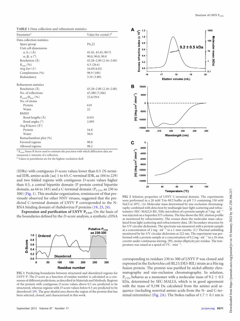
corresponding to residues 230 to 300 of LNYV P was cloned andexpressed in the Escherichia coli BL21(DE3-RIL) strain as a His tagfusion protein. The protein was purified by nickel-affinity chro-matography and size-exclusion chromatography. In solution,PCTD behaves as a monomer with a molecular mass of 9.2 � 0.5kDa, determined by SEC-MALLS, which is in good agreementwith the mass of 9,198 Da calculated from the amino acid se-quence (including nonviral amino acids from the N- and C-ter-minal extremities) (Fig. 2A). The Stokes radius of 1.7 � 0.1 nm is
TABLE 1 Data collection and refinement statistics
Parametera Value for crystal 1b
Data collection statisticsSpace group P4122Unit cell dimensions
a, b, c (Å) 43.45, 43.45, 89.71�, �, � (°) 90.0, 90.0, 90.0
Resolution (Å) 43.28–2.00 (2.16–2.00)Rsym (%) 4.3 (26.6)Avg I/�I 16.05(4.03)Completeness (%) 98.9 (100)Redundancy 3.76 (3.80)
Refinement statisticsResolution (Å) 43.28–2.00 (2.16–2.00)No. of reflections 47,380 (7,366)Rwork/Rfree (%) 22.6/29.0No. of atoms
Protein 610Water 22
RMSDBond lengths (Å) 0.031Bond angles (°) 2.095
Avg B factor (Å2)Protein 54.8Water 58.0
Ramachandran plot (%)Favored regions 90.8Allowed regions 98.2
a Rsym, linear R factor used to estimate the precision with which diffraction data aremeasured; I, intensity of a reflection.b Values in parentheses are for the highest-resolution shell.
FIG 1 Predicting boundaries between structured and disordered regions forLNYV P. The D score as a function of residue number is calculated as a con-sensus of different predictions, as described in Materials and Methods. Regionsof the protein with contiguous D-score values above 0.5 are predicted to bestructured, whereas regions with D-score values below 0.5 are predicted to bedisordered (19). The gray shaded area shows the region of the protein that hasbeen selected, cloned, and characterized in this work.
FIG 2 Solution properties of LNYV C-terminal domain. The experimentswere performed in a 20 mM Tris-HCl buffer at pH 7.5 containing 150 mMNaCl at 20°C. (A) Molecular mass determined by size-exclusion chromatog-raphy combined with detection by multiangle laser light scattering and refrac-tometry (SEC-MALLS-RI). Fifty microliters of a protein sample at 7 mg · ml�1
was injected on a Superdex S75 column. The line shows the SEC elution profileas monitored by refractometry. The crosses show the molecular mass calcu-lated from light scattering and refractometry data. (B) Secondary structure byfar-UV circular dichroism. The spectrum was measured with a protein sampleat a concentration of 2 mg · ml�1 in a 1-mm cuvette. (C) Thermal unfoldingmonitored by far-UV circular dichroism at 222 nm. The experiment was per-formed with a protein sample at a concentration of 0.2 mg · ml�1 in a 10-mmcuvette under continuous stirring. [�], molar ellipticity per residue. The tem-perature was raised at a speed of 1°C · min�1.
Structure of LNYV PCTD
September 2013 Volume 87 Number 17 jvi.asm.org 9571
Dow
nloa
ded
from
http
s://j
ourn
als.
asm
.org
/jour
nal/j
vi o
n 19
Jan
uary
202
2 by
167
.250
.166
.217
.

close to that expected for a globular protein of this molecular mass(1.6 nm) (31). The far-UV circular dichroism spectrum with twominima at 208 and 222 nm indicated an �-helix-rich protein (Fig.2B). The reversible thermal unfolding exhibited a sigmoid transi-tion typical of a well-folded protein with a hydrophobic core (Fig.2C). These results clearly demonstrated that LNYV PCTD consistsof an autonomous structural unit that adopts a globular fold witha large content of �-helical structure even in the absence of theremaining part of the protein.
Crystal structure of LNYV PCTD. A selenomethionine deriva-tive of the LNYV PCTD was crystallized at 20°C in 20 mM sodiumcacodylate buffer at pH 6.5 containing 200 mM magnesium ace-tate and 15% MPD. Diffraction data on frozen crystals were col-lected at the Se-K edge on beam line ID14-4 (ESRF, Grenoble,France). The crystal diffracted to a 2.0-Å resolution and belongedto space group P4122 with one monomer in the asymmetric unit.The phases were solved by the SAD method using the ShelX-C-D-E pipeline (40). A preliminary model including residues 230 to300 of LNYV and the first amino acid (Leu) of the C-terminal Histag was automatically generated in the electron density with thesoftware Resolve (41) and was refined with the REFMAC5 pro-gram (42). The quality of the model was evaluated with thePROCHEK program (32) (Table 1). The high B-factor values as-sociated with numerous residues in the structure and the absenceof visible density for eight of the nonviral residues that are part ofour construct likely contribute to the discrepancy found betweenthe quality of the data (resolution of 2 Å) and the quality of thederived model (Rwork � 22.6%; Rfree � 29.0%) (Table 1).
The C-terminal domain of LNYV P comprises five � helices,named �1 to �5 (Fig. 3A). The overall structure of PCTD is made ofa globular region formed by helices �1 to �4 and by the N-terminalpart of helix �5 and of a stalk formed by the C-terminal part ofhelix �5. The globular region has one flat face formed by helices �4
and �5 and one rounded face formed by the three remaining he-lices. Side chains from residues in helices �1 to �4 and residues inthe N-terminal moiety of helix �5 form a hydrophobic core. Ahydrophobic groove including a hole is localized on the proteinsurface between helices �1, �2, and �5 (Fig. 3B, hydrophobicgroove 1). It includes Trp244, Tyr252, Ile256, Phe259, Leu260,and Leu292. A second deep groove at the top of the domain is linedwith Leu238, Leu242, Leu266, and Leu273 (Fig. 3B, hydrophobicgroove 2). Analysis of the distribution of electrostatic potentials(Fig. 3C) showed the presence of two patches of negative chargeson opposite faces of the molecule. Asp263, Asp264, Glu284, andGlu287 form an elongated negative patch on the flat face (negativepatch 1), whereas Asp243, Glu245, Glu248, Glu276, and Glu279(negative patch 2) line along a ridge on the round face of PCTD.
Structural comparison with other PCTDs. The three-dimen-sional structure of the corresponding C-terminal domain of P hasbeen solved previously for three rhabdoviruses (20, 23, 24), threeparamyxoviruses (43–45), and the VP35 protein of a filovirus (46,47). In the absence of sequence conservation between LNYV P andthese other P proteins, we performed pairwise alignment of theLNYV PCTD structure with the PCTD structures from the otherviruses using the PDBeFold server (48) and the TM-align server(49). These programs use the optimization of empirical scores foraligning structures. These scores, the quality function score (Qscore) associated with the probability P score used by PDBeFoldand the template-modeling score (TM score) used by TM-align,account for both alignment length and the distance between
aligned residues and, therefore, are less sensitive to local structuralvariations than the root mean square deviation (RMSD) alone(Table 2). The value of these scores increases from 0 to 1 withincreasing structural similarity. Typically, a high Q-score valueassociated with a P value larger than 3 or a TM value larger than 0.5indicates that there is a similarity in the fold of the aligned mole-cules (48, 49). Both procedures yielded values below the limitsconsidered to indicate structural homology (Table 2). TheQ-score values were between 0.10 and 0.25, but the associated Pvalues were low, indicating a low significance. The TM-score val-ues approached the 0.5 limit but remained slightly lower. In con-clusion, these alignment procedures suggest that the structure ofthe LNYV PCTD domain is different from that of the PCTD domainsof other NNVs.
Search of PDB. Searches of PDB using the DALI server (50) orPDBeFold server (48) yielded only hits that did not meet the sig-nificance criteria set by default in these processes and failed torecognize the C-terminal domains of other NNV phosphopro-teins as relatives of the LNYV PCTD.
DISCUSSIONA common modular architecture for NNV phosphoproteins. AllNNVs share a similar organization of their genome, similar mech-anisms for transcription and replication in the host cell, and sim-ilar functions for the different viral proteins in the replicationprocess, arguing for a descent from a common ancestor. However,the high rate of mutations due to the absence of proofreading inRNA replication produces large divergences in protein lengthsand sequences. For some proteins, homology between the differ-ent NNV families remains detectable at the level of the amino acidsequence, allowing the construction of phylogenetic trees frommultiple-sequence alignments (3, 51). Structural comparison ofthe nucleoproteins and matrix proteins clearly reveals conserva-tion of protein folds (52, 53). For the phosphoprotein, however,the length of the protein varies greatly between viruses, and se-quence similarity is generally undetectable beyond the family leveland even sometimes beyond the genus level (3, 39, 54).
Predictions and experimental studies revealed that the phos-phoproteins from different NNV families share a modular archi-tecture with disordered regions alternating with folded domains(19, 27, 55, 56). Disordered regions and folded domains playingsimilar roles in the replication and transcription processes are po-sitioned in the same order along the polypeptide chain. The pro-tein can be grossly divided into two parts. The N-terminal part isglobally disordered and in some cases contains transiently popu-lated helices in regions involved in binding viral or cellular part-ners. In particular, this N-terminal region contains a binding sitefor the RNA-free nucleoprotein (N0) conserved in the amino acidsequence (39). For VSV, the D score predicted this region to bestructured (19), but it is globally disordered in isolation and formsa stable helix only upon binding to N0 (57, 58). The C-terminalpart contains a homo-oligomerization domain and the C-termi-nal N-RNA binding domain (PCTD) connected by a flexible linkerof variable length. The P protein of the Paramyxoviridae formstetramers (28, 59), whereas that of the Rhabdoviridae formsdimers (18, 21, 22). The D score calculated from the amino acidsequence of LNYV (Fig. 1) supports a modular organization sim-ilar to that of the other NNVs and predicts the presence of a longN-terminal disordered region (aa 1 to 63) and a central foldedregion (aa 64 to 184) separated from PCTD (aa 230 to 300) by a
Martinez et al.
9572 jvi.asm.org Journal of Virology
Dow
nloa
ded
from
http
s://j
ourn
als.
asm
.org
/jour
nal/j
vi o
n 19
Jan
uary
202
2 by
167
.250
.166
.217
.

disordered linker (aa 185 to 229). The predicted central domain ofLNYV may correspond to the homo-oligomerization domain.
Evolutionary relationships within NNV families. The scoresobtained for pairwise structural alignments of the PCTDs fromthe Rhabdoviridae, Filoviridae, and Paramyxovirinae are below thethreshold commonly accepted for validating homology. Even thestructural comparisons between LNYV PCTD and the correspond-ing domain from vesiculoviruses or lyssaviruses show no evidencefor structural conservation. Nevertheless, because different evi-dence supports a divergent evolution of these viral families (54,
60) and because in all NNV P proteins this domain is located at theC-terminal end and fulfills the same function of binding to thenucleocapsid, it is tempting to speculate that they diverged from acommon ancestor. As such, structural comparisons can provideclues about the mechanisms by which this domain has evolved.Indeed, the pairwise alignments of the LNYV PCTD structure withthe PCTD structures from the other viruses showed a remarkableconservation of the topology of the backbone and of secondarystructure elements (Fig. 4). Helices �1, �2, �3, �4, and �5 of LNYVPCTD are aligned with helices �1, 1 (310 helix), �2, �3, and �4 of
FIG 3 Crystal structure of LNYV PCTD. (A) Schematic representation of LNYV PCTD. Helices are numbered from �1 to �5. The numbers indicate the positionsof the N- and C-terminal residues. The figure was drawn with the PyMOL program. The positions of the � helices along the amino acid sequence are shown. Theunderlined residues are not visible in the crystal. The residues of LNYV are shown in black or red, and the nonviral residues added in the construct are coloredin gray. (B) Hydrophobic surface map. The distribution of hydrophobic residues on the surface is shown; yellow indicates hydrophobic sidechains. (C)Electrostatic surface potential. The surface potential calculated with the Delphi program is plotted on the solvent-accessible surface of the protein from red(negatively charged regions in electrostatic potential units, where kB is Boltzmann’s constant, T is the temperature, and e is the electron charge; �10 kBTe�1) toblue (positively charged regions; �10 kBTe�1).
Structure of LNYV PCTD
September 2013 Volume 87 Number 17 jvi.asm.org 9573
Dow
nloa
ded
from
http
s://j
ourn
als.
asm
.org
/jour
nal/j
vi o
n 19
Jan
uary
202
2 by
167
.250
.166
.217
.

RAV and MOKV PCTD (Fig. 4B), respectively, suggesting that theoverall topological organization of these secondary structure ele-ments is conserved among rhabdoviruses. Previously, we hadshown that the topology of PCTD is also conserved between RAVand VSV but that the latter lacks helices �4 and �6 of the RAVprotein (24). In accordance, helices �1, �2, �3, and �5 of LNYVPCTD are matched with �1, 1, �2, and �4 of VSV, respectively (Fig.4C). These alignments suggest that the PCTD domain of rhabdovi-ruses may have conserved the backbone topology of an ancientfold and may have evolved by acquiring or losing secondary struc-ture elements.
The C-terminal domain of paramyxovirus P proteins consistsof an antiparallel triple-helix bundle that seems to be well con-served within this family and is different from the structure ofrhabdovirus PCTD. Although it may be ambiguous to align a do-main comprising only three consecutive helices, the four helices ofLNYV PCTD can be aligned with the three helices of measles virus,mumps virus, and Sendai virus PCTD (Fig. 4D). Helices �2 and �5
of LNYV PCTD match helices �1 and �3 of the paramyxoviruses,respectively. The pair of short helices of LNYV, �3 and �4, thencorresponds to helix �2 of the paramyxoviruses, although onlyone of these helices may be conserved and the other may corre-spond to an additional structural element. These structural align-ments suggest some similarities in the general topology of thepolypeptide backbone and in the arrangement of secondary struc-ture elements, as already suggested (24, 25, 39), even if our struc-tural alignment of PCTD from the Rhabdoviridae and Paramyxo-viridae is different from that previously reported (25). Similarly,
the LNYV PCTD structure can also be aligned with the C-terminaldomain of the Ebola virus (EBOV) VP35 protein. This domain iscomposed of an �-helical subdomain and a �-sheet subdomain,and the five helices of LNYV can be aligned with the four helices ofthe helical subdomain of EBOV PCTD, again, with the short helices�3 and �4 of LNYV matching the longer �3 helix of EBOV(Fig. 4E).
To translate these structural similarities into evolutionary dis-tances and to build a phylogenetic tree based on the PCTD struc-tures, we used the structure homology program (34, 35). Accord-ing to this procedure, pairwise alignments were performed, andprobabilities of equivalence were calculated for every residue ofboth structures by comparing the positions of their C� atoms andthe local shape of the polypeptide chains around each residue. Aglobal parameter characteristic of the similarity between the twostructures was then computed as the sum of the probabilities forall residues that have a structural equivalent in both structures.This parameter was further converted into an evolutionary dis-tance using an empirical logarithmic function established withmetrics for sequence alignments, and these distances were thenused to build a phylogenetic tree. The structure-based phyloge-netic tree for the NNVs obtained from the comparison of theirPCTD domains (Fig. 5) is similar to the trees obtained on the basisof the amino acid sequence of the N or L proteins (3, 52). The treesuggests a monophyletic origin for the PCTD domain. It shows thatcytorhabdoviruses are as evolutionarily distant from other Rhab-doviridae of the genera Vesiculovirus and Lyssavirus as from theFiloviridae and Paramyxovirinae and suggests that the C-terminal
TABLE 2 Structural alignmentsa
Virus PDB accession no. Ntarget
PDBeFold TM-align
Q P RMSD (Å) Nalign TM RMSD (Å) Nalign
RAV 1VYI 111 0.18 0.00 3.24 55 0.43 3.31 50MOKV 2WZL 107 0.15 0.06 3.15 50 0.47 3.32 58VSV 2K47 73 0.13 0.03 3.37 39 0.36 3.26 72MEV 10KS 53 0.25 0.00 3.41 47 0.41 3.53 47SEV 1R4G 53 0.20 0.02 3.42 42 0.40 3.01 45MUV 3BBZ 48 0.19 0.00 3.94 42 0.40 2.98 48EBOV 3FKE 123 0.10 0.00 3.26 44 0.34 3.84 43WASP 1EJ5 107 0.18 0.14 3.73 59 0.47 3.24 61a LNYV PCTD, used as the query, contains 71 residues. Ntarget, number of residues in the target structure; Nalign, number of residues aligned in the best three-dimensionalsuperposition. RMSD is calculated between C� atoms of matched residues in the best three-dimensional superposition according to the following equation:
RMSD �� 1
Nres�i�1
Nres
di
where di is the distance between N pairs of equivalent C� atoms, i is the residue number, and Nres is the number of residues. The Q score evaluates the quality of the C� atomalignment and takes both RMSD and alignment length into account according to the following equation:
Nalign1 Nalign2
�1 � (RMSD ⁄ R0)2�Nres1 Nres2
where Nalign1 and Nalign2 are the number of aligned residues in the query and target structures, respectively; Nres1 and Nres2 are the number of residues in each protein; and R0 is anempirical parameter set equal to 3 Å. Q takes values between 0 and 1, and identical structures have a score of 1. The P-score parameter represents the minus logarithm of the Pvalue, where the P value measures the probability of achieving a match of the same or better quality as that obtained by chance. This P score was calibrated using a set of 700structures, and P scores less than 3 indicate statistically insignificant matches. The TM score evaluates the quality of the C� alignment, which takes both RMSD and alignmentlength into account according to the following equation:
TM � max�1 ⁄ Nres�i
N
1 ⁄ [1 � (di ⁄ d0)2]�where max is maximum and d0 is an empirical parameter depending on the length of the protein:
d0 � 1.24�3
Nres � 15 � 18.
TM takes values between 0 and 1. TM values of 0.17 correspond to random similarities, whereas TM values of 0.5 indicate an equivalent fold. MEV, measles virus; SEV, Sendaivirus; MUV, mumps virus; WASP, Wiskott–Aldrich syndrome protein.
Martinez et al.
9574 jvi.asm.org Journal of Virology
Dow
nloa
ded
from
http
s://j
ourn
als.
asm
.org
/jour
nal/j
vi o
n 19
Jan
uary
202
2 by
167
.250
.166
.217
.

domain of P has evolved after the NNVs diverged into the differ-ent families. Most plant rhabdoviruses are dependent for theirtransmission on insects, in which they also multiply, and arethereby influenced in their evolution by replication in their vec-
tors (61). In this context, it is notable that, within the Rhabdoviri-dae, LNYV is slightly more closely related to VSV, which is also anarbovirus, than to the lyssaviruses (RAV and MOKV).
In the hypothesis that P from the different NNV families
FIG 4 Comparison of the LNYV PCTD structure with the PCTD structures of other NNVs. (A) Topology of LNYV PCTD. (B to E) Pairwise structural alignments.The structure of LNYV was aligned pairwise with the structure of the C-terminal domains of different rhabdoviruses (RAV, VSV, MOKV), paramyxoviruses(measles virus, mumps virus, Sendai virus), and filoviruses (EBOV) (Table 2). Representative superpositions for RAV (B), VSV (C), measles virus (MEV) (D),and EBOV (E) are shown. In each panel, the structure of LNYV PCTD is shown in red and that of the aligned domain is shown in blue. The topology of thecompared protein domain is shown by the side.
Structure of LNYV PCTD
September 2013 Volume 87 Number 17 jvi.asm.org 9575
Dow
nloa
ded
from
http
s://j
ourn
als.
asm
.org
/jour
nal/j
vi o
n 19
Jan
uary
202
2 by
167
.250
.166
.217
.

evolved from a common ancestor, we can ask the question of whyits C-terminal domain varies so much in sequence and length. TheC-terminal domain plays multiple roles in the different viruses,but its common function is to mediate the binding of P to thenucleocapsid (62, 63), an interaction that seems to be essential forthe processivity of the viral polymerase (64, 65). The large varia-tion in PCTD sequences can be explained by weak selective pressureacting on the sequence, as the only functional requirement is theconservation of an interaction with the nucleocapsid that is suffi-ciently strong for attaching and positioning the polymerase ontoits template. Such selective pressure is much less stringent than itis, for example, in the case of an enzyme active site, and therefore,the PCTD sequence is likely to tolerate extensive sequence varia-tions. In addition, the topological complexity of the polypeptidechain is low, as judged by values of the contact order parameter ofless than 0.2 (66), and therefore also suggests low selective pres-sure on the amino acid sequence from folding. It is worth pointingout that this variability extends even further to the mechanism ofcomplex formation between P and the N-RNA template. PCTD ofthe rhabdoviruses is a well-folded domain that binds to the top ofthe C-terminal domain of one N protomer, with the flexible NC-terminal loop of the same protomer and of the adjacent onepinching PCTD on two sides (62, 63). In the Filoviridae, the sub-domain of PCTD that is directly involved in the interaction with thenucleocapsid has been localized, but no structural informationabout the formation of the complex is available. In the subfamilyParamyxovirinae, PCTD binds to the C-terminal disordered tail ofN that protrudes from the nucleocapsid (67), inducing the forma-
tion of a helix in this disordered region of N upon formation of thecomplex (68–70). In the subfamily Pneumovirinae, the region cor-responding to PCTD is a short, disordered segment that binds tothe surface of the N-terminal domain of N (71, 72). In the lattercase, it is even more difficult to detect structural homology. Onemay speculate that, for the Pneumovirinae, the C-terminal regionof P has been extensively reduced to a minimal segment whichremains capable of binding to N, though at a different site on thesurface of N.
In addition, the structural comparison of PCTD from the differ-ent NNV families suggests that the variation in the length of thisdomain results not only from mutations but also from the exten-sion or reduction of secondary structure elements and potentiallyalso from the acquisition or loss of some secondary structure ele-ments. In the Paramyxoviridae, the only known function of PCTD
is to attach P to the nucleocapsid, whereas in the Filoviridae andRhabdoviridae, PCTD is involved in other functions. We can there-fore speculate that the larger size of the C-terminal domain iscorrelated with its implication in other functions. In EBOV PCTD
(VP35), two entities performing different functions are clearlydistinguishable in the structure (46): a four-helix subdomain thatcontains the structurally homologous helices of the Paramyxoviri-nae and interacts with the nucleocapsid (73) and a C-terminalsubdomain made of a � sheet, an � helix, and a type II polyprolinehelix involved in double-stranded RNA binding and in evadingthe host immune system (46, 47). In animal rhabdoviruses (VSVand RAV), the functional regions are not as clearly separated fromeach other (23, 24). VSV PCTD interacts with L (74), is phosphor-ylated by a cellular kinase (75), and regulates viral transcriptionand replication (76). RAV PCTD is larger than VSV PCTD and isinvolved in supplementary activities. In addition to its role in thefixation of P to the nucleocapsid and in the regulation of viraltranscription and replication through phosphorylation by cellularkinases (77, 78), RAV PCTD is also involved in the evasion of thehost immune system through interactions with different cellularfactors (79–81) and in the regulation of the nucleocytoplasmictransport of P (77). In conclusion, the P protein may be less con-served than other NNV proteins because it has served as a plat-form for acquiring new functions through the recruitment of ad-ditional structural modules and thereby for allowing viruses toadapt to their different host cell environments.
ACKNOWLEDGMENTS
This work was supported by grants from the French ANR (ANR-07-001-01 [ANRAGE], ANR-12-BSV8-0025-01). E.A.R. was supported by apostdoctoral fellowship from the ANR. N.M. was supported by a fellow-ship from the Région Rhone-Alpes.
We thank the Partnership for Structural Biology for the excellentstructural biology environment.
REFERENCES1. Jackson AO, Dietzgen RG, Goodin MM, Bragg JN, Deng M. 2005.
Biology of plant rhabdoviruses. Annu. Rev. Phytopathol. 43:623– 660.2. Kormelink R, Garcia ML, Goodin M, Sasaya T, Haenni AL. 2011.
Negative-strand RNA viruses: the plant-infecting counterparts. Virus Res.162:184 –202.
3. Dietzgen RG, Callisher CH, Kurath G, Kuzmin IV, Rodriguez LL, StoneDM, Tesh RB, Tordo N, Walker PJ. 2011. Family Rhabdoviridae. In KingAM, Adams MJ, Carstens EB, Lefkowitz EJ (ed), Virus taxonomy, VIIIthReport of the ICTV. Elsevier Academic Press, Oxford, United Kingdom.
4. Stubbs LL, Grogan RG. 1963. Lettuce necrotic yellows virus. Nature197:1229.
FIG 5 Structure-based phylogenetic tree showing the three NNV families.Using the structure homology program for superposition, the tree shows thePCTD structure of the following viruses: vesicular stomatitis virus (VSV; PDBaccession no. 2K47), rabies virus (RAV; PDB accession no. 1VYI), Mokolavirus (MOKV; PDB accession no. 2WZL), lettuce necrotic yellows virus(LNYV; PDB accession no. 3T4R), Ebola virus (EBOV; PDB accession no.3FKE), measles virus (MEV; PDB accession no. 1OKS), mumps virus (MUV;PDB accession no. 3BBZ), and Sendai virus (SEV; PDB accession no. 1R4G).The length of the branches is scaled to structural differences, such that thedistance between two species is the sum of the length of all branches connect-ing them. The numbers relative to the distance derived from this analysis areindicated above each branch.
Martinez et al.
9576 jvi.asm.org Journal of Virology
Dow
nloa
ded
from
http
s://j
ourn
als.
asm
.org
/jour
nal/j
vi o
n 19
Jan
uary
202
2 by
167
.250
.166
.217
.

5. Heim F, Lot H, Delecolle B, Bassler A, Krczal G, Wetzel T. 2008.Complete nucleotide sequence of a putative new cytorhabdovirus infect-ing lettuce. Arch. Virol. 153:81–92.
6. Martin KM, Dietzgen RG, Wang R, Goodin MM. 2012. Lettuce necroticyellows cytorhabdovirus protein localization and interaction map, andcomparison with nucleorhabdoviruses. J. Gen. Virol. 93:906 –914.
7. Wolanski BS, Chambers TC. 1971. The multiplication of lettuce necroticyellows virus. Virology 44:582–591.
8. Dietzgen RG, Callaghan B, Wetzel T, Dale JL. 2006. Completion of thegenome sequence of lettuce necrotic yellows virus, type species of thegenus Cytorhabdovirus. Virus Res. 118:16 –22.
9. Callaghan B, Dietzgen RG. 2005. Nucleocapsid gene variability revealstwo subgroups of lettuce necrotic yellows virus. Arch. Virol. 150:1661–1667.
10. Dietzgen RG, Francki RI. 1988. Analysis of lettuce necrotic yellows virusstructural proteins with monoclonal antibodies and concanavalin A. Vi-rology 166:486 – 494.
11. Wetzel T, Dietzgen RG, Dale JL. 1994. Genomic organization of lettucenecrotic yellows rhabdovirus. Virology 200:401– 412.
12. Wetzel T, Dietzgen RG, Geering AD, Dale JL. 1994. Analysis of thenucleocapsid gene of lettuce necrotic yellows rhabdovirus. Virology 202:1054 –1057.
13. Ivanov I, Yabukarski F, Ruigrok RW, Jamin M. 2011. Structural insightsinto the rhabdovirus transcription/replication complex. Virus Res. 162:126 –137.
14. Emerson SU, Yu Y. 1975. Both NS and L proteins are required for in vitroRNA synthesis by vesicular stomatitis virus. J. Virol. 15:1348 –1356.
15. Arnheiter H, Davis NL, Wertz G, Schubert M, Lazzarini RA. 1985. Roleof the nucleocapsid protein in regulating vesicular stomatitis virus RNAsynthesis. Cell 41:259 –267.
16. Masters PS, Banerjee AK. 1988. Complex formation with vesicular sto-matitis virus phosphoprotein NS prevents binding of nucleocapsid pro-tein N to nonspecific RNA. J. Virol. 62:2658 –2664.
17. Peluso RW, Moyer SA. 1988. Viral proteins required for the in vitroreplication of vesicular stomatitis virus defective interfering particle ge-nome RNA. Virology 162:369 –376.
18. Gérard FCA, Ribeiro E, Albertini A, Zaccai G, Ebel C, Ruigrok R, JaminM. 2007. Unphosphorylated Rhabdoviridae phosphoproteins form elon-gated dimers in solution. Biochemistry 46:10328 –10338.
19. Gérard FCA, Ribeiro EA, Leyrat C, Ivanov I, Blondel D, Longhi S,Ruigrok RWH, Jamin M. 2009. Modular organization of rabies virusphosphoprotein. J. Mol. Biol. 388:978 –996.
20. Assenberg R, Delmas O, Ren J, Vidalain PO, Verma A, Larrous F,Graham SC, Tangy F, Grimes JM, Bourhy H. 2010. The structure of thenucleoprotein binding domain of the Mokola virus phosphoprotein. J.Virol. 84:1089 –1096.
21. Ding H, Green TJ, Lu S, Luo M. 2006. Crystal structure of the oligomer-ization domain of the phosphoprotein of vesicular stomatitis virus. J. Vi-rol. 80:2808 –2814.
22. Ivanov I, Crepin T, Jamin M, Ruigrok R. 2010. Structure of thedimerization domain of the rabies virus phosphoprotein. J. Virol. 84:3707–3710.
23. Mavrakis M, McCarthy AA, Roche S, Blondel D, Ruigrok RW. 2004.Structure and function of the C-terminal domain of the polymerase co-factor of rabies virus. J. Mol. Biol. 343:819 – 831.
24. Ribeiro EA, Jr, Favier A, Gerard FC, Leyrat C, Brutscher B, Blondel D,Ruigrok RW, Blackledge M, Jamin M. 2008. Solution structure of theC-terminal nucleoprotein-RNA binding domain of the vesicular stomati-tis virus phosphoprotein. J. Mol. Biol. 382:525–538.
25. Delmas O, Assenberg R, Grimes JM, Bourhy H. 2010. The structure ofthe nucleoprotein binding domain of lyssavirus phosphoprotein reveals astructural relationship between the N-RNA binding domains of Rhabdo-viridae and Paramyxoviridae. RNA Biol. 7:322–327.
26. Leyrat C, Schneider R, Ribeiro EA, Jr, Yabukarski F, Yao M, Gerard FC,Jensen MR, Ruigrok RW, Blackledge M, Jamin M. 2012. Ensemblestructure of the modular and flexible full-length vesicular stomatitis virusphosphoprotein. J. Mol. Biol. 423:182–197.
27. Karlin D, Ferron F, Canard B, Longhi S. 2003. Structural disorder andmodular organization in Paramyxovirinae N and P. J. Gen. Virol. 84:3239 –3252.
28. Llorente MT, Garcia-Barreno B, Calero M, Camafeita E, Lopez JA,Longhi S, Ferron F, Varela PF, Melero JA. 2006. Structural analysis of thehuman respiratory syncytial virus phosphoprotein: characterization of an
alpha-helical domain involved in oligomerization. J. Gen. Virol. 87:159 –169.
29. Hock M, Kraus I, Schoehn G, Jamin M, Andrei-Selmer C, Garten W,Weissenhorn W. 2010. RNA induced polymerization of the Borna diseasevirus nucleoprotein. Virology 397:64 –72.
30. Leung DW, Prins KC, Basler CF, Amarasinghe GK. 2010. EbolavirusVP35 is a multifunctional virulence factor. Virulence 1:526 –531.
31. Uversky VN. 1993. Use of fast protein size-exclusion liquid chromatog-raphy to study the unfolding of proteins which denature through the mol-ten globule. Biochemistry 32:13288 –13298.
32. Laskowski RA, Macarthur MW, Moss DS, Thornton JM. 1993.PROCHECK: a program to check the stereochemical quality of proteinstructures. J. Appl. Crystallogr. 26:283–291.
33. Larkin MA, Blackshields G, Brown NP, Chenna R, McGettigan PA,McWilliam H, Valentin F, Wallace IM, Wilm A, Lopez R, ThompsonJD, Gibson TJ, Higgins DG. 2007. Clustal W and Clustal X version 2.0.Bioinformatics 23:2947–2948.
34. Abrescia NG, Bamford DH, Grimes JM, Stuart DI. 2012. Structureunifies the viral universe. Annu. Rev. Biochem. 81:795– 822.
35. Stuart DI, Levine M, Muirhead H, Stammers DK. 1979. Crystal struc-ture of cat muscle pyruvate kinase at a resolution of 2.6 Å. J. Mol. Biol.134:109 –142.
36. Rocchia W, Sridharan S, Nicholls A, Alexov E, Chiabrera A, Honig B.2002. Rapid grid-based construction of the molecular surface and the useof induced surface charge to calculate reaction field energies: applicationsto the molecular systems and geometric objects. J. Comput. Chem. 23:128 –137.
37. Pei J, Grishin NV. 2001. AL2CO: calculation of positional conservation ina protein sequence alignment. Bioinformatics 17:700 –712.
38. DeLano WL. 2002. The PyMOL molecular graphics system. DeLano Sci-entific, Palo Alto, CA.
39. Karlin D, Belshaw R. 2012. Detecting remote sequence homology indisordered proteins: discovery of conserved motifs in the N-termini ofMononegavirales phosphoproteins. PLoS One 7:e31719. doi:10.1371/journal.pone.0031719.
40. Hubschle CB, Sheldrick GM, Dittrich B. 2011. ShelXle: a Qt graphicaluser interface for SHELXL. J. Appl. Crystallogr. 44:1281–1284.
41. Terwilliger TC. 2003. Automated main-chain model building by templatematching and iterative fragment extension. Acta Crystallogr. D Biol. Crys-tallogr. 59:38 – 44.
42. Murshudov GN, Skubak P, Lebedev AA, Pannu NS, Steiner RA, Nich-olls RA, Winn MD, Long F, Vagin AA. 2011. REFMAC5 for the refine-ment of macromolecular crystal structures. Acta Crystallogr. D Biol. Crys-tallogr. 67:355–367.
43. Blanchard L, Tarbouriech N, Blackledge M, Timmins P, BurmeisterWP, Ruigrok RW, Marion D. 2004. Structure and dynamics of the nu-cleocapsid-binding domain of the Sendai virus phosphoprotein in solu-tion. Virology 319:201–211.
44. Johansson K, Bourhis JM, Campanacci V, Cambillau C, Canard B,Longhi S. 2003. Crystal structure of the measles virus phosphoproteindomain responsible for the induced folding of the C-terminal domain ofthe nucleoprotein. J. Biol. Chem. 278:44567– 44573.
45. Kingston RL, Gay LS, Baase WS, Matthews BW. 2008. Structure of thenucleocapsid-binding domain from the mumps virus polymerase; an ex-ample of protein folding induced by crystallization. J. Mol. Biol. 379:719 –731.
46. Leung DW, Ginder ND, Fulton DB, Nix J, Basler CF, Honzatko RB,Amarasinghe GK. 2009. Structure of the Ebola VP35 interferon inhibitorydomain. Proc. Natl. Acad. Sci. U. S. A. 106:411– 416.
47. Leung DW, Prins KC, Borek DM, Farahbakhsh M, Tufariello JM,Ramanan P, Nix JC, Helgeson LA, Otwinowski Z, Honzatko RB, BaslerCF, Amarasinghe GK. 2010. Structural basis for dsRNA recognition andinterferon antagonism by Ebola VP35. Nat. Struct. Mol. Biol. 17:165–172.
48. Krissinel E, Henrick K. 2004. Secondary-structure matching (SSM), anew tool for fast protein structure alignment in three dimensions. ActaCrystallogr. D Biol. Crystallogr. 60:2256 –2268.
49. Zhang Y, Skolnick J. 2005. TM-align: a protein structure alignment al-gorithm based on the TM-score. Nucleic Acids Res. 33:2302–2309.
50. Roostaee A, Barbar E, Lavigne P, LeHoux JG. 2009. The mechanism ofspecific binding of free cholesterol by the steroidogenic acute regulatoryprotein: evidence for a role of the C-terminal alpha-helix in the gating ofthe binding site. Biosci. Rep. 29:89 –101.
51. Poch O, Sauvaget I, Delarue M, Tordo N. 1989. Identification of four
Structure of LNYV PCTD
September 2013 Volume 87 Number 17 jvi.asm.org 9577
Dow
nloa
ded
from
http
s://j
ourn
als.
asm
.org
/jour
nal/j
vi o
n 19
Jan
uary
202
2 by
167
.250
.166
.217
.

conserved motifs among the RNA-dependent polymerase encoding ele-ments. EMBO J. 8:3867–3874.
52. Assenberg R, Delmas O, Morin B, Graham SC, De Lamballerie X,Laubert C, Coutard B, Grimes JM, Neyts J, Owens RJ, Brandt BW,Gorbalenya A, Tucker P, Stuart DI, Canard B, Bourhy H. 2010. Genom-ics and structure/function studies of Rhabdoviridae proteins involved inreplication and transcription. Antiviral Res. 87:149 –161.
53. Ruigrok RW, Crepin T, Kolakofsky D. 2011. Nucleoproteins and nu-cleocapsids of negative-strand RNA viruses. Curr. Opin. Microbiol. 14:504 –510.
54. Lamb RA. 2007. Mononegavirales, p 1357–1361. In Knipe DM, HowleyPM, Griffin DE, Lamb RA, Martin MA, Roizman B, Straus SE (ed), Fieldsvirology, 5th ed, vol 1. Lippincott Williams & Wilkins, Philadelphia, PA.
55. Castagne N, Barbier A, Bernard J, Rezaei H, Huet JC, Henry C, DaCosta B, Eleouet JF. 2004. Biochemical characterization of the respiratorysyncytial virus P-P and P-N protein complexes and localization of the Pprotein oligomerization domain. J. Gen. Virol. 85:1643–1653.
56. Reid SP, Cardenas WB, Basler CF. 2005. Homo-oligomerization facili-tates the interferon-antagonist activity of the ebolavirus VP35 protein.Virology 341:179 –189.
57. Leyrat C, Jensen MR, Ribeiro EA, Gérard F, Ruigrok R, Blackledge M,Jamin M. 2011. The N0-binding region of the vesicular stomatitis virusphosphoprotein is globally disordered but contains transient �-helices.Protein Sci. 20:542–556.
58. Leyrat C, Yabukarski F, Tarbouriech N, Ribeiro EA, Jr, Jensen MR,Blackledge M, Ruigrok RW, Jamin M. 2011. Structure of the vesicularstomatitis virus N-P complex. PLoS Pathog. 7:e1002248. doi:10.1371/journal.ppat.1002248.
59. Tarbouriech N, Curran J, Ruigrok RW, Burmeister WP. 2000. Tetra-meric coiled coil domain of Sendai virus phosphoprotein. Nat. Struct.Biol. 7:777–781.
60. Pringle CR. 1997. The order Mononegavirales— current status. Arch.Virol. 142:2321–2326.
61. Hogenhout SA, Redinbaugh MG, Ammar el-D. 2003. Plant and animalrhabdovirus host range: a bug’s view. Trends Microbiol. 11:264 –271.
62. Green TJ, Luo M. 2009. Structure of the vesicular stomatitis virus nucleo-capsid in complex with the nucleocapsid-binding domain of the smallpolymerase cofactor, P. Proc. Natl. Acad. Sci. U. S. A. 106:11721–11726.
63. Ribeiro EA, Leyrat C, Gérard FC, Albertini AA, Falk C, Ruigrok RW,Jamin M. 2009. Binding of rabies virus polymerase cofactor to recombi-nant circular nucleoprotein-RNA complexes. J. Mol. Biol. 394:558 –575.
64. Morin B, Rahmeh AA, Whelan SP. 2012. Mechanism of RNA synthesisinitiation by the vesicular stomatitis virus polymerase. EMBO J. 31:1320 –1329.
65. Stillman EA, Whitt M. 1999. Transcript initiation and 5=-end modifica-tions are separable events during vesicular stomatitis virus transcription. J.Virol. 73:7199 –7209.
66. Plaxco KW, Simons KT, Baker D. 1998. Contact order, transition stateplacement and the refolding rates of single domain proteins. J. Mol. Biol.277:985–994.
67. Jensen MR, Communie G, Ribeiro EA, Jr, Martinez N, Desfosses A,Salmon L, Mollica L, Gabel F, Jamin M, Longhi S, Ruigrok RW,Blackledge M. 2011. Intrinsic disorder in measles virus nucleocapsids.Proc. Natl. Acad. Sci. U. S. A. 108:9839 –9844.
68. Gely S, Lowry DF, Bernard C, Jensen MR, Blackledge M, Costanzo S,Bourhis JM, Darbon H, Daughdrill G, Longhi S. 2010. Solution struc-ture of the C-terminal X domain of the measles virus phosphoprotein andinteraction with the intrinsically disordered C-terminal domain of thenucleoprotein. J. Mol. Recognit. 23:435– 447.
69. Jensen MR, Bernado P, Houben K, Blanchard L, Marion D, Ruigrok
RW, Blackledge M. 2010. Structural disorder within Sendai virus nucleo-protein and phosphoprotein: insight into the structural basis of molecularrecognition. Protein Pept. Lett. 17:952–960.
70. Kingston RL, Hamel DJ, Gay LS, Dahlquist FW, Matthews BW. 2004.Structural basis for the attachment of a paramyxoviral polymerase to itstemplate. Proc. Natl. Acad. Sci. U. S. A. 101:8301– 8306.
71. Galloux M, Tarus B, Blazevic I, Fix J, Duquerroy S, Eleouet JF. 2012.Characterization of a viral phosphoprotein binding site on the surface ofthe respiratory syncytial nucleoprotein. J. Virol. 86:8375– 8387.
72. Tran TL, Castagne N, Bhella D, Varela PF, Bernard J, Chilmonczyk S,Berkenkamp S, Benhamo V, Grznarova K, Grosclaude J, Nespoulos C,Rey FA, Eleouet JF. 2007. The nine C-terminal amino acids of the respi-ratory syncytial virus protein P are necessary and sufficient for binding toribonucleoprotein complexes in which six ribonucleotides are contactedper N protein protomer. J. Gen. Virol. 88:196 –206.
73. Prins KC, Binning JM, Shabman RS, Leung DW, Amarasinghe GK,Basler CF. 2010. Basic residues within the ebolavirus VP35 protein arerequired for its viral polymerase cofactor function. J. Virol. 84:10581–10591.
74. Takacs AM, Das T, Banerjee AK. 1993. Mapping of interacting domainsbetween the nucleocapsid protein and the phosphoprotein of vesicularstomatitis virus by using a two-hybrid system. Proc. Natl. Acad. Sci.U. S. A. 90:10375–10379.
75. Barik S, Banerjee AK. 1992. Sequential phosphorylation of the phospho-protein of vesicular stomatitis virus by cellular and viral protein kinases isessential for transcription activation. J. Virol. 66:1109 –1118.
76. Das T, Pattnaik AK, Takacs AM, Li T, Hwang LN, Banerjee AK. 1997.Basic amino acid residues at the carboxy-terminal eleven amino acid re-gion of the phosphoprotein (P) are required for transcription but not forreplication of vesicular stomatitis virus genome RNA. Virology 238:103–114.
77. Moseley GW, Filmer RP, DeJesus MA, Jans DA. 2007. Nucleocytoplas-mic distribution of rabies virus P-protein is regulated by phosphorylationadjacent to C-terminal nuclear import and export signals. Biochemistry46:12053–12061.
78. Pasdeloup D, Poisson N, Raux H, Gaudin Y, Ruigrok RW, Blondel D.2005. Nucleocytoplasmic shuttling of the rabies virus P protein requires anuclear localization signal and a CRM1-dependent nuclear export signal.Virology 334:284 –293.
79. Blondel D, Regad T, Poisson N, Pavie B, Harper F, Pandolfi PP, De TheH, Chelbi-Alix MK. 2002. Rabies virus P and small P products interactdirectly with PML and reorganize PML nuclear bodies. Oncogene 21:7957–7970.
80. Brzozka K, Finke S, Conzelmann KK. 2006. Inhibition of interferonsignaling by rabies virus phosphoprotein P: activation-dependent bindingof STAT1 and STAT2. J. Virol. 80:2675–2683.
81. Vidy A, Chelbi-Alix M, Blondel D. 2005. Rabies virus P protein interactswith STAT1 and inhibits interferon signal transduction pathways. J. Virol.79:14411–14420.
82. Kabsch W. 2010. XDS. Acta Crystallogr. D Biol. Crystallogr. 66:125–132.83. Heldrick GM. 2010. Experimental phasing with SHELXC/D/E: combin-
ing chain tracing with density modification. Acta Crystallogr. D Biol.Crystallogr. 66:479 – 485.
84. Terwilliger TC. 2003. Automated main-chain model building by templatematching and iterative fragment extension. Acta Crystallogr. D Biol. Crys-tallogr. 59:38 – 44.
85. Murshudov GN, Skubak P, Lebedev AA, Pannu NS, Steiner RA, Nich-olls RA, Winn MD, Long F, Vagin AA. 2011. REFMAC5 for the refine-ment of macromolecular crystal structures. Acta Crystallogr. D Biol. Crys-tallogr. 67:355–367.
Martinez et al.
9578 jvi.asm.org Journal of Virology
Dow
nloa
ded
from
http
s://j
ourn
als.
asm
.org
/jour
nal/j
vi o
n 19
Jan
uary
202
2 by
167
.250
.166
.217
.