Embed Size (px)
Citation preview

Structure of the MUR1 GDP-Mannose 4,6-Dehydratase fromArabidopsis thaliana:Implications for Ligand Binding and Specificity
Anne M. Mulichak,‡ Christopher P. Bonin,§,|| Wolf-Dieter Reiter,§ and R. Michael Garavito*,‡
Department of Biochemistry and Molecular Biology, Michigan State UniVersity, East Lansing, Michigan 48824-1319, andDepartment of Molecular and Cell Biology, UniVersity of Connecticut, Storrs, Connecticut 06269
ReceiVed August 19, 2002
ABSTRACT: GDP-D-mannose 4,6-dehydratase catalyzes the first step in the de novo synthesis of GDP-L-fucose, the activated form ofL-fucose, which is a component of glycoconjugates in plants known to beimportant to the development and strength of stem tissues. We have determined the three-dimensionalstructure of the MUR1 dehydratase isoform fromArabidopsis thalianacomplexed with its NADPH cofactoras well as with the ligands GDP and GDP-D-rhamnose. MUR1 is a member of the nucleoside-diphosphosugar modifying subclass of the short-chain dehydrogenase/reductase enzyme family, havinghomologous structures and a conserved catalytic triad of Lys, Tyr, and Ser/Thr residues. MUR1 is thefirst member of this subfamily to be observed as a tetramer, the interface of which reveals a close andintimate overlap of neighboring NADP+-binding sites. The GDP moiety of the substrate also binds in anunusual syn conformation. The protein-ligand interactions around the hexose moiety of the substratesupport the importance of the conserved triad residues and an additional Glu side chain serving as ageneral base for catalysis. Phe and Arg side chains close to the hexose ring may serve to confer substratespecificity at the O2 position. In the MUR1/GDP-D-rhamnose complex, a single unique monomer withinthe protein tetramer that has an unoccupied substrate site highlights the conformational changes thataccompany substrate binding and may suggest the existence of negative cooperativity in MUR1 function.
The 6-deoxy monosaccharideL-fucose is found as acomponent of glycoconjugates in organisms from bacteriato mammals and has a diverse range of functions. In humans,L-fucose is most notably an important constituent of glyco-proteins such as the blood group antigens as well as cellsurface carbohydrate ligands of the cell adhesion family ofselectins involved in functions such as inflammation and theimmune response (1). Among other organisms,L-fucose-containing glycoconjugates are involved in developmentalsignaling in Drosophila (2), are critical components ofbacterial cell walls where they may play a role in pathogen-icity, and among rhizobial organisms, are components of Nodfactors, influencing nodulation efficiency and host specificity(3, 4). In plants,L-fucose has important structural functionsas a component of glycoproteins and cell wall polysaccha-rides, such as xyloglucan and rhamnogalacturonans I andII. Xyloglucan molecules cross-link cellulose microfibrils,one of the major load-bearing elements of the cell wall, andmay be involved in the regulation of extension growth. Ithas been proposed thatL-fucose may stabilize conformationsthat effectively bind cellulose (5, 6); however, this hypothesishas recently been challenged on the basis of the normal
growth habit and wall strength of anArabidopsismutantspecifically deficient in xyloglucan fucosylation (7). Al-though the function of rhamnogalacturonan II is unknown,the presence of fucose appears to be important for formationof the normal borate di-ester cross-linked form of thispolysaccharide (8).
L-Fucose is incorporated into these glycoconjugates fromthe activated donor form, GDP-L-fucose. In all organismsstudied thus far, the de novo biosynthesis of GDP-L-fucoseoccurs via a conserved, two-step biosynthetic pathway(Figure 1): the formation of GDP-4-keto-6-deoxy-D-mannosefrom GDP-D-mannose, which is catalyzed by the enzymeGDP-mannose 4,6-dehydratase (GMD), followed by epimer-ization and reduction reactions catalyzed by a second enzymeto form the GDP-L-fucose product.
Enzymes having GMD activity have been identified indiverse organisms, including bacteria, plants, and mammals.GMDs are highly similar in amino acid sequences and belongto a sugar-modifying subclass of the NAD(P)-dependentshort-chain dehydrogenase/reductase (SDR) family of en-zymes (9-13). SDRs have quite homologous three-dimen-sional structures and share a small number of highlyconserved amino acids, including a triad of Tyr-XXX-Lysand Ser/Thr residues that are important for catalysis. Thethree-dimensional structures of several enzymes from thissubfamily have been determined by X-ray crystallography(14-22), including the GMD fromEscherichia coli(23).Of the closely related enzymes, UDP-galactose epimerase(GalE) and dTP-glucose 4,6-dehydratase (dTGDH) have been
* To whom correspondence should be addressed. E-mail: [email protected]. Phone: (517)355-9724. Fax: (517)353-9334.
‡ Michigan State University.§ University of Connecticut.|| Present address: Department of Biochemistry and Molecular
Biophysics, Columbia University, New York, NY 10032.1 Abbreviations: dTGDH, dTDP-glucose 4,6-dehydratase; GalE,
UDP-galactose epimerase; GMD, GDP-D-mannose 4,6-dehydratase;SDR, short-chain dehydrogenase/reductase.
15578 Biochemistry2002,41, 15578-15589
10.1021/bi0266683 CCC: $22.00 © 2002 American Chemical SocietyPublished on Web 11/26/2002

investigated extensively by crystallographic (15, 16, 19, 24-31), mutagenesis and kinetic (32-49) studies.
In Arabidopsis thaliana, two isoforms of GDP-mannosedehydratase, having 92% amino acid sequence identity, havebeen identified: MUR1, which is expressed throughout theplant tissues, and GMD1, which appears to be root-specific(50). Mutations of themur1 gene produce plants with anearly complete deficiency of fucose in above-ground tissuesand a 40% reduction of fucose in root tissue. AlthoughL-fucose may be replaced byL-galactose to some extent (51),mur1plants are characterized by markedly dwarfed growthand compromised tensile stem strength (52). The recentstructure ofapo-GMD from E. coli (23) established itsgeneral tertiary structure, but the absence of crystal structureswith bound cofactor or substrate provided little insight intothe mechanisms of ligand binding and catalysis. To inves-tigate these issues, we have determined the X-ray crystalstructure of the MUR1 enzyme in complexes with theNADP(H) cofactor as well as GDP and GDP-rhamnose, abyproduct formed from the natural MUR1 GDP-4-keto-6-deoxy-D-mannose product during overexpression. The MUR1structure also reveals ligand-induced conformational changes,a novel oligomeric state among enzymes of this family, andan unexpected conformation for the bound nucleotide-sugarsubstrate.
EXPERIMENTAL PROCEDURES
Expression and Purification.The mur1 gene was clonedinto a pET28b vector (Novagen), adding a Leu-Glu sequenceand hexahistidine tag at the C-terminus, and transformed intoE. coli expression strain BL21(DE3). Starting cultures (100mL) were grown overnight at 37°C from glycerol stocks.Several 1-L cultures were then grown at 37°C until 0.5-0.6 o.d. at 600 nm was reached. The cultures were theninduced with 0.12 mM IPTG and grown at room temperaturefor 16 h. Harvested cells were lysed by sonication in bufferA (50 mM HEPES, 300 mM NaCl, and 2 mMâ-mercap-toethanol at pH 8.0) and centrifuged at 10000g for 30 minto remove cell debris. The resulting supernatant fraction wasloaded onto an Ni-NTA column (Qiagen), washed withbuffer B (50 mM HEPES, 300 mM NaCl, 2 mMâ-mercap-toethanol, and 20 mM imidazole at pH 7.5), and eluted with
a buffer similar to buffer B but having 200 mM imidazole.Selenomethionine (Se-Met) labeled MUR1 was prepared bygrowingE. coli BL21(DE3) initially in LB media, followedby transfer into minimal media supplemented withL-selenomethionine (Anatrace, Inc., Maumee, OH). Se-Met-labeled MUR1 was isolated and purified following the sameprotocol used on the native protein.
Crystallization.The purified MUR1 enzyme was crystal-lized using the hanging drop vapor diffusion method: theprotein was mixed with a reservoir solution in a 1:1 volumeratio, and the resulting droplet was placed against theundiluted reservoir solution. Crystallization screening experi-ments based on the sparse matrix methodology (53) wereperformed, in the presence and absence of ligands, to identifyconditions that supported crystal growth. In the absence ofany added NADP(H) cofactor or substrate, prismatic crystalsgrew in∼2.1 M ammonium sulfate, 2-2.5% (w/v) PEG 400,and 0.1 M imidazole buffer (pH 6.4). These type I crystalsbelong to the space groupP212121 (a ) 113.2 Å,b ) 119.0Å, c ) 118.9 Å) and contain four protein molecules perasymmetric unit. In the presence of 2 mM NADPH and 10mM GDP, crystals of similar morphology were obtainedunder identical conditions. However, this type II crystal formbelong to the related space groupC2221 (a ) 116.9 Å,b )124.7 Å,c ) 110.6 Å) and has two molecules per asymmetricunit. Se-Met-labeled MUR1, in the presence of 2 mMNADPH, was also crystallized under reservoir conditionssimilar to those used to grow type I crystals. In this case,the optimized crystallization conditions for the Se-Met-substituted protein required the addition of 0.5%â-octylglucoside to minimize crystal growth defects. The resultingcrystals, hexagonal bipyramids, belongs to space groupP62-22 (a ) b ) 98.0 Å,c ) 150.2 Å) with one molecule in theasymmetric unit. Prior to data collection, all crystals weretransferred to a solution similar to the reservoir conditionsfor growth but supplemented with 30% glycerol as acryoprotectant and flash-frozen in liquid propane. To preventcracking of the crystals, transfer to cryo conditions wasperformed incrementally, increasing the glycerol concentra-tion in steps of 5% at intervals of 1 min.
Data Collection.Multiwavelength anomalous X-ray dif-fraction data at 3.5 Å resolution were collected from Se-Met-labeled MUR1 crystals at the Structural Biology Centerbeamline 19-ID at the Advanced Photon Source, ArgonneNational Laboratory. Four data sets were measured at theselenium K-absorption edge peak (0.9793 Å), inflection point(0.9795 Å), and high (0.9464 Å) and low (1.01626 Å) energyremote wavelengths. Data were processed using HKL2000and scaled using SCALEPACK software (54).
For the type I crystal form of the native MUR1 enzyme,data at 2.2 Å resolution were collected at the AdvancedPhoton Source beamline 5-ID (DND-CAT) at 100° K on aMAR CCD detector using radiation at 1.0 Å wavelength.For the MUR1 ternary complex with NADPH and GDP (typeII crystals), a 1.8 Å resolution data set was measured at theNational Synchrotron Light Source beamline X-25 at 95 Kon a Brandeis B4 area detector and using radiation at 1.1 Åwavelength. Both latter data sets were integrated and scaledusing HKL version 1.96.6 software (54). The statistics ofdata collection for the three crystal forms are summarizedin Table 1.
FIGURE 1: Schematic diagram of the mechanism proposed forcatalysis by GDP-mannose dehydratase, whereby GDP-mannose(a) is converted to GDP-4-keto-6-deoxymannose (d) via 4-keto (b)and 4-keto-5,6-ene (c) intermediates.
GDP-mannose 4,6-Dehydratase Structure fromA. thaliana Biochemistry, Vol. 41, No. 52, 200215579

Phasing and Structure Refinement.Despite its moderateresolution limit, the hexagonal Se-MUR1 crystal form wassuitable for phasing using multiwavelength anomalousdispersion methods. CNSsolve v0.9a (55) was used todetermine 6 of 8 Met positions for the hexagonal Se-MUR1crystal form, producing initial phases at 3.5 Å resolutionhaving an overall figure of merit value of 0.78. Phases wereimproved through density modification using density trunca-tion and solvent flipping techniques, yielding a final figureof merit of 0.87. Although the initial MAD phased electrondensity maps were not of high quality, they were sufficientto position a model based on the known structures ofhomologous enzymes. Chain interactive graphics software(56) was then used to build a MUR1 model containing oneprotein molecule and one bound NADPH molecule in theasymmetric unit. The structure was partially refined at 3.2Å resolution against the low-energy remote wavelength dataset using CNSsolve v0.9a software with an overall B-factorto a R-factor of 0.254 (R-free ) 0.339).
The resulting atomic model of Se-MUR1/NADPH com-plex was then used for molecular replacement phasing ofthe types I and II MUR1 crystals using the AMoRe programs(57) in the CCP4 crystallographic program suite (58). Theatomic models for both orthorhombic crystal forms wererefined using similar strategies. Using CNSsolve v0.9a,several initial cycles of simulated annealing were followedby simple positional refinement; each refinement cyclealternated with a cycle of manual model-building using Chaininteractive graphics software. Noncrystallographic symmetryrestraints were imposed between the two independentmonomers of the type II form; in the asymmetric unit of thetype I crystals, similar restraints were imposed between thethree equivalent molecules. In later stages, individual B-factors were refined, and water molecules were included inthe models at positions having|Fo - Fc| electron densityabove the 3σ level and appropriate geometry to a hydrogen-bonding partner. In the type I crystal structure, electrondensity clearly indicated the presence of bound NADPH inall independent monomers as well as a GDP-sugar ligandbound to three of the four subunits,despitethe absence ofany added ligands in the crystallization conditions. The finaltype I model (Rfactor ) 0.201,Rfree ) 0.247) includes a MUR1tetramer with four bound NADPH molecules, three boundGDP-D-rhamnose molecules, and 475 water molecules. Forthe type II crystal structure, good electron density wasobserved for bound NADPH cofactors as well as for boundGDP. The final type II model (Rfactor ) 0.180,Rfree ) 0.200)includes an independent dimer of the MUR1 tetramer, one
NADPH and one GDP in each monomer, and 440 watermolecules. For both crystal forms, the MUR1 modelsgenerally include residues 28-367 of 373 possible aminoacids and contain a short 6-8 residue break in the main chainoccurring at approximately residue 76. The single uniquemonomer of the type I crystal form has two additionaldisordered regions at segments 246-250 and 310-318, andthe C-terminus is only observed to residue 362. Refinementresults for both crystal forms are summarized in Table 2.The atomic coordinates and structure factors for the MUR1/NADPH/GDP-D-rhamnose (type I) and the MUR1/NADPH/GDP (type II) crystal structures have been deposited in theProtein Data Bank as entries 1N7G and 1N7H, respectively.
RESULTS AND DISCUSSION
General Structure and Oligomeric State.As expected,MUR1 has a bi-domain structure (Figure 2), observed in theknown structures of theE. coli GMD (23) and other relatedNDP-sugar modifying SDRs. The core of the large domain,which is responsible for binding the NADP(H) cofactor, iscomprised of a Rossmann fold, a motif of parallelâ-sheetinterleaved by flankingR-helices that is commonly associatedwith dinucleotide binding sites. As is characteristic for allenzymes of the SDR family, the sixth and typically finalâ-strand of the canonical Rossmann fold motif is followedby a peptide extension that adds a seventhâ-strand andadditional long connectingR-helix. In GMD, as well as othermore closely related NDP-sugar modifying enzymes, thisC-terminal portion is greatly elaborated to form a distinctsecond small domain, which binds the sugar-nucleotidesubstrate. The small domain is formed largely of a bundleof threeR-helices with a short section of mixed parallel andantiparallelâ-sheet.
In all three crystal forms, which represent crystal latticeswith one, two, and four protein molecules per asymmetricunit, the MUR1 enzyme is found to be a homotetramerhaving 222 molecular symmetry (Figure 2b). Gel filtrationresults (data not shown) are consistent with the hypothesisthat the MUR1 tetramer exists in solution and is not acrystallization artifact. This is the first direct observation of
Table 1: Statistics of X-ray Data Collection
data setspacegroup
resoln(Å)
uniquereflns
completeness(%) Rmerge
a
MUR1 type I P212121 2.2 76177 92 (78)b 3.8 (20.2)MUR1 type II C2221 1.8 70780 95 (88) 6.2 (7.7)SeMet-MUR1 P6522peak 3.2 13242 100 (100) 7.6 (31.9)inflection 3.2 13265 100 (100) 8.2 (31.5)low remote 3.2 13299 100 (100) 7.9 (31.7)high remote 3.2 13262 100 (100) 8.1 (32.0)
a Rmerge ) ∑|I - ⟨I⟩|/∑I, where I is the observed intensity of anindividual reflection, and⟨I⟩ is the mean intensity of that reflection.b Values in parentheses represent values in last shell.
Table 2: Statistics of Structure Refinement
data set type I type II
resolution (Å) 30-2.2 10-1.8total atoms 11035 5761
4 MUR1 2 MUR14 NADPH 2 NADPH3 GDP-D-rhamnose 2 GDP475 waters 440 waters
reflectionsworking set 68,520 66,421test set 3,521 3,557
R-factora (%) 20.1 18.0R-freeb 24.7 20.1rmsdc
bonds (Å) 0.005 0.006angles (deg) 1.2 1.4dihedrals (deg) 23 23impropers (deg) 0.8 1.0
a R-factor ) ∑|Fo - Fc|/∑Fo, whereFo and Fc are observed andcalculated structure factors, respectively.b R-free is the cross validationR-factor computed for the test set of reflections (5% of the total wereused).c rmsd is the root-mean-square deviation.
15580 Biochemistry, Vol. 41, No. 52, 2002 Mulichak et al.

a tetramer among the enzymes of the nucleotide-sugarmodifying SDR subfamily. Moreover, the observed crystalforms for MUR1 demonstrate that the tetramer can assumeperfect 222 crystallographic symmetry (i.e., all subunitsassume the same conformational state) or can break the 222molecular symmetry as seen in the type I crystals (see below).
Within the MUR1 tetramer, two monomers are associatedby a back-to-back packing interface in which the two longparallelR4 andR5 helices of the large domains from eachmonomer form an extensive, four-helix bundle. With theexceptions of ADP-L-glycero-D-mannoheptose 6-epimerase(20) and dTDP-6-deoxy-L-lyxo-4-hexulose reductase (21),this dimer interface is found in all structures of closely relatedenzymes as well as in nearly all other SDR enzymes. Twocanonical “SDR dimers” then combine to form the MUR1tetramer, creating an extended interface of parallel helicesand burying approximately 3100 Å2 of accessible surface
area of each dimer. At the dimer-dimer interface, thedinucleotide binding clefts of neighboring monomers abutagainst each other, such that the adenosyl phosphate moietiesof bound NADPH molecules are within 7.5 Å (Figure 3).The cofactor binding sites of the two monomers areintimately overlapped in this region (as discussed below).However, the region around the nicotinamide ring andadjacent substrate binding site remain accessible and unaf-fected by tetramer formation.
Among the crystal structures of the more distantly relatedSDRs, many are observed to be homotetramers that incor-porate the same conserved dimer interface as the dimericfamily members. However, the dimer-dimer interactions inthese tetramers are not equivalent to those found in MUR1.This situation is analogous to the NAD-linked dehydrogen-ases (59) where two different modes for tetramer formationexist: one that produces noninteracting binding sites (e.g.,
FIGURE 2: Ribbon drawings showing structures of (a) the MUR1 monomer in stereoview and (b) the arrangement of four MUR1 molecules(A-D) to form a tetramer. For the monomer, secondary structure is highlighted by color, theâ-strands of the Rossmann motif are numberedconsecutively, and an asterisk indicates the disordered peptide 76-81. The positions of bound NADPH (yellow) and GDP-D-rhamnose(red) molecules are shown as space-filling models. Arrows indicate the boundary between the large (above) and the small (below) domains.In panel b, helices in green and violet differentiate the two characteristic SDR dimers that combine to form the MUR1 tetramer. Within thetetramer, one MUR1 monomer (D) is unique in having an unoccupied substrate binding site. Figure was prepared using Molscript (72) andRaster3D (73).
GDP-mannose 4,6-Dehydratase Structure fromA. thaliana Biochemistry, Vol. 41, No. 52, 200215581
lactate dehydrogenase) and another that permits cooperativeinteractions (e.g., glyceraldehyde 3-phosphate dehydrogen-ase). The extensive interactions between the NADP(H)binding sites observed in the MUR1 tetramer clearly suggestthat a dimer-tetramer transition might occur in many GMDsand may be dependent on NADP(H) binding. Interestingly,the apo-form ofE. coli GMD was found to be a dimer inthe crystal structure and in solution (23). Moreover, recom-binant human GMD depleted of NADP(H) was also foundto be mainly a dimer (60). On the other hand, largeroligomeric species of GMD were observed inH. pylori (61),in porcine thyroid (62), and even inE. coli when a sufficientamount of NADP(H) remained bound (63). The flattenedand elongated ellipsoidal shape (∼100 Å × 74 Å × 57 Å)of the MUR1 tetramer may explain the larger oligomericsizes seen for GMD complexed with NADP(H).
In the MUR1/NADPH/GDP complex (type II crystals),both independent monomers bind GDP; in the type I crystalform, three monomers in the asymmetric unit contain a boundGDP-sugar. The enzyme conformation of the MUR1 mol-ecules containing bound GDP or a GDP-sugar is virtuallyidentical (∼0.35 Å rmsd on main-chain atoms). These MUR1molecules each include residues 28-366, with a small breakin the main chain where residues 76-81 are disordered andunobserved in electron density maps. This disordered seg-ment immediately precedes theâ3 strand of the Rossmannfold, in a loop which is highly variable in both size and aminoacid sequence among the enzymes of the nucleotide-sugarmodifying SDR subfamily. The GDP-sugar binding site ofthe fourth monomer in the asymmetric unit of the type Icrystals is unoccupied. This “empty” monomer differssignificantly in conformation from the other three monomersof the tetramer in the type I crystals, with a rmsd of
approximately 1 Å overall in the main-chain atoms (discussedin more detail below). Although the molecular packing withintheC2221 andP212121 crystal lattices is equivalent, the typeI P212121 crystal lattice allows the breakdown of the 222molecular symmetry tetramer with only minor changes incrystal contacts.
Generally, the structure of MUR1 corresponds well withthat of the apoE. coli GMD (23), where a slightly betteragreement (rmsd of 1.1 Å on CR atoms) occurs with theMUR1 monomer that also lacks bound substrate. Theconformations of the small, substrate-binding domains aremost variable overall between the two structures and includea large peptide insertion in theE. coli enzyme; however,the position of the threeR-helix bundle remains reasonablywell conserved. The large domains superimpose more closely(0.8 Å rmsd) with two notable exceptions that arise fromthe differences in NADP(H) binding and oligomerizationstate. The first significant difference is the loop formed byresidues 60-75, which makes interactions important for bothNADP(H) binding and tetramer formation in the MUR1structures but is completely disordered in theE. coli apo-GMD. The second is a large deviation in the loop followingtheâ4 strand (MUR1 residues 114-124), which forms partof the cofactor binding cleft. In theE. coli GMD structurethis loop is shifted by up to 9 Å and partially occupies theposition of bound cofactor. TheR4 helix, which immediatelyfollows this segment, is also shifted by approximately 1.3Å. Thus, cofactor binding appears to be critical both fortetramer formation and to induce the active conformation ofthe GMD enzymes.
Cofactor Binding.All GMDs studied thus far are knownto utilize NADP+ exclusively or preferentially over NAD+.The MUR1 crystal structures reveal well-ordered NADPH
FIGURE 3: (a) View of the novel MUR1 tetramer interface where loops 60-69 of two monomers, shown in blue and green, extend to makeseveral hydrogen-bonding interactions with the opposite protein molecule as well as with its bound NADPH cofactor (yellow). Watermolecules that bridge the NADPH phosphate moieties are depicted as blue spheres. (b) Stereoview showing detail of the overlappingNADPH binding sites. Dashed lines indicate potential hydrogen bonds. For clarity, not all protein-protein interactions between the 60-69loops are shown. Figures 4-6 were prepared using SETOR (74).
15582 Biochemistry, Vol. 41, No. 52, 2002 Mulichak et al.

molecules bound in all monomers, even when no cofactorwas included in crystallization experiments, demonstratingthat the cofactor remains tightly bound to the MUR1 enzymethroughout purification. The observed nonplanar electrondensity at the nicotinamide moiety (Figure 4) is consistentwith the reduced form of the cofactor ring. The conformationof bound NADPH is identical in all the MUR1 structuresdetermined, regardless of whether GDP, GDP-sugar, or noligand at all is bound to the enzyme. This is in contrast tocrystallographic studies of GalE, which found the conforma-tion of the cofactor to vary depending on oxidation state andthe presence or absence of a sugar moiety in the substratebinding site (16, 24). Attempts to exchange the NADPHbound in MUR1 by cocrystallization with excess NADP+
or NAD(H) were unsuccessful; the reduced NADPH cofactorwas still clearly observed in electron density maps.
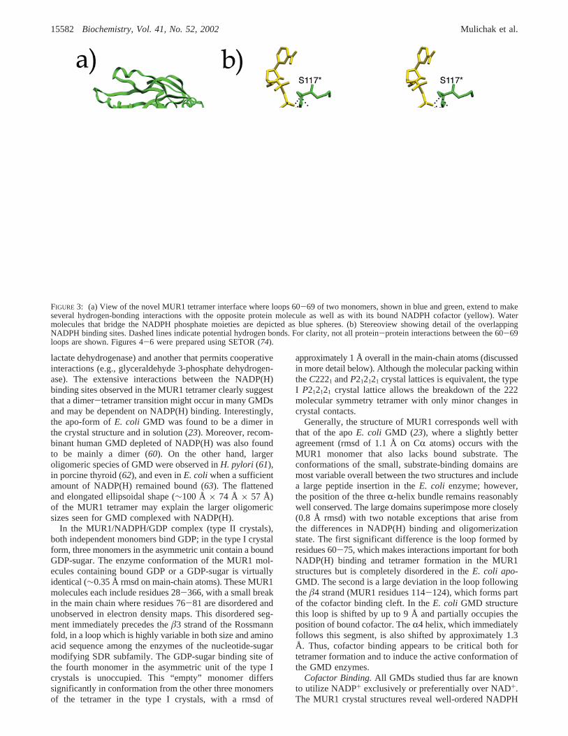
Binding interactions of the cofactor in the MUR1 bindingsite are shown schematically in Figure 5. Consistent withprevious homologous structures, the NADPH cofactor bindsin an extended conformation to the MUR1 large domain,with the pyrophosphate bridge positioned at the N-terminalend (or positive dipole) of theR1 helix, in a mannercharacteristic of the Rossmann dinucleotide-binding motif.
The glycine-rich signature sequence of the classical Ross-mann motif, Gly35-XX-Gly38-XX-Gly41 (where X is anyamino acid) allows a close approach of the cofactor withthe enzyme backbone with direct hydrogen-bonds formedbetween the pyrophosphate and the amide nitrogen atomsof Gln39 and Asp40. Additional hydrogen bonds areprovided to the pyrophosphate by the side chains of Ser117and Arg220.
The most striking feature of the MUR1 cofactor bindingsite is that it, as well as the bound NADPH itself, is anintegral and intimate part of the tetramer interface thatcompletely buries the adenosyl phosphate portion of thebound cofactor (Figure 3). Interactions at this interface areformed predominantly by the peptide segment Arg60-Arg69,part of the loop between the large domainâ2 strand andR2helix, just preceding the disordered region at Asp76-Asn81.This loop, highly variable in size and amino acid sequenceamong related enzymes, closes over the adenosyl end ofbound cofactor in the structures of several other NAD-binding homologues. In the MUR1 tetramer, the Arg60-Arg69 loops from adjacent monomers instead extend acrossthe dimer-dimer interface to interact with each other in asymmetric “fireman’s grip” manner. The two Arg60 side
FIGURE 4: (a) Stereoview showing 2Fo - Fc electron density (1σ level) at the MUR1 active site with GDP-D-rhamnose (red) and NADPH(yellow) bound. Catalytic residues are highlighted in magenta, and spheres represent buried water molecules. (b) Stereoview illustrating thedetailed interactions between the MUR1 catalytic residues, substrate hexose, and cofactor nicotinamide moieties. Dashed lines indicatehydrogen bonds. The nicotinamide C4 atom is positioned midway between the hexose C4 and C6 atoms, the donor and acceptor positionsof hydride transfer, respectively.
GDP-mannose 4,6-Dehydratase Structure fromA. thaliana Biochemistry, Vol. 41, No. 52, 200215583
chains adopt a nearly parallel stacked arrangement, with aplanar separation of approximately 4.5 Å. A number of directinteractions are formed between the two loops, includinghydrogen bonds between the side chains of Arg60, Asn64,and Arg69 with the main-chain carbonyl oxygen atoms ofArg61*, Asn66*, and Ser63*, respectively, from the secondmonomer. The Arg69 side chain simultaneously forms ahydrogen bond with the Ser63* side chain hydroxyl as well.
Although the loops participate somewhat in cofactorbinding within the same monomer via hydrogen-bonding ofthe Arg60 side chain to the adenosyl phosphate moiety, theyactually contribute more extensive interactions to the NAD-PH molecule bound in the neighboring binding cleft. Forexample, the Ser63 residue hydrogen bonds to the neighbor-ing NADPH phosphate moiety via both main-chain amideand side-chain hydroxyl groups, the latter of which is insertedso as to form a bridged interaction between the cofactor andthe Arg69 side chain from the host monomer. In addition,the Arg61 side chain is extended to form a direct hydrogenbond with the pyrophosphate bridge as well as a solvent-bridged interaction with the phosphate of the secondNADPH. This places the Arg side chain in a position tosimultaneously form protein-protein hydrogen bonds withthe main-chain carbonyl oxygen atom of Ala115* as wellas the side-chain hydroxyl of Ser117*.
In addition to the unique intermonomer contributions tocofactor binding, MUR1 also maintains a number of interac-tions with bound NADPH that are conserved with relatedenzymes. The Arg60 side chain packs against the planarsurface of the adenine moiety while hydrogen bonding tothe phosphate group, as has been observed for the GDP-fucose synthase (17, 18). Hydrogen bonding of the adenineby both the side chain of Asp91 and the main-chain amidegroup of the subsequent residue are also highly conservedinteractions, both in NAD+- and NADP+-binding enzymes.The adenyl ribose forms direct hydrogen bonds with enzymebackbone via the free hydroxyl group with the amide nitrogenatoms of both Thr37 and Gly38. The Thr37 side chain alsoforms hydrogen bonds to both the ribose hydroxyl andphosphate moieties. The ribose ring oxygen is also withinhydrogen-bonding distance of the main chain at the Ala115amide nitrogen. These main-chain interactions are conservedin the structure of closely related GDP-L-fucose synthase (17,18), which also utilizes NADP+ rather than NAD+.
In the region of the enzyme active site, one element ofcofactor binding that is highly conserved among relatedenzymes is hydrogen bonding of the ribose hydroxyls bythe Lys and Tyr side chains of the catalytic triad. Buriedwater molecules mediate additional interactions of theenzyme with one ribose hydroxyl as well as the pentose ringoxygen atom. The nicotinamide moiety of the cofactor isbound in a syn conformation, with the carboxyamide nitrogenwithin hydrogen-bonding distance of a phosphoryl oxygenatom. This conformation is consistent with NAD(P)Hcomplexes of related enzymes and is the appropriate orienta-tion for B-side hydride transfer during catalysis. The synconformation may be further stabilized by hydrogen bondswith the surrounding MUR1 enzyme between the carboxya-mide oxygen and His215 main-chain amide nitrogen as wellas between ring hydrogen atoms and the carbonyl oxygenatoms of Gly161 and Leu212.
Substrate Binding Site.The MUR1 complexes presentedhere offer the first crystallographic observation of ligandbinding among enzymes of this family that are specific forGDP-sugars. Moreover, the observation of a bound GDP-sugar (identified as GDP-D-rhamnose) in the active sites ofthree monomers in the type I crystals allows characterizationof GDP-sugar interactions with MUR1. Binding interactionsat the MUR1 substrate site are shown schematically in Figure5 for GDP-D-rhamnose; however, the GDP moiety is boundidentically in both GDP and GDP-D-rhamnose complexes.Interestingly, the GDP moiety binds in the syn conformation,whereas this nucleotide more typically binds to enzymes inthe anti conformation. This conformation is stabilized by anintramolecular hydrogen bond between guanine aminonitrogen atom and phosphoryl oxygen atoms, with the N3ring nitrogen atom also within hydrogen-bonding distance(3.1 Å) of the O5′ oxygen atom. The ability of GDP to adoptthis unusual conformation may be an important determinantin substrate specificity for GMD enzymes.
The GDP moiety is bound entirely within the small domainof MUR1, with the pyrophosphate bridge hydrogen-bondingto the amide nitrogen of Val225, again at the positive dipoleof an R-helix. This mode of substrate binding, reminiscentof dinucleotide binding by the Rossmann motif, appears asa common feature among closely related enzymes. GDPforms hydrogen bonds with the side chains of Asn214,Lys228, Arg253, Arg314, and Glu317 (Figure 5), residues
FIGURE 5: Schematic diagram of potential hydrogen-bonding interactions (dashed lines) of bound NADPH and GDP-D-rhamnose withMUR1 binding site. For clarity, interactions of NADPH with residues from the neighboring MUR1 monomer and some water molecules(W) are not included.
15584 Biochemistry, Vol. 41, No. 52, 2002 Mulichak et al.

that are all highly conserved among the amino acid sequencesof GMDs. Moreover, the hydrogen bonding of Arg314 tooxygen atoms of both phosphoryl groups and the hydrogenbonding of the carboxylate oxygens of Glu317 to eitherribose hydroxyl group are interactions also observed in UDP-sugar binding SDR enzymes as well (16, 22). The guaninering hydrogen bonds to the MUR1 main chain at the Glu223carbonyl and Gly247 amide groups. In addition to these directprotein/ligand hydrogen bonds, GDP also makes severalinteractions mediated by buried water molecules in thebinding pocket.
In the type I crystal form, a GDP-sugar was observed inthe substrate binding site for three monomers of the MUR1tetramer, despite the absence of any added ligand in thecrystallization experiments. Thus, a nucleotide-sugar fromthe expression host can remain tightly associated with theenzymethroughout purification. Similar behavior has previ-ously been reported for GalE (41) and dTDP-D-glucoseoxidoreductase (32), which were purified as naturally oc-curring abortive complexes because of tight binding of theircofactor and substrate/product. In the case of MUR1, theligand observed in the binding site is most probably not thenatural substrate, GDP-D-mannose, but rather GDP-D-rham-nose formed as a dead-end product during the overexpressionof MUR1 in E. coli. Electron density for the sugar moiety(Figure 4) clearly indicates a single axial hydroxyl group atthe O2 position, but no density is observed for an O6hydroxyl group, consistent withD-rhamnose. Furthermore,analysis of the sugar content of the enzyme sample used forcrystallization (data not shown) revealed the presence ofGDP-rhamnose predominantly (87%), with lesser amountsof GDP-mannose.
D-Rhamnose is a rare sugar, thus far identified in natureonly as a constituent of bacterial lipopolysaccharides. OnlyL-rhamnose has been detected in plant tissues; however, theformation of GDP-D-rhamnose in plant extracts has beenknown since the 1960s (64). In the presence of excessNADPH, MUR1 forms this product in theE. coli expressionstrain (50) by acting as a reductase on its natural 4-keto6-deoxymannose product. The absence of detectableD-rhamnose in vivo suggests that these conversions by plantGMDs may be merely experimental artifacts; however, itremains possible, given the diverse range of carbohydratesproduced by plants, that this unusual sugar may be a naturalproduct formed only in low levels. Interestingly, the naturalpathways of GDP-D-rhamnose biosynthesis have been elu-cidated for a number of bacterial species and are found toinvolve conversion of GDP-D-mannose in a two-step process,utilizing a homologous GDP-mannose dehydratase andsecond reductase enzyme (65). A GMD from Aneuriniba-cillus thermoaerophilusthiswas similarly reported to catalyzeboth dehydratase and reductase reactions to produce arhamnose product, albeit less efficiently than the naturalreductase enzyme (66).
The presence of bound GDP-D-rhamnose provides anunexpected view of ligand binding, which should closelyapproximate that of the natural GDP-D-mannose substrate.The sugar moiety extends into the crevice between large andsmall domains of the MUR1 structure, where it is stabilizedby extensive hydrogen-bonding interactions with the enzyme.At the catalytic center, the reactive O4 hydroxyl of the hexosemoiety hydrogen bonds to the side chains of both Ser162
and Tyr185, residues of the catalytic site. The hexose O3hydroxyl interacts with both Tyr185 and the main-chaincarbonyl oxygen of Ser117, while the O5 ring oxygen is ingood hydrogen-bonding distance to the Asn214 side-chainamino group. Specificity for the axial hydroxyl of themannose substrate at the O2 position appears to be largelya result of steric hindrance by the adjacent Phe224 side chain(Figure 4a). In the GDP-D-rhamnose complex, the phenylring makes a van der Waals contact of 3.4 Å with C2 of thebound hexose and would be expected to obstruct binding ofan equatorial hydroxyl at this position. A second importantfeature of binding appears to be the Arg220 side chain. Foundin a parallel stacking arrangement with the Phe224 ring,Arg220 is positioned above the plane of the hexose ring, ingood position to make stabilizing hydrogen bonds with anaxial O2 hydroxyl group. The position of the Arg side chainitself is rigidly constrained by two hydrogen bonds with theGlu216 side chain as well as additional bonds to two buriedwater molecules, both of which interact simultaneously withNADPH pyrophosphate oxygen atoms.
Although not observed in the MUR1/GDP-D-rhamnosecomplex, the likely position of the O6 hydroxyl of the GDP-mannose can be reasonably deduced from the crystalstructure. Two low-energy rotamer positions for the O6hydroxyl are possible within the substrate binding site. Onewould place the hydroxyl within hydrogen-bonding distanceof the side chain of Ser162, part of the catalytic triad; thisorientation has previously been observed in the complex ofSQD1 with UDP-glucose (22). However, this is the moresterically strained of the two orientations. It is more likelythat the O6 hydroxyl adopts the second possible orientation,as is observed for UDP-glucose in GalE (16), which placesit in position to hydrogen bond to both Ser163 and Asn214side-chain oxygen atoms. In fact, a water molecule is foundnear this potential O6 position in the MUR1 complex, within2.5 Å of the GDP-D-rhamnose C5 atom and makinghydrogen-bonding contacts with the side chains of Ser163,Asn214, and Glu164. Notably, as discussed below, the latterresidue is believed to serve as a catalytic base in thedehydratase reaction.
Conformational Changes upon Ligand Binding.In bothGDP and GDP-D-rhamnose complexes, the conformation ofthe MUR1 enzyme is essentially identical, with a rmsd onmain-chain atoms of 0.35 Å. Only a slight shift (0.5 Å) isobserved in the small domain where loops 244-251 and304-315 are more tightly closed over the bound substratewhen GDP-D-rhamnose is the ligand. The structure of thesubstrate binding site itself as well as of the bound NADPHcofactor are also nearly indistinguishable in the two com-plexes. In the GDP complex, the ligand sugar moiety isreplaced by four additional buried water molecules. One ofthese water molecules is found within 0.5 Å of the positionoccupied by the hexose O4 hydroxyl and makes identicalhydrogen-bonding interactions, illustrating the strong drivingforce for binding at this position. The buried water moleculefound near the presumed position of the mannose O6hydroxyl group is conserved in the MUR1 complexes withboth GDP-D-rhamnose and GDP but is shifted by 0.85 Å inthe active site to accommodate the hexose moiety.
In contrast, the unoccupied MUR1 monomer in theasymmetric unit of the type I crystals reveals significantdifferences in the main-chain conformation. A comparison
GDP-mannose 4,6-Dehydratase Structure fromA. thaliana Biochemistry, Vol. 41, No. 52, 200215585

of monomers in bound and unbound states in shown in Figure6. Although the large domain remains unchanged (rmsd of0.45 Å on all atoms), the small domain of the unbound formhas a more open conformation, with the helices shifted byup to 2.4 Å. Moreover, loops 245-250 and 309-318, whichclose over the bound ligand in the GDP and GDP-sugarcomplexes, are completely disordered and unobserved inelectron density maps. Even when well-ordered in the MUR1ternary complexes, these loops are not stabilized by hydrogenbonds with the rest of the protein structure but rather onlyby interactions with the bound ligand. Thus, these loops serveas flexible flaps that may open to allow access of substrateto the catalytic site; once the site is occupied, these loops inthe closed conformation contribute important binding inter-actions and isolate the substrate from the surrounding solvent.
Implications for Catalysis. The SDR enzymes share ahighly conserved catalytic triad of Y-XXX-K and Ser/Thrresidues (67) and moreover appear to share a conservedgeneral mechanism for the initiation of catalysis. Theproposed mechanism for the MUR1 reaction (68) is shownschematically in Figure 1. In the first step, common to allSDR enzymes, the deprotonation of the O4 hydroxyl by ageneral base, coupled with concerted hydride abstractionfrom the C4 atom by the NAD(P)+ cofactor, yields a 4-ketointermediate (2). The MUR1 reaction then proceeds byelimination of a water molecule from C6, yielding a 4-keto-5,6-ene species (3), followed by reduction of the C5-C6double bond to form the final 4-keto-6-deoxymannoseproduct (4). This mechanism is supported by the recentdetection of a 4-keto-5,6-glucoseen intermediate for thehomologous dTGDH reaction (34). It has been demonstratedfor a bacterial GMD (68) as well as for dTGDH (33) that itis the mannose C4 hydride that is transferred via NADPHto C6 in the final reduction step.
The importance of the Tyr, Lys, Ser/Thr triad for catalysishas been demonstrated by mutagenesis and kinetic studies
for E. coli GMD (23) as well as for other related SDRenzymes. The Tyr side chain, in the form of a negativelycharged tyrosinate, has been identified as the catalytic basethat acts on the reactive substrate O4 hydroxyl. In the MUR1complex, a hydrogen bond between the phenolic oxygen ofTyr185 and the rhamnose O4 hydroxyl implies the directattack on O4 by the Tyr base during catalysis, consistentwith similar results derived from the recent crystal structuresof SQD1 (22), GalE (28), dTGDH (35), and RmlB (31). Theadjacent Lys side chain in the SDR triad is believed to servein modifying the Tyr pKa, stablizing the negative charge ofthe tyrosinate moiety, as well as contributing to cofactorbinding (36, 69, 70). The role of the conserved Ser/Thr isless clearly defined. The Ser/Thr side chain may play a rolein the proper orientation of bound substrate and/or in thestabilization of reaction intermediates or transition states.Unusually short (<2.5 Å) hydrogen bonds between this sidechain and the hexose O4 hydroxyl were noted for theproductive ternary complex in SQD1 (22) and the abortivecomplexes of GalE (28) and Rm1B (31). The presence ofshort, low-barrier hydrogen bonds at this position has beenproposed to facilitate proton transfer during catalysis (21,22, 28). The average hydrogen-bonding distance of 2.4 Åbetween hexose O4 and Ser hydroxyl groups in the MUR1complex is consistent with these observations.
It has recently been proposed for the broader class SDRenzymes that the catalytic triad should be expanded to atetrad, including as well a highly conserved Asn residue (71).For these enzymes, the Asn backbone carbonyl is postulatedto participate in a proton relay involving the catalytic Lysand Tyr side chains, a cofactor ribose hydroxyl, and a pocketof buried water molecules, thus facilitating proton transferand serving as a bridge for exchange between the catalyticsite and the bulk solvent. Although the correspondingposition is substituted by a Val residue in the MUR1structure, a solvent-bridged interaction is maintained between
FIGURE 6: Superposition of MUR1 monomers in which substrate binding site is occupied (green) and empty (blue). Positions of boundNADPH (yellow) and GDP-D-rhamnose (red) ligands are depicted in ball-and-stick representation. When the substrate binding site isunoccupied, the small domain is shifted to a more open conformation, and two loops following residues 244 and 308 (arrows) are completelydisordered. View is rotated approximately 90° from that of Figure 2a.
15586 Biochemistry, Vol. 41, No. 52, 2002 Mulichak et al.
the main-chain carbonyl oxygen and the catalytic Lys sidechain. However, unlike the more distant SDR relatives, inthe complexes of MUR1 and most other closely relatedenzymes, the catalytic Lys and Tyr side chains are notdirectly networked via mutual hydrogen bonding to acommon NAD(P) ribose hydroxyl (Figure 5), as requiredby the proton relay hypothesis.
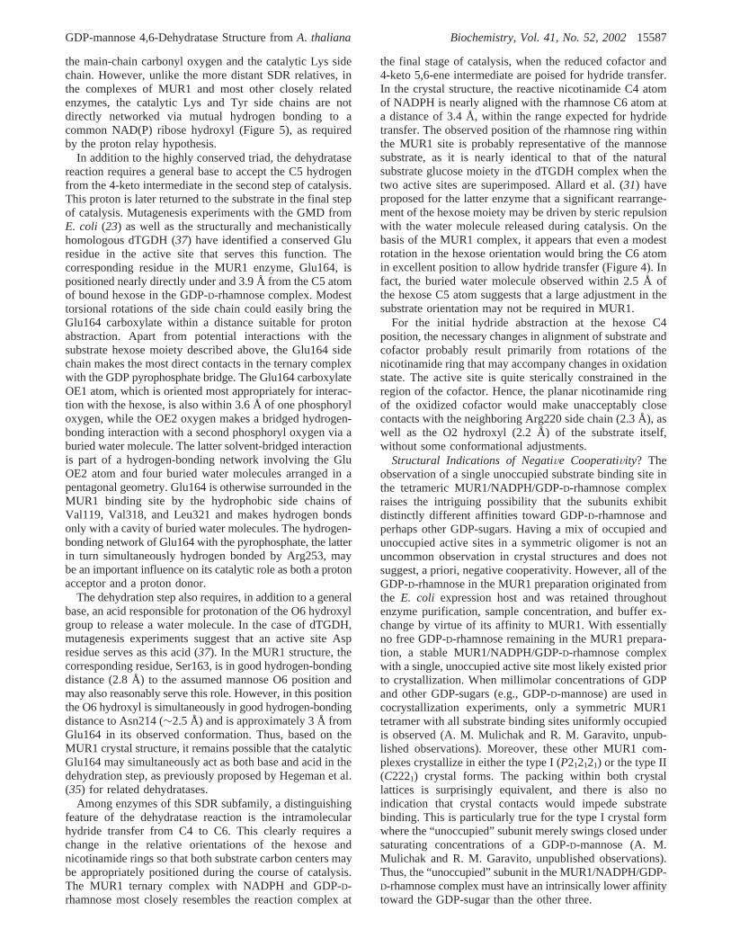
In addition to the highly conserved triad, the dehydratasereaction requires a general base to accept the C5 hydrogenfrom the 4-keto intermediate in the second step of catalysis.This proton is later returned to the substrate in the final stepof catalysis. Mutagenesis experiments with the GMD fromE. coli (23) as well as the structurally and mechanisticallyhomologous dTGDH (37) have identified a conserved Gluresidue in the active site that serves this function. Thecorresponding residue in the MUR1 enzyme, Glu164, ispositioned nearly directly under and 3.9 Å from the C5 atomof bound hexose in the GDP-D-rhamnose complex. Modesttorsional rotations of the side chain could easily bring theGlu164 carboxylate within a distance suitable for protonabstraction. Apart from potential interactions with thesubstrate hexose moiety described above, the Glu164 sidechain makes the most direct contacts in the ternary complexwith the GDP pyrophosphate bridge. The Glu164 carboxylateOE1 atom, which is oriented most appropriately for interac-tion with the hexose, is also within 3.6 Å of one phosphoryloxygen, while the OE2 oxygen makes a bridged hydrogen-bonding interaction with a second phosphoryl oxygen via aburied water molecule. The latter solvent-bridged interactionis part of a hydrogen-bonding network involving the GluOE2 atom and four buried water molecules arranged in apentagonal geometry. Glu164 is otherwise surrounded in theMUR1 binding site by the hydrophobic side chains ofVal119, Val318, and Leu321 and makes hydrogen bondsonly with a cavity of buried water molecules. The hydrogen-bonding network of Glu164 with the pyrophosphate, the latterin turn simultaneously hydrogen bonded by Arg253, maybe an important influence on its catalytic role as both a protonacceptor and a proton donor.
The dehydration step also requires, in addition to a generalbase, an acid responsible for protonation of the O6 hydroxylgroup to release a water molecule. In the case of dTGDH,mutagenesis experiments suggest that an active site Aspresidue serves as this acid (37). In the MUR1 structure, thecorresponding residue, Ser163, is in good hydrogen-bondingdistance (2.8 Å) to the assumed mannose O6 position andmay also reasonably serve this role. However, in this positionthe O6 hydroxyl is simultaneously in good hydrogen-bondingdistance to Asn214 (∼2.5 Å) and is approximately 3 Å fromGlu164 in its observed conformation. Thus, based on theMUR1 crystal structure, it remains possible that the catalyticGlu164 may simultaneously act as both base and acid in thedehydration step, as previously proposed by Hegeman et al.(35) for related dehydratases.
Among enzymes of this SDR subfamily, a distinguishingfeature of the dehydratase reaction is the intramolecularhydride transfer from C4 to C6. This clearly requires achange in the relative orientations of the hexose andnicotinamide rings so that both substrate carbon centers maybe appropriately positioned during the course of catalysis.The MUR1 ternary complex with NADPH and GDP-D-rhamnose most closely resembles the reaction complex at
the final stage of catalysis, when the reduced cofactor and4-keto 5,6-ene intermediate are poised for hydride transfer.In the crystal structure, the reactive nicotinamide C4 atomof NADPH is nearly aligned with the rhamnose C6 atom ata distance of 3.4 Å, within the range expected for hydridetransfer. The observed position of the rhamnose ring withinthe MUR1 site is probably representative of the mannosesubstrate, as it is nearly identical to that of the naturalsubstrate glucose moiety in the dTGDH complex when thetwo active sites are superimposed. Allard et al. (31) haveproposed for the latter enzyme that a significant rearrange-ment of the hexose moiety may be driven by steric repulsionwith the water molecule released during catalysis. On thebasis of the MUR1 complex, it appears that even a modestrotation in the hexose orientation would bring the C6 atomin excellent position to allow hydride transfer (Figure 4). Infact, the buried water molecule observed within 2.5 Å ofthe hexose C5 atom suggests that a large adjustment in thesubstrate orientation may not be required in MUR1.
For the initial hydride abstraction at the hexose C4position, the necessary changes in alignment of substrate andcofactor probably result primarily from rotations of thenicotinamide ring that may accompany changes in oxidationstate. The active site is quite sterically constrained in theregion of the cofactor. Hence, the planar nicotinamide ringof the oxidized cofactor would make unacceptably closecontacts with the neighboring Arg220 side chain (2.3 Å), aswell as the O2 hydroxyl (2.2 Å) of the substrate itself,without some conformational adjustments.
Structural Indications of NegatiVe CooperatiVity? Theobservation of a single unoccupied substrate binding site inthe tetrameric MUR1/NADPH/GDP-D-rhamnose complexraises the intriguing possibility that the subunits exhibitdistinctly different affinities toward GDP-D-rhamnose andperhaps other GDP-sugars. Having a mix of occupied andunoccupied active sites in a symmetric oligomer is not anuncommon observation in crystal structures and does notsuggest, a priori, negative cooperativity. However, all of theGDP-D-rhamnose in the MUR1 preparation originated fromthe E. coli expression host and was retained throughoutenzyme purification, sample concentration, and buffer ex-change by virtue of its affinity to MUR1. With essentiallyno free GDP-D-rhamnose remaining in the MUR1 prepara-tion, a stable MUR1/NADPH/GDP-D-rhamnose complexwith a single, unoccupied active site most likely existed priorto crystallization. When millimolar concentrations of GDPand other GDP-sugars (e.g., GDP-D-mannose) are used incocrystallization experiments, only a symmetric MUR1tetramer with all substrate binding sites uniformly occupiedis observed (A. M. Mulichak and R. M. Garavito, unpub-lished observations). Moreover, these other MUR1 com-plexes crystallize in either the type I (P212121) or the type II(C2221) crystal forms. The packing within both crystallattices is surprisingly equivalent, and there is also noindication that crystal contacts would impede substratebinding. This is particularly true for the type I crystal formwhere the “unoccupied” subunit merely swings closed undersaturating concentrations of a GDP-D-mannose (A. M.Mulichak and R. M. Garavito, unpublished observations).Thus, the “unoccupied” subunit in the MUR1/NADPH/GDP-D-rhamnose complex must have an intrinsically lower affinitytoward the GDP-sugar than the other three.
GDP-mannose 4,6-Dehydratase Structure fromA. thaliana Biochemistry, Vol. 41, No. 52, 200215587

While a detailed structural basis for lower binding affinityfor GDP-D-rhamnose is not immediately apparent, contactsexist between neighboring monomers that could mediate suchcommunication between the substrate-binding domains. Forinstance, across the tetramer interface, the long C-terminalhelices from the small domains of adjacent monomers packagainst one another (Figure 2b). This interaction is signifi-cantly perturbed in the case of the single unoccupiedmonomer. Additionally, close contacts occur between seg-ments 169-182 of monomers related by the highly conservedback-to-back dimer interface. These loops contribute to asmall section ofâ-sheet including threeâ-strands of the smalldomain, all of which are shifted in response to the bindingof substrate.
In summary, the X-ray crystal structures of MUR1complexes presented here confirm the presence of catalyticresidues that are conserved among the sequences of relatedenzyme and further suggest residues that are likely deter-minants in substrate binding and specificity. Beyond this,however, the structures present unexpected and intriguingissues, including the syn binding mode of GDP, thefunctional role of tetrameric GMD, and the possible existenceof negative cooperativity, which all warrant further investiga-tion. Efforts also continue toward successful displacementof tightly bound NADPH in the purified enzyme to addressissues such as the effect of cofactor oxidation state on activesite conformation and the role of NADP(H) binding intetramer formation.
ACKNOWLEDGMENT
We gratefully acknowledge M. Dumond for preparationand purification of the MUR1 enzyme. Studies related herewere supported by the U.S. Department of Energy Grant DE-FG02-95ER20203 (W.-D.R.) and the Michigan Life ScienceCorridor grant to the Center for Structural Biology (R.M.G.).Research was carried out in part at the National SynchrotronLight Source, Brookhaven National Laboratory, which issupported by the U.S. Department of Energy. Portions ofthis work were performed at the DuPont-Northwestern-DowCollaborative Access Team (DND-CAT) Synchrotron Re-search Center located at Sector 5 of the Advanced PhotonSource (APS). DND-CAT is supported by E.I. DuPont deNemours & Co., Dow Chemical Company, NSF GrantDMR-9304725, and a grant from the State of Illinois (IBHEHECA NWU 96). Use of the Argonne National LaboratoryStructural Biology Center 19ID beamline at the APS wassupported by the U.S. Department of Energy, under ContractW-31-109-ENG-38. We thank the staffs of the NSLS X25and APS DND and SBC beamlines for their assistance.
REFERENCES
1. Varki, A. (1994)Proc. Natl. Acad. Sci. U.S.A. 91, 7390-7497.2. Roos, C., Kolmer, M., Mattila, P., and Renkonen, R. (2001)J.
Biol. Chem. 277, 3168-3175.3. Lamrabet, Y., Bellogin, R. A., Cubo, T., Espuny, R., Gil, A.,
Krishnan, H. B., Megias, M., Ollero, F. J., Pueppke, S. G., Ruiz-Sainz, J. E., Spaink, H. P., Tejero-Mateo, P., Thomas-Oates, J.,and Vinardell, J. M. (1999)Mol. Plant-Microbe Interact. 12, 207-217.
4. Lopez-Lara, I. M., Blok-Tip, L., Quinto, C., Garcia, M. L.,Bloemberg, G. V., Lamers, G. E., Lugtenberg, B. J., Thomas-Oates, J., and Spaink, H. P. (1996)Mol. Microbiol. 21, 397-408.
5. Levy, S., York, W. S., Stuike-Prill, R., Meyer, B., and Staehelin,L. A. (1991) Plant J. 1, 195-215.
6. Levy, S., Maclachlan, G., and Staehelin, L. A. (1997)Plant J.11, 373-386.
7. Vanzin, G. F., Madson, M., Carpita, N. C., Raikhel, N. V.,Keegstra, K., and Reiter, W.-D. (2002)Proc. Natl. Acad. Sci.U.S.A. 99, 3340-3345.
8. O’Neill, M. A., Eberhard, S., Albersheim, P., and Darvill, A. G.(2001)Science 294, 846-849.
9. Baker, M. E., and Blasco, R. (1992)FEBS Lett. 301, 89-93.10. Labesse, G., Vidal-Cros, A., Chomilier, J., Gaudry, M., and
Mornon, J.-P. (1994)Biochem. J. 304, 95-99.11. Krozowski, Z. (1994)J. Steroid Biochem. Mol. Biol. 51, 125-
130.12. Jornvall, H., Persson, B., Krook, M., Atrian, S., Gonzalez-Duarte,
R., Jeffery, J., and Ghosh, D. (1995)Biochemistry 34, 6003-6013.
13. Jornvall, H., Hoog, J.-O., and Persson, B. (1999)FEBS Lett. 445,261-264.
14. Bauer, A. J., Rayment, I., Frey, P. A., and Holden, H. M. (1992)Proteins 12, 372-381.
15. Thoden, J. B., Frey, P. A., and Holden, H. M. (1996)Protein Sci.5, 2149-2161.
16. Thoden, J. B., Frey, P. A., and Holden, H. M. (1996)Biochemistry35, 5137-5144.
17. Somers, W. S., Stahl, M. L., and Sullivan, F. X. (1998)Structure6, 1601-1612.
18. Rizzi, M., Tonetti, M., Vigevani, P., Sturla, L., Bisso, A., De Flora,A., Bordo, D., and Bolognesi, M. (1998)Structure 6, 1453-1465.
19. Allard, S. T. M., Giraud, M.-F., Whitfield, C., Graninger, M.,Messner, P., and Naismith, J. H. (2001)J. Mol. Biol. 307, 283-295.
20. Deacon, A. M., Ni, Y. S., Coleman, W. G., Jr., and Ealick, S. E.(2000)Structure 8, 453.
21. Blankenfeldt, W., Kerr, I. D., Giraud, M.-F., McMiken, H. J.,Leonard, G. A., Whitfield, C., Messner, P., Graninger, M., andNaismith, J. H. (2002)Structure 10, 773-786.
22. Mulichak, A. M., Theisen, M. J., Essigmann, B., Benning, C.,and Garavito, R. M. (1999)Proc. Natl. Acad. Sci. U.S.A. 96,13097-13102.
23. Somoza, J. R., Menon, S., Schmidt, H., Joseph-McCarthy, D.,Dessen, A., Stahl, M. L., Somers, W. S., and Sullivan, F. X. (2000)Structure 8, 123-135.
24. Thoden, J. B., Frey, P. A., and Holden, H. M. (1996)Biochemistry35, 2557-2566.
25. Thoden, J. B., Gulick, A. M., and Holden, H. M. (1997)Biochemistry 36, 10685-10695.
26. Thoden, J. B., Hegeman, A. D., Wesenberg, G., Chapeau, M. C.,Frey, P. A., and Holden, H. M. (1997)Biochemistry 36, 6294-6304.
27. Thoden, J. B., and Holden, H. M. (1998)Biochemistry 37, 11469-11477.
28. Thoden, J. B., Wohlers, T. M., Fridovich-Keil, J. L., and Holden,H. M. (2000)Biochemistry 39, 5691-5701.
29. Thoden, J. B., Wohlers, T. M., Fridovich-Keil, J. L., and Holden,H. M. (2001)J. Biol. Chem. 276, 20617-20623.
30. Thoden, J. B., Wohlers, T. M., Fridovich-Keil, J. L., and Holden,H. M. (2001)J. Biol. Chem. 276, 15131-15136.
31. Allard, S. T. M., Beis, K., Giraud, M.-F., Hegeman, A. D., Gross,J. W., Wilmouth, R. C., Whitfield, C., Graninger, M., Messner,P., Allen, A. G., Maskell, D. J., and Naismith, J. H. (2002)Structure 10, 81-92.
32. Zarkowsky, H., Lipkin, E., and Glaser, L. (1970)J. Biol. Chem.245, 6599-6606.
33. Snipes, C. E., Brillinger, G.-U., Sellers, L., Mascaro, L., and Floss,H. G. (1977)J. Biol. Chem. 252, 8113-8117.
34. Gross, J. W., Hegeman, A. D., Vestling, M. M., and Frey, P. A.(2000)Biochemistry 39.
35. Hegeman, A. D., Gross, J. W., and Frey, P. A. (2001)Biochemistry40, 6598-6610.
36. Gerratana, B., Cleland, W. W., and Frey, P. A. (2001)Biochemistry40, 9187-9195.
37. Gross, J. W., Hegeman, A. D., Gerratana, B., and Frey, P. A.(2001)Biochemistry 40, 12497-12504.
38. Hegeman, A. D., Gross, J. W., and Frey, P. A. (2002)Biochemistry41, 2797-2804.
39. Berger, E., Arabshahi, A., Wei, Y., Schilling, J. F., and Frey, P.A. (2001)Biochemistry 40, 6699-6705.
15588 Biochemistry, Vol. 41, No. 52, 2002 Mulichak et al.

40. Liu, Y., Thoden, J. B., Kim, J., Berger, E., Gulick, A. M., Ruzicka,F. J., Holden, H. M., and Frey, P. A. (1997)Biochemistry 36,10675-10684.
41. Vanhooke, J. L., and Frey, P. A. (1994)J. Biol. Chem. 269,31496-31504.
42. Liu, Y., Vanhooke, J. L., and Frey, P. A. (1996)Biochemistry 35,7615-7620.
43. Swanson, B. A., and Frey, P. A. (1993)Biochemistry 32, 13231-13236.
44. Burke, J. R., and Frey, P. A. (1993)Biochemistry 32, 13220-13230.
45. Flentke, G. R., and Frey, P. A. (1990)Biochemistry 29, 2430-2436.
46. Arabshahi, A., Flentke, G. R., and Frey, P. A. (1988)J. Biol. Chem.263, 2638-2643.
47. Wee, T. G., Davis, J., and Frey, P. A. (1972)J. Biol. Chem. 247,1339-1342.
48. Wee, T. G., and Frey, P. A. (1973)J. Biol. Chem. 248, 33-40.49. Wong, S. S., and Frey, P. A. (1977)Biochemistry 16, 298-305.50. Bonin, C. P., Potter, I., Vanzin, G. F., and Reiter, W.-D. (1997)
Proc. Natl. Acad. Sci. U.S.A. 94, 2085-2090.51. Zablackis, E., York, W. S., Pauly, M., Hantus, S., Reiter, W.-D.,
Chapple, C. C. S., Albersheim, P., and Darvill, A. G. (1996)Science 272, 1808-1810.
52. Reiter, W.-D., Chapple, C. C. S., and Somerville, C. R. (1993)Science 261, 1032-1035.
53. Jancarik, J., and Kim, S.-H. (1991)J. Appl. Crystallogr. 24, 409-411.
54. Otwinowski, Z., and Minor, W. (1997)Methods Enzymol. 276,307-326.
55. Brunger, A. T. (1998)Acta Crystallogr. D54, 905-921.56. CHAIN (1995) CHAIN: Crystallographic Modeling Program
Version 7.0, Baylor College of Medicine, Waco, TX.57. Navaza, J. (1994)Acta Crystallogr. A50, 157-163.58. Collaborative Computational Project N. (1994)Acta Crystallogr.
D50, 760-763.
59. Rossmann, M. G., Liljas, A., Branden, C.-I., and Banaszak, L. J.(1975)Enzymes 11, 61-102.
60. Bisso, A., Sturla, L., Zanardi, D., De Flora, A., and Tonetti, M.(1999)FEBS Lett. 456, 370-374.
61. Wu, B., Zhang, Y., and Wang, P. G. (2001)Biochem. Biophys.Res. Commun. 285, 364-371.
62. Broschat, K. O., Chang, S., and Serif, G. (1985)Eur. J. Biochem.153, 397-401.
63. Sturla, L., Bisso, A., Zanardi, D., Benatti, U., De Flora, A., andTonetti, M. (1997)FEBS Lett. 412, 126-130.
64. Barber, G. A. (1968)Biochim. Biophys. Acta 165, 68-75.65. Rocchetta, H. L., Pacan, J. C., and Lam, J. S. (1998)Mol.
Microbiol. 29, 1419-1434.66. Kneidinger, B., Graninger, M., Adams, G., Puchberger, M., Kosma,
P., Zayni, S., and Messner, P. (2001)J. Biol. Chem. 276, 5577-5583.
67. Persson, B., Krook, M., and Jornvall, H. (1991)Eur. J. Biochem.200, 537-543.
68. Oths, P. J., Mayer, R. M., and Floss, H. G. (1990)Carbohydr.Res. 198, 91-100.
69. Varughese, K. I., Xuong, N. H., Kiefer, P. M., Matthews, D. A.,and Whiteley, J. M. (1994)Proc. Natl. Acad. Sci. U.S.A. 91,5582-5586.
70. Winberg, J. O., Brendskag, M. K., Sylte, I., Lindstad, R. I., andMcKinley-McKee, J. S. (1999)J. Mol. Biol. 294, 601-616.
71. Filling, C., Berndt, K. D., Benach, J., Knapp, S., Prozorovski, T.,Nordling, E., Ladenstein, R., Jornvall, H., and Oppermann, U.(2002)J. Biol. Chem. 277, 25677-25684.
72. Kraulis, P. J. (1991)J. Appl. Crystallogr. 24, 946-950.73. Merrit, E., and Murphy, M. (1994)Acta Crystallogr. D50, 869-
873.74. Evans, S. V. (1993)J. Mol. Graphics 11, 134-138.
BI0266683
GDP-mannose 4,6-Dehydratase Structure fromA. thaliana Biochemistry, Vol. 41, No. 52, 200215589


![The Dehydratase ADT3 Affects ROS Homeostasis and … · The Dehydratase ADT3 Affects ROS Homeostasis and Cotyledon Development1[OPEN] Alessia Para, DurreShahwar Muhammad, Danielle](https://img.pdfslide.net/doc/110x75/5cca391988c993570d8d8d21/the-dehydratase-adt3-affects-ros-homeostasis-and-the-dehydratase-adt3-affects.jpg)